A retinoic acid receptor expressed in the early ...
12
A retinoic acid receptor expressed in the early development of Xenopus laevis Heidrun Ellinger-Ziegelbauet and Christine Dreyer Max-Planck-Institut fiir Entwicklungsbiologie, D-7400 Tubingen, Germany We have isolated cDNAs coding for a putative retinoic acid receptor (RAR) of the 7-type from a Xenopus laevis neurula cDNA library. By transient cotransfection of COS cells with an expression vector and a reporter plasmid, this cDNA is shown to direct the synthesis of a retinoic acid-dependent transcription factor. In embryos of X. laevis, transcription of the corresponding gene is greatly enhanced during gastrulation and early neurulation. Two distinct areas with high abundance of RAR 7 mRNA are located at the anterior and at the posterior end of the neurula. The two maxima have emerged by the end of gastrulation and they become more pronounced during neurulation. At tailbud and early tadpole stages, the RAR transcripts are found mainly in the head mesenchyme and in the tailbud. The expression of this RAR is region-specific but not germ-layer-specific. The strong and stage-specific activation of zygotic transcription of this RAR gene, and the specific localization of the mRNA are consistent with the temporal and spatial pattern of retinoic acid sensitivity of X. laevis embryos. Therefore it is likely that the gene product mediates the effects of endogenous and of exogenous retinoic acid on early embryogenesis of Xenopus. The significance of these findings for the specification of the anteroposterior axis is discussed. [Key Words: Xenopus-, retinoic acid receptor; anteroposterior axis; in situ hybridization] Received August 30, 1990; revised version accepted November 8, 1990. All-trans retinoic acid (RA), a derivative of vitamin A, is a potent teratogen, and affects the differentiation of many cells in vitro (Sporn and Roberts 1983; Wolf 1984). In vertebrates, RA is apparently involved in the specifi- cation of the anteroposterior axis of the developing limb (Thaller and Eichele 1987; Eichele 1989) and in the de- velopment of the central nervous system (Durston et al. 1989). Endogenous RA has been detected during early embryogenesis in Xenopus laevis (Durston et al. 1989) and in chickens (Wagner et al. 1990), and in limb buds of the chick (Thaller and Eichele 1987). Exogenous RA applied on Xenopus embryos before neurulation causes truncation of anterior head struc- tures (Durston et al. 1989; Sive et al. 1990) and micro- cephaly due to transformation of anterior to more poste- rior neural structures (Durston et al. 1989). Both cellular RA-binding proteins (CRABP) (Chytyl and Ong 1984) and nuclear retinoic acid receptors (RARs) have been identified in vertebrates (Petkovich et al. 1987; Brand et al. 1988; Giguere et al. 1987; Krust et al. 1989; Ragsdale et al. 1989; Zelent et al. 1989; Kastner et al. 1990). The discovery that RARs belong to the superfamily of li- gand-dependent DNA-binding proteins with a Zn-finger domain implies that RA acts as a modulator of the ac- tivity of specific target genes (Evans 1988). With the aim of investigating the role of ligand-depen- dent transcription factors in amphibian development, we have screened a cDNA library of X. laevis neurula with a mixture of oligonucleotides constructed ac- cording to the consensus between different genes en- coding nuclear receptors of steroid and thyroid hor- mones and of RA (Evans 1988). We have found that at least two different classes of nuclear receptor-encoding genes are expressed during neurulation. One codes for a putative receptor that has not been described previously, and whose ligand is unknown, while the second encodes a RAR of the 7 subclass. In this discussion we describe the identification of the RAR cDNA and the temporal and spatial pattern of expression of the corresponding gene during development. The results are discussed in the context of recent reports concerning the effects of RA on the specification of the anteroposterior axis. Results Isolation and characterization of a cDNA coding for a putative X. laevis RAR We have screened a XgtlO cDNA library from X. laevis stage 17 neurulae (Kintner and Melton 1987) with con- sensus oligonucleotides that would encode part of the first Zn finger of the DNA-binding domain of known nuclear receptors for thyroid hormones and RA (Evans 1988). Of 22 recombinant phages whose DNA hybrid- ized with the consensus oligonucleotides, 14 were shown to be closely related by cross hybridization under stringent conditions (data not shown). The inserts of four of these were subcloned into a plasmid vector and one was totally and three were partially sequenced from both ends and were shown to be almost identical. The cDNA shown in Figure lb is 2077 nucleotides long and 94 GENES & DEVELOPMENT 5:94-104 © 1991 by Cold Spring Harbor Laboratory Press ISSN 0890-9369/91 $1.00 Cold Spring Harbor Laboratory Press on April 5, 2022 - Published by genesdev.cshlp.org Downloaded from
Transcript of A retinoic acid receptor expressed in the early ...

A retinoic acid receptor expressed in the early development of Xenopus laevis Heidrun Ellinger-Ziegelbauet and Christine Dreyer
Max-Planck-Institut fiir Entwicklungsbiologie, D-7400 Tubingen, Germany
We have isolated cDNAs coding for a putative retinoic acid receptor (RAR) of the 7-type from a Xenopus laevis neurula cDNA library. By transient cotransfection of COS cells with an expression vector and a reporter plasmid, this cDNA is shown to direct the synthesis of a retinoic acid-dependent transcription factor. In embryos of X. laevis, transcription of the corresponding gene is greatly enhanced during gastrulation and early neurulation. Two distinct areas with high abundance of RAR 7 mRNA are located at the anterior and at the posterior end of the neurula. The two maxima have emerged by the end of gastrulation and they become more pronounced during neurulation. At tailbud and early tadpole stages, the RAR transcripts are found mainly in the head mesenchyme and in the tailbud. The expression of this RAR is region-specific but not germ-layer-specific. The strong and stage-specific activation of zygotic transcription of this RAR gene, and the specific localization of the mRNA are consistent with the temporal and spatial pattern of retinoic acid sensitivity of X. laevis embryos. Therefore it is likely that the gene product mediates the effects of endogenous and of exogenous retinoic acid on early embryogenesis of Xenopus. The significance of these findings for the specification of the anteroposterior axis is discussed.
[Key Words: Xenopus-, retinoic acid receptor; anteroposterior axis; in situ hybridization]
Received August 30, 1990; revised version accepted November 8, 1990.
All-trans retinoic acid (RA), a derivative of vitamin A, is a potent teratogen, and affects the differentiation of many cells in vitro (Sporn and Roberts 1983; Wolf 1984). In vertebrates, RA is apparently involved in the specification of the anteroposterior axis of the developing limb (Thaller and Eichele 1987; Eichele 1989) and in the development of the central nervous system (Durston et al. 1989). Endogenous RA has been detected during early embryogenesis in Xenopus laevis (Durston et al. 1989) and in chickens (Wagner et al. 1990), and in limb buds of the chick (Thaller and Eichele 1987).
Exogenous RA applied on Xenopus embryos before neurulation causes truncation of anterior head structures (Durston et al. 1989; Sive et al. 1990) and microcephaly due to transformation of anterior to more posterior neural structures (Durston et al. 1989). Both cellular RA-binding proteins (CRABP) (Chytyl and Ong 1984) and nuclear retinoic acid receptors (RARs) have been identified in vertebrates (Petkovich et al. 1987; Brand et al. 1988; Giguere et al. 1987; Krust et al. 1989; Ragsdale et al. 1989; Zelent et al. 1989; Kastner et al. 1990). The discovery that RARs belong to the superfamily of li-gand-dependent DNA-binding proteins with a Zn-finger domain implies that RA acts as a modulator of the activity of specific target genes (Evans 1988).
With the aim of investigating the role of ligand-depen-dent transcription factors in amphibian development, we have screened a cDNA library of X. laevis neurula with a mixture of oligonucleotides constructed according to the consensus between different genes en
coding nuclear receptors of steroid and thyroid hormones and of RA (Evans 1988). We have found that at least two different classes of nuclear receptor-encoding genes are expressed during neurulation. One codes for a putative receptor that has not been described previously, and whose ligand is unknown, while the second encodes a RAR of the 7 subclass. In this discussion we describe the identification of the RAR cDNA and the temporal and spatial pattern of expression of the corresponding gene during development. The results are discussed in the context of recent reports concerning the effects of RA on the specification of the anteroposterior axis.
Results Isolation and characterization of a cDNA coding for a putative X. laevis RAR
We have screened a XgtlO cDNA library from X. laevis stage 17 neurulae (Kintner and Melton 1987) with consensus oligonucleotides that would encode part of the first Zn finger of the DNA-binding domain of known nuclear receptors for thyroid hormones and RA (Evans 1988). Of 22 recombinant phages whose DNA hybridized with the consensus oligonucleotides, 14 were shown to be closely related by cross hybridization under stringent conditions (data not shown). The inserts of four of these were subcloned into a plasmid vector and one was totally and three were partially sequenced from both ends and were shown to be almost identical. The cDNA shown in Figure lb is 2077 nucleotides long and
94 GENES & DEVELOPMENT 5:94-104 © 1991 by Cold Spring Harbor Laboratory Press ISSN 0890-9369/91 $1.00
Cold Spring Harbor Laboratory Press on April 5, 2022 - Published by genesdev.cshlp.orgDownloaded from

Xenopus retinoic acid receptor
a xRAR-39
T3,SK sense
^ T7,KS antisense
_ < en CO _ — o 3 x : - c o « o. °-
UJ c/5 c/> W
L 3 O = 3 tO-C to (0 M X 0 3 0 5
< m —
A ^ CL
FPT .100bp.
1 5 30 45 60 75 !«/ ATCAGAGCCCACACTGCTGCTTTCCCTTCCTCTTCTGAAAACTTCAGTGCCCACTTACCCGCTCCCCCTACTCCC
90 105 120 135 150 AAAATGTACGACTGCATGGAAGCCTTCCCTCTGATGCCTCGGCCGCTCTATGACATGAGTCCTCAGGGTCCCTGC
1 M Y D C M E A F P L M P R P L Y D M S P Q G P C 1 6 5 1 8 0 1 9 5 2 1 0 2 2 5
ATGCTGCGCAAAGCCGGCTGTTTTGGGGGGCTGGACCCCTTTGGATGGATGCAGAGCCACAGCATGCAATCTGTA 2 5 M L R K A G C F G G L D P F G W M Q S H S M Q S V
2 4 0 2 5 5 2 7 0 2 8 5 3 0 0 GAAACACAAAGTACCAGCTCGGAGGAAATGGTGCCCAGTTCACCCTCGCCGCCGCCACCTCCACGTGTTTACAAG
5 0 E T Q S T S S E E M V P S S P S P P P P P R V Y K 3 1 5 3 3 0 3 4 5 360 3 7 5
CCCTGCTTCGTCTGCAATGACAAGTCTTCAGGTTATCACTACGGGGTCAGTTCATGCGAGGGCTGCAAGGGCTTT 7 5 P I C ] F V | C ] N D K S S G Y H Y G V S S [c] i G [Cj K G F
3 9 0 4 0 5 4 2 0 4 3 5 4 5 0 T T T C G G C G C A G T A T A C A G A A A A A C A T G G T A T A T A C A T G T C A C C G T G A C A A G A A C T G C C A A A T C A A C A A G G T C A C A
1 0 0 F R R S I Q K N M V Y T [ £ | H R D K N [ C ] Q I N K V T
465 480 495 510 525 C G G A A C C G T T G C C A G T T T T G C C G A C T G C A G A A A T G C T T C C A G G T C G G A A T G T C T A A A G A G G C G G T T A G A A A T G A C
1 2 5 R N R | C ] Q F [ C ] R L Q K [ C ] F Q V G M S K E A V R N D 5 4 0 5 5 5 5 7 0 5 8 5 6 0 0
AGAAACAAGAAGAAAAAGGAGATAAAAGAGGAAGTGGTGCTACCAGACAGCTATGAGATGCCCCCAGAAATGGAA 150 R N K K K K E I K E E V V L P D S Y E M P P E M E
6 1 5 6 3 0 6 4 5 660 6 7 5 G A G C T A A T T C A G A A A G T C A G C A A A G C A C A T C A A G A A A C C T T T C C T T C T C T C T G C C A G C T A G G C A A A T A C A C C A C G
1 7 5 E L I Q K V S K A H Q E T F P S L C Q L G K Y T T 6 9 0 7 0 5 7 2 0 7 3 5 7 5 0
AACTCCAGTGCTGATCAAAGAGTGCAGTTGGACTTGGGCTTGTGGGATAAATTCAGTGAACTGTCCACCAAGTGT 200 N S S A D Q R V Q L D L G L W D K F S E L S T K C
765 780 795 810 825 ATCATCAAGATTGTGGAGTTTGCCAAACGTTTGCCTGGATTCACCACACTGACCATCGCCGACCAAATCACCTTA
225 I I K I V E F A K R L P G F T T L T I A D Q I T L 840 855 870 885 900
TTGAAGTCTGCCTGCCTGGATATACTGATGCTCCGGATTTGCACACGCTACACTCCAGAACAAGACACCATGACC 250 L K S A C L D I L M L R I C T R Y T P E Q D T M T
915 930 945 960 975 TTCTCAGATGGCCTTACTCTAAACCGTACCCAGATGCACAATGCTGGTTTTGGGCCACTTACAGACCTGGTTTTT
2 7 5 F S D G L T L N R T Q M H N A G F G P L T D L V F 990 1005 1020 1035 1050
TCATTTGCAGACCAGCTACTCCCATTAGAAATGGATGACACAGAGACAGGACTTCTGAGCGCCATCTGTTTAATT
300 S F A D Q L L P L E M D D T E T G L L S A I C L I
1065 1080 1095 1110 1125 TGTGGAGATCGGATGGATTTAGAAGAGCCAGAAAAAGTGGAGAAGTTACAAGAACCACTTCTCGAGGGCCTGAAA
325 C G D R M D L E E P E K V E K L Q E P L L E G L K 1140 1155 1170 1185 1200
TTTTATGCTCGTCGACGAAGGCCTGACAAACCTTACATGTTTCCTCGTATGCTTATGAAAATCACTGACTTACGT 350 F Y A R R R R P D K P Y M F P R M L M K I T D L R
1215 1230 1245 1260 1275 GGGATCAGTACAAAAGGTGCTGAGCGAGCAATCACATTGAAGCTGGAGATACCAGGACCTATGCCTCCGCTGATT
375 G I S T K G A E R A I T L K L E I P G P M P P L I 1290 1305 1320 1335 1350
CGGGAGATGCTGGAGAATCCAGAAGCCTTTGAAGATGGAGCTGCCACCCCTAAACCCAGCGAGCGTTCTTCCAGT 400 R E M L E N P E A F E D G A A T P K P S E R S S S
1365 1380 1395 1410 1425 GAGAGCAGTAATGGAAGTCCTACAGGAGAAGATAGCAGTGGCTCGAAGACCCCTTAGTTGTCCTTAGCATATTGG
425 E S S N G S P T G E D S S G S K T P * 1440 1455 1470 1485 1500
GGAAAGATATCCCAAAGCCTAAACACTTTGTATTGAGCCACGTCAGTTCTACCCATTGTATGAGGCATTCTGGGG 1515 1530 1545 1560 1575
GGACCTCAAATCAGCAGGGTTCCGCATACCATTCTAGTGGCTCAATACTGCTCTTATGTACTGAGGCAAAACCTC 1590 1605 1620 1635 1650
TTTGAGAGGCAGACTGGAATAGACAACATCTTTTTCCTAGGAACCTGGAGATCACTATGCATTCCTTTGCCTTGT 1665 1680 1695 1710 1725
CAGGCAGCTGAAATATTTAAAGACTCATTACAGTACAAGCACAGGAGTCCCTGATGTTTTAAAGGGAACTTGGAT 1740 1755 1770 1785 1800
ATTTCTAAAGAGTCTTCTTCACCCTCAGCATTTTGCCAAAGATTATGATGACCTTTAACCCTCTAAAAAGAGAAA 1815 1830 1845 1860 1875
GAAAAAAAAAAGGTCTTGCAGCTGAATGACAGAAGCATCTCAAGTACAATAATACCAGCAGCAAAGAGATGGCCT 1890 1905 1920 1935 1950
TTATCTCTGGAAGAGTCTGAAGCACATTGCTCTGTGTGTGTGTGTGTGTACATATGCATATACATATTAATIVAAC 1965 1980 1995 2010 2025
AAGCACACACATTTATATATAAATATATACACACATGTATGTATGTGCGTTATTAAATATATTTGCTATTGCATT 2040 2055 2070
GTTTTTATGCAAAAAAACCCATTGGCTGAAACGAGGTCATGTGACTATAAAT
Figure 1. (See following page for legend.)
GENES & DEVELOPMENT 95
Cold Spring Harbor Laboratory Press on April 5, 2022 - Published by genesdev.cshlp.orgDownloaded from
EUinger-Ziegelbauer and Dreyei
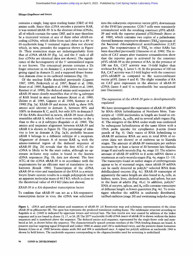
contains a single, long open reading frame (ORF) of 442 amino acids. Since this cDNA encodes a putative RAR, it was termed xRAR-39. It is the shortest of four cDNAs, all of which contain the same ORF, and it may therefore be a truncated version of any of three other xRAR-en-coding cDNAs, which differ from each other in a 40- to 65-nucleotide-long 5'-terminal region (data not shown) which, in turn, precedes the sequence shown in Figure lb . Their restriction maps are indistinguishable from that of cDNA xRAR-39 (Fig. la), and the 3 ' ends of all four cDNAs are identical (data not shown). The significance of the heterogeneity of the 5'-untranslated region is not known. The conceptual protein contains a Zn finger structure in its domain C, a putative nuclear targeting signal in domain D, and a supposed dimer formation domain close to its carboxyl terminus (Fig. lb).
Of the nuclear RARs described previously (Giguere 1987, 1990; Petkovich et al. 1987; Brand et al. 1988; Krust et al. 1989; Ragsdale et al. 1989; Zelent et al. 1989; Kastner et al. 1990), the deduced amino acid sequence of xRAR-39 most closely resembles that of the y^ subtypes of RAR found in mice and humans (Krust et al. 1989; Zelent et al. 1989; Giguere et al. 1990; Kastner et al. 1990) (Fig. 2a). XRAR-39 and mouse RAR 7B show 50% amino acid identity in domain A, between 90% and 100% in domains B, C, and E, but only 33% in domain F. Of the RARs described in newts, xRAR-39 most closely resembles nRAR 5, which itself is more similar to the y than to the a or 3 subtypes (Ragsdale et al. 1989). The amino acid sequence alignment between xRAR-39 and nRAR 8 is shown in Figure 2b. The percentage of identity is low in domain A (Fig. 2a,b), probably because nRAR 8 belongs to a different subtype of 7 receptors. Alignment of domain A of mouse RAR 7B with the amino-terminal region of the deduced sequence of xRAR-39 (Fig. 2b) reveals that the first ATG of the cDNA is likely to be the start codon, although no upstream in-frame stop codon is found in the known cDNA sequences (Fig. lb; data not shown). The first ATG of the cDNA xRAR-39 is in accordance with the requirements for an efficient start of translation in eu-karyotes (Kozak 1986). Transcription of the cDNA xRAR-39 in vitro and translation of the RNA in a reticulocyte lysate system results in a single polypeptide with an apparent molecular mass of 48.5 kD, which is close to the theoretical value of 49.9 kD (data not shown).
XRAR-39 is a RA-dependent tiansciiption factor
To confirm that xRAR-39 can act as a RA-responsive transcription factor in vivo, the cDNA was subcloned
into the eukaryotic expression vector pSVL downstream of the SV40 late promoter. COS-7 cells were transiently cotransfected with the resulting construct pSVL-xRAR-39 and with the reporter plasmid pTK28mult (Brent et al. 1989), which contains two copies of a palindromic thyroid hormone responsive element (TREp) upstream of the bacterial chloramphenicol acetyl transferase (CAT) gene. The responsiveness of TREp to other RARs has been described previously (Umesono et al. 1988). The results of CAT assays after transient cotransfection show that the reporter gene is activated by the construct pSVL-xRAR-39 in the presence of RA. In the presence of 100 UM RA, CAT activity was 15-fold higher than without RA (Fig. 3, lanes 5-8). In the presence of 100 UM RA, CAT activity was 3.5-fold higher with the construct pSVL-xRAR39 as compared to the nonrecombinant vector pSVL (lanes 4 and 8). The slight stimulus of RA on CAT gene expression in the absence of xRAR-39 cDNA (lanes 3 and 4) is reproducible but unexplained (see Discussion).
The expression of the xRAR-39 gene is developmentally regulated
We have investigated the expression of xRAR-39 mRNA by RNA-RNA hybridization of Northern blots. Transcripts of —2500 nucleotides in length are found in embryos, tadpoles, A^ cells, and in several adult organs (Fig. 4). The integrity of the RNA under study was confirmed by subsequent hybridization of the same filters with a DNA probe specific for cytoplasmic p-actin (lower panels of Fig. 4). Only traces of RNA hybridizing to xRAR-39 antisense RNA are seen in total RNA of oocytes (data not shown) or of embryos up to bias tula stages. The amount of xRAR-39 transcripts per embryo increases by at least a factor of 30 between late blastula (stage 9) and early neurula (Fig. 4a; stage 15). The relative amount of xRAR-39 mRNA to p actin mRNA shows a maximum at early neurula stages (Fig. 4a; stages 12-15). The transcripts found at earlier stages of embryogenesis appear to be of maternal origin, since xRAR-39 mRNA can be easily detected in poly(A)'^-selected RNA from defolliculated oocytes (Fig. 4c). XRAR-39 transcripts of apparently the same length are also found in A^ cells, in kidney, testis, liver, skeletal muscle, and spleen, but not in the brain of adults (Fig. 4b, c). In addition, poly(A) + RNA of oocytes, spleen, and Ag cells contains transcripts of different length in lower quantities (Fig. 4c). To investigate whether the mRNA is uniformly distributed, tailbud embryos (stage 28) and swimming tadpoles (stage
Figure 1. cDNA and predicted amino acid sequences of xRAR-39. [A] Restriction map and schematic representation of the clone xRAR-39 in pBluescript SK. The open box represents the predicted translation reading frame. The subdomain structure according to Ragsdale et al. (1989) is indicated by uppercase letters and vertical bars. The first Sau3A site was created by addition of the linker sequence and is not foimd in clones 12, 17, or 26. [B] The 2077-nucleotide £coRI cDNA insert of xRAR-39 is shown without the linker sequences and is numbered above the sequence. The deduced amino acid sequence, represented by the single-letter code and numbered at left, is shown below the sequence. The characteristically spaced cysteine residues of the DNA-binding domain are boxed. A putative nuclear targeting signal (Dingwall and Laskey 1986) at amino acid 150 is underlined twice, and the putative dimer formation domain (Glass et al. 1989) between amino acids 363 and 394 is underlined once. A signal for poly(A) addition at nucleotide 1944 is shown by bold letters. The nucleotide sequence corresponding to the oligonucleotides used for screening is underlined.
96 GENES & DEVELOPMENT
Cold Spring Harbor Laboratory Press on April 5, 2022 - Published by genesdev.cshlp.orgDownloaded from

Xenopus retinoic acid receptor
nRARa
mRAR/3
mRAR 7 g x R A R - 3 9
( D o m a i n A)
9 3
1 0 9
1 3 3
1 4 9
1 7 3
1 8 8
2 1 3
2 2 8
2 5 3
2 6 8
2 9 3
308
333
348
373
388
4 1 3
4 2 8
Y G A A Y D M S
G P G P Q G
A G L P C M
L R R L R K
S S C F C F
A G L G G L
Y D C Y D C
M E S M E A
F V P F P L
G P R R L H P R P L
I M S S S K D R I C S T S T Q L S Q L H G F P P S M Y
A T G
P F A F S S N M R
E S F D P F
A W A G W M
Q P A Q S H
S L Q B M Q
P F D L T N G A Y F R S F P T D L P K E M A S L
S V E I I I S V E
S T S S
I M I S T S S
E E M I I I E E M
V P S I I I
V P S
S P S
I 1 I S P S
p P P
I I I p p p
P P R V
I I I I P P R V
C E G C
M M C E G C
L Q K C
M M L Q K C
K G F
I I I K G F
F Q V
I I F E V
F R R
I I I F R R
S I Q I I I
S I Q
K N M
I I I K N M
V Y T C
M M V Y T C
H R D I I I
H R D
K N C I I I
K N C
C N D
I I I C N D
Q I N
I I I Q I N
K S S
I I I K S S
K V T
I I I K V T
G Y H
I I I G Y H
R N R
I I I R N R
Y G V
I I I Y G V
C Q F
I I : C Q Y
S S
I 1 s s C R
I I C R
G M
I I G M
M E E L
I I I M E A L
I Q K
I I 1 I Q K
V S K
I I I V S K
K E A
I I I K E A
A H Q
I I I A H Q
V R N
I I I V R N
E T F
I I I E T F
D R N K
M M D R N K
K K K
I I I K K K
E I K I I I E I K
E E V i I I E E V
V L P
I D S Y I I I D S Y
E M P
I I I E M P
L W D K
I I I L W H K
A C L D
M M A C L D
P L T D
I I I I
E P E K
M M E P E K
L R G I
M M L R G I
F S E
I I I F S E
I L M
I I I I L M
L V F
! I I L V F
V E K
I : I V D K
S T K
I I I S T K
L S T
I : I L A T
L R I
I I I L R I
S F A
: I I A F A
L Q E
I I I L Q E
G A E
I I I G A E
K C I
I I I K C I
C T R
I I I C T R
D Q L
: ! I E Q L
I K I
I I I I K I
Y T P
i I I Y T P
L P L I ! !
P S L C
M M P S L C
V E F A
M M V E F A
E Q D T
M M E Q D T E M D D
M M
Q L G
I I I Q L G
K R L
I I I K R L
M T F
I I I M T F
T E T
I ! ! L P L E M D D T E T
E G L I I E A L
K Y T
I I I K Y T P G F
I I I P G F S D G
I I I S D G G L L
! ! ! G L L P L L
I I I P L L R A I
I I I R A I
K F Y A I I I K I Y A
R R R I I I R R R
T L K I I I T L K
L E I P M M M E I P
G P M I I I G P M
R P D I I R P N
P P L i I I P P L
T N S
I I I T N S T T L
: I I A T L L T L
I I I L T L S A I
! ! ! S A I K P Y
I I I K P Y I R E I I I I R E
S A D I I I S A D
T I A
I I I T I A N R T
I I I N R T C L I I I ! C L i
M F P
I I I M F P
I I I
Q R V
I I H R V D Q I
I I I D Q I Q M H
I I I Q M H C G D I I ] C G b R M L
I I I R M L N P E
I I N P G
Q L D
I I I Q L D T L L
I I I T L L N A G I I I N A G
R M D
! I I R M D M K I
I I I M K I
K P S I
P K S
E R S
I E Q
S S E
K P I
S S N
K V E I I
G E D I I G E K
S K T : I T K D
L G
I I L G K S
I : K A F G
I I F G L E I ! L E T D
I I T D
I I
T Q I I T Q
P E I I P E
D G I D D
Figure 2. Primary amino acid sequence comparison of xRAR-39 and members of the RAR subfamily, [a] Primary sequences were subdivided in domains according to Ragsdale et al. (1989) and aligned using the computer program Microgenie (Beck-mann). Regions of similarity between xRAR-39 and other RARs are presented schematically as percent amino acid identity. (nRAR a, nRAR 8) Newt RAR a and RAR 8 (Ragsdale et al. 1989); (mRAR 7B) mouse RAR 7B (Kastner et al. 1990). (i>) Sequence alignment of mouse RAR 7B (only domain A is shown), xRAR-39, and newt RAR 8 (nRAR). In domain A, amino acids identical in mouse RAR 7B and in xRAR-39 are shown in bold letters. Domains A, C, and E are boxed.
39) were dissected, and mRNA was extracted from the anterior (A), middle (M), and posterior (P) thirds. The Northern blot reveals that at both stages head and tail fragments contain more xRAR-39 mRNA than the fractions from the midbody (Fig. 4d,e). Northern hybridization with full-length antisense RNA (Fig. 4d), with a 3 ' -specific antisense RNA (data not shown), and with a cDNA fragment specific of the 5' end (Fig. 4e) gave very similar results.
XRAR-39 shows a region-specific, but not germ layer-specific, expression pattern
A more detailed analysis of the location of xRAR-39 mRNA in embryos and tadpoles has been performed by in situ hybridization of antisense RNA on tissue sections. At gastrula stages lOVi and 11, the mRNA is
mainly found in the ectoderm and mesoderm, and it is slightly more abundant at the prospective dorsal as compared to the ventral side (data not shown). By the end of gastrulation, the mRNA is most highly concentrated posteriorly in the dorsal blastopore lip, and the ventral lip is also significantly labeled by xRAR-39 antisense RNA (Fig. 5a) but not by sense RNA (Fig. 5c). Significant levels of label are also found in the anterior region, as seen on the sagittal section shown in Figure 5 a. On parasagittal sections, label is seen mainly in the nonecto-dermal layers below and in front of the neural plate (Fig. 5b and data not shown). At later stages of neurulation, the two maxima of expression at the ends of the embryo are more pronounced (Fig. 6). Anteriorly, most of the xRAR-39 mRNA is found in the head mesoderm and in the archenteron roof, whereas the anterior part of the neural plate is almost unlabeled. Figure 6a shows a
GENES & DEVELOPMENT 97
Cold Spring Harbor Laboratory Press on April 5, 2022 - Published by genesdev.cshlp.orgDownloaded from

Ellinget-Ziegelbauer and Dieyer
« 0 ,
pSVL 1
100 , RA(rtVI)
pSVL-xfW?
pTK28mdt
Figure 3. RA induces pSVL-xRAR-39-dependent transcription of a TREp-CAT reporter in COS-7 cells. The reporter pTK28mult (Brent et al. 1989) was cotransfected into COS-7 cells with the expression plasmid pSVL-xRAR-39 (lanes 5-8\ or the vector pSVL (lanes 3 and 4). After transfection, cells were treated without (lanes 3 and 5), or with 1, 10, or 100 nM RA as indicated at bottom. (Lane 1] Blank control; (lane 2) CAT assay with 8 units of purified enzyme. Activation of the CAT reporter gene was assayed by incubating cell extracts with [''*C]chlor-amphenicol. Chloramphenicol and its acetylated products were separated by thin-layer chromatography and detected by autoradiography.
transverse section through a neurula of stage 17, at the level of the prospective eyes, which are separated by prechordal mesoderm. Whereas the eye anlagen are not significantly labeled, a strong signal is seen in the subjacent archenteron roof and in the prechordal mesoderm. The epidermis and the lateral mesoderm are labeled more weakly. A transverse section from the midbody (Fig. 6b) shows a higher concentration of xRAR-39 mRNA in the neural plate as compared to the adjacent ectoderm. The archenteron roof is also significantly labeled, whereas labeling of the notochord and the somitogenic mesoderm is hardly above background. Sagittal sections of neurulae of stage 17 (data not shown) and of stage 20 (Fig. 6c) reveal that the anterior zone of maximal xRAR-39 expression comprises head mesoderm and endoderm. The labeled endodermal derivative is the anterior archenteron roof, including the mesoderm-free zone in front of the prosencephalic anlage, where the endoderm is directly underlying the ectoderm (Nieuwkoop and Florschiitz 1950; Hausen and Riebesell 1991). The labeled head mesoderm is the prechordal plate subjacent to the prospective forebrain. In contrast, the differentiating notochord (Fig. 6c) and somites (Fig. 6d) are almost unlabeled. The archenteron floor and the vegetal yolk mass do not contain significant amounts of xRAR-39 mRNA. Posteriorly, most of the mRNA is found in the circumblasto-poral collar (Fig. 6c). In this region all three germ layers are labeled: the endoderm forming the archenteron roof, the undifferentiated mesoderm, and the posterior neuroectoderm (Fig. 6c). On posterior transverse sections of neurulae of stage 17, the blastoporal endoderm appears to be unlabeled (data not shown). The anterior and the posterior area of xRAR mRNA expression are apparently
not delimited by defined borders; rather the intensity of the signal decreases gradually toward the midbody (Fig. 6c).
In early tadpoles (stage 31} the xRAR-39 mRNA is found in head mesenchyme but is at the limit of detection in the brain and the eye (Fig. 7a). At stage 25 (data not shown) and at stage 31 the highest concentration of xRAR-39 mRNA is seen in the tailbud (Fig. 7c), which is derived from the most posterior dorsal region of the neurula (Davidson 1988). Likewise in the tailbud (Fig. 7c), the expression of xRAR-39 is apparently not germ layer-specific. In the midbody, at stage 25 as well as at stage 31, the hybridization signal of the antisense RNA (Fig. 7b) is hardly stronger than that of the sense RNA control (not shown) and does not appear to be locally restricted. This result is in agreement with the results of Northern blots showing the low abundance of xRAR-39 mRNA in the midbody (Fig. 4d,e).
Discussion
The cDNA xRAR-39 encodes a RA-dependent transcription factor
Four independent isolates of slightly different cDNAs of a neurula stage 17 library all contain the same ORF (Fig. 1), which encodes a RAR most similar to the RARs of the subtype 73 (Fig- 2.), which have been described previously in humans and in mice (Krust et al. 1989; Zelent et al. 1989; Giguere et al. 1990; Kastner et al. 1990). For a functional assay, COS-7 cells were cotransfected with an expression plasmid containing the cDNA xRAR-39 and the reporter plasmid pTK28 mult (Fig. 3). Addition of 10"'' M RA led to a 15-fold increase of the reporter gene activity. This compares to a factor of 5 reported for newt RAR 6 (Ragsdale et al. 1989) and a factor of 20 found for mouse RAR y^ (Giguere et al. 1990). We find that the CAT gene activity of the reporter is also slightly stimulated by RA in the presence of the nonrecombinant expression plasmid pSVL (Fig. 3). Comparison of RA-dependent CAT activity in the presence or absence of the xRAR-encoding gene in pSVL leads to the minimum estimate of a 3.5-fold receptor-dependent activation. A certain degree of RA-dependent activation of reporter genes in the absence of an exogenous receptor-encoding gene has been reported previously (Petkovich et al. 1987; Graupner et al. 1989; Sucov et al. 1990); however, controls with the nonrecombinant expression vector, but in the presence of RA, are not always shown. Recently, Sucov et al. (1990) have presented evidence that several widely used cell lines, including CV-1 cells, the progenitors of COS cells, express a low level of endogenous RAR. We have selected COS-7 cells because they were previously reported to be free of receptors for T3 and for RA and because they replicate and express exogenous genes in SV40-derived vectors very efficiently (Glass et al. 1989). The observed effect may also be related to the high copy number of SV40-derived vectors achieved after transient transfection, which might lead to an artificial derepression of certain genes. Despite this difficulty, we
98 GENES & DEVELOPMENT
Cold Spring Harbor Laboratory Press on April 5, 2022 - Published by genesdev.cshlp.orgDownloaded from

Xenopus retinoic acid receptor
1 5 8 9 10 11 12 15 ?0 ?4 28 38 Ar, K T B O A.; K L M
18S»
28S«
18S»
1 2 3 4 5 6
-* * •
1 2 3
iNif V ^ IJF ^ ^
_ A M P A M P A M P
Figure 4. Northern blot analysis of xRAR-39 mRNA in X. laevis embryos and tissues of adults. Full-length xRAR-39 antisense RNA was hybridized to total RNA of embryos of the stages as indicated above each lane [a); total RNA [b], and poly(A)+ RNA of tissue culture cells and of adult tissues [c], and total RNA of parts of dissected embryos {d, e). Embryos in a, d, and e were staged according to Nieuwkoop and Faber (1967). (Stage 1) Fertilized egg; (stage 5) 16-cell stage; (stages 8 and 9) blastula; (stages 10-12) gastrula; (stages 15-20) neurula; (stages 24-28) tailbud; (stages 38 and 39) early tadpoles. The sources of RNA analyzed in b and c were Ag cells (A )̂, kidney (K), testis (T), brain (B), defolhculated oocytes of stages I-III (Dumont 1972) (O) liver (L), skeletal muscle (M), and spleen (S). [d and e) The anterior (A), middle (M), or posterior (P) thirds of embryos of stage 28 (lanes 1-3] or stage 39 (lanes 4-6) were dissected, (e) The probe was a 5'-specific cDNA fragment that contained the first 157 bp upstream of the first Sphl site (Fig. la). Each lane contained the amount of total RNA equivalent to 1.5 embryos [a, d, and e], or 10 |i,g of total RNA [b], or ~5 |xg of poly(A) +-selected RNA (c). The positions of 28S and 18S rRNAs are indicated by arrowheads. Bottom panels show the result of subsequent hybridization of each filter with a DNA probe specific for cytoplasmic p-actin.
have reproducibly observed a RA-dependent activation of the reporter CAT gene. The effective concentrations of RA were between 10~' and 10"^ M. This is in the range of effective concentrations reported for other nu
clear RARs (Petkovich et al. 1987; Giguere et al. 1987; Ragsdale et al. 1989). Endogenous RA has been estimated to occur at a concentration of 1.5 x 10"'^ M in embryos of X. laevis (Durston et al. 1989).
Figure 5. In situ hybridization of xRAR-39 anti-sense RNA to sections of X laevis late gastrula (stage 12.5). [a] Sagittal section photographed with bright-field illumination after staining with azure B [left] and with epipolarization to show the hybridization signals [liglit]. [b] The dorsal anterior part of a parasagittal section was photographed with polarized UV light and dimmed translucent light, (c) Dorsal posterior part of a sagittal section hybridized with the control sense xRAR-39 probe, photographed with epipolarization. (A) anterior; (AR) archenteron; (EN) endoderm; (DL) dorsal lip; (NP) neural plate; (P) posterior; (VL) ventral lip; (YP) yolk plug. Bars, 100 jxm.
GENES & DEVELOPMENT 99
Cold Spring Harbor Laboratory Press on April 5, 2022 - Published by genesdev.cshlp.orgDownloaded from

Ellinger-Ziegelbauer and Dreyer
Figure 6. Localization of xRAR-39 transcripts on sections of neurulae. [a] Anterior transverse section of a neurula of stage 17 at the level of the eye anlagen. (b) Dorsal part of a transverse section of the trunk region of the same embryo (stage 17). (c) Sagittal section of a neurula of stage 20. [d] Detail of a parasagittal section of the same embryo (stage 20), showing the somites, [a and b, left) c, top). Bright-field illumination, {a and b, light) C; bottom) hybridization signals shown by epipolarization. [d] Hybridization signals and morphology shown simultaneously. (A) anterior; (AR) archenteron? (BP) blastoporus; (E) eye anlagen; (EN) endoderm; (D) dorsal; (N) notochord; (P) posterior; (PE) prosencephalic anlagen; (PM) prechordal mesoderm; (S) somites; (V) ventral. Bars, 100 jxm. 100 GENES & DEVELOPMENT
Cold Spring Harbor Laboratory Press on April 5, 2022 - Published by genesdev.cshlp.orgDownloaded from

Xenopus letinoic acid receptor
Figure 7. XRAR-39 mRNA distribution in early tadpoles (stage 31). Transverse sections at the level of the eyes {a], the trunk {b], and the tip of the tail (c) are shoM n̂ with bright-field illumination [left] and v^ith epipolarization {right). The hybridization signals on sense controls (not shown) were in intensity comparable to those over the brain and the eyes in a. (B) brain; (DT) digestive tract; (E) eye. Bars, 100 |xm.
Expression of xRAR-39 is found at both ends of the anteroposterior axis
Northern blots and in situ hybridization have revealed a defined spatial and temporal pattern of expression of xRAR-39. After the midblastula transition the small amount of maternal xRAR-39 mRNA is greatly increased due to zygotic transcription of the corresponding gene (Fig. 4a). The transcripts are much shorter than those of RAR 8 found in the limb buds of newts (Rags-dale et al. 1989). This may reflect differences between species and between different subclasses of RAR y. By the end of gastrulation, both an anterior and a posterior area of abundant expression of xRAR-39 begin to emerge (Fig. 5). These two distinct maxima become much more pronounced during neurulation (Fig. 6) and can be followed at least up to early tadpole stages (Figs. 4d,e and 7). Since we see transcripts of apparently uniform size on Northern blots and since their length is the same in anterior and posterior parts of tailbud embryos (Fig. 4), we suppose that the same species of xRAR y is expressed at both ends of the anteroposterior axis. To confirm that the transcripts that hybridize to full-length antisense RNA are specific of xRAR-39, we have repeated some experiments with probes lacking any sequences specific of the highly conserved domain C. Northern blots of dissected tailbud embryos have also been probed with anti-sense RNA representing the 3'-end downstream of the Sail site (data not shown), and with a cDNA representing the 5'-untranslated region and part of domain A (Fig. 4e). In situ hybridization of sagittal sections of stage 20 embryos with an antisense RNA specific for the 3'-SaiI/£coRI fragment of xRAR-39 also showed the localization of xRAR-39 transcripts at both ends of the anteroposterior axis (data not shown). Although the controls showed that the full-length antisense RNA bound specifically to xRAR-39 transcripts, we cannot exclude the presence of differentially spliced variants of the same mRNA, which nevertheless might have the same length.
Although a more detailed analysis of the expression of xRAR-39 in frog embryogenesis is required, the
emerging picture strikingly resembles that of the restricted expression of RAR 7 in the mouse embryo (for details, see Ruberte et al. 1990).
The effects of exogenous RA on embryogenesis might be mediated by xRAR-39
Treatment of Xenopus embryos with a pulse of RA before neurulation leads to disorders at both the anterior and the posterior end of the tadpole. The head anlagen appear to be most sensitive to RA, which causes truncation of anterior facial structures (Durston et al. 1989; Sive et al. 1990) and microcephaly due to formation of hindbrain at the expense of forebrain (Durston et al. 1989). RA-treated embryos that have no pigmented eyes also frequently have a disturbed zone in their tail (Fig. 1 in Durston et al. 1989; C. Dreyer, unpubl.).
The timing and the local restriction of the burst of zygotic expression of xRAR-39 during gastrulation and neurulation of X. laevis make it likely that this subtype of RAR is involved in mediating the effects of endogenous and artificially applied RA (Durston et al. 1989; Sive et al. 1990). Our findings on the predominant expression of xRAR-39 at both ends of the embryo may explain the relative resistance of the trunk region to RA. The effects of low doses of RA are possibly caused by the action of RA on cells that are susceptible due to the presence of receptors but are normally distant from the putative source of endogenous retinoids (Thaller and Ei-chele 1990; Wagner et al. 1990). The much higher RA sensitivity of the head anlagen as compared to the tail, despite comparable amounts of xRAR-39 mRNA at both ends of the embryo (Figs. 4d,e and 6c), could be explained if an endogenous source of retinoids were normally located closer to the posterior end of the embryo, at least during the RA-sensitive phase of specification of the anteroposterior axis (Durston et al. .1989; Sive et al. 1990). Moreover, the local concentration of free RA might be regulated by the abundance of CRABP (Chytyl and Ong 1984; Eichele 1989) or of RA-metabolizing enzymes. In addition, the same receptor in the presence or absence of
GENES & DEVELOPMENT 101
Cold Spring Harbor Laboratory Press on April 5, 2022 - Published by genesdev.cshlp.orgDownloaded from

Ellinger-Ziegelbauer and Dreyer
its ligand could have antagonistic effects, as documented in the instance of the thyroid hormone receptor (Ban-iahmad et al. 1990). Alternatively, the tail structures may be less affected by pulses of RA given at an early stage because they differentiate later than the affected head structures (Davidson 1988).
Externally visible effects of low doses of RA are diminished or even missing cement glands, noses, and eyes (Durston et al. 1989; Sive et al. 1990; C. Dreyer, unpubl.). Gastrula ectoderm can be induced to express cement gland markers by combination with mesoderm in vitro (Sive et al. 1989, 1990). In situ, the mechanism of cement gland specification might be different, the more so since prolonged contact of mesoderm with "precement gland" in vitro changes the fate of the latter to neural tissue (Sive et al. 1989, 1990). In the embryo, the cement gland is induced in the most anterior ectoderm in a mesoderm-free zone, where the endoderm of the archenteron roof is directly underlying the ectoderm (Nieuwkoop and Florschutz 1950; Hansen and Riebesell 1991). Since the anterior archenteron roof expresses the xRAR-39 mRNA so abundantly, the inhibitory effect of RA on the differentiation of the cement gland might be mediated by the directly underlying endoderm. Substantial contributions of the anterior endoderm to the induction of the nose and the lens have been reported previously (Jacobson 1963).
Neural induction in amphibians is believed to occur in two steps. First, the involuting mesoderm induces the overlying competent ectoderm to become anterior-specified neural tissue. Subsequently, most of the induced neural tissue is transformed to become posterior neural tissue (Eyal-Giladi 1954; Nieuwkoop 1973, 1985). It is most likely the second step, neural transformation, that is affected when exogenous RA is applied to embryos of X. laevis before early neurula stages (Durston et al. 1989). Since we find expression of xRAR-39 mRNA mainly in the cell layers adjacent to the anterior neural anlagen, the effect of RA on neural transformation might be mediated by the subjacent mesoderm. This notion is in agreement with many findings suggesting that the regional specification of the nervous system is directed by the underlying mesoderm (Eyal-Giladi 1954; Toivonen and Saxen 1968; Nieuwkoop 1973, 1985). Alternatively, xRAR-39 might be expressed in the anterior part of the neural plate before it is specified. Of course we cannot exclude the simultaneous presence of other types of RAR, whose mRNA might be of a different structure (Mangelsdorf et al. 1990) and not hybridize to xRAR-39 antisense RNA.
Potential target genes might be regulated by RAR according to a positional value in the embryo
The finding that expression of xRAR-39 is apparently region-specific but not germ layer-specific implies that potential target genes of the nuclear receptor of RA may be regulated according to a positional value along the anteroposterior axis, irrespective of the germ layer. The local concentration of endogenous RA and of a given
RAR might together specify a positional value. Certain homeo box-containing genes have been discussed as potential targets of RA (Durston et al. 1989; Wright et al. 1989, and references therein; Sive et al. 1990). Some homeo box genes are expressed in Xenopus embryos in a spatial pattern (Condie and Harland 1987; Ruiz i Altaba and Melton 1989; Wright et al. 1989) that could be explained if they were regulated by a factor such as RAR, which occurs in both the ectoderm and the mesoderm, but has restricted expression along the anteroposterior axis (Wright et al. 1989; Sive et al. 1990).
Materials and methods
Screening of cDNA library and cDNA sequence analysis
A mixture of four different 4-S-mer consensus oligonucleotides was derived of previously described cDNA sequences. The structure was 5'-GAAGAAGICCTTGCAGCCCTCACAG-GIG^^GACCCCATAGTGGTAGCC. Oligonucleotides were end-labeled with [7-3^P]ATP and T4-polynucleotide kinase (New England Biolabs). It was used to screen a XgtlO X. laevis neurula cDNA library (Kintner and Melton 1987) at low stringency. Hybridization was at 42°C in 20% formamide, 5 x SSPE, 5x Denhardt's, 50 niM sodium phosphate (pH 7.0), 0.1% SDS, 10 mM sodium pyrophosphate, and 0.1% herring sperm DNA [20 X SSPE contains 3.6 M NaCl, 0.225 M NaH2P04, 0.175 M NaOH, and 20 mM EDTA; 50 x Denhardt's is 1 % bovine serum albumin (BSA), 1% polyvinylpyrrohdone, and 1% FicoU]. The filters were washed with 4x SSPE, 0.1% SDS, at 54°C. Of 22 positive clones that were isolated, 14 were shown to be structurally related by cross hybridization under stringent conditions. The hybridization probe was the Pstl-Sall fragment of clone 39 (Fig. la), and it was labeled by oligonucleotide-primed synthesis (Feinberg and Vogelstein 1983). cDNAs were sub-cloned into pBluescript SK vectors (Stratagene), restriction mapped, and overlapping restriction fragments of appropriate sizes were subcloned into M13mpl8 and M13mpl9 sequencing vectors. DNA sequences were determined from single-stranded M13 or double-stranded plasmid DNA by the dideoxy method with Sequenase (U.S. Biochemicals) and analyzed by the computer program Microgenie (Beckmann).
Transient cotransfection of COS-7 cells
The expression vector pSVL-xRAR-39 contains the complete cDNA insert of xRAR-39 under the control of the SV40 late promoter in pSVL (Pharmacia). To construct the expression vector pSVL-xRAR-39, the cDNA xRAR-39 was excised from a Bluescript SK vector with Xbal and Hifldlll, and after refilling of the Hindlll site, the cDNA was ligated into pSVL (Pharmacia), using the Smal and Xbal restriction sites of the vector. The reporter plasmid pTK28mult (Brent et al. 1989) contains two copies of the palindromic TREp upstream of the thymidine kinase promoter linked to the bacterial CAT gene. COS-7 cells (3 X 10^/60-mm^ tissue culture dish) were seeded in Dul-becco's modified Eagle medium (DMEM) supplemented with 0.02 U/ml insulin, 4 mM glutamine, and 10% fetal calf serum (GEBCO). Twenty-four hours later, cells were transiently co-transfected with 0.5 ml per dish of 0.25 mg/ml DEAE-dextran (Sigma) in Tris-buffered saline, containing 2 |xg pSVL (Pharmacia) or 2 |xg of expression vector pSVL-xRAR-39, 2 |xg of the reporter plasmid pTK28mult, and 1 fjig of the p-galactosidase expression vector pCHllO (Pharmacia), which was used to control transfection efficiency. After 30 min incubation at room
102 GENES & DEVELOPMENT
Cold Spring Harbor Laboratory Press on April 5, 2022 - Published by genesdev.cshlp.orgDownloaded from

Xenopus retinoic acid receptor
temperature, cells were treated with 15% dimethylsulfoxide (DMSO) in DMEM for 2 min. Cells were then incubated with DMEM containing 10% FCS and 0.1 mM chloroquine (Sigma) at 37°C for 3 hr, subsequently with DMEM containing 10% of charcoal-stripped FCS for 24 hr and, finally, for 24 hr with DMEM with or without RA as indicated. Cell extracts were assayed for CAT activity as described by Gorman et al. (1982).
RNA isolation and Noithem blots
Total RNA from staged embryos (Nieuwkoop and Faber 1967) was extracted as described by Krieg and Melton (1984). Total RNA of adult tissues was prepared by a LiCl-urea method (Fey and Hansen 1990) and selected for poly(A)+ RNA on oligo(dT)-cellulose (Pharmacia). RNA was separated on denaturing formaldehyde agarose gels and transferred to GeneScreen nylon filters. Hybridization of ^^P-labeled antisense RNA was performed overnight at 58°C in a buffer containing 50% form-amide, 10% dextran sulfate, 10 x Denhardt's solution, 1 M NaCl, 1% SDS, and 0.15 mg/ml salmon sperm DNA. The filters were then washed three times with 2x SSC, 0.5% SDS, and twice with 0.1 x SSC, 0.1% SDS at 70°C. Exposure to X-ray films was at - 70°C with intensifying screens. A genomic DNA probe specific of X. laevis cytoplasmic p-actin was ^^P-labeled by oligonucleotide-primed synthesis (Feinberg and Vogelstein 1983). Hybridization of DNA probes was at 42°C; washing was at 55°C.
In situ hybridization
Embryos were fixed with 3% paraformaldehyde for 1 hr at room temperature, embedded in acrylamide (Hausen and Dreyer 1981), and sectioned on a cryostat. Hybridization of [̂ ^SjUTP-labeled full-length antisense or sense RNA (4 x 10* to 5 x 10* cpm/770-mm^ coverslip in 50 |JL1 of solution) was essentially as described (Poting et al. 1990), except that the wash in buffer containing 50% formamide was at 55°C. Exposure was on Ilford L4 nuclear emulsion for 10 days (Figs. 6 and 7) or on Ilford K5 for 4 days (Fig. 6). After film development, sections were coun-terstained with 0.25% azure B in 0.1 M phosphate buffer (pH 4.0) for 4 min, and photographed on a Zeiss axioplan photomi-croscope. Silver grains were visualized in the dark field by epi-polarization. In some instances sections were photographed with dimmed bright-field illumination in combination with polarized UV light to facilitate the superposition of morphology and hybridization signals.
Acknowledgments
We thank Brigitte Glaser and Conny Stoltz for excellent technical assistance, Dr. D.A. Melton for the X. laevis neuiula cDNA library. Dr. D. Tautz for oligonucleotide synthesis, Dr. D.D. Moore for the plasmid pTK28mult, Dr. G. Spohr for a Xenopus p-actin DNA probe, Dr. R. Stick for oocyte polyA+ RNA, Drs. Stefan Jentsch, Doris Wedlich, and Metta Riebesell for helpful suggestions, Drs. Peter Hausen, Nancy Hopkins, Amo MuUer, David Stott, and Rudolf Winklbauer for comments on the manuscript, Margot Heller for typing it, and Roswitha Gromke-Lutz and Doris Eder for help with the figures.
The publication costs of this article were defrayed in part by payment of page charges. This article must therefore be hereby marked "advertisement" in accordance with 18 USC section 1734 solely to indicate this fact.
References
Baniahmad, A., C. Steiner, A.C. Kohne, and R. Renkawitz. 1990. Modular structure of a chicken lysozyme silencer: Involvement of an unusual thyroid hormone receptor binding site. Ceii 61: 505-514.
Brand, N., M. Petkovich, A. Krust, P. Chambon, H. de The, A. Marchio, P. Tiollais, and A. Dejean. 1988. Identification of a second human retinoic acid receptor. Nature 332: 850—853.
Brent, G.A., J.W. Harney, Y. Chen, R.L. Warne, D.D. Moore, and P.R. Larsen. 1989. Mutations of the rat GH promoter which increase and decrease response to thyroid hormone define a consensus thyroid hormone response element. Mol. Endocrinol. 3: 1996-4004.
Chytyl, F. and D.E. Ong. 1984. Cellular retinoic acid binding proteins, in The retinoids (ed. M.B. Sporn, A.B. Roberts, and D.S. Goodman), vol. 2, pp. 89-123. Academic Press, Orlando, Florida.
Condie, B.G. and R.M. Harland. 1987. Posterior expression of a homeobox gene in early Xenopus embryos. Development 101:93-105.
Davidson, D. 1988. Segmentation in frogs. Development 1048:221-229.
Dingwall, D. and R.A. Laskey. 1986. Protein import into the cell nucleus. Annu. Rev. Cell Biol. 2: 367-390.
Dumont, J.N. 1972. Oogenesis in Xenopus laevis (Daudin). I. Stages of oocyte development in laboratory maintained animals. /. Morphol. 136: 153-180.
Durston, A.J., J.P.M. Timmermans, W.J. Hage, H.F.J.Hendriks, N.J. de Vries, M. Heideveld, and P.D. Nieuwkoop. 1989. Retinoic acid causes an anteroposterior transformation in the developing central nervous system. Nature 340: 140-144.
Eichele, G. 1989. Retinoids and vertebrate limb pattern formation. Trends Genet. 5: 246-251.
Evans, R.E. 1988. The steroid and thyroid hormone receptor su-perfamily. Science 240: 889-895.
Eyal-Giladi, H. 1954. Dynamic aspects of neural induction in amphibia. Arch. Biol. 65: 179-259.
Feinberg, A.P. and B. Vogelstein. 1983. A technique for radiola-belling DNA restriction endonuclease fragments to a high specific activity. Anal. Biochem. 123: 13-16.
Fey, J. and P. Hausen. 1990. Appearance and distribution of laminin during development of Xenopus laevis. Development 42: 144-152.
Giguere, V., E.S. Ong, P. Segui, and R.M. Evans. 1987. Identification of a receptor for the morphogen retinoic acid. Nature 330: 624-629.
Giguere, V., M. Shago, R. Zimgibl, P. Tate, J, Rossant, and S. Varmuza. 1990. Identification of a novel isoform of the retinoic acid receptor gamma expressed in the mouse embryo. Mol. Cell Biol. 10: 2335-2340.
Glass, C.K., S.M. Lipkin, O.V. Devary, and M.G. Rosenfeld. 1989. Positive and negative regulation of gene transcription by a retinoic acid-thyroid hormone receptor heterodimer. Cell 59: 697-708.
Gorman, CM., L.F. Moffat, and B.H. Howard. 1982. Recombinant genomes which express chloramphenicol acetyltrans-ferase in mammalian cells. Mol. Cell. Biol. 2: 1044-1051.
Graupner, G., K.N. Wills, M. Tzukerman, K. Zhang, and M. Pfahl. 1989. Dual regulatory role for thyroid-hormone receptors allows control of retinoic-acid receptor activity. Nature 340: 653-656.
Hausen, P. and C. Dreyer. 1981. The use of polyacrylamide as an embedding medium for immunohistochemical studies of embryonic tissues. Stain Technol. 56: 287-293.
Hausen, P. and M. Riebesell. 1991. The early development of Xenopus laevis. An atlas of histology. Springer-Verlag,
GENES & DEVELOPMENT 103
Cold Spring Harbor Laboratory Press on April 5, 2022 - Published by genesdev.cshlp.orgDownloaded from

Ellinget-Ziegelbauei and Dieyer
Berlin/Heidelberg/New York. (In press.) Jacobson, A.G. 1963. The determination and positioning of the
^ nose, lens and ear. II. The role of endoderm. /. Exp. Zool. 4 : 2 8 5 - 2 9 1 .
Kastner, P., A. Krust, C. Mendelsohn, J.M. Gamier, A. Zelent, P. Leroy, and A. Staub. 1990. Murine isoforms of retinoic acid receptor gamma with specific patterns of expression. Pioc. Natl. Acad. Sci. 78: 2700-2704.
Kintner, C.R. and D.A. Melton. 1987. Expression of Xenopus N-CAM RNA in ectoderm is an early response to neural induction. Development 99: 311-325.
Kozak, M. 1986. Point mutations define a sequence flanking the AUG initiator codon that modulates translation by eu-karyotic ribosomes. Cell 44: 283-292 .
Krieg, P. and D.A. Melton. 1984. Functional messenger RNAs are produced by SP6 in vitro transcription of cloned cDNAs. Nucleic Acids Res. 12: 7057-7070.
Krust, A., P. Kastner, M. Petkovich, A. Zelent, and P. Chambon. 1989. A third human retinoic acid receptor, hRAR-gamma. Pioc. Natl. Acad.Sci. 86: 5310-5314.
Mangelsdorf, D.J., S.E. Ong, J.A. Dyck, and R.M. Evans. 1990. Nuclear receptor that identifies a novel retinoic acid response pathway. Nature 345: 224-229.
Nieuwkoop, P.D. 1973. The "organization center" of the amphibian embryo: Its origin, spatial organization and morpho-genetic action. /. Morphol. 10: 1-37 .
. 1985. Inductive interactions in early amphibian development and their general nature. /. Embiyol. Exp. Morphol. 89S: 333-347.
Nieuwkoop, P.D. and P.A. Florschiitz. 1950. Quelques carac-teres speciaux de la gastrulation et de la neurulation de I'oeuf de Xenopus laevis, Daud. et de quelques autres An-oures. Arch. Biol. 61 : 113-150.
Nieuwkoop, P.D. and J. Faber. 1967. Normal table of Xenopus laevis (Daudin). North-Holland Publishing Company, Amsterdam.
Petkovich, M., N.J. Brand, A. Krust, and P. Chambon. 1987. A human retinoic acid receptor which belongs to the family of nuclear receptors. Nature 330: 444-450.
Poting, A., K. Danker, L. Hartmann, M. Koster, D. Wedlich, and W. Knochel. 1990. Two different mRNAs coding for identical elongation factor EF-la polypeptides are present in early embryos of Xenopus laevis. Differentiation 44: 1 0 3 -110.
Ragsdale, C.W., M. Petkovich, P.B. Gates, P. Chambon, and J.M. Brockes. 1989. Identification of a novel retinoic acid receptor in regenerative tissues of the newt. Nature 341: 6 5 4 -657.
Ruberte, E., P. Dolle, A. Krust, A. Zelent, G, Morriss-Kay, and P. Chambon. 1990. Specific spatial and temporal distribution of retinoic acid receptor gamma transcripts during mouse embryogenesis. Development 108: 213-222.
Ruiz i Altaba, A. and D.A. Melton. 1989. Bimodal and graded expression of the Xenopus gene Xhox3 during embryonic development. Development 106: 173-183.
Sive, H.L., K. Hattori, and H. Weintraub. 1989. Progressive determination during formation of the anteroposterior axis in Xenopus laevis. Cell 58: 171-180.
Sive, H.L., B.W. Draper, R.M. Harland, and H. Weintraub. 1990. Identification of a retinoic acid-sensitive period during primary axis formation in Xenopus laevis. Genes Dev. 4: 9 3 2 -942.
Spom, M.B. and A.B. Roberts 1983. Role of retinoids in differentiation and carcinogenesis. Cancer Res. 43: 3034-3039.
Sucov, H.M., K.K. Murakami, and R.M. Evans. 1990. Characterization of an autoregulated response element in the mouse
retinoic acid receptor type p gene. Proc. Natl. Acad. Sci. 87: 5392-5396.
Thaller, C. and G. Eichele. 1987. Identification and spatial distribution of retinoids in the developing chick limb bud. Nature 327: 625-628.
. Isolation of 3,4-didehydroretinoic acid, a novel morpho-genetic signal in the chick wing bud. Nature 345: 815-819.
Toivonen, S. and L. Saxen. 1968. Morphogenetic interaction of presumptive neural and mesodermal cells mixed in different ratios. Science 159: 539-540.
Umesono, K., V. Giguere, C.K. Glass, M.G. Rosenfeld, and R.M. Evans. 1988. Retinoic acid and thyroid hormone induce gene expression through a common responsive element. Nature 336: 262-265.
Wagner, M., C. Thaller, T. Jessell, and G. Eichele. 1990. Polarizing activity and retinoid synthesis in the floor plate of the neural tube. Nature 345: 819-822.
Wolf, G. 1984. Multiple functions of vitamin A. Physiol. Rev. 64: 873-937.
Wright, C.V.E., K.W.Y. Cho, G. Oliver, and E.M. De Robertis. 1989. Vertebrate homeodomain proteins: Families of region-specific transcription factors. Trends Biochem. Sci. 14: 5 2 -56.
Zelent, A., A. Krust, M. Petkovich, P. Kastner, and P. Chambon. 1989. Cloning of murine a and p retinoic acid receptors and a novel receptor gamma predominantly expressed in skin. Nature 339: 714-717.
104 GENES & DEVELOPMENT
Cold Spring Harbor Laboratory Press on April 5, 2022 - Published by genesdev.cshlp.orgDownloaded from

10.1101/gad.5.1.94Access the most recent version at doi: 5:1991, Genes Dev.
H Ellinger-Ziegelbauer and C Dreyer Xenopus laevis.A retinoic acid receptor expressed in the early development of
References
http://genesdev.cshlp.org/content/5/1/94.full.html#ref-list-1
This article cites 45 articles, 13 of which can be accessed free at:
License
ServiceEmail Alerting
click here.right corner of the article or
Receive free email alerts when new articles cite this article - sign up in the box at the top
Copyright © Cold Spring Harbor Laboratory Press
Cold Spring Harbor Laboratory Press on April 5, 2022 - Published by genesdev.cshlp.orgDownloaded from
![Retinoic acid receptor gamma impacts cellular adhesion ......Integrin-mediated adhesion to the extracellular matrix stringently regulates cell cycle pro-gression [21, 22]. Integrins,](https://static.fdocuments.us/doc/165x107/5e92de562b69f522913c3786/retinoic-acid-receptor-gamma-impacts-cellular-adhesion-integrin-mediated.jpg)