A Rare Case of CNS Mineralization in End Stage Renal … · Mohamed A, Farah Y (2016) A Rare Case...
3
Cite this article: Mohamed A, Farah Y (2016) A Rare Case of CNS Mineralization in End Stage Renal Disease. J Clin Nephrol Res 3(4): 1043. Central Journal of Clinical Nephrology and Research *Corresponding author Amira Mohamed, Department of Internal Medicine, Mount Sinai hospital, 1500 S Fairfield Ave, Chicago IL 60608, USA, Tel: 1312-415-8809; Email: amira. Submitted: 18 April 2016 Accepted: 06 May 2016 Published: 08 May 2016 ISSN: 2379-0652 Copyright © 2016 Mohamed et al. OPEN ACCESS Case Report A Rare Case of CNS Mineralization in End Stage Renal Disease Amira Mohamed 1 * and Yasir Farah 2 1 Department of Internal Medicine, Mount Sinai Hospital, USA 2 University of Medical Sciences and Technology, Khartoum, Africa Abstract Dystrophic calcifications in ESRD have been reported extensively in the past including calcifications of the CNS. In these patients, calcium level is usually elevated but the intact parathyroid hormone level always is. iPTH level starts increasing at the initiation of dialysis and sometimes continues to increase to very high levels (>1000pg/ mg). The level of this hormone may correlate to the likelihood of calcification in certain patients. We present a 29 year old male with a past medical history of type I diabetes, hypertension and ESRD on hemodialysis for 6 months developed tonic-clonic seizures on the way to dialysis. He was found to have a pontine lesion later found to be most consistent with dystrophic calcification. Keywords • CNS calcification • CNS mineralization • Intact parathyroid hormone • End stage kidney disease ABBREVIATIONS ESRD: End Stage Kidney Disease; CNS: Central Nervous System; Ca: Calcium; P: Phosphorus; iPTH: Intact Parathyroid Hormone; RF: Rheumatoid Factor; BP: Blood Pressure INTRODUCTION Visceral, cutaneous and even ocular calcifications in patients with ESRD have been documented but one of the rare complications is CNS calcifications [1]. There have been multiple reports on dystrophic CNS calcification in patients with ESRD and secondary hyperparathyroidism [2,3] but no means of prevention have been identified. The pathogenesis is poorly understood even though the increased incidence of these metastatic calcifications in the recent years suggests that the widespread use of calcium based phosphate binders and vitamin D might be associated. Other factors including race, advanced age and low turn over bone disease are also possible contributors. In several studies, patients with significant soft tissue calcifications had a Ca x P product below 70. The iPTH level however, is usually increased in these patients [4] in a way that is proportional to the duration of dialysis. Other complications related to increased iPTH level which have been studied include bone marrow fibrosis, myocardial hypertrophy and fibrosis, vascular calcification and impaired endothelial vasodilation [5]. Our patient is a 29 year old male who presented with 1 episode of a generalized tonic clonic seizure which resulted in a long and perplexing hospital stay. CASE PRESENTATION A 29 year old male with type one diabetes, hypertension and ESRD on hemodialysis developed tonic clonic seizures on the way to a dialysis session. He was thought to have developed ESRD secondary focal segmental glomerulosclerosis 6 months prior to this presentation based on biopsies preformed at an outside institution. The patient had no history of seizure disorder and this episode was described by the family as generalized tonic clonic with no urine or fecal incontinence. The patient was on multivitamins but was not taking any vitamin D or phosphate binders prior to this episode. The family noted that he was not always compliant with his medication for diabetes or hypertension; he was however, compliant with his dialysis sessions. On admission the patient’s vitals were stable except for a blood pressure of 211/130. He was minimally responsive to verbal stimuli with a Glasgow coma scale of 10 at presentation. A full neurological exam could not be completed due to the patient’s clinical condition however; he was moving all 4 extremities and did not appear to have any focal weakness. The rest of his physical exam was negative. He was intubated for altered mental status and started on nicardipine drip for BP controlwhich was gradually titrated offit in less than 24 hours using oral medications including Lisinopril, hydralazine, isosorbide dinitrate and coreg. His blood glucose was elevated at 700mg/dl (normal: 65- 99mg/dl); Total calcium on admission was 8.8mg/dl (normal
Transcript of A Rare Case of CNS Mineralization in End Stage Renal … · Mohamed A, Farah Y (2016) A Rare Case...

Cite this article: Mohamed A, Farah Y (2016) A Rare Case of CNS Mineralization in End Stage Renal Disease. J Clin Nephrol Res 3(4): 1043.
CentralBringing Excellence in Open Access
Journal of Clinical Nephrology and Research
*Corresponding authorAmira Mohamed, Department of Internal Medicine, Mount Sinai hospital, 1500 S Fairfield Ave, Chicago IL 60608, USA, Tel: 1312-415-8809; Email: amira.
Submitted: 18 April 2016
Accepted: 06 May 2016
Published: 08 May 2016
ISSN: 2379-0652
Copyright© 2016 Mohamed et al.
OPEN ACCESS
Case Report
A Rare Case of CNS Mineralization in End Stage Renal DiseaseAmira Mohamed1* and Yasir Farah2
1Department of Internal Medicine, Mount Sinai Hospital, USA2University of Medical Sciences and Technology, Khartoum, Africa
Abstract
Dystrophic calcifications in ESRD have been reported extensively in the past including calcifications of the CNS. In these patients, calcium level is usually elevated but the intact parathyroid hormone level always is. iPTH level starts increasing at the initiation of dialysis and sometimes continues to increase to very high levels (>1000pg/mg). The level of this hormone may correlate to the likelihood of calcification in certain patients. We present a 29 year old male with a past medical history of type I diabetes, hypertension and ESRD on hemodialysis for 6 months developed tonic-clonic seizures on the way to dialysis. He was found to have a pontine lesion later found to be most consistent with dystrophic calcification.
Keywords• CNS calcification• CNS mineralization• Intact parathyroid hormone• End stage kidney disease
ABBREVIATIONSESRD: End Stage Kidney Disease; CNS: Central Nervous
System; Ca: Calcium; P: Phosphorus; iPTH: Intact Parathyroid Hormone; RF: Rheumatoid Factor; BP: Blood Pressure
INTRODUCTIONVisceral, cutaneous and even ocular calcifications in
patients with ESRD have been documented but one of the rare complications is CNS calcifications [1]. There have been multiple reports on dystrophic CNS calcification in patients with ESRD and secondary hyperparathyroidism [2,3] but no means of prevention have been identified. The pathogenesis is poorly understood even though the increased incidence of these metastatic calcifications in the recent years suggests that the widespread use of calcium based phosphate binders and vitamin D might be associated. Other factors including race, advanced age and low turn over bone disease are also possible contributors.
In several studies, patients with significant soft tissue calcifications had a Ca x P product below 70. The iPTH level however, is usually increased in these patients [4] in a way that is proportional to the duration of dialysis. Other complications related to increased iPTH level which have been studied include bone marrow fibrosis, myocardial hypertrophy and fibrosis, vascular calcification and impaired endothelial vasodilation [5]. Our patient is a 29 year old male who presented with 1 episode of a generalized tonic clonic seizure which resulted in a long and perplexing hospital stay.
CASE PRESENTATIONA 29 year old male with type one diabetes, hypertension
and ESRD on hemodialysis developed tonic clonic seizures on the way to a dialysis session. He was thought to have developed ESRD secondary focal segmental glomerulosclerosis 6 months prior to this presentation based on biopsies preformed at an outside institution. The patient had no history of seizure disorder and this episode was described by the family as generalized tonic clonic with no urine or fecal incontinence. The patient was on multivitamins but was not taking any vitamin D or phosphate binders prior to this episode. The family noted that he was not always compliant with his medication for diabetes or hypertension; he was however, compliant with his dialysis sessions.
On admission the patient’s vitals were stable except for a blood pressure of 211/130. He was minimally responsive to verbal stimuli with a Glasgow coma scale of 10 at presentation. A full neurological exam could not be completed due to the patient’s clinical condition however; he was moving all 4 extremities and did not appear to have any focal weakness. The rest of his physical exam was negative.
He was intubated for altered mental status and started on nicardipine drip for BP controlwhich was gradually titrated offit in less than 24 hours using oral medications including Lisinopril, hydralazine, isosorbide dinitrate and coreg.
His blood glucose was elevated at 700mg/dl (normal: 65-99mg/dl); Total calcium on admission was 8.8mg/dl (normal
CentralBringing Excellence in Open Access
Mohamed et al. (2016)Email:
J Clin Nephrol Res 3(4): 1043 (2016) 2/3
8.4-10.2mg/dl)with albumin of 3.8g/dl (normal: 3.4-4.8g/dl)and remained within normal limits throughout the admission. Phosphorous was a low 1.8mg/dl (normal: 2.8-4.1 mg/dl on admission but was replaced and remained within normal limits afterwards. The patient did not have any other electrolyte abnormalities. Nonetheless, his intact parathyroid hormone (iPTH) level was found to be elevated at 213pg/ml (normal: 12- 88pg/ml). No previous labs were available for comparison.
The patients WBC count remained normal and he did not have any fevers. At this time the most likely cause of his seizures and altered mental status was thought to be either metabolic encephalopathy, hypertensive encephalopathy or secondary to hyperglycemia.
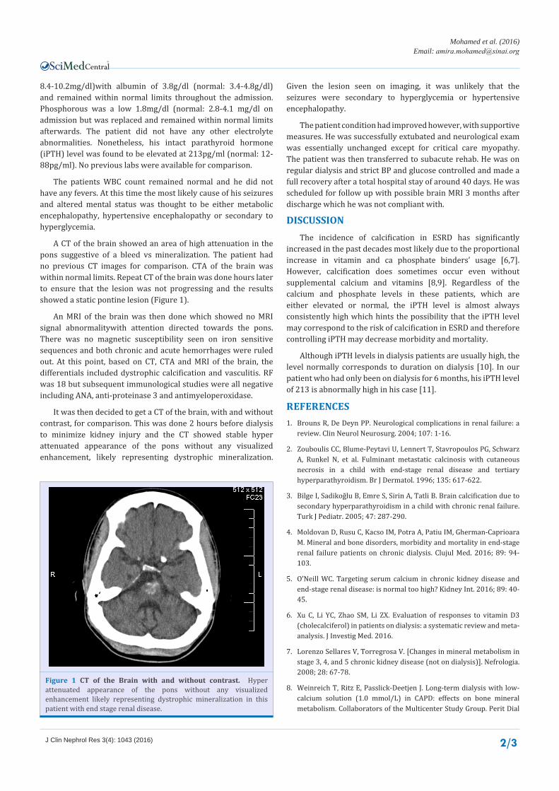
A CT of the brain showed an area of high attenuation in the pons suggestive of a bleed vs mineralization. The patient had no previous CT images for comparison. CTA of the brain was within normal limits. Repeat CT of the brain was done hours later to ensure that the lesion was not progressing and the results showed a static pontine lesion (Figure 1).
An MRI of the brain was then done which showed no MRI signal abnormalitywith attention directed towards the pons. There was no magnetic susceptibility seen on iron sensitive sequences and both chronic and acute hemorrhages were ruled out. At this point, based on CT, CTA and MRI of the brain, the differentials included dystrophic calcification and vasculitis. RF was 18 but subsequent immunological studies were all negative including ANA, anti-proteinase 3 and antimyeloperoxidase.
It was then decided to get a CT of the brain, with and without contrast, for comparison. This was done 2 hours before dialysis to minimize kidney injury and the CT showed stable hyper attenuated appearance of the pons without any visualized enhancement, likely representing dystrophic mineralization.
Given the lesion seen on imaging, it was unlikely that the seizures were secondary to hyperglycemia or hypertensive encephalopathy.
The patient condition had improved however, with supportive measures. He was successfully extubated and neurological exam was essentially unchanged except for critical care myopathy.The patient was then transferred to subacute rehab. He was on regular dialysis and strict BP and glucose controlled and made a full recovery after a total hospital stay of around 40 days. He was scheduled for follow up with possible brain MRI 3 months after discharge which he was not compliant with.
DISCUSSIONThe incidence of calcification in ESRD has significantly
increased in the past decades most likely due to the proportional increase in vitamin and ca phosphate binders’ usage [6,7]. However, calcification does sometimes occur even without supplemental calcium and vitamins [8,9]. Regardless of the calcium and phosphate levels in these patients, which are either elevated or normal, the iPTH level is almost always consistently high which hints the possibility that the iPTH level may correspond to the risk of calcification in ESRD and therefore controlling iPTH may decrease morbidity and mortality.
Although iPTH levels in dialysis patients are usually high, the level normally corresponds to duration on dialysis [10]. In our patient who had only been on dialysis for 6 months, his iPTH level of 213 is abnormally high in his case [11].
REFERENCES1. Brouns R, De Deyn PP. Neurological complications in renal failure: a
review. Clin Neurol Neurosurg. 2004; 107: 1-16.
2. Zouboulis CC, Blume-Peytavi U, Lennert T, Stavropoulos PG, Schwarz A, Runkel N, et al. Fulminant metastatic calcinosis with cutaneous necrosis in a child with end-stage renal disease and tertiary hyperparathyroidism. Br J Dermatol. 1996; 135: 617-622.
3. Bilge I, Sadikoğlu B, Emre S, Sirin A, Tatli B. Brain calcification due to secondary hyperparathyroidism in a child with chronic renal failure. Turk J Pediatr. 2005; 47: 287-290.
4. Moldovan D, Rusu C, Kacso IM, Potra A, Patiu IM, Gherman-Caprioara M. Mineral and bone disorders, morbidity and mortality in end-stage renal failure patients on chronic dialysis. Clujul Med. 2016; 89: 94-103.
5. O’Neill WC. Targeting serum calcium in chronic kidney disease and end-stage renal disease: is normal too high? Kidney Int. 2016; 89: 40-45.
6. Xu C, Li YC, Zhao SM, Li ZX. Evaluation of responses to vitamin D3 (cholecalciferol) in patients on dialysis: a systematic review and meta-analysis. J Investig Med. 2016.
7. Lorenzo Sellares V, Torregrosa V. [Changes in mineral metabolism in stage 3, 4, and 5 chronic kidney disease (not on dialysis)]. Nefrologia. 2008; 28: 67-78.
8. Weinreich T, Ritz E, Passlick-Deetjen J. Long-term dialysis with low-calcium solution (1.0 mmol/L) in CAPD: effects on bone mineral metabolism. Collaborators of the Multicenter Study Group. Perit Dial
Figure 1 CT of the Brain with and without contrast. Hyper attenuated appearance of the pons without any visualized enhancement likely representing dystrophic mineralization in this patient with end stage renal disease.

CentralBringing Excellence in Open Access
Mohamed et al. (2016)Email:
J Clin Nephrol Res 3(4): 1043 (2016) 3/3
Mohamed A, Farah Y (2016) A Rare Case of CNS Mineralization in End Stage Renal Disease. J Clin Nephrol Res 3(4): 1043.
Cite this article
Int. 1996; 16: 260-268.
9. Thompson B, Towler DA. Arterial calcification and bone physiology: role of the bone-vascular axis. Nat Rev Endocrinol. 2012; 8: 529-546.
10. Endocrine Abstracts. 2010.
11. Stevens LA, Djurdjev O, Cardew S, Cameron EC, Levin A. Calcium, phosphate, and parathyroid hormone levels in combination and as a function of dialysis duration predict mortality: evidence for the complexity of the association between mineral metabolism and outcomes. J Am Soc Nephrol. 2004; 15: 770-779.


![Indigenous Enhanced Mineralization Pyrene, Benzo[a]pyrene ...Indigenous soil microorganism mineralization experiments. All of the mineralization experiments were performed by using](https://static.fdocuments.us/doc/165x107/5e7c41b0b7c4ef64181e5e16/indigenous-enhanced-mineralization-pyrene-benzoapyrene-indigenous-soil-microorganism.jpg)