a rapid quantitative in vitro serum neutralization test for rabies antibody
7
A RAPID QUANTITATIVE IN VITRO SERUM NEUTRALIZATION TEST FOR RABIES ANTIBODY D. A. King,* D. L. Croghan,* and E. L. Shaw* INTRODUCTION THE QUANTITATIVE DETERMINATION of neutralizing rabies antibody has been ham- pered by the lack of a rapid in vitro method of assay. At the present time, the accepted technique is an in vivo mouse serum neutralization test (2). This method has several inherent disadvantages, includ- ing the fourteen day test period, the prob- lem of individual resistance, and the nonspecific mortality associated with any animal test. Thomas et al. (9) describes a rapid qualitative in vitro rabies antibody test that utilizes an indirect fluorescent anti- body technique; however, it has not been evaluated quantitatively on a basis com- parable to the in vivo neutralization test (8). Because rabies virus can be propagated in monolayer cell culture (1, 3, 5) and its growth observed by fluorescent antibody techniques (4), studies were undertaken to determine if these techniques could be employed in a quantitative in vitro assay for rabies antibody. MATERIALS Anti-rabies Fluorescent Antibody Conju- gate Following the technique described by Lennette (6), conjugated gamma globulin was prepared from hyperimmunized ham- ster anti-rabies serum. The conjugate was not adsorbed with tissue powder, but in- stead, diluted with normal hamster brain homogenate prior to staining. This tech- nique has resulted in brilliant specific fluorescence with little or no background staining. 'National Animal Disease Laboratory, Bio- logics Services, Animal Inspection and Quaran- tine Division, Agricultural Research Service, United States Department of Agriculture, Ames, Iowa. Cell Cultures Various cell cultures were employed in determining the one most suitable for rabies growth. Primary kidney cultures of guinea pig, hamster, rabbit, dog, cat, and swine were tested. Chick fibroblast cul- tures and a hamster kidney cell line (BHK) were also used. Viruses Several strains of rabies virus were employed in selecting the one most suit- able to be used as an indicator virus for cell culture infectivity and antigenicity in neutralization studies. These included the LEP Flury strain (20% chick embryo suspension), CVS-25 fixed virus strain (10% mouse brain suspension), and a cell culture adapted strain of CVS virus (50% culture fluids). Serum Prevaccination and postvaccination sera from humans and guinea pigs were used to study immunological response. Com- mercial hyperimmune horse serum1 was also used to correlate antibody titers ob- tained from in vivo and in vitro assays. METHODS Medium Primary cell cultures were prepared by the method of Youngner et al. (10). The cells were planted onto 10.5 X 35 mm. coverslips in Leighton tubes and incubated at 370 C. in a medium consisting of Hank's BSS, 0.5% lactoalbumin hydrolysate (LAH) and enriched with 10% sterile calf serum. At the time of assay inoculations, the medium was changed to Earle's Balanced Salt Solution (EBS) containing 0.5% LAH and 2% sterile calf serum. Incorporated in all media, at the time of use, were antibiotics at the following lLederle Laboratories, Pearl River, New York. 87 CAN. VET. JouR., vol. 6, no. 8, August, 1965
Transcript of a rapid quantitative in vitro serum neutralization test for rabies antibody

A RAPID QUANTITATIVE IN VITRO SERUM NEUTRALIZATIONTEST FOR RABIES ANTIBODY
D. A. King,* D. L. Croghan,* and E. L. Shaw*
INTRODUCTION
THE QUANTITATIVE DETERMINATION ofneutralizing rabies antibody has been ham-pered by the lack of a rapid in vitromethod of assay. At the present time, theaccepted technique is an in vivo mouseserum neutralization test (2). This methodhas several inherent disadvantages, includ-ing the fourteen day test period, the prob-lem of individual resistance, and thenonspecific mortality associated with anyanimal test.Thomas et al. (9) describes a rapid
qualitative in vitro rabies antibody testthat utilizes an indirect fluorescent anti-body technique; however, it has not beenevaluated quantitatively on a basis com-parable to the in vivo neutralization test(8).Because rabies virus can be propagated
in monolayer cell culture (1, 3, 5) and itsgrowth observed by fluorescent antibodytechniques (4), studies were undertakento determine if these techniques could beemployed in a quantitative in vitro assayfor rabies antibody.
MATERIALS
Anti-rabies Fluorescent Antibody Conju-gate
Following the technique described byLennette (6), conjugated gamma globulinwas prepared from hyperimmunized ham-ster anti-rabies serum. The conjugate wasnot adsorbed with tissue powder, but in-stead, diluted with normal hamster brainhomogenate prior to staining. This tech-nique has resulted in brilliant specificfluorescence with little or no backgroundstaining.
'National Animal Disease Laboratory, Bio-logics Services, Animal Inspection and Quaran-tine Division, Agricultural Research Service,United States Department of Agriculture,Ames, Iowa.
Cell CulturesVarious cell cultures were employed in
determining the one most suitable forrabies growth. Primary kidney cultures ofguinea pig, hamster, rabbit, dog, cat, andswine were tested. Chick fibroblast cul-tures and a hamster kidney cell line(BHK) were also used.
VirusesSeveral strains of rabies virus were
employed in selecting the one most suit-able to be used as an indicator virus forcell culture infectivity and antigenicity inneutralization studies. These included theLEP Flury strain (20% chick embryosuspension), CVS-25 fixed virus strain(10% mouse brain suspension), and a cellculture adapted strain of CVS virus (50%culture fluids).
SerumPrevaccination and postvaccination sera
from humans and guinea pigs were usedto study immunological response. Com-mercial hyperimmune horse serum1 wasalso used to correlate antibody titers ob-tained from in vivo and in vitro assays.
METHODS
MediumPrimary cell cultures were prepared by
the method of Youngner et al. (10). Thecells were planted onto 10.5 X 35 mm.coverslips in Leighton tubes and incubatedat 370 C. in a medium consisting of Hank'sBSS, 0.5% lactoalbumin hydrolysate (LAH)and enriched with 10% sterile calf serum.At the time of assay inoculations, themedium was changed to Earle's BalancedSalt Solution (EBS) containing 0.5% LAHand 2% sterile calf serum.
Incorporated in all media, at the timeof use, were antibiotics at the following
lLederle Laboratories, Pearl River, NewYork.
87CAN. VET. JouR., vol. 6, no. 8, August, 1965
CANADIAN VETERINARY JOURNAL
levels: dihydrostreptomycin, 0.2 mg./ml.;potassium penicillin G, 200 U/ml.; andNystatin2 20 U/ml.
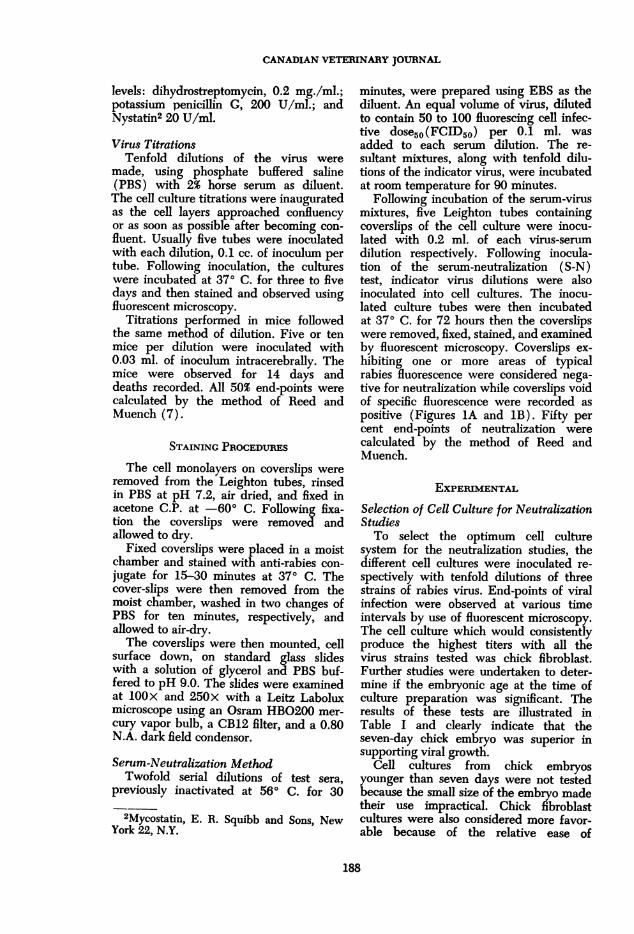
Virus TitrationsTenfold dilutions of the virus were
made, using phosphate buffered saline(PBS) with 2% horse serum as diluent.The cell culture titrations were inauguratedas the cell layers approached confluencyor as soon as possible after becoming con-fluent. Usually five tubes were inoculatedwith each dilution, 0.1 cc. of inoculum pertube. Following inoculation, the cultureswere incubated at 370 C. for three to fivedays and then stained and observed usingfluorescent microscopy.
Titrations performed in mice followedthe same method of dilution. Five or tenmice per dilution were inoculated with0.03 ml. of inoculum intracerebrally. Themice were observed for 14 days anddeaths recorded. All 50% end-points werecalculated by the method of Reed andMuench (7).
STAINING PROCEDURES
The cell monolayers on coverslips wereremoved from the Leighton tubes, rinsedin PBS at pH 7.2, air dried, and fixed inacetone C.P. at -60° C. Following fixa-tion the coverslips were removed andallowed to dry.
Fixed coverslips were placed in a moistchamber and stained with anti-rabies con-jugate for 15-30 minutes at 370 C. Thecover-slips were then removed from themoist chamber, washed in two changes ofPBS for ten minutes, respectively, andallowed to air-dry.The coverslips were then mounted, cell
surface down, on standard glass slideswith a solution of glycerol and PBS buf-fered to pH 9.0. The slides were examinedat IOOX and 250x with a Leitz Laboluxmicroscope using an Osram HBO200 mer-cury vapor bulb, a CB12 filter, and a 0.80N.A. dark field condensor.
Serum-Neutralization MethodTwofold serial dilutions of test sera,
previously inactivated at 560 C. for 30
2Mycostatin, E. R. Squibb and Sons, NewYork 22, N.Y.
minutes, were prepared using EBS as thediluent. An equal volume of virus, dilutedto contain 50 to 100 fluorescing cell infec-tive dose50(FCID50) per 0.1 ml. wasadded to each serum dilution. The re-sultant mixtures, along with tenfold dilu-tions of the indicator virus, were incubatedat room temperature for 90 minutes.
Following incubation of the serum-virusmixtures, five Leighton tubes containingcoverslips of the cell culture were inocu-lated with 0.2 ml. of each virus-serumdilution respectively. Following inocula-tion of the serum-neutralization (S-N)test, indicator virus dilutions were alsoinoculated into cell cultures. The inocu-lated culture tubes were then incubatedat 370 C. for 72 hours then the coverslipswere removed, fixed, stained, and examinedby fluorescent microscopy. Coverslips ex-hibiting one or more areas of typicalrabies fluorescence were considered nega-tive for neutralization while coverslips voidof specific fluorescence were recorded aspositive (Figures IA and IB). Fifty percent end-points of neutralization werecalculated by the method of Reed andMuench.
EXPERIMENTALSelection of Cell Culture for NeutralizationStudiesTo select the optimum cell culture
system for the neutralization studies, thedifferent cell cultures were inoculated re-spectively with tenfold dilutions of threestrains of rabies virus. End-points of viralinfection were observed at various timeintervals by use of fluorescent microscopy.The cell culture which would consistentlyproduce the highest titers with all thevirus strains tested was chick fibroblast.Further studies were undertaken to deter-mine if the embryonic age at the time ofculture preparation was significant. Theresults of these tests are illustrated inTable I and clearly indicate that theseven-day chick embryo was superior insupporting viral growth.
Cell cultures from chick embryosyounger than seven days were not testedbecause the small size of the embryo madetheir use impractical. Chick fibroblastcultures were also considered more favor-able because of the relative ease of
188

RABIES ANTIBODY TEST
FIGURE IA. Fluorescing cells in chick fibroblast cell culture monolayer observed 72 hoursafter inoculation of a virus-serum mixture. Cellular infection indicates no neutralization.
FIGURE 1B. Chick fibroblast cell culture monolayer observed 72 hours after inoculation of avirus-serum mixture. The absence of specific fluorescence indicates neutralization.
189

CANADIAN VETERINARY JOURNAL
TABLE I
EFFECT OF EMBRYO AGE ON VIRAL GROWTH
50% End-points of:Embryonic Age at Timeof Culture Preparation CVS Virus LEP Virus C.C.CVS Strain
7 day 1065 10-5° 10^09 day 10-4.6 10-4-0 10-3.512 day 10-2.0 10-30 10-30
handling, rapid cellular growth, andavailability.
Selection of an Indicator VirusAfter selection of a suitable culture
system for neutralization, studies weredirected to the selection of an indicatorvirus for the assays. Since all of the strainsappeared to propagate readily in chickfibroblast cell cultures, only the questionof antigenicity needed to be resolved. Itwas felt that the CVS fixed virus strainwould offer the most favorable comparableantigenicity since it is the virus most com-monly used in animal SN tests andimmunity-challenge studies. Multiple titra-tions of the virus were performed, bothin mice and cell culture, to obtain anaccurate 50% end-point for neutralizationstudies. Although the amount of inoculumvaried between the two systems, the ad-justed end-points were comparable.
Effect of Virus Concentrations uponNeutralizationTo determine a suitable viral popula-
tion for neutralization studies, SN testswere performed with various concentra-tions of virus. Hyperimmune anti-rabieshorse serum was used as the test serum.The results are tabulated in Table II andclearly indicate that the viral concentrationdirectly affects the neutralization index50-(NI50) of the serum. Further tests withsera of low antibody content indicatedthat false negative readings could be ob-tained if the indicator viral concentrationwas higher than 100 FCID50's per 0.1 ml.To allow for minor errors in diluting tech-niques and bottle variability, the indicatorvirus is routinely diluted to contain be-tween 50 to 100 FCID50/0.1 ml.
TABLE II
EFFECT OF VIRAL CONCENTRATION ONNEUTRALIZATION
Serum-Neutralization FCID60's of VirusTiter of Serum Used to Challenge Serum
>1:3,200 311:1,600 3161:566 3,162
< 1:50 31,620
Determination of Optimal Time ofObservationSome serum-neutralization tests, em-
ploying cell culture for viral assays can beobserved periodically and final resultstabulated when the cytopathogenic effectof the viral indicator is clearly visible.Since this test for rabies antibody utilizesfluorescent antibody staining, it was neces-sary to determine the optimum time forobserving viral activity. To determinethis, a series of identical SN tests werestained and observed at various time inter-vals. It was found that viral propagationcould be detected as early as 72 hours andthat observation at later intervals did notchange the NI50 of the serum. It wasfurther observed that the virus did not"spread" from the original infected cellsregardless of the amount of time elapsedbetween observations. Thus, only the virusintroduced at the time of inoculation pro-pagated sufficiently to exhibit fluorescencewhen stained with specific anti-rabiesconjugate. Because of these results, allfuture neutralization studies were termi-nated at 72 hours post-innoculation.
Time of Viral AttachmentWhen assaying sera with little or no
antibody content, serum dilutions as lowas 1 in 2 had to be employed. Such serumconcentrations were often toxic to the
190

RABIES ANTIBODY TEST
TABLE IIICOMPARISON OF SERUM NEUTRALIZATION RESULTS BETWEEN in vitro AND
in vivo METHODS
Serum SN Titer by SN Titer byIdentification in vitro Method in vivo Method*
8 1:12 1:239 1:3 1:610 1:3 1:311 1:3 1:1312 1:4 1:613 1:192 1:40614 1:288 1:21515 1:288 1:15216 1:192 1:51217 1:576 1:30418 1:288 1:43221 > 1:768 1:89723 1:96 < 1:20024 < 1:48 < 1:20026 > 1:6,400 1:3,36827 > 1:6,400 1:5,40828 1:6,400 1:5,957
*Tests conducted by Dr. G. R. Sharpless, Lederle Laboratories, PearlRiver, New York.
cell cultures after exposure for severaldays and in some instances, within a fewhours. It became necessary to remove thevirus-serum inoculum as soon as viralattachment was complete.To determine the time of viral attach-
ment, titrations of the indicator virus wereconducted. At intervals of 1, 2, 4, 8, 16,and 24 hours, culture tubes, representinga titration, were washed with EBS andfresh media was added to replace themedia containing the virus inoculum.Seventy-two hours postinoculation alltitrations were observed for specific viralfluorescence and the end-points weretabulated. The only titration with amarked decrease in titer was that titrationwhich received a media change in onehour following inoculation. This titrationwas tenfold lower than the other titrationswhich were, for practical purposes, identi-cal. Thus, virus attachment appears com-plete after two hours; and making a mediachange after this time reduced cell toxicity.
RESULTS
Following the determination of methodsand materials to be used for the in vitroassay, studies were begun to evaluatethe practicality of the technique. Somestudies were concerned with the measure-
ment of antibody responses following vac-cination while other evaluations comparedresults obtained from SN tests conductedin mice.
Serum samples, representing a spectrumof antibody responses, were obtained andassayed by the in vitro SN method. Neu-tralization titers had been determined pre-viously by mouse inoculation SN assay.3Table III shows the comparison of NI50'sobtained by both methods.
Postvaccination sera were obtained fromguinea pigs representing an experimentalrabies vaccine test.4 The various groupsof animals had been vaccinated with dilu-tions of an experimental rabies vaccine.The serum samples were coded to preventbiased reading of the SN tests. Table IVdemonstrates the average antibody re-sponse of each group and the dilution ofvaccine received.
DISCUSSION
In evaluating the in vitro serumneutralization assay for rabies antibody,the most critical consideration was capa-bility for detection of varying levels of
3Supplied by Lederle Laboratories, PearlRiver, New York.
4Supplied by Fort Dodge Laboratories, Inc.,Fort Dodge, Iowa.
191

CANADIAN VETERINARY JOURNAL
TABLE IVANTIBODY RESPONSE TO VARIOUS CONCENTRATION OF RABIES ANTIGEN
Dilution of NI50 of PostvaccinationAnimal Group Vaccine Received Sera (Average)
A Undiluted > 1:128B 1:5 1:128C 1:10 1:64D 1:20 1:32
Control < 1:2
antibody. This was measured by determin-ing the antibody response of animals re-ceiving various concentrations of rabiesantigen. The animals receiving less anti-gen responded with a lower antibody level(Table IV). The next consideration was tocompare results obtained by in vivo assays,and the data in Table III demonstratesthat the two methods are comparable.
At the start of the neutralization studies,there was concern whether the antigen-antibody reaction might be reversible.There is no evidence, however, to showthis occurs within the periods the test isconducted.As noted previously, a slide taken from
the same dilution appeared to haveapproximately the same number of in-fected cells, regardless of the elapsedtime between inoculation and observation.This was true in all strains tested, althoughthere may be adapted strains capable ofspreading from a focus of infection.The early reading of the test is a
distinct advantage over the in vivomethod. The specificity of the fluorescentantibody completely eliminates the prob-lem of false readings which must beconsidered when testing in animals. Theonly technical problem encountered withthe in vitro assays was serum toxicity tothe cell cultures. This appeared occasion-ally when serum was tested at low dilu-tions; however, a media change followingviral attachment reduced the cellulardamage to a minimum.
Regardless of the technique used inantibody determination, little can begained until the correlation between anti-body level and animal protection againstrabies is resolved. It is hoped that thedevelopment of a rapid quantitativeassay of rabies antibody will help speedstudies toward that end.
SUMMARY
A rapid quantitative assay for rabiesantibody, employing cell culture and fluo-rescent antibody techniques, is described.The determination of the most suitablematerials and methods is reported.
Results obtained by in vitro assays areevaluated by detecting varying antibodylevels with experimental animals. The invivo and in vitro methods were compared.
RESUME
L'auteur decrit un essai quantitatif ra-pide pour les anticorps de la rage aumoyen de cultures de cellules et de tech-niques fluorescentes. II fait rapport sur ladetennination des methodes et des mate-riaux les plus appropries.
Les resultats obtenus par les essais invitro sont evalues par la detection desdivers niveaux d'anticorps avec les ani-maux servant "a titre d'experience. On afait des comparaisons entre les methodesin vivo et in vitro.
REFERENCES
1. ABELSETH, M. K. Propagation of rabiesvirus in pig kidney cell culture. Can.Vet. Jour. 5: 4. 1964.
2. ATANASIU, R., BAHMANYAU, M., BALTA-ZARD, M., Fox, J. P., HABEL, K., KAPLAN,M. M., KISSLING, R. E., KoMoRov, A.,KoPROWSKI, H., LEPINE, P., GALLARDO,F. P., and SCHAEFFER, M. Bull. WorldHealth Organ. 14: 593. 1956.
3. FENJE, P. Propagation of rabies virusin cultures of hamster kidney cells. Can.J. Microbiol. 6: 479. 1960.
4. KAPLAN, M. M., FoRsRE, Z., and Kop-ROWSRI, H. Demonstration of rabiesvirus in tissue culture with fluorescentantibody technique. Bull. World HealthOrganization. 22: 434. 1960.
192

RABIES ANTIBODY TEST
5. KISSLING, R. E. Growth of rabies virusin non-nervous tissue culture. Proc. Soc.Exptl. Biol. Med. 98: 223. 1958.
6. LENNETTE, E. H. Rabies FluorescentAntibody (FRA) Test. Standard Proce-dures. Viral and Rickettsial DiseaseLaboratory, Berkeley, California StateDepartment of Public Health. 1962.
7. REED, L. J., and MUENCH, H. A simplemethod of estimating fifty per cent end-points. Am. J. Hyg. 27: 493. 1938.
8. SYKEs, R. K. Personal communication.1965.
9. THOMAS, J. B., SYKES, R. K., and RICKER,A. S. Evaluation of indirect fluorescentantibody technique for detection ofrabies antibody in human sera. J. Im-munol. 91(6): 721. 1963.
10. YOUNGNER, J. S. Monolayer Tissue Cul-tures; Preparation and Standardizationof Suspensions of Trypsinized MonkeyKidney Cells. Proc. Soc. Exptl. Biol.Med. 85: 202. 1954.
ABSTRACTS
Brinkman, D. C., and Burch, G. R.(1964). Metofane (methoxyflurane) anes-thesia in a variety of animal species.-Allied Vet. 36, 36-43, and 64.
FROM CASE REPORTS of 144 investigators,data were amassed for 4,129 animalsanesthetized with methoxyflurane. Theprocedures employed and results obtainedare described in general terms for pri-mates, pigs, rats, mice, birds, and a fewindividuals of other species. The advan-tages of this new inhalant anesthetic werelow toxicity and marked muscle relaxingproperties. No specific contraindicationwas found in any species. Details of theproperties, dosage and administration, sideeffects and precautions are given, and thedrug is recommended for wide usage as ageneral anesthetic.
Reprinted from The Veterinary BulletinVol. 34, No. 11, November, 1964.
Jenkins, J. I. & Fletcher, F. J. (1964). Theeradication of lice from a rat colony bymeans of a malathion dipping routine.-J. Anim. Tech. Ass. 15, 1-5.
As attempts to eradicate the rat louse,Polyplax spinulosa, from a lab. colony byperiodically dusting with 4% melathionpowder had failed, dipping in a bath of0.125% malathion prepared from MalastanE-C50, a commercial emulsifiable prepara-tion designed specifically for animal use,
was undertaken after trials had provedthat malathion at this concentration didnot lower the cholinesterase activity of theblood and was harmless to rats. Weanlingsand breeding stock were completely freedfrom lice by one wetting in a speciallydesigned dipping box, and since then onlybreeding stock have been so treated.
Reprinted from "The Veterinary Bulletin",Vol. 34, No. 12, December, 1964.
Anon. (1964). Vaccination against Johne'sdisease.-Vet. Rec. 76, 423-424.
The Ministry of Agriculture has con-ducted two field trials and experimentalwork at the Central Veterinary Laboratory,Weybridge, to investigate the practicalvalue of vaccination against Johne's dis-ease. The main findings are that the use ofa single dose of vaccine in calves underone month of age gives significant protec-tion against the development of clinicaldisease, but losses may be experienced invaccinated herds if good hygiene is notpractised. After vaccination skin sensitivityto tuberculin is shown. It has been decidedthat as from May 1st 1964 Johne's diseasevaccine may be used for calves in herds inwhich the disease is a problem and inwhich there is a satisfactory tuberculintesting history. The conditions under whichthe vaccine may be used are given.
Reprinted from "The Veterinary Bulletin",Vol. 34, No. 12, December, 1964.
193