A Quantitative Ultrastructural Analysis of Parasitized
17
Human Cerebral Malaria A Quantitative Ultrastructural Analysis of Parasitized Erythrocyte Sequestration G. G. MACPHERSON, MA, BM, BCh, DPhil, M. J. WARRELL, MB, BS, MRCP, MRCPath, N. J. WHITE, MD, BSc, MRCP, SORNCHAI LOOAREESUWAN, MD, DTM&H, and D. A. WARRELL, MA, DM, FRCP For investigation of the pathogenesis of cerebral malaria, immediate postmortem samples from brain and other tis- sues of patients dying with Plasmodium falciparum malaria, with (CM) or without (NCM) cerebral malaria, were processed for electron microscopy. Counts of parasi- tized erythrocytes (PRBCs) in cerebral and other vessels showed that the proportion of PRBCs was higher in CM than in NCM, and also that the proportion of PRBCs was higher in the brain than in other organs examined in both CM and NCM. Cerebral vessels from CM patients were more tightly packed with RBCs than those from NCM patients, but there was no significant difference in the amount or degree of endothelial damage or numbers of vessels with endothelial pseudopodia. Fibrillar (fibrin) CEREBRAL MALARIA is a diffuse symmetric en- cephalopathy occurring in a proportion of patients in- fected with Plasmodiumfalciparum. It is the most im- portant severe manifestation of Pfalciparum infection and carries a mortality of between 20%o and 50%16.1 2 The clinical features of cerebral malaria are well documented,3`7 but some aspects of its pathogenesis remain obscure. Several mechanisms have been pro- posed. These include mechanical obstruction of cere- bral vessels resulting from the decreased deformability of parasitized erythrocytes (PRBCs)8 or from the adhe- sion of PRBCs to vascular endothelium9 l0; the release of toxic factors from PRBCs, leading to increased vas- cular permeability, the breakdown of the blood-brain barrier, and cerebral edema1112; and the induction of an inflammatory response in and around cerebral ves- sels, leading to increased vascular permeability and ce- rebral edema.13 The pathology of cerebral malaria in humans has been reviewed at gross and light-microscopic levels,'4 From the Sir William Dunn School of Pathology and the Nuffield Department of Clinical Medicine, University of Oxford, United Kingdom, The Faculty of Tropical Medicine, Mahidol University, Bangkok, Thailand, and the Liverpool School of Tropical Medicine, United Kingdom deposits were present in a small proportion of vessels, but no thrombosis was present. There was neither acute nor chronic inflammation, and leukocytes were absent within or outside cerebral vessels. There was no immune com- plex deposition in cerebral vessels. Parasites in cerebral vessels were mainly trophozoites or schizonts. Occasional RBC remnants following parasite release were seen. Some parasites were degenerate, resembling crisis forms. PRBCs adhered to endothelium via surface knobs. It is concluded that there is no evidence for an inflammatory or immune pathogenesis for human cerebral malaria and that the clinical effects probably relate to anoxia and the metabolic activities of the parasites (Am J Pathol 1985, 119:385-401) and ultrastructural studies of the pathologic features of human Pfalciparum infection in the liver and kid- neys have been reported. 15.16 No animal model has suc- cessfully mimicked the characteristic clinical and patho- logic features of human cerebral malaria, but detailed ultrastructural studies have been made of the patho- logic features of Pfalciparum in the owl monkey, Ao- tus. 17 Several studies have described cerebral disease in rodents following infection with various species of Plasmodium. 13,18 In the majority of these reports, the disease is related to inflammatory intravascular and Supported by the Wellcome Trust of Great Britain as part of the Wellcome-Mahidol University, Oxford Tropical Medi- cine Research Programme. Accepted for publication January 16, 1985. Address reprint requests to Dr. G. G. MacPherson, Sir Wil- liam Dunn School of Pathology, University of Oxford, South Parks Road, Oxford OXI 3RE, England. 385
Transcript of A Quantitative Ultrastructural Analysis of Parasitized
Human Cerebral Malaria
A Quantitative Ultrastructural Analysis of Parasitized
Erythrocyte Sequestration
G. G. MACPHERSON, MA, BM, BCh, DPhil,M. J. WARRELL, MB, BS, MRCP, MRCPath,
N. J. WHITE, MD, BSc, MRCP,SORNCHAI LOOAREESUWAN, MD, DTM&H, and
D. A. WARRELL, MA, DM, FRCP
For investigation ofthe pathogenesis ofcerebral malaria,immediate postmortem samples from brain and other tis-sues of patients dying with Plasmodium falciparummalaria, with (CM) or without (NCM) cerebral malaria,were processed for electron microscopy. Counts ofparasi-tized erythrocytes (PRBCs) in cerebral and other vesselsshowed that the proportion ofPRBCs was higher in CMthan in NCM, and also that the proportion ofPRBCs washigher in the brain than in other organs examined in bothCM and NCM. Cerebral vessels from CM patients weremore tightly packed with RBCs than those from NCMpatients, but there was no significant difference in theamount or degree of endothelial damage or numbers ofvessels with endothelial pseudopodia. Fibrillar (fibrin)
CEREBRAL MALARIA is a diffuse symmetric en-cephalopathy occurring in a proportion of patients in-fected with Plasmodiumfalciparum. It is the most im-portant severe manifestation of Pfalciparum infectionand carries a mortality of between 20%o and 50%16.1 2The clinical features of cerebral malaria are welldocumented,3`7 but some aspects of its pathogenesisremain obscure. Several mechanisms have been pro-posed. These include mechanical obstruction of cere-bral vessels resulting from the decreased deformabilityof parasitized erythrocytes (PRBCs)8 or from the adhe-sion of PRBCs to vascular endothelium9 l0; the releaseof toxic factors from PRBCs, leading to increased vas-cular permeability, the breakdown of the blood-brainbarrier, and cerebral edema1112; and the induction ofan inflammatory response in and around cerebral ves-sels, leading to increased vascular permeability and ce-rebral edema.13The pathology of cerebral malaria in humans has
been reviewed at gross and light-microscopic levels,'4
From the Sir William Dunn School of Pathology and the NuffieldDepartment of Clinical Medicine, University of Oxford,United Kingdom, The Faculty of Tropical Medicine, Mahidol University,Bangkok, Thailand, and the Liverpool School of Tropical Medicine,United Kingdom
deposits were present in a small proportion ofvessels, butno thrombosis was present. There was neither acute norchronic inflammation, and leukocytes were absent withinor outside cerebral vessels. There was no immune com-plex deposition in cerebral vessels. Parasites in cerebralvessels were mainly trophozoites or schizonts. OccasionalRBC remnants following parasite release were seen.Some parasites were degenerate, resembling crisis forms.PRBCs adhered to endothelium via surface knobs. It isconcluded that there is no evidence for an inflammatoryor immune pathogenesis for human cerebral malaria andthat the clinical effects probably relate to anoxia and themetabolic activities of the parasites (Am J Pathol 1985,119:385-401)
and ultrastructural studies of the pathologic featuresof human Pfalciparum infection in the liver and kid-neys have been reported. 15.16 No animal model has suc-cessfully mimicked the characteristic clinical and patho-logic features of human cerebral malaria, but detailedultrastructural studies have been made of the patho-logic features of Pfalciparum in the owl monkey, Ao-tus. 17 Several studies have described cerebral disease inrodents following infection with various species ofPlasmodium. 13,18 In the majority of these reports, thedisease is related to inflammatory intravascular and
Supported by the Wellcome Trust of Great Britain as partof the Wellcome-Mahidol University, Oxford Tropical Medi-cine Research Programme.
Accepted for publication January 16, 1985.Address reprint requests to Dr. G. G. MacPherson, Sir Wil-
liam Dunn School of Pathology, University of Oxford, SouthParks Road, Oxford OXI 3RE, England.
385
386 MACPHERSON ET AL
perivascular changes. The relationship of these modelsto human cerebral malaria is not clear.Any explanation of the etiology of human cerebral
malaria must take into account the morphological as
well as the clinical features of the disease. Previousstudies have examined routine autopsy specimens,where the possibility of significant postmortem degener-ation could not be excluded. The present study was un-
dertaken in an attempt to detect differences between theultrastructure of freshly fixed specimens of brain andsome other organs in patients dying from cerebralmalaria and control subjects dying with noncerebralmalaria. Specimens were examined for specific featureswith a semiquantitative procedure, and the results werecorrelated with the clinical diagnosis.The sequestration of red cells containing mature
forms of the parasite in capillaries and postcapillaryvenules demonstrated in this study appears to be a con-
sistent feature of severe P falciparum malaria. Thisresults in large discrepancies between peripheral bloodparasite counts and the parasite count observed in histo-logic specimens -these discrepancies are most evidentin cerebral malaria. Blood smears taken from capillaryfinger-prick or venous samples may therefore consider-ably underestimate the total parasite load in the infectedpatient. The possibility that sequestration might occur
in the dermal capillaries, and that parasite counts ob-tained from skin smear or skin biopsies would there-fore more accurately represent the total parasite loadthan conventional blood smears was investigated in pa-tients with cerebral and uncomplicated Pfalciparummalaria.
Materials and Methods
Clinical
Seven patients with cerebral malaria who died in theintensive care unit of PraPokklao Hospital, Chantaburi,Eastern Thailand, were studied. Investigation andmanagement of these patients has already been de-scribed.2 All were treated with parenteral quinine. Ce-rebral malaria was defined as unrousable coma in a pa-
tient with asexual forms of Pfalciparum detectable inthe peripheral blood smear. Other causes of coma were
excluded. Clinical, biochemical, and hematologic de-tails of these cases are given in Table 1. Control speci-mens were taken from 6 patients who died during thecourse of a Pfalciparum infection, but without cere-
bral malaria.The study of skin sequestration of PRBCs included
nine patients with cerebral malaria and seven with un-
complicated Pfalciparum malaria whose venous bloodparasitemia exceeded 1%. Informed consent to skin bi-opsy was obtained from the patients or their accom-
panying relatives. These patients were studied as soonafter admission to the hospital as possible.
Collection and Fixation of Specimens
Samples of tissues were taken from patients within90 minutes of death. Brain tissue was obtained fromevery case and kidney, liver, lung, and heart tissue wasobtained from some patients with a Franklin-modifiedvim Silverman biopsy needle. Pieces were discharged,cut into 1-mm cubes with a scalpel, and fixed. Areasof gray matter in brain specimens were selected for fixa-tion, and smears were also made on microscope slidesand stained with Giemsa or Wright's stain. Blood sam-ples were discharged into stirred fixative. Fixation waswith 2.5% glutaraldehyde in 0.1 M sodium cacodylatebuffer, pH 7.4, at 4 C for 2 hours. Samples were thenwashed three times, coded, and flown to Oxford in abuffer solution. All subsequent processing was donewithout knowledge of the clinical diagnosis. Specimenswere postfixed in 1% osmium tetroxide in 0.1 M sodiumcacodylate, pH 7.4, dehydrated in graded ehtanols, andembedded in Araldite. One-micron sections were stainedfor light microscopy.19 Thin sections cut with a dia-mond knife and stained with lead citrate20 were exam-ined on a JEOL IOOXC electron microscope. Some spec-imens were stained prior to postfixation to revealendogenous peroxidatic activity2" with the use of di-aminobenzidine (1 mg/ml) in Tris-HCl buffer, pH 7,containing 0.005Gb H202.
Capillary blood samples were obtained by prickingthe finger pulp with a 25-gauge needle. Venous sam-ples were obtained from the antecubital veins. Standardthin films were prepared. Slit skin smears were obtainedfrom the lobe of an ear, as described for the diagnosisof leprosy. Skin biopsies were taken with a standard4-mm circular punch. Care was taken not to squeezethe skin beforehand. Local anesthetic was infiltratedsubcutaneously. The base of the biopsy sample was thensmeared repeatedly on a glass slide. Duplicate slides wereprepared and stained with Giemsa. The number of para-sites in 2000 red cells was counted on each slide. Theresults were compared by paired t tests.
Analysis of Sections
Thick (1-,u) sections of brain and other tissues wereexamined by light microscopy. Blood vessels wereidentified, intravascular RBCs were counted, and theproportion containing recognizable asexual parasiteswas scored.Thin sections were examined systematically with the
squares on the support grids as reference areas. At least20 blood vessels (or all vessels on small or sparsely vas-
AJP * June 1985

HUMAN CEREBRAL MALARIA 387
CO00
E0
0
oEQ a)
C>
a) c 0 U)E acE @E
E 0,mD " )
*nc -O F
(U
C E
2
cu
0 ) 0)
E
a_ E
0
L CD
C
o o oE
CZU
_,
CE(s
(U
CD0
()
cnE EU) CD -C
CL CD~-
0 CL
CD
0)
coEU0
-c
X -
a) COCCD = cc$
E%Ea
<o C
E EU)
U)
co co _ a_)cU
C"coE E, Q E:3, CL E E75 75o (Um
C CCOC*
C CC C ° 0 c
Z'6-0 0a _ _ca OE
I I I I <al
+ + + I I
N 0
Ca l
cm q(ND
c( c0
+
o
0
U)EQ)
'T CII
IT C\j0)
N-
ODU
N'-M
N (U
N- N
r0 0Nc
U) tCM U)
N 0NNC
C\-
N cD_CN
0 0D CO)
N LU N
~0)
N D NCf)C\i CO _ CO
lp CC\j CD It rlCD U') L
(UC\l-
0
(0N-
(U
0
U)
N
+ +
cl.
Uj)-i a)
U)U
(1 'OCL0 n _
(-U (U-
't CO
'-(0C0) CO
CN C)
(D CDN
0o CDCN CD C(CO V) _
0) N
(0
C0 N\
(0 C)
U)0 N CMC\ C-_
U)
CO3U)
0(U
CV
(U
-
co
U)
C Q. C
0 E*ai .5
U) 0
0 U)O o
U)
a E~OL
II I I
+ + + I I
- CO co1tN 0a t
(a -
t U)
NN
+ CM
0
CM
(0
CO
CM
CM
0)l
O 0I
0 (0
N N
0) _~
N C
r-
N
U)-U U
CM
"i CM U)
U)CM0o-C\J_ CM
CD CII CD C--Ft
n0U1) (U)
U)
0;) N C0) 0 C e" C .i U D 't
(U CD N -a co a-) C
U .C
uL E n o
-0
(U u Q
D U) o
E E-
E E ct)U) 1)(Co
+ +
0 LI a)0o D0 V t-co LO 1t lq 0) c\j ( LO CM_ c0) CN _ _
U) C% 0 C\ o0) 0C 0o
C\l
CII U') CII o)- "t 04 C\J cn
0 0 (U 0 -
0) ) U') NO
V
CY) (U C) 0CD 'I 0 (004 C\j C\j CM
0 _ 0 0 (U0 0)0o 0 NMD(0D - CN (0
N N CN 0)
- O
N CD
_ ), t U') O)0_ )
c (0 0
CM(U C\ -0
0 D0C) C\N
0D CD N 0)0 14 0 0
CM
CN 0) CD -
Co 0 (U 0v)V (D - V
VCO '-V0 0 N1 N1
a O r- C\J
U) CO CN 0LO LO CM
00 O0
NY) (C)C\l CO _ CO
C\ 't LO C\j(Ut 0)N 0)_C\J
+ + + + +
LO LO It c) o14 ,q ) CD C\lU) CD C\ N -
FL LLc co(U c0)0; 0
.r(U
Ei a un
(U0
~ C 0 ' .rc_ E D2c( CJ Cv cm
- C\l Cl) ) Ct LoXw U (D - co
0 % 0;CM C\j C\ -
0 0 - CM'-N CM CN
Vol. 119 * No. 3
co(U
(U
.9-co
U)
co
n(U
C-)
.U._
()a-

388 MACPHERSON ET AL
Table 2-Comparison of Cerebral Vessels From Patients With and Without Cerebral Malaria*
Degree of% RBCs packingparasitized (0-3)
% Vesselswith 3 +packing
Extravascular RBCs(no. of occasions,not individual cells)
% Vessels withendothelialdamage
Group 1:cerebral malaria
1 34.12 25.23 53.04 55.05 48.36 43.87 43.8Mean ± SD 43.3 ± 10.6
Group 2:noncerebral malaria
8 09 30.010 7.011 52.512 4.713 5.5Mean ± SD 16.6 ± 20.5
1.61.232.582.822.672.692.11
2.24 ± 0.62
0.471.250.131.250.670.50
0.71 ± 0.45
2.72<0.01
25.025.965.090.977.876.950.0
58.8 ± 26.0
025.00
12.508.3
7.63 ± 10.0
2.95<0.01
0421300
000
0
1.25NS
93.726.930.0
100.055.529.470.0
57.9 ± 40.0
70.012.512.558.820.00
29.0 ± 28.4
1.79NS
25.033.370.0
100.080.076.955.5
63.0 + 26.7
5.025.00
52.950
14.65 + 20.9
2.63<0.01
6.33.909.1
44.407.1
10.1 ± 15.5
00
12.504.80
2.88 ± 5.09
1.20NS
* Specimens from individual patients were coded before examination and scored without knowledge of the clinical diagnosis. The significance of differ-ences between the groups was tested with the Mann-Whitney test.
cularized specimens) were identified and scored for a
variety of characteristics. These included1) Type of vessel (arteriole, capillary, or venule)2) Presence of PRBCs (+ or -)3) Tightness of packing of RBCs (scored as 0-3 +)
0 = no RBCs1+ = loose, free-floating RBCs2+ = vessel filled with RBCs but little distortionof cell outlines3 + = vessel filled with RBCs with obvious dis-tortion of cell outlines
4) State of RBCs (density of cytoplasm, presence ofsurface excrescences [knobs], apparent adhesion toendothelium)5) State of endothelium (intact or damaged, pres-ence of gaps between endothelial cells, presence ofpseudopodia, presence of deposits on basementmembrane)6) State of parasites ("healthy" or "damaged," stateof maturation)7) Presence of leukocytes and platelets8) Presence of extravascular RBCs9) Presence of intravascular deposits
Statistical Analysis
Data from the different groups were compared withthe Student t test or the Mann-Whitney test.
Results
Clinical DetailsClinical details of the 30 patients studied are given
in Table 1.
Preservation of Specimens
In general, the tissues showed very good preserva-tion of ultrastructural detail in comparison with speci-mens obtained during open brain surgery. Artifacts werepresent in some cases - eg, neuronal degeneration,gaps between blood vessels and surrounding brain tis-sue, and damage to renal tubular epithelium- but bloodvessels and their contents were generally well preserved.In particular, membranes and mitochondria showed fewsigns of artifactual damage.
Controls
Apart from the samples obtained from Pfalciparum-infected patients dying without overt cerebral malaria,specimens obtained at open brain surgery and fixed im-mediately in glutaraldehyde were also examined (kindlysupplied by Dr. M. Esiri, Radcliffe Infirmary, Oxford).
Comparison of Brain Specimens From PatientsWith (CM) or Without (NCM) Cerebral Malaria
Sections of brain from Pfalciparum-infected patientswere examined "blind," as described above, and scoredfor abnormalities. Those abnormalities amenable tosemiquantitative analysis are presented in Table 2 and
Patient
% VesselscontainingPRBCs
% Vesselswith densedeposits
T
P
2.15<0.05
AJP * June 1985
HUMAN CEREBRAL MALARIA 389
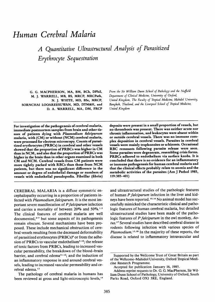
Figure 1-Comparison of blood vessels from pa-tients with and without cerebral malaria. Electronmicrographs of vessels from CM and NCM patientswere scored "blind" for several parameters. Thepoints represent the average score for each pa-rameter in an individual patient. The bars representmeans for all patients.
100 -
90 -
80-
70-
60-
50-
40-
30-
20
10
3+packing Endothelial damage Vessels with
parasitised RBCs
CM NCM CM NCM CM NCM
Vessels with 'Healthy' vessels with.. Fdtflo lA S .
fibrillar deposts
I T-CM NCM CM NCM
Figure 1. Several major differences between the twogroups are apparent:
1) More than three times as many vessels in the CMgroup contained PRBCs (P< 0.05). The great majorityof parasitized vessels were capillaries and venules. Onlyoccasional arterioles were seen, and these were almostalways empty of cells.
2) The average tightness of RBC packing in vessels(on a 0-3 + score) was significantly higher in the CMgroup (2.24) than in the NCM group (0.71) (P< 0.005).
3) The percentage of tightly packed vessels (3 +) was
significantly higher in the CM group (58.707o) than inthe NCM group (7.6070) (P < 0.001).
4) Endothelial damage was observed in many speci-mens. In CM, the proportion of vessels showing dam-age ranged from 27% to 100o and in NCM from 0%to 58%o but the difference is not statistically significant.Few gaps were seen between endothelial cells in un-damaged vessels, and there was no difference in theirfrequency between the groups.
5) Ten percent of the vessels in the CM group con-tained dense fibrillar deposits (range, 0-44%70), whereas
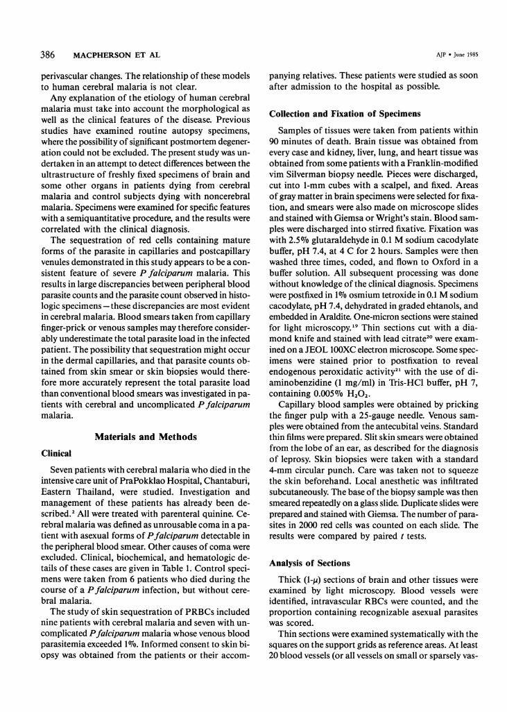
FpFigure 2-Cerebral malaria. Aparasitized RBC within a cerebralvenule. The parasites are under-going schizogony (P), and mem-branes appear to be attached tothe parasitophorous vacuole at onepoint (*). Dense fibrillar material ispresent within the lumen of the ves-sel (F). The endothelium is relative-ly undamaged, but mitochondriaare not well preserved (M). Numer-ous fingerlike endothelial pseu-dopodia are present (-). Astrocytefootpads are swollen (FP).(x 10,000)
I
Vol. 119 * No. 3
cndouthelial pseudopodal
t
t
.k
*s-

390 MACPHERSON ET AL
A B
Figure 3A-Cerebral malaria. An irregularly shaped parasitized RBC in a cerebral venule. The RBC shows numerous surface knobs. The parasite ap-pears degenerate (P). The vascular endothelium is damaged close to the apparent point of attachment (-). (x 9000) B-Cerebral malaria. A pre-sumed "ghost" of a parasitized RBC within a cerebral venule. The "ghost" is recognizable as such because of the numerous surface knobs. Some densedeposits are present within the cell (-). Extracellular spaces are shown (*). The nature of the large dense body (D) is unknown. (x 16,900)
only two vessels in all the NCM specimens containedsimilar deposits. On some occasions the deposits werestriated, suggesting that they were fibrin.
6) Endothelial pseudopodia were conspicuous insome vessels (see below). For comparative purposes,only those vessels with undamaged endothelium werescored. A higher proportion of vessels in the NCMgroup possessed pseudopodia, but the difference wasnot statistically significant (P > 0.5). Two patients inthe NCM group (Patients 9 and 11) differed conspicu-ously from the rest of the group in terms of the per-centage of parasitism of RBCs, the tightness of pack-ing of cerebral vessels, and the proportion of vesselscontaining PRBCs. Although in coma, their conditionwas not diagnosed as CM because of other clinical fea-tures.
Structure of PRBCs
PRBCs displayed several distinct abnormalities.
ShapeSome PRBCs possessed relatively regular outlines
(Figure 2), but the majority were very irregular. The de-gree of irregularity appeared to correlate with the timeafter parasitization, in that the most bizarre outlinesand largest parasites were present in cells with the leastremaining hemoglobin, as judged by cytoplasmic elec-tron density and peroxidatic activity (Figure 3A). Oc-casionally cells were seen with extremely distorted out-lines (Figure 3B). They were recognizable as RBCs only
because of the presence of many knobs on their sur-face. They are thought to represent RBC "ghosts" re-maining after the escape of parasites.
Plasma Membrane
The majority of PRBC possessed conspicuous sur-face irregularities (knobs). When fully developed, theknobs showed a conical outline with a circular base (Fig-ure 4A). Some tangential profiles of knobs showed arelatively electron-lucent core. The numbers of knobsappeared to correlate with the length of time afterparasitization. In some cells the knobs were rather reg-ularly arranged, with a similar gap between knobs. Onoccasion, rather than discrete knobs, there appeared tobe larger areas of dense material underlying the plasmamembrane, perhaps due to the coalescence of individualknobs (Figure 4B). In many instances it was difficultto follow the bilayered plasma membrane because oftangential sectioning, but where it was distinct, it wasclearly overlying the knobs (Figure 4C).
CytoplasmThe cytoplasm of parasitized cells was usually less
electron-dense than that of normal RBCs. In many casesthe cytoplasm had a granular appearance (see Figure3A). Few organelles were seen in PRBCs apart fromthe parasitophorous vacuole. Maurer's clefts were seeninfrequently. Occasionally, volumes of PRBC cyto-plasm were seen to be enclosed within membranes.These membranes showed the typical trilaminar struc-ture but appeared to be discontinous. It is not clear
AJP * June 1985
IWO 0. -Z
. "kI
A-

HUMAN CEREBRAL MALARIA 391
A B C
l
Figure 4A-Cerebral malaria. Tangential section through the edge of a parasitized RBC. Surface knobs are separated by regular gaps. They appearto have a less electron-dense central core. (x 80,000) B-Cerebral malaria. A cerebral venule tightly packed with parasitized RBC. The endothelium(-~) is almost totally destroyed. Dense material is aggregated under the RBC plasma membrane. (x 16,000) C-Cerebral malaria. Cerebral venule.Strands of electron-dense material (t) connect the endothelial cell (E) with the RBC (R). The plasma membrane of the RBC clearly overlies the knobs.(x 132,000)
whether this represents natural fenestration or a post-mortem artifact. In some cases the membranes were sin-gle (Figure 5A), but in many cases they consisted oftwo distinct trilaminar layers (Figure 5B). These mem-branes usually showed no connection with the parasi-
x~
\~~~~~~~~~~i
tophorous vacuole or the cell surface, but occasionallythey were clearly attached to the parasitophorous vacu-ole (Figures 5A and 6A). On one occasion multiplemembranes were seen to be attached to the vacuole (Fig-ure 6A). At the site of attachment a complex arrange-
Jr qwO~|. . 't 41 w ..v/.
Figure 5A-Trophozoite in an RBC. Volumes of cytoplasm are enclosed by apparently fenestrated membranes. One membrane is clearly attached tothe parasite. The endothelium shows postmortem damage (-). (x 20,800) B-Membranes within PRBCs. The outer set of membranes is clearlydouble. The inner membranes are sectioned tangentially. Electron-dense material is associated with both membranes. (x 46,200)
Vol. 119 * No. 3
AJP * June 1985
!*2.0,4
_ .5 r !,.Ad.
,.
v1 ", X.
B
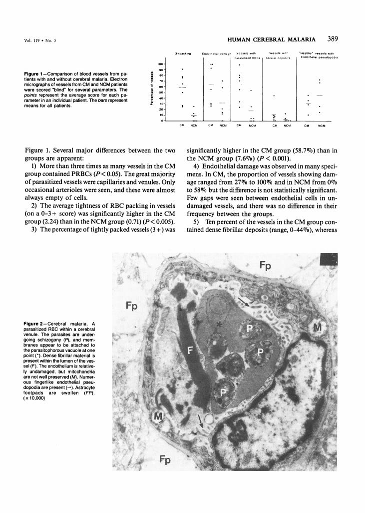
Figure 6A-Four sets of membranes are associated with a single trophozoite. (x 33,800) B-Complex arrangement of membranes adjacent to para-site. Five distinct trilaminar membranes can be distinguished (detail of 6A). (x 99,000)
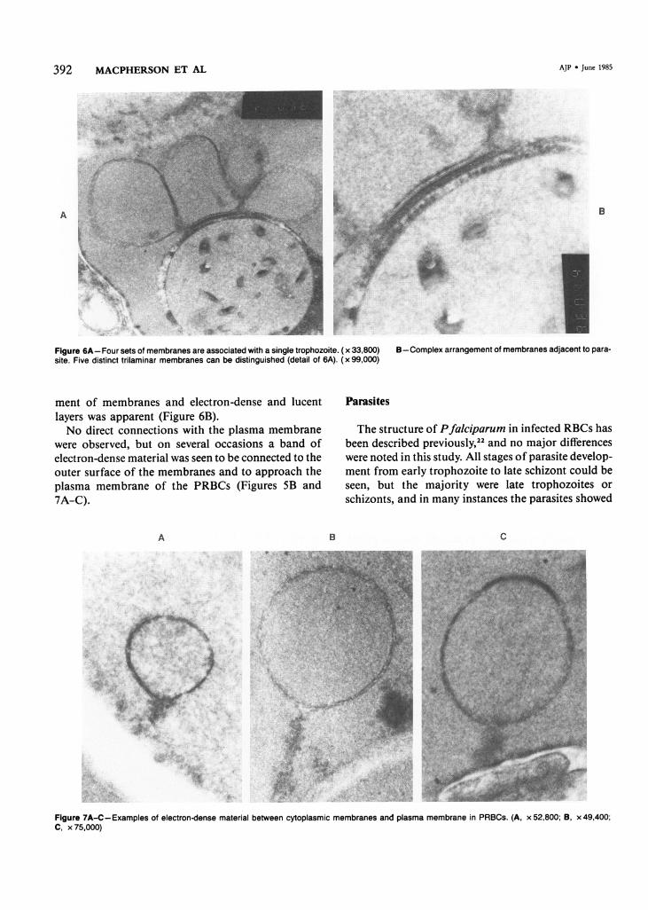
ment of membranes and electron-dense and lucentlayers was apparent (Figure 6B).No direct connections with the plasma membrane
were observed, but on several occasions a band ofelectron-dense material was seen to be connected to theouter surface of the membranes and to approach theplasma membrane of the PRBCs (Figures 5B and7A-C).
A
*~~~~ ~ ~~~~~~~~~~~~~~~~~~~~~~~~~~ .. . - x
V.~~~~~~~~~~~~~~~~~~~~,; 4.,- '*}o *.v A ........ *X>w.z iX $bML
- ,-<:aW~~~~~~~~~~~~~~~~~~~~~~~~~~~~~~~~~~~~~~~~~~~~~~~~~~~~~~~~~2
Parasites
The structure of Pfalciparum in infected RBCs hasbeen described previously,22 and no major differenceswere noted in this study. All stages of parasite develop-ment from early trophozoite to late schizont could beseen, but the majority were late trophozoites or
schizonts, and in many instances the parasites showed
B C
Figure 7A-C-Examples of electron-dense material between cytoplasmic membranes and plasma membrane in PRBCs. (A, x 52,800; B, x 49,400;C, x 75,000)
392 MACPHERSON ET AL
A
-A

HUMAN CEREBRAL MALARIA 393
A B
Figure 8A-Three PRBCs showing schizonts in a cerebral blood vessel. Pigment granules are conspicuous (-). (x 5000) B-Cerebral malaria. Acerebral venule is filled with a mass of degenerating material in which parasitic remains including malarial pigment are clearly visible (-). The endotheli-um of the venule appears to be intact. (x 8600)
morphologic signs of degeneration (Figure 3A). Inmany instances pigment was clearly present within theparasite (Figure 8A). On occasion, vessels appeared tobe blocked by a mass of necrotic material in which para-sites and RBCs could both be identified (Figure 8B).In these vessels, the parasites and RBCs showed moreadvanced degeneration than the blood vessels, indicat-ing that the degeneration was not a postmortem artifact.These degenerate parasites are somewhat similar mor-phologically to "crisis forms."
Interaction of RBCs With Vascular Endothelium
It has been suggested previously that PRBCs mayadhere to endothelial cells by their "knobs."',10 Wesought evidence to test this hypothesis. The observa-tions were as follows:
1) PRBCs were seen in intimate contact with en-dothelial cells at sites with and without knobs.
2) In the majority of points of contact a gap wasclearly visible between plasma membranes of thePRBCs and endothelial cells. Where no gap was visi-ble, it seemed likely that this was the result of tangen-tial sectioning. No clear evidence for membrane fusionwas obtained.
3) On several occasions, the contours of PRBC fol-lowed closely those of an endothelial cell but with arelatively large gap, probably due to shrinkage duringfixation. In such cases, strands of electron-dense mate-rial could sometimes be seen running between the two
cells, and the point of origin of such strands on thePRBC was very frequently a knob or area of increaseddensity (Figure 4C).
In an attempt to show that such strands arose pref-erentially from knobs, the points of origin of electron-dense strands between PRBCs and endothelial cells werecounted. On electron micrographs of 10 randomly cho-sen PRBCs displaying knobs, measurements were madeof the length of plasma membrane in the profile, thenumber of knobs, and the average length of the knobs.On average, knobs occupied 21.6% (± 5.5%) of the cellperimeter, whereas 64.1% of electron-dense strands (n= 64) arose from knobs, thus showing a 3-fold increasein the observed, compared with expected, frequencies.
4) In the majority of cases, the endothelial cellsshowed no changes at the point of contact, but occa-sionally an area of increased density was observed.Sometimes the endothelium appeared to be damagedopposite a PRBC knob, and it is thought that this mayrepresent damage occurring during tissue processing be-cause of the adhesion of PRBCs to endothelium.
Cells
Occasional neutrophils were seen, but other leuko-cytes were not observed in significant numbers eitherin or outside blood vessels. A striking negative obser-vation was the total absence of blood platelets, eithercirculating or in thrombi.
Vol. 119 * No. 3

394 MACPHERSON ET AL
A_ 4A 4 B
Figure 9A-Cerebral malaria. The endothelium of a cerebral venule is damaged, and a gap (-)extends to the basement membrane (B). (x 18,200) B3-Cerebral malaria. A ruptured cerebral venule has allowed the escape of PRBCs. The ends of the venule wall are marked by arrows. A PRBC ghostis present (*). (x4300)
Endothelial Cells
In many specimens, endothelial cell morphology waswell preserved, but in some vessels the cells showed ad-vanced degenerative changes with loss of the luminalmembrane and disorganization of the cytoplasm (Fig-ure 4B). In some specimens, all vessels showed suchchanges; but in others, vessels within 100, of a damagedvessel were unaffected, indicating that the degenerationwas unlikely to be postmortem. Occasionally, gaps in
... . . A h'4 .41.
otherwise healthy endothelium could be seen, and insome cases the basement membrane was exposed (Fig-ure 9A).On only one occasion was a total defect in a venule
wall observed (Figure 9B). In this case, PRBCs had es-caped in small numbers into the surrounding tissue. Noplatelets were seen in the damaged area.A striking feature of endothelial cells in many ves-
sels from patients with and without cerebral marlariawas the presence of numerous projections extending
_~~. B
,k -h,
s:trC~~~~~~~~~~~~a"; ft.KFigure IOA-Noncerebral malaria. Numerous fingerlike pseudopodia in a cerebral venule. (x 13,000) B-Cerebral malaria. Endothelial pseudopo-dia are apparently attached to a PRBC in a cerebral venule (-). (x 10,000)
AJP * June 1985
t

VOl. 119 * No. 3
Table 3-Frequency of PRBCs in Different Organs*
Organ
BrainHeartLungLiverKidney
Cerebral malaria
45.1 (2185) (7)19.8 (333) (3)4.2 (568) (4)
17.0 (200) (1)4.70 (830) (5)
Noncerebral malaria
19.4 (643) (6)23.5 (34) (1)35.7 (14) (1)0.67 (150) (1)5.03 (397) (3)
* Percentage of RBCs parasitized. Total number of RBCs counted isin first set of parentheses, followed by the number of patients.One-micron sections were examined with light microscopy. Twenty ves-
sels, or all vessels if the specimen was small, were examined. The totalnumber of RBCs and the proportion containing parasites were counted.
into the lumen of the vessel (Figure IOA). In some cases,such projections were seen in vessels apparently blockedby RBCs, and on many occasions the projections werein intimate contact with the parasitized cells (FigurelOB). Infrequently, endothelial cell projections were seenin vessels from control specimens, but these were neveras frequent nor as long as in malarial specimens.
Basement Membrane
The basement membrane was not noticeably thickerin blood vessels containing PRBCs than in control ves-sels, and there was no sign of deposits resembling im-mune complexes.
Frequency of Parasitized RBCs inBrain and Other Organs
Araldite-embedded sections l, thick were examinedunder oil immersion, and all blood vessels within thesection were identified. Profiles of all intravascularRBCs and the proportion containing recognizable para-sites were counted. Two hundred RBCs per section orthe total number in sections containing less than 200was counted. The results are shown in Table 3. Figure11 shows the relationship between the proportions of
80-
Figure 11 -Comparison of the frequencies ofparasitized RBCs in different organs from individualpatients. The percentage of parasitized RBCs with-in vessels in different organs was counted for in-dividual patients. The points represent thesepercentages. The lines join points obtained fromdifferent organs in the same patient.
08
coalco
0
m
bR
HUMAN CEREBRAL MALARIA 395
PRBCs in brain and other organs where samples wereavailable from the same patient. The lines join pointsindicating the percentage of parasitization in two or-gans from the same patient.The difference between the sizes of parasites and
erythrocytes means that not all sections through aPRBC will include part of the parasite. Because para-site size and shape change during development, it is notpracticable to make an accurate estimate of the correc-tion factor needed. Ratios of parasite/RBC diameterswere measured on 12 randomly selected electron micro-graphs. The mean ratio was 0.56, and only two cellshad ratios of over 0.7. This would suggest that at least30%o of sections through parasitized cells would not in-clude the parasite and that therefore the frequency ofparasitization in some cerebral vessels might approach70%o.
In all cases where samples were available, the orderof the degree of parasitization was brain > heart > liver,lung, kidney >> blood. It was apparent that somePRBCs in the liver were within Kupffer cells, but theproportion could not be determined (Figure 12A) be-cause of the small sample size.The great majority of PRBCs in the kidney were in
glomeruli, where some were clearly adherent to glomer-ular endothelium (Figure 12B). Very few PRBCs wereseen elsewhere in the kidney. There were no othersignificant abnormalities in the glomeruli.Few specimens of lung which included alveoli were
available. In the one case in which a detailed compari-son with brain was possible, some interesting differenceswere apparent. Thus, some alveolar blood vessels con-tained PRBCs, but the parasites appeared to be almostall in ring or early trophozoite stages (Figure 13A).Other vessels contained large numbers of inflammatorycells, including neutrophils, monocytes, and lympho-cytes (Figure 13B). In the example shown, there was noclinical evidence for aspiration pneumonia. On the cor-responding brain specimens, the majority of parasites
Brain Heart Brain LungBrain Kidney Brain Kidney

396 MACPHERSON ET AL
Figure 12A-Noncerebral malaria. Liver. A Kupffercell (K) contains several RBCs, of which some areparasitized (P). ( x 3600) B-Cerebral malaria.Kidney glomerulus. Several parasitized RBCs arepresent and appear adherent to the glomerular en-dothelium. A monocyte (M) is present in the lumen.(x 2600)
B
Figure 13A-Cerebral malaria. Lung. Parasitized RBCs are frequent but do not display conspicuous surface knobs. The parasites are relatively imma-ture; ring forms or early trophozoites (T). Compare this figure with Figure 15, from the sarme patient. (x 3600) B-Cerebral malaria. Lung. Samepatient as previous figure. Venules (V) are packed with large numbers of inflammatory cells, including neutrophil polymorphs (P), monocytes (M), andlymphocytes (L). (x 4600)
A
B
A
AJP -a June 1985
HUMAN CEREBRAL MALARIA 397
were late trophozoites or schizonts, PRBC "ghosts" werepresent, and no leukocytes were seen.
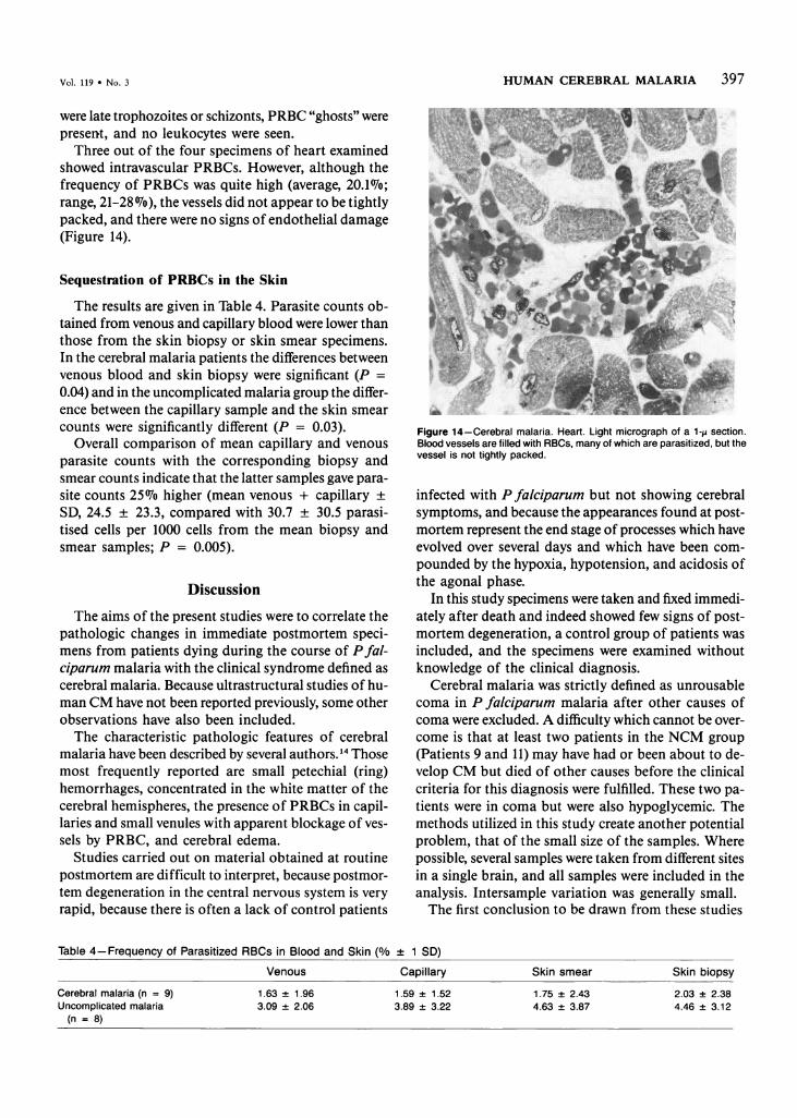
Three out of the four specimens of heart examinedshowed intravascular PRBCs. However, although thefrequency of PRBCs was quite high (average, 20.1%7o;range, 21-28 7o), the vessels did not appear to be tightlypacked, and there were no signs of endothelial damage(Figure 14).
Sequestration of PRBCs in the Skin
The results are given in Table 4. Parasite counts ob-tained from venous and capillary blood were lower thanthose from the skin biopsy or skin smear specimens.In the cerebral malaria patients the differences betweenvenous blood and skin biopsy were significant (P =0.04) and in the uncomplicated malaria group the differ-ence between the capillary sample and the skin smearcounts were significantly different (P = 0.03).
Overall comparison of mean capillary and venousparasite counts with the corresponding biopsy andsmear counts indicate that the latter samples gave para-site counts 25%o higher (mean venous + capillary ±SD, 24.5 ± 23.3, compared with 30.7 ± 30.5 parasi-tised cells per 1000 cells from the mean biopsy andsmear samples; P = 0.005).
Discussion
The aims of the present studies were to correlate thepathologic changes in immediate postmortem speci-mens from patients dying during the course of Pfal-ciparum malaria with the clinical syndrome defined ascerebral malaria. Because ultrastructural studies of hu-man CM have not been reported previously, some otherobservations have also been included.The characteristic pathologic features of cerebral
malaria have been described by several authors. 14 Thosemost frequently reported are small petechial (ring)hemorrhages, concentrated in the white matter of thecerebral hemispheres, the presence of PRBCs in capil-laries and small venules with apparent blockage of ves-sels by PRBC, and cerebral edema.
Studies carried out on material obtained at routinepostmortem are difficult to interpret, because postmor-tem degeneration in the central nervous system is veryrapid, because there is often a lack of control patients
:xr;'T . .NWFigure 14-Cerebral malaria. Heart. Light micrograph of a 1-p section.Blood vessels are filled with RBCs, many of which are parasitized, but thevessel is not tightly packed.
infected with P falciparum but not showing cerebralsymptoms, and because the appearances found at post-mortem represent the end stage of processes which haveevolved over several days and which have been com-pounded by the hypoxia, hypotension, and acidosis ofthe agonal phase.
In this study specimens were taken and fixed immedi-ately after death and indeed showed few signs of post-mortem degeneration, a control group of patients wasincluded, and the specimens were examined withoutknowledge of the clinical diagnosis.
Cerebral malaria was strictly defined as unrousablecoma in P falciparum malaria after other causes ofcoma were excluded. A difficulty which cannot be over-come is that at least two patients in the NCM group(Patients 9 and 11) may have had or been about to de-velop CM but died of other causes before the clinicalcriteria for this diagnosis were fulfilled. These two pa-tients were in coma but were also hypoglycemic. Themethods utilized in this study create another potentialproblem, that of the small size of the samples. Wherepossible, several samples were taken from different sitesin a single brain, and all samples were included in theanalysis. Intersample variation was generally small.The first conclusion to be drawn from these studies
Table 4-Frequency of Parasitized RBCs in Blood and Skin (%0/+ 1 SD)
Venous
Cerebral malaria (n = 9)Uncomplicated malaria
(n = 8)
1.63 ± 1.96
Capillary
1.59 ± 1.52
Skin smear
1.75 ± 2.43
Skin biopsy
2.03 ± 2.384.63 ± 3.87 4.46 ± 3.12
Vol. 119 * No. 3
3.09 -,-. 2.06 3.89 t 3.22
398 MACPHERSON ET AL
is that there is little evidence of extravascular pathol-ogy, either hemorrhagic or inflammatory. Few extravas-cular RBCs were seen, and some were present in theNCM group. No areas of hemorrhage compatible withthose described as "ring" hemorrhages were observed,probably because of the small size of samples and be-cause ring hemorrhages are most common in white mat-ter, whereas the majority of samples we examined camefrom gray matter.
Extensive destruction of the cerebral microvascula-ture is also unlikely on clinical grounds; survivors ofcerebral malaria make a complete recovery without evi-dence of neurologic deficit in more than 95% of cases,red cells are not found in the cerebrospinal fluid, andcomputed tomography of the brain is usually normal.23
Leukocytes were seen very rarely outside blood ves-sels in CM and NCM samples. Some, mainly neu-trophils, were seen within vessels, but their frequencydid not appear to be increased above normal. Theaccumulation of mononuclear cells, variously calledneuroglia, microglia, or macrophages, described in ce-rebral malaria24'25 was not observed; but such accumu-lations appear to be related mainly to areas of hemor-rhage and may represent a secondary phenomenon.The absence of granular deposits along basement
membranes of cerebral venules and of other evidenceof vasculitis, together with the absense of inflamma-tory cells, suggests that immune complexes are not pres-ent in the cerebral vessels of CM patients. Immuno-peroxidase staining of protease-digested formalin-fixedbrain sections from 10 cerebral malaria patients revealedno IgG or IgM deposition (M. J. Warrell, unpublishedobservations). The absence of any clinical evidence ofglomerulonephritis and the finding that serum comple-ment levels are normal except in the most seriously illpatients26 (S. Tharavanij and P. Malasit, unpublishedobservations) also argue against an immune-complex-mediated etiology.We conclude that there is no evidence for either acute
or chronic inflammation in human cerebral malaria,and that it is most unlikely that any immune processis involved.
These data contrast strongly with those seen in somerodent malarias,'3 where monocyte accumulation andemigration are conspicuous features of the early stagesof the disease. This suggests that the pathogenesis ofnervous system involvement in such models differs fromthat in humans and that there is no satisfactory animalmodel for human cerebral malaria.The essential structural components of thrombi are
platelets and fibrin,27 but platelets were strikingly ab-sent from cerebral vessels in CM and NCM patients.
In the one example of a breached vessel seen, there wereno platelets present. This correlates with the throm-bocytopenia described in Pfalciparum malaria.26 Stri-ated fibrillar deposits, probably of fibrin, were presentin a small proportion of cerebral vessels from both CMand NCM patients, but in general, deposits were notseen in vessels with 3 + packing of RBCs, suggestingthat thrombosis plays no part in the etiology of CM.Widespread thrombosis of small vessels would also notbe compatible with the complete neurologic recoveryseen in most CM patients.
Overt endothelial damage was present in a propor-tion of vessels from all CM specimens, and also in somevessels from some non-CM specimens. In 2 CM cases(Patients 1 and 4) the overall morphology of the braintissue suggested postmortem degeneration, and almostall vessels were affected. In the others, only a propor-tion of vessels were damaged, and adjacent vessels hadapparently normal endothelium. Because this patternwas observed in both CM and non-CM cases, it isdifficult to relate it to the etiology of CM.The strongest correlation between vascular pathol-
ogy and CM in the present study is the tightness of pack-ing of RBCs in capillaries and venules, where it is quiteclear that CM patients have a higher average densityof packing and a considerably larger proportion oftightly packed vessels. It is difficult to account for thisdegree of packing and for the higher proportion ofPRBCs seen in CM, compared with NCM, patients, to-gether with the striking differences in the frequency ofPRBCs between the brain and other organs, unless itis due to selective adhesion of PRBCs to cerebral en-dothelium. Leakage of blood plasma from small ves-sels in amounts large enough to account for the tightpacking might be expected to cause cerebral edema,which is apparently absent except as an agonalphenomenon23 and would be expected to occur in otherorgans, without barriers to diffusion, to at least as greatan extent as in the brain.
Decreased deformability of PRBCs8 might similarlybe predicted to lead to their accumulation equally invessels of all organs, and to their retention in capillariesand upstream vessels, rather than in venules.We conclude that selective adhesion of PRBCs to ce-
rebral vascular endothelium is the most probable ex-planation.
It is widely accepted that the "knobs" on parasitizedRBCs may be involved in adhesion to endothelialcells,9"0 and studies of the binding of infected RBCsto cultured human endothelial cells34 and amelanoticmelanoma cells35 strongly suggest that knobs form thesites of attachment of sequestered RBCs, but it is of-
AJP * June 1985
HUMAN CEREBRAL MALARIA 399
ten difficult to be certain that the morphologic changesdescribed at regions of RBC/endothelial cell apposi-tion, where the radius of curvature of the cells is lo-cally decreased, are not due to local tangential section-ing resulting in the loss of the appearance of normalmembrane relationships. The observations in the pres-ent study of strands of electron-dense material runningpreferentially between "knobs" and endothelial mem-branes do provide concrete evidence that the knobs areindeed related to adhesion "in viva"
For adhesion to occur, PRBCs need to come into con-tact with endothelium. A striking and unexpected ob-servation was the presence of numerous endothelialpseudopodia in cerebral vessels of both CM and NCMpatients, many in intimate contact with and apparentlyadherent to PRBCs. Similar pseudopodia were not seenin control brain sections or in blood vessels from otherorgans in CM and NCM patients.
Clark29 has suggested that cerebral malaria may becaused by increased vascular permeability due to en-dothelial damage, which may be caused by active oxy-gen metabolites such as hydrogen peroxide, superoxideanion, and the hydroxyl radical and that one manifesta-tion of endothelial damage is the development of pseu-dopodia. Such metabolites can be released by neu-trophils and by activated macrophages,30 but there isa conspicuous absence of these cells in the brains ofCM patients. Although it is known that some malarialparasites can generate H202, production is inhibited atlow oxygen tensions,31 and there is no evidence at pres-ent that they can produce other oxygen metabolites. Inaddition, the endothelial pseudopodia described byClark differ morphologically from those observed inthe present study.
Cerebral endothelial pseudopodia have been de-scribed in at least two other situations. Yu et a132 fedchickens on a diet high in linoleic acid and deficientin vitamin E and observed flaplike pseudopodia in ce-rebral vessels. Schmahl et a133 induced endotoxin shockin cats and observed microvilluslike structures develop-ing from cerebral vessels, whereas vessels in other or-gans showed overt endothelial damage.The development of pseudopodia by cerebral en-
dothelium may be peculiar to these vessels and repre-sent a nonspecific response to injurious stimuli. Thegreater endothelial surface area available for PRBC con-tact in cerebral vessels because of pseudopodia mightexplain the higher frequency of PRBCs in these ves-sels. It is also possible that plasma membrane speciali-zation in cerebral endothelium leads to the specificityof interactions, but in the absence of cultured en-dothelium from the brain this hypothesis is difficult to
test. The expression of binding molecules by PRBCscould be dependent on the availability of anabolic sub-strates such as purines and glucose, and purines maybe more readily available in hypoxic cerebral capillaries.
Thus, we suggest that the high degree of red cellparasitization seen in the cerebral capillaries resultsfrom selective accumulation of PRBCs within these ves-sels due to the increased adhesiveness of PRBCs forcerebral endothelial cells, perhaps related to the pres-ence of endothelial pseudopodia, and that the neu-rologic syndrome results from hypoxia and otherbiochemical disturbances caused by mechanical ob-struction of the vessels by cells containing activelymetabolizing parasites. Immune or inflammatory pro-cesses appear to play no part in the pathogenesis ofthe syndrome.
Other Observations
The mechanism by which parasite proteins are trans-ported to the plasma membrane of the RBC is not clear.The observation of parasite-associated membraneswithin the cytoplasm of PRBCs and the presence ofelectron-dense material between these membranes andthe plasma membrane suggests that they may possiblybe involved in transport of material from the parasiteto the plasma membrane of the cell, but morphologicobservations alone cannot provide decisive evidence.The term "crisis form" describes degenerative changes
in intraerythrocytic parasites at the time of crisis in mon-key malarias, possibly caused by tumor necrosis factoror free oxygen radicals.38 In this study a proportion ofPRBCs within cerebral vessels contained parasites show-ing morphologic signs of damage similar to crisis forms.Inr the absence of local inflammatory cells able to re-lease mediators, it is possible that the parasites weredamaged elsewhere in the circulation before becomingarrested in the cerebral vessels.Our observation that parasite counts were higher in
skin smears or biopsies than in peripheral blood sam-ples lends some support to the claim by Chinese work-ers that blood sampled by intradermal skin pricks isa more sensitive method of diagnosing Pfalciparummalaria than the conventional peripheral blood smearor even bone-marrow aspiration.39
Conclusions
1) The clinical syndrome of cerebral malaria corre-lates closely with the presence of tight packing of cere-bral venules and capillaries by PRBCs. In the absenceof thrombosis or inflammation, the most likely cause
Vol. 119 * No. 3

400 MACPHERSON ET AL AJP * June 1985
is selective adhesion of PRBCs to cerebral endothelialcells.
2) There is a selective accumulation of PRBCs in ce-rebral vessels, as compared with vessels in other organsexamined. Sequestration is least apparent in the skin.
3) There is morphologic evidence of adhesion ofPRBCs to cerebral endothelium via their surface knobs.
4) There is no evidence that overt endothelial dam-age plays an important part in this condition, althoughthe presence of endothelial pseudopodia may be relatedto the selective accumulation of PRBCs in the brain.
5) There may be no need to involve factors other thanmetabolic disturbances consequent upon a reductionin cerebral microcirculatory flow to account for the clin-ical features of CM.
References
1. Daroff RB, Deller JJ, Kastl AJ, Blocker WW: Cerebralmalaria. J Am Med Assoc 1967, 202:679-682
2. Warrell DA, Looareesuwan S, Warrell MJ: Dexametha-sone proves deleterious in cerebral malaria: A doubleblind study in 100 comatose patients. N Engl J Med 1982,306:313-319
3. Rigdon RH: The pathological lesions in the brain inmalaria. South Med J 1944, 37:687-694
4. Sheehy TW, Reba RC: Complications of falciparummalaria and their treatment. Ann Intern Med 1967, 66:807-809
5. Thomas JD: Cerebral malaria: Clinical histopathologi-cal correlation. Trop Geogr Med 1971, 23:232-238
6. Marsden PD, Bruce-Chwatt LJ: Cerebral malaria, Topicsin Tropical Neurology. Edited by RW Hornabrook.Philadelphia, F.A. Davis, 1975, pp 29-44
7. Warrell DA: The impact of clinical investigation on twoThird World diseases: cerebral malaria and louse-bornerelapsing fever, Advanced Medicine. Edited by KB Saun-ders. Pitman Medical, 1983, pp 99-111
8. Miller LH, Chien S, Usami S: Decreased deformabilityof Plasmodium coatneyi-infected red cells and its possi-ble relationship to cerebral malaria. Am J Trop Med Hyg1972, 21:133-137
9. Trager W, Rudzinska MA, Bradbury PC: The fine struc-ture of Plasmodiumfalciparum and its host erythrocytesin natural malarial infections in man. Bull WHO 1966,35:883-885
10. Luse SA, Miller LH: Plasmodium falciparum malaria:Ultrastructures of parasitized erythrocytes in cardiac ves-sels. Am J Trop Med Hyg 1971, 20:655-660
11. Maegraith BG: Pathogenic processes in malaria, ThePathology of Parasitic Diseases. Edited by AER Taylor.Oxford, Blackwell, 1966, pp 15-32
12. Migasena P, Maegraith BG: The blood brain barrier inP. knowlesi infection. Trans R Soc Trop Med Hyg 1965,59:2
13. Rest JR: Cerebral malaria in inbred mice: I. A new modeland its pathology. Trans R Soc Trop Med Hyg 1982,76:410-415
14. Aikawa M, Suzuki M, Gutierrez Y: Pathology of malaria,Malaria. Edited by JP Kreier. Vol 2. New York, AcademicPress, 1980, pp 47-102
15. de Brito T, Barone AA, Faria RM: Human liver biopsyin P. falciparum and P. vivax: A light and electron mi-croscopy study. Virchows Arch [Pathol Anat] 1969,348:220-229
16. Hartenbower DL, Kantor GL, Rosen VJ: Renal failuredue to acute glomerulonephritis during falciparummalaria: Case report. Mil Med 1972, 137:74-76
17. Gutierrez Y, Aikawa M, Fremount HN, Sterling CR: Ex-perimental infection of Aotus monkeys with Plasmodiumfalciparum: Light and electron microscopic changes. AnnTrop Med Parasitol 1976 70:25-44
18. Yoeli M, Hargreaves BJ: Brain capillary blockage pro-duced by a virulent strain of rodent malaria. Science 1974,184:572-573
19. Richardson KC, Jarret L, Finke EH: Embedding in epoxyresins for ultrathin sectioning in electron microscopy.Stain Technol 1960, 35:313
20. Reynolds ES: The use of lead citrate at high pH as anelectron-opaque stain in electron microscopy. J Cell Biol1963, 17:208
21. Graham RC Jr, Karnovsky MJ: The early stages of ab-sorbtion of injected horse-radish peroxidase in the prox-imal tubules of mouse kidney: Ultrastructural cytochemis-try by a new technique. J Histochem Cytochem 1966,14:291
22. Aikawa M, Seed TM: Morphology of plasmodia, Malaria.Edited by JP Krier. Vol 1. New York Academic Press,1980, pp 285-344
23. Looareesuwan S, Warrell DA, White NJ, SutharasamaiP, Chanthavanich P, Sundaravej K, Juel-Jensen BE, Bun-nag D, Harinasuta T: Do patients with cerebral malariahave cerebral oedema? A computed tomography study.Lancet 1983, 1:434-437
24. Durck H: Uber die bei Malaria perniciosa comatosaaufretenden Veranderungen des Zentralnervensystems.Arch Schiffs-Trop Hyg 1917, 21:117-132
25. Dhayagude RG, Puranare NM: Autopsy study of cere-bral malaria with special reference to malarial granuloma.Arch Pathol 1943, 36:550-558
26. Dennis LH, Eichelberger JW, Inman MM, Conrad M:Depletion of coagulation factors in drug-resistant Plas-modium falciparum malaria. Blood 1967, 29:713-721
27. French JE: Formation and fate of a thrombus, Athero-sclerosis: Proceedings of the Second International Sym-posium. Edited by RJ Jones. New York, Springer-Verlag,1970, pp 80-88
28. Devakul KA, Harinasuta T, Reid HA: l25l-labelled fibrino-gen in cerebral malaria. Lancet 1966, 2:886
29. Clark IA, Hunt NH, Cowden WB: Immunopathologyof malaria, Immunology, Immunopathology and Immu-noprophylaxis of Parasitic Infectins. Edited by EJLSoulsby. CRC press (In press)
30. Badwey JA, Karnovsky ML: Active oxygen species andthe function of phagocytic leukocytes. Ann Rev Biochem1980, 49:695
31. Friedman MJ: Expression of inherited resistance tomalaria in culture, Malaria and the Red Cell. Ciba Foun-dation Symposium. Vol 14. London, Pitman, 1983, pp196-201
32. Yu WA, Yu MC, Young PA: Ultrastructural changes inthe cerebrovascular endothelium induced by a diet highin linoleic acid and deficient in vitamin E. Exp Mol Pathol1974, 21:289-299
33. Schmahl, FW, Schlote W, Urbascher B, Betz E, HeuserD, Heckers H: Reactions of endothelial cells of cerebralvessels in endotoxic shock. Proceedings of the 5th An-nual Conference on Shock, 1982, pp 192-193
34. Udeinya IJ, Schmidt JA, Aikawa M, Miller LH, GreenI. Falciparum malaria-infected erythrocytes specificallybind to cultured human endothelial cells. Science 1981,213:555-557
35. Schmidt JA, Udeinya IJ, Leech JH, Hay RJ, Aikawa M,Barnwell J, Green I, Miller LH. Plasmodiumfalciparummalaria: A amelanotic melanoma cell lines bears recep-

Vol. 119 * No. 3 HUMAN CEREBRAL MALARIA 401
tors for the knob ligand on infected erythrocytes. J ClinInvest 1982, 70:379-386
36. Smith DH, Theakston RDG: Comments on the ultrastruc-ture of human erythrocytes infected with Plasmodiummalariae. Ann Trop Med Parasitol 1970, 64:329
37. David PH, Hommel M, Miller LH, Udeinya IJ, OliginoLD. Surface alterations of erythrocytes in Plasmodiumfalciparum malaria: II. Spleen- and antibody-modulationof infected erythrocyte cytoadherence. Proc Nat Acad SciUSA 1983, 80:507-509
38. Jensen JB, Boland MT, Allan JS, Carlin JM, van de WaaJA, Divo AA, Akood MAS: Association between humanserum-induced crisis forms in cultured Plasmodiumfal-ciparum and clinical immunity to malaria in Sudan. In-fect Immun 1983, 41:1302
39. Li QQ, Guo X, Jian H, Fan T, Huang W: Developmen-tal state of Plasmodium falciparum in the intradermal,peripheral, and medullary blood of patients with cere-bral malaria. Natl Med J China 1983, 63:692-693
AcknowledgmentsWe are extremely grateful for the excellent technical as-
sistance of Mrs. Kamolrat Silamut in Bangkok and Ms. M.Bergin-Cartwright and Mr. C. Jenkins in Oxford. Mrs. P. R.Woodward typed the manuscript both accurately and quickly.Dr. F H. C. Marriott, Department of Biomathematics, helpedwith the statistical analysis of data.