NIF (Neurite-Inducing Factor): A Novel Peptide Inducing Neurite ...
Upload
sandra-perezCategory
view
213download
0
Journal of NeuroVirology, 13: 139–149, 2007c© 2007 Journal of NeuroVirologyISSN: 1355-0284 print / 1538-2443 onlineDOI: 10.1080/13550280701191459
A protein encoded by the bovine herpesvirus 1 openreading frame E gene induces neurite-likemorphological changes in mouse neuroblastoma cellsand is expressed in trigeminal ganglionic neurons
Sandra Perez,1,2 Florencia Meyer,2,3 Gail Henderson,1,2 Yunquan Jiang,1,2 Simon Sherman,2,4 Alan Doster,1,2
Melissa Inman,1,2 and Clinton Jones1,2
1Department of Veterinary and Biomedical Sciences, 2Nebraska Center for Virology, and 3School of Biological Sciences,University of Nebraska, Lincoln, Nebraska, USA 4Nebraska Informatics Center for the Life Sciences, Eppley CancerInstitute, University of Nebraska Medical Center, Omaha, Nebraska, USA
Bovine herpes virus 1 (BHV-1), like other α-herpesvirinae subfamily members,establishes latency in sensory neurons. Periodically BHV-1 reactivates from la-tency, resulting in virus shedding and spread to uninfected cattle. Although re-activation from latency does not usually lead to recurrent disease, the latency-reactivation cycle is crucial for virus transmission. The latency-related (LR)RNA is abundantly expressed during latency, and expression of a LR encodedprotein is necessary for dexamethasone-induced reactivation from latency incattle. Within LR promoter sequences, a small open reading frame (ORF) wasidentified (ORF-E) that is antisense to the LR-RNA, and downstream of thebICP0 gene. ORF-E transcription is consistently detected in trigeminal ganglia(TG) of latently infected calves, suggesting ORF-E expression plays a role inthe latency-reactivation cycle. Polyclonal antiserum directed against an ORF-Epeptide or the entire ORF-E protein specifically recognizes the nucleus of sen-sory neurons in TG of latently infected calves. The ORF-E peptide–specific an-tiserum also recognizes a protein when mouse neuroblastoma cells (neuro-2A)are transfected with an ORF-E expression construct. In contrast to the growthinhibiting properties of the LR gene, stably transfected ORF-E–expressing cellswere obtained. Neuro-2A cells stably transfected with a plasmid expressingORF-E induced morphological changes that resembled neurite-like projections.In contrast, neurite-like projections were not observed following transfectionof neuro-2A cells with an empty vector. These studies suggest that a proteinencoded by ORF-E has the potential to alter the physiology or metabolism ofneuronal cell types, which may be important for long-term latency. Journal ofNeuroVirology (2007) 13, 139–149.
Keywords: bovine herpesvirus type 1; latency; neurite outgrowth; ORF-E
Introduction
Bovine herpes virus 1 (BHV-1) establishes life-long latency in ganglionic neurons of the periph-
Address correspondence to Clinton Jones, Deparment of Veteri-nary and Biomedical Sciences, University of Nebraska, Lincoln,Fair Street at East Campus Loop, Lincoln, NE 68583-0905, USA.E-mail: [email protected]
This work was supported by two USDA grants (2005-01554 and2006-01627), and a Public Health Service grant 1P20RR15635 tothe Nebraska Center for Virology.
Received 24 October 2006; revised 21 November 2006; accepted20 December 2006.
eral nervous system, primarily the trigeminal ganglia(TG), after initial replication in mucosal epithelium.Virus reactivation and spread to susceptible calvesoccur after natural or corticosteroid-induced stress(Rock et al, 1992; Sheffy and Davies, 1972). In-fection can cause conjunctivitis, pneumonia, gen-ital disorders, abortions, and an upper respiratoryinfection referred to as “shipping fever” (Tikooet al, 1995). Because BHV-1–associated pathogene-sis and shipping fever cost the U.S. cattle industryat least $500 million/year (Bowland and Shewen,2000), developing better modified live vaccines isdesirable.

BHV-1 ORF-E protein and neurite outgrowth140 S Perez et al
The latency-related (LR)-RNA is abundantly ex-pressed in latently infected neurons (Kutish et al,1990; Rock et al, 1987, 1992). A fraction of LR-RNA ispolyadenylated and alternatively spliced in TG, sug-gesting this RNA is translated into more than one pro-tein (Devireddy and Jones, 1998; Hossain et al, 1995).LR gene products inhibit cell growth (Hossain et al,1995; Jiang et al, 1998) and apoptosis (Ciacci-Zanellaet al, 1999). A mutant BHV-1 strain that containsthree stop codons near the beginning of the LR-RNAwas constructed to test whether LR proteins play arole in virus growth in cattle (Inman et al, 2001).Calves infected with the LR mutant exhibit dimin-ished clinical symptoms, and virus shedding in theeye (Inman et al, 2001), TG (Inman et al, 2002), or ton-sils (Perez et al, 2005), compared to calves infectedwith wild-type (wt) BHV-1 or the LR rescued virus.wt BHV-1 or the LR rescued virus, but not the LRmutant virus, reactivate from latency following treat-ment with dexamethasone (DEX). During the transi-tion from acute infection to latency (establishment oflatency), higher levels of apoptosis occur in TG neu-rons of calves infected with the LR mutant relative tocalves infected with wt BHV-1 (Lovato et al, 2003).These studies indicate that wt expression of LR geneproducts is required for the latency-reactivation cyclein cattle.
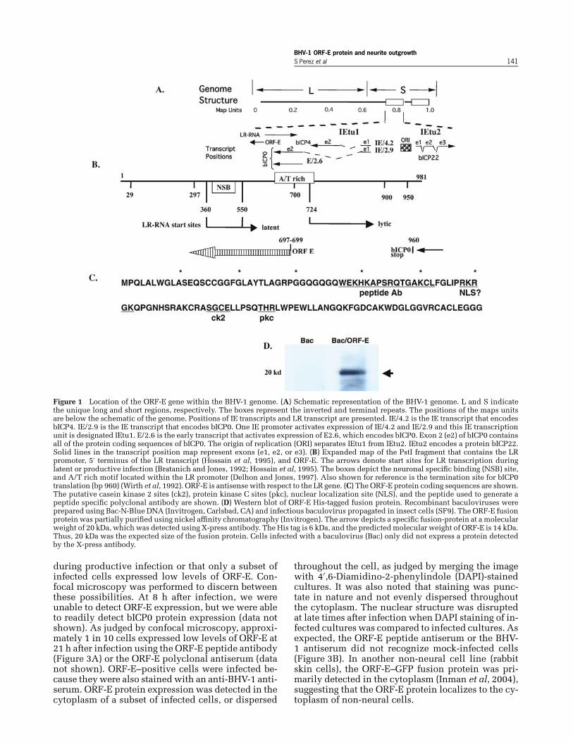
A small open reading frame (ORF) within theLR promoter was identified, and designated ORF-E (Figure 1B and C) (Inman et al, 2004). ORF-E isantisense to the LR transcript, downstream of thebICP0 ORF, but does not overlap bICP0 (Figure 1B).A transcript encompassing ORF-E is expressed in6/6 calves latently infected with BHV-1 (Inman et al,2004). The LR promoter contains a neuronal-specificbinding domain (NSB; Figure 1B) and has neuronal-specific transcriptional activity (Bratanich et al, 1992;Bratanich and Jones, 1992; Delhon and Jones, 1997;Jones et al, 1990). Sequences that activate expressionof the LR-RNA during productive infection contain along AT-rich motif (Figure 1B), suggesting these samepromoter sequences also activate ORF-E RNA expres-sion. ORF-E is 134 amino acids (aa) long and con-tains potential casein kinase 2 (ck2) as well as pro-tein kinase C (pkc) phosphorylation sites (Figure 1C).A BLAST analysis did not reveal strong similarity toknown cellular proteins.
In this study, we generated antiserum directedagainst the entire ORF-E or an ORF-E peptide. TheORF-E antiserum recognized a specific protein intransfected mouse neuroblastoma cells (neuro-2A).Unlike the LR gene, ORF-E does not induce cell cy-cle arrest, and consequently stably transfected cellswere obtained. Neuro-2A cells stably transfected withORF-E frequently have neurite-like projections, andare morphologically distinct compared to neuro-2Acells stably transfected with the empty vector. TheORF-E antiserum specifically reacted with TG neu-rons of infected calves, but when reactivation from la-tency was induced by addition of DEX, ORF-E protein
expression was not readily detected. These studiessuggest that the ORF-E protein is expressed in latentlyinfected neurons, and that ORF-E alters the morphol-ogy and/or physiology of neuronal cell types.
Results
ORF-E protein expression in transfectedneuro-2A cellsTo test whether the ORF-E gene encodes a protein, itwas necessary to generate specific antiserum directedagainst ORF-E. With this objective in mind, a peptideidentified as the most immunogenic region of ORF-Ewas synthesized (Figure 1C), and injected into rab-bits to generate an ORF-E specific antiserum (ORF-Epeptide antiserum). In addition, the entire ORF-E wascloned into a baculovirus expression system, ORF-Ewas overexpressed (Figure 1D), purified, and the pu-rified ORF-E protein used to generate rabbit poly-clonal antiserum (ORF-E polyclonal antiserum).
The protein coding sequences of ORF-E (Figure 1C)were cloned upstream of the green fluorescent protein(GFP) coding sequences of phMGFP. The ORF-E/GFPplasmid or the blank GFP vector was transfectedinto mouse neuroblastoma cells (neuro-2A). As pre-viously demonstrated (Inman et al, 2004), the ORF-E/GFP fusion protein was localized to the nucleus whenexpressed in neuro-2A cells (Figure 2A). In con-trast, neuro-2A cells transfected with the GFP expres-sion vector (phMGFP) contained GFP randomly dis-tributed throughout the cell (Figure 2B). The ORF-Epeptide antiserum specifically recognized a proteinwith an approximate molecular weight of 40 kDa at24 h after transfection (Figure 2C). Based on the pre-dicted size of ORF-E (approximately 15 kDa) and GFP(25 kDa), this was the expected size of the fusionprotein. At 48 or 72 h after transfection, the ORF-E/GFP fusion protein was detectable, but at lower lev-els. As expected, the ORF-E peptide antiserum didnot specifically recognize a 40 kDa protein whenneuro-2A cells were transfected with the blank ex-pression vector. When neuro-2A cells were trans-fected with an expression vector containing onlyORF-E, the ORF-E peptide antiserum (Figure 2D) orthe ORF-E polyclonal antiserum (data not shown)specifically detected a protein that migrates at ap-proximately 14 kDa. Similar levels of protein werepresent in samples because when the blots werestripped and reprobed with a β-actin antibody simi-lar levels of protein were detected (Figure 2C and D;β-actin panels).
ORF-E protein expression during productiveinfectionIn contrast to the results in transiently transfectedcells, the ORF-E polyclonal or peptide antiserum didnot consistently detect ORF-E expression in produc-tively infected Madin Darby bovine kidney (MDBK)cells. This suggested that ORF-E was not expressed
BHV-1 ORF-E protein and neurite outgrowthS Perez et al 141
Figure 1 Location of the ORF-E gene within the BHV-1 genome. (A) Schematic representation of the BHV-1 genome. L and S indicatethe unique long and short regions, respectively. The boxes represent the inverted and terminal repeats. The positions of the maps unitsare below the schematic of the genome. Positions of IE transcripts and LR transcript are presented. IE/4.2 is the IE transcript that encodesbICP4. IE/2.9 is the IE transcript that encodes bICP0. One IE promoter activates expression of IE/4.2 and IE/2.9 and this IE transcriptionunit is designated IEtu1. E/2.6 is the early transcript that activates expression of E2.6, which encodes bICP0. Exon 2 (e2) of bICP0 containsall of the protein coding sequences of bICP0. The origin of replication (ORI) separates IEtu1 from IEtu2. IEtu2 encodes a protein bICP22.Solid lines in the transcript position map represent exons (e1, e2, or e3). (B) Expanded map of the PstI fragment that contains the LRpromoter, 5′ terminus of the LR transcript (Hossain et al, 1995), and ORF-E. The arrows denote start sites for LR transcription duringlatent or productive infection (Bratanich and Jones, 1992; Hossain et al, 1995). The boxes depict the neuronal specific binding (NSB) site,and A/T rich motif located within the LR promoter (Delhon and Jones, 1997). Also shown for reference is the termination site for bICP0translation (bp 960) (Wirth et al, 1992). ORF-E is antisense with respect to the LR gene. (C) The ORF-E protein coding sequences are shown.The putative casein kinase 2 sites (ck2), protein kinase C sites (pkc), nuclear localization site (NLS), and the peptide used to generate apeptide specific polyclonal antibody are shown. (D) Western blot of ORF-E His-tagged fusion protein. Recombinant baculoviruses wereprepared using Bac-N-Blue DNA (Invitrogen, Carlsbad, CA) and infectious baculovirus propagated in insect cells (SF9). The ORF-E fusionprotein was partially purified using nickel affinity chromatography (Invitrogen). The arrow depicts a specific fusion-protein at a molecularweight of 20 kDa, which was detected using X-press antibody. The His tag is 6 kDa, and the predicted molecular weight of ORF-E is 14 kDa.Thus, 20 kDa was the expected size of the fusion protein. Cells infected with a baculovirus (Bac) only did not express a protein detectedby the X-press antibody.
during productive infection or that only a subset ofinfected cells expressed low levels of ORF-E. Con-focal microscopy was performed to discern betweenthese possibilities. At 8 h after infection, we wereunable to detect ORF-E expression, but we were ableto readily detect bICP0 protein expression (data notshown). As judged by confocal microscopy, approxi-mately 1 in 10 cells expressed low levels of ORF-E at21 h after infection using the ORF-E peptide antibody(Figure 3A) or the ORF-E polyclonal antiserum (datanot shown). ORF-E–positive cells were infected be-cause they were also stained with an anti-BHV-1 anti-serum. ORF-E protein expression was detected in thecytoplasm of a subset of infected cells, or dispersed
throughout the cell, as judged by merging the imagewith 4′,6-Diamidino-2-phenylindole (DAPI)-stainedcultures. It was also noted that staining was punc-tate in nature and not evenly dispersed throughoutthe cytoplasm. The nuclear structure was disruptedat late times after infection when DAPI staining of in-fected cultures was compared to infected cultures. Asexpected, the ORF-E peptide antiserum or the BHV-1 antiserum did not recognize mock-infected cells(Figure 3B). In another non-neural cell line (rabbitskin cells), the ORF-E–GFP fusion protein was pri-marily detected in the cytoplasm (Inman et al, 2004),suggesting that the ORF-E protein localizes to the cy-toplasm of non-neural cells.

BHV-1 ORF-E protein and neurite outgrowth142 S Perez et al
Figure 2 ORF-E expression in transfected mouse neuro-2A cells. Neuro-2A cells were transfected with the ORF-E/GFP plasmid (2 μgof plasmid; A) or the GFP blank expression vector (phMGFP, 2 μg; B). At 24 h after transfection, cells were viewed under a fluorescentmicroscope as described in Materials and Methods. The magnification for A and B is 100×. The image on the left is fluorescence onlyand the right image is the merge between fluorescence and phase contrast. These images are representative of several independentstudies. (C) Neuro-2A cells were transfected with the ORF-E/GFP plasmid or the GFP empty expression vector (10 μg/100-mm dish). Atthe designated times after transfection, cells were processed for Western blotting. (D) Neuro-2A cells were transfected with an ORF-Eexpression plasmid, or an empty expression vector (pcDNA3.1–). For C and D, the ORF-E peptide antiserum was used to probe the Westernblot. Approximately 50 μg protein was loaded per lane. The blot was stripped, and reprobed with an antibody directed against β-actin(Santa Cruz Biotechnology, Santa Cruz, CA).
The ORF-E gene induces morphological changesin neuro-2A cellsThe LR gene inhibits apoptosis and cell growthfollowing transfection of certain cell types(Ciacci-Zanella et al, 1999; Geiser and Jones,2005; Schang et al, 1996). Conversely, ORF-E over-expression did not have a dramatic effect on thefrequency of apoptosis (induction or inhibition), nor
Figure 3 ORF-E protein expression in productively infected bovine kidney cells. Confocal microscopy was used to test whether ORF-Ewas expressed in productively infected bovine kidney (MDBK) cells. MDBK cells were infected with BHV-1 (MOI = 0.75 pfu/cell), andat 21 h after infection (A) cultures were fixed and confocal microscopy performed as described previously (Zhang and Jones, 2005). TheORF-E peptide antiserum or a BHV-1 antiserum was used to detect infected cells. Nuclei were detected by DAPI staining. As a control,mock-infected cells were used (B).
did it inhibit cell growth. Following transfection ofneuro-2A cells with an ORF-E expression plasmidcontaining a neomycin resistance gene (pcDNA3.1–),we were able to readily select stably transfected cellsusing G418. In these cultures, low levels of ORF-Eprotein expression were detected. When ORF-Estably transfected neuro-2A cells were subcultured,we consistently observed that these cells contained

BHV-1 ORF-E protein and neurite outgrowthS Perez et al 143
Figure 4 Stable transfection of neuro-2A cells with an ORF-E expression plasmid. A mammalian expression plasmid (pcDNA3.1–)containing ORF-E or the empty vector (pcDNA) was transfected into neuro-2A cells. At 24 h after transfection, stably transfected cellswere incubated with the antibiotic G418 to select for transfected cells. Shown are representative fields (100× magnification) following3 weeks of incubation with medium containing G418.
long extensions resembling neurites (Figure 4; ORF-Epanels). In many cases, multiple extensions wereobserved, and many of these neurite-like extensionswere quite long. Cells that contained neurite-likeprojections were viable as judged by trypan blueexclusion (data not shown), and they appeared togrow in culture. In sharp contrast to ORF-E, the LRgene inhibited growth of neuro-2A cells, but doesnot induce neurite-like growth (Geiser and Jones,2005). When neuro-2A cells were transfected witha blank expression vector, neurite-like projectionswere not frequently observed (Figure 4; pcDNA).Furthermore, there was a morphological differencein cells stably transfected with ORF-E versus cellsstably transfected with pcDNA3.1–. In contrast to theresults obtained with neuro-2A cells, ORF-E did notalter the morphology of a bovine kidney or testiclecell line (unpublished data).
The ORF-E antiserum recognizes a protein in TGneurons of latently infected calvesThin sections were prepared from TG of latently in-fected calves (60 days after infection), and the ability
of the ORF-E antisera to specifically recognize thesesections was tested. The ORF-E peptide antiserum(Figure 5A and B) or the ORF-E polyclonal antiserum(Figure 5C) specifically recognized a protein presentin the nucleus of TG neurons. Neither ORF-E anti-serum reacted with all neurons from latently infectedcalves. In contrast to the results obtained with TG sec-tions prepared from latently infected calves, the ORF-E polyclonal antiserum (Figure 5D) as well as theORF-E peptide antiserum (data not shown) did notreact with the nucleus of sensory neurons from mock-infected calves. Even when the section cut throughthe nucleus of neurons from mock-infected calves,only the counter-staining of nucleoli was detected.
The TG used for this study were obtained fromcalves at 60 days after infection, and these calves wereused in previously published studies. We know thesecalves are latently infected because infectious virusis not detected in nasal or ocular swabs (Inman et al,2002; Perez et al, 2005; Winkler et al, 2000a, 2000b,2002). Furthermore, we are unable to detect a latetranscript (gC) by reverse transcriptase–polymerasechain reaction (RT-PCR) using total RNA prepared

BHV-1 ORF-E protein and neurite outgrowth144 S Perez et al
Figure 5 ORF-E is expressed in TG of latently infected calves. TG thin sections were prepared from two different latently infectedcalves (60 days after infection). (A and B) Sections were stained with the ORF-E peptide antiserum (1:1000 dilution). (C) Sections werestained with the ORF-E polyclonal antiserum (1:200 dilution). (D) TG sections from mock-infected calves were probed with the ORF-Epolyclonal antiserum (1:100 dilution). (E and F) A bICP0-specific antibody (1:100 dilution) was incubated with TG thin sections preparedfrom latently infected calves. This antibody specifically reacts with the bICP0 protein (Inman et al, 2002; Zhang and Jones, 2001, 2005).Magnification was 20× for A to D, and 40× for E and F. Arrows denote neurons that were positively stained.
from TG of these calves at 60 days after infection(Inman et al, 2004). As another confirmatory assay toprove these calves were latently infected, TG sectionswere stained with an antibody that specifically recog-nizes bICP0 (Zhang and Jones, 2005). The bICP0 anti-serum did not react with TG sections prepared fromlatently infected calves (Figure 5E and F), adding fur-ther support that these calves were latently infected.In summary, this study suggested that a protein en-coded by ORF-E was expressed in TG neurons ofcalves latently infected with BHV-1.
ORF-E protein expression during acute infectionand reactivation from latencyAdditional studies were performed to test whetherORF-E was expressed in TG of acutely infectedcalves. Seven days after infection is the peak of acuteinfection in calves (Inman et al, 2001, 2002; Schangand Jones, 1997; Winkler et al, 2002), and thus thistime was used for examining ORF-E expression inTG. For these studies, the ORF-E peptide antiserumwas used. ORF-E protein expression was detected in asubset of neurons at 7 days after infection (Figure 6Aand B). As was observed during latency (Figure 6D),nuclei of positive neurons were stained with theORF-E peptide antiserum. If TG sections preparedfrom mock-infected calves were incubated with theORF-E peptide antiserum, staining was not detected(Figure 6C).
Reactivation from latency can be initiated in la-tently infected calves or rabbits if a single injec-tion of DEX is given (Ackermann et al, 1982; Inman
et al, 2002; Jones et al, 2000; Rock et al, 1992; Sheffyand Davies, 1972). Because LR-RNA expression isdramatically reduced during reactivation from la-tency (Rock et al, 1992), we tested whether ORF-Eprotein expression was reduced in calves latently in-fected with BHV-1 following DEX treatment. At 24 hafter DEX treatment, we detected ORF-E protein ex-pression near the periphery of TG neurons using theORF-E peptide antiserum (Figure 6E). It did not ap-pear that the neuron itself was stained; rather satelliteor infiltrating lymphocytes were apparently stained.At 48 h after DEX treatment, a few neurons were de-tected in which ORF-E was detected in the nucleus(Figure 6F).
Previous studies demonstrated that a subset of TGneurons express a protein recognized by a peptideantibody directed against the LR ORF-2 (Jiang et al,1998). Studies were performed to compare the per-centage of neurons that express ORF-E versus ORF-2.A polyclonal antibody directed against ORF-2 (Jianget al, 2004) was used for these studies because this isthe first ORF downstream of the LR-RNA, and ORF-2–specific antibodies specifically recognize a subsetof neurons during latency (Jiang et al, 1998). Thesestudies indicated that during acute infection (7 daysafter infection), approximately 6% of the total neu-rons were stained with the ORF-E peptide antiserum,but less than 1% of the total neurons were stainedwith the ORF-2 antiserum (Figure 7). During latency(60 days after infection), approximately 8% of the to-tal neurons were stained with the ORF-E antiserum.In contrast, only 2% of the total neurons were ORF-2

BHV-1 ORF-E protein and neurite outgrowthS Perez et al 145
Figure 6 Analysis of ORF-E protein expression during acute infection, and reactivation from latency. TG thin sections were preparedfrom acutely infected calves (7 days after infection; A, B). (C) TG from mock-infected calf. (D) TG section from a latently infected calf(60 days after infection). (E) TG section from a latently infected calf (60 days after infection) that was treated with 100 mg DEX (IV injection)for 24 h to initiate reactivation from latency (Inman, 2002). (F) TG section from a latently infected calf (60 days after infection) that wastreated with 100 mg DEX (IV injection) for 48 h to initiate reactivation from latency. TG sections were stained with the ORF-E peptideantibody (1:1000 dilution). Magnification for all sections was 20×. The images are representative of many slides that were obtained fromat least two calves for each time point. Arrows denote cells positively stained by the antiserum.
positive. At 48 h after dexamethasone induced reac-tivation from latency, ORF-2 protein expression wasnot detected, which correlated with a dramatic re-duction in the levels of LR-RNA (Rock et al, 1992).To avoid the uncertainty of whether ORF-E was de-tected in neurons or non-neuronal cells at 24 h DEXtreatment, only those samples at 48 h after DEX treat-ment were used to count ORF-E positive neurons.Although the percentage of neurons that expressedORF-E was reduced, we were able to detect ORF-Eprotein expression in TG neurons following DEX-
0
1
2
3
4
5
6
7
8
posi
tive
neu
rons
(%
)
acuteinfection
latency 48h-DEX
ORF-E positive
ORF 2 positive
Figure 7 Estimation of the % of neurons that express ORF-E. Thenumber of neurons expressing ORF-E or ORF-2 in four fields ofthree different TG sections was counted to estimate the % of neu-rons expressing each of the respective proteins. Approximately1000 neurons were counted for each antibody. ORF-E–positiveneurons are denoted by the black column, and ORF-2 by the whitecolumn. For these studies, TG from two different latently infectedcalves were used.
induced reactivation from latency. In summary, thesestudies suggested that a higher percentage of TG neu-rons expressed detectable levels of ORF-E protein rel-ative to ORF-2.
Discussion
Collectively, this study and other published studies(Jiang et al, 1998; Perez et al, 2006) suggested at leasttwo viral proteins, ORF-E and proteins encoded bythe LR gene, are expressed in a subset of TG neuronsduring BHV-1 latency. This is in sharp contrast toHSV-1 latency because the latency-associated tran-script does not appear to encode a protein (Jones,2003). We hypothesize that proteins encoded byORF-E and the LR gene have the potential to be recog-nized by the immune system. For long-term latency tooccur, a mechanism must exist that inhibits immunerecognition of neurons expressing ORF-E or LR pro-teins. LR gene products, directly or indirectly, maybe one factor that inhibits immune recognition in TGbecause higher inflammatory infiltrates are detectedin TG of calves acutely infected with the LR mutantversus wt BHV-1 (Perez et al, 2006).
The ORF-E protein was primarily expressed inthe nucleus of TG sensory neurons and transfectedneuro-2A cells, whereas in non-neural cell typesORF-E was primarily in the cytoplasm. Most nu-clear proteins contain a nuclear localization signal(NLS) that has one or two clusters of basic aminoacids (K/R) (Kalderon et al, 1984; Lanford and Butel,1984; Richter et al, 1985; Robbins et al, 1991). Al-though ORF-E contains one motif (RKRGK) that re-sembles a NLS (Figure 1C), it is not known if this

BHV-1 ORF-E protein and neurite outgrowth146 S Perez et al
motif functions as a NLS or whether neuronal factorspromote ORF-E nuclear localization. The presenceof ORF-E in the nucleus of neuronal cells suggestedORF-E is a regulatory protein that may stimulateneurite-like growth in neuro-2A cells. However, ORF-E probably does not promote true neurite outgrowthbecause neurite formation is generally associatedwith terminal differentiation and cell cycle with-draw. Thus, ORF-E may cooperate with other cellu-lar factors to promote neurite outgrowth followinginfection.
We assumed ORF-2 would be expressed in a higherpercentage of TG neurons during latency because it isthe first ORF within the LR gene. However, our stud-ies suggested ORF-E was expressed in a higher per-centage of neurons. Although one could argue thatthe ORF-E antibody had a higher titer than the ORF-2 antibody, this was not the case when Western blotstudies were performed with ORF-E or ORF-2 fusionproteins overexpressed from a Baculovirus vector(data not shown). Unless the ORF-E antiserum worksbetter with formalin-fixed and paraffin-embeddedsections of TG, there is not a dramatic difference inthe titers of the ORF-E or ORF-2 antiserum used forthese studies.
The HSV-1 AL (antisense to LAT) gene is presentin an analogous position on the HSV-1 genome as theORF-E gene (Perng et al, 2002). An AL protein hasbeen described (Perng et al, 2002); but there is virtu-ally no similarity between ORF-E and the AL protein.To date, it is not known if the AL protein or RNA isexpressed in latently infected TG, or if the AL genestimulates neurite-like projections in neuro-2A cells.
Does ORF-E play a role in the latency-reactivationcycle of BHV-1? Although we believe that ORF-Eplays a role in the latency-reactivation cycle, it is notessential because mutating the LR gene inhibits reac-tivation from latency (Inman et al, 2002), in part, byreducing the number of apoptotic neurons at the endof acute infection (Lovato et al, 2003). The LR mutantvirus that was constructed contains stop codons at the5′ terminus of the first LR ORF (Inman et al, 2001),and consequently the LR mutant virus can synthesizeORF-E transcripts. When latently infected calves orrabbits are treated with DEX for 24 h, extensive vi-ral gene expression occurs in TG neurons, and theninfectious virus is detected in nasal or ocular swabs(Rock et al, 1992; Winkler et al, 2000b, 2002). It isunlikely that ORF-E directly enhances reactivationfrom latency because the number of neurons express-ing ORF-E decrease during the first 24 h after DEXtreatment of latently infected calves. ORF-E also doesnot appear to directly inhibit apoptosis in infectedneurons because ORF-E does not inhibit apoptosis intransient transfection assays (data not shown). Col-lectively, these studies suggest that ORF-E has an an-cillary role in mediating certain aspects of the estab-lishment and or maintenance of of long-term latency.We further predict that the ability of ORF-E to in-duce neurite-like growth in neurons plays a role in
restoring mature neuronal functions after infection.To definitively prove what role ORF-E plays in thelatency-reactivation cycle of BHV-1, it will be neces-sary to construct an ORF-E–null mutant BHV-1 strain,and an ORF-E/LR mutant virus. These respective mu-tant virus strains will then be compared to the phe-notype of the LR mutant virus and wt BHV-1.
Materials and methods
Cells and virusBovine kidney cells (MDBK, ATCC CCL-22) weregrown in Earl’s modified Eagle’s medium supple-mented with 5% fetal bovine serum (FBS), penicillin(10 U/ml), and streptomycin (100 μg/ml). MDBK cellswere split 1:6 every 2 to 3 days.
The BHV-1 Cooper strain (wt virus) was obtainedfrom the National Veterinary Services Laboratory,Animal and Plant Health Inspection Services, Ames,Iowa. Viral stocks were prepared by infecting MDBKcells with an multiplicity of infection (MOI) of0.001 from a plaque-purified virus and subsequentlytitrated on MDBK cells.
Neuro-2A cells (ATCC catalogue CCL131) aremouse neuroblastoma cells, which were grown inEarle’s minimal essential medium supplementedwith 10% FBS, penicillin (10 U/ml), and strepto-mycin (100 μg/ml).
Animal experimentsBHV-1–free crossbred calves (∼250 kg) were ran-domly assigned and housed in isolation rooms toprevent cross contamination. Calves were sedatedwith xylazine (approximately 50 mg/50 kg bodyweight; Bayer, Shawnee Mission, KS). Calves werethen inoculated with 1 ml of a solution containing1 × 107 pfu/ml of virus in each nostril and eye, drop-wise without scarification, for a total of 4 × 10⁷ pfuper animal as described previously (Inman et al,2001, 2002; Schang and Jones, 1997; Winkler et al,2002). Experiments using animals were performed inaccordance with the American Association of Labora-tory Animal Care guidelines. Calves were housed un-der strict isolation containment and given antibioticsbefore and after BHV-1 infection to prevent secondarybacterial infection. All calves designated as being la-tently infected were euthanized at 60 days after infec-tion, and these calves were not shedding infectiousvirus from the nasal cavity or the eye (Inman et al,2002; Perez et al, 2005; Winkler et al, 2000a, 2000b,2002). DEX was used to initiate reactivation from la-tency as described previously (Inman et al, 2002). TGwere fixed in formalin, paraffin embedded, and thinsections cut for immunohistochemistry.
Cloning ORF-E into plasmid vectorsA plasmid containing the entire LR region (Figure 1)was digested to completion with PstI. The fragmentcontaining ORF-E was agarose gel purified and used

BHV-1 ORF-E protein and neurite outgrowthS Perez et al 147
for ligation. The correct orientation of the PstI in-sert was determined by restriction digest. To studycellular localization of ORF-E expression, ORF-Ewas cloned into the mammalian expression vector,Monster Green Fluorescent Protein (phMGFP Vec-tor; Promega, catalogue number E6421) to generatean ORF-E–GFP fusion protein. PCR was performedon viral genomic DNA using primers that containedunique restriction sites for amplification of ORF-E aspreviously described (Inman et al, 2004).
Antibody productionTo generate a recombinant Baculovirus that expressesORF-E, a PCR-amplified product (Inman et al, 2004)was cloned into pBlueBacHis2 such that ORF-E wasin frame with the histidine tag. A recombinant bac-ulovirus was then constructed that expressed theORF-E fusion protein. The recombinant baculovirusstrain was grown in SF9 insect cells using proceduresdescribed by Invitrogen. Recombinant baculovirusORF-E constructs were characterized by testing forORF-E protein expression using the Express antibodythat recognizes the X-press epitope at the 5′ termi-nus of LR protein sequences (R910-25; Invitrogen).Large-scale expression was carried out in 2-L flasksseeded with SF9 cells at a density of 2 × 10⁶ cells/mlin a total volume of 1000 ml. At 4 days after infec-tion, cells were pelleted by centrifuging for 30 minat 8000 rpm (Beckman J2-21 centrifuge, JA-10 rotor),and suspended in 20 ml of Guanidinium Lysis Buffer(6 M guanidine hydrochloride, 20 mM sodium phos-phate, 500 mM NaCl, pH 7.8). Suspended cells werethen passed through an 18-gauge needle four timesto lyse cells and shear cellular DNA. The ORF-E fu-sion protein was partially purified using nickel chro-matography. The ORF-E fusion protein was furtherpurified using preparative sodium dodecyl sulfate–polyacrylamide gel electrophoresis (SDS-PAGE), thefusion protein band excised, eluted, and then injectedinto rabbits to generate a specific polyclonal anti-serum (Animal Pharm Services; CA). This serum is re-ferred to as the ORF-E polyclonal antiserum through-out the text. The antiserum directed against LR ORF-2was previously described (Jiang et al, 2004).
A peptide corresponding to the immunogenic re-gion of ORF-E was synthesized and a polyclonalantibody directed against this peptide generated inrabbits by Biosource (Camarillo, CA). The immuno-genic region of ORF-E was predicted using programson the ExPASy website (ProtScale) and the Predict-Protein server (http://www.predictprotein.org). Thisserum is referred to as the ORF-E peptide antiserumthroughout the text.
Confocal microscopy to examine subcellularlocalization of ORF-EMDBK cells were cultured in 4-well Lab-Tek slides,and then infected for 21 h with wt BHV-1 (MOI = 0.75pfu/cell). Cultures were washed twice with EMEM(without serum) and fixed in cold 100% ethanol at
−20◦C for 5 min. After a brief wash with Tris-bufferedsaline (TBS), slides were blocked in 3% bovine serumalbumin (BSA) in TBS for 1 h and then incubatedwith the primary antibodies for 2 h at room tempera-ture (RT). The primary antibody consisted of a mix-ture of anti-ORF-E peptide antibody (dilution 1:100)and anti-BHV-1 antibody (dilution 1:100) (AmericanBioResearch Laboratories) in TBS 0.05% Tween 20(TBS-T), 1% BSA. Three washes of 10 min each withTBS-T were followed by incubation with secondaryantibody mix for 1 h at RT in the dark. The secondaryantibody mixture consisted of donkey anti-rabbitimmunoglobulin G (IgG) conjugated to Cy2 (dilution1:50) and goat anti-donkey IgG conjugated to Cy5(dilution 1:50). Three washes of 10 min each withTBS-T were followed by incubation with DAPI stainfor 10 min. Slides were then mounted with gel(Sigma) and a coverslip. Images were collected usinga Bio-Rad confocal laser-scanning microscope (MRC-1024ES) with excitation/emission at 488/520 nm.
Transient expression and Western blot analysisNeuro-2A cells were transfected with 10 μg of therespective expression plasmids using TransIT trans-fection reagents (Mirus; Madison, WI) as described bythe manufacturer. At the designated times after trans-fection, cells were collected, and lysed in 500 μl of1 × SDS sample buffer (50 mM Tris-HCl, pH 6.8, 10%glycerol, 2% SDS, 5% β–mecaptoethanol). Cell ex-tract was boiled for 5 minutes and the supernatantused for SDS-PAGE. Western blots were performed asdescribed previously using the designated antibodies(Henderson et al, 2002; Jiang et al, 1998).
ImmunohistochemistryTissue sections were deparaffinized, rehydrated ingraded ethanol, and then treated with 3% hydrogenperoxide in phosphate-buffered saline (PBS) (pH 7.4)for 10 min to inactivate endogenous peroxidase. Afterwashing in PBS, tissue sections were digested withproteinase K (DAKO) for 20 minutes at 37◦C. Non-specific binding was blocked by incubation with 5%normal goat serum, 0.25% BSA in PBS for 45 min atroom temperature. Endogenous biotin was blockedby treatment of sections with avidin/biotin block-ing reagents (Vector Labs). Tissue samples were in-cubated overnight at 4◦C with the ORF-E polyclonalantiserum (1:100 or 1:200 dilution), or with the ORF-E peptide antiserum (1:1000 dilution). Slides werewashed three times in PBS before addition of bi-otinylated secondary anti-rabbit immunoglobulin G(IgG) antibody (Vector Labs) for 30 min at room tem-perature. Slides were then incubated with freshlyprepared substrate (Vector NovaRed substrate kit forperoxidase; Vector Labs), rinsed with distilled wa-ter, and counterstained in methyl green. Tissues frommock-infected calves, and incubation of infected tis-sue sections in the absence of primary or secondaryantibodies, were used as controls to confirm thespecificity of the test.

BHV-1 ORF-E protein and neurite outgrowth148 S Perez et al
References
Ackermann M, Peterhans E, Wyler R (1982). DNA of bovineherpesvirus type 1 in the trigeminal ganglia of latentlyinfected calves. Am J Vet Res 43: 36–40.
Bowland SL, Shewen PE (2000). Bovine respiratorydisease: commercial vaccines currently available inCanada. Can Vet J 41: 33–48.
Bratanich AC, Hanson ND, Jones CJ (1992). The latency-related gene of bovine herpesvirus 1 inhibits the activityof immediate-early transcription unit 1. Virology 191:988–991.
Bratanich AC, Jones CJ (1992). Localization of cis-actingsequences in the latency-related promoter of bovineherpesvirus 1 which are regulated by neuronal cell typefactors and immediate-early genes. J Virol 66: 6099–6106.
Ciacci-Zanella J, Stone M, Henderson G, Jones C (1999).The latency-related gene of bovine herpesvirus 1 in-hibits programmed cell death. J Virol 73: 9734–9740.
Delhon G, Jones C (1997). Identification of DNA sequencesin the latency related promoter of bovine herpes virustype 1 which are bound by neuronal specific factors.Virus Res 51: 93–103.
Devireddy LR, Jones C (1998). Alternative splicing of thelatency-related transcript of bovine herpesvirus 1 yieldsRNAs containing unique open reading frames. J Virol 72:7294–7301.
Geiser V, Jones C (2005). The latency related gene en-coded by bovine herpesvirus 1 encodes a small regula-tory RNA that inhibits cell growth. J NeuroVirol 11: 563–570.
Henderson G, Peng W, Jin L, Perng G-C, Nesburn AB,Wechsler SL, Jones C. (2002). Regulation of caspase 8-and caspase 9-induced apoptosis by the herpes simplexvirus latency-associated transcript. J NeuroVirol 8: 103–111.
Hossain A, Schang LM, Jones C (1995). Identification ofgene products encoded by the latency-related gene ofbovine herpesvirus 1. J Virol 69: 5345–5352.
Inman M, Lovato L, Doster A, Jones C (2001). A mutationin the latency-related gene of bovine herpesvirus 1 leadsto impaired ocular shedding in acutely infected calves.J Virol 75: 8507–8515.
Inman M, Lovato L, Doster A, Jones C (2002). A mutationin the latency related gene of bovine herpesvirus 1 in-terferes with the latency-reactivation cycle of latency incalves. J Virol 76: 6771–6779.
Inman M, Zhou J, Webb H, Jones C (2004). Identification ofa novel transcript containing a small open reading framethat is expressed during latency, and is antisense to thelatency related gene of bovine herpes virus 1 (BHV-1).J Virol 78: 5438–5447.
Jiang Y, Hossain A, Winkler MT, Holt T, Doster A, Jones C(1998). A protein encoded by the latency-related geneof bovine herpesvirus 1 is expressed in trigeminal gan-glionic neurons of latently infected cattle and interactswith cyclin-dependent kinase 2 during productive in-fection. J Virol 72: 8133–8142.
Jiang Y, Inman M, Zhang Y, Posadas NA, Jones C (2004).A mutation in the latency related gene of bovine her-pesvirus 1 (BHV-1) inhibits protein expression of a pro-tein from open reading frame 2 (ORF-2) and an adjacentreading frame during productive infection. J Virol 78:3184–3189.
Jones C (2003). Herpes simplex virus type 1 and bovineherpesvirus 1 latency. Clin Micro Rev 16: 79–95.
Jones C, Delhon G, Bratanich A, Kutish G, Rock D (1990).Analysis of the transcriptional promoter which regulatesthe latency-related transcript of bovine herpesvirus 1.J Virol 64: 1164–1170.
Jones C, Newby TJ, Holt T, Doster A, Stone M,Ciacci-Zanella J, Webster CJ, Jackwood MW (2000).Analysis of latency in cattle after inoculation with atemperature sensitive mutant of bovine herpesvirus 1(RLB106). Vaccine 18: 3185–3195.
Kalderon D, Roberts BL, Richardson WD, Smith AE (1984).A short amino acid sequence able to specify nuclear lo-cation. Cell 39: 499–509.
Kutish G, Mainprize T, Rock D (1990). Characterizationof the latency-related transcriptionally active region ofthe bovine herpesvirus 1 genome. J Virol 64: 5730–5737.
Lanford RE, Butel JS (1984). Construction and characteri-zation of an SV40 mutant defective in nuclear transportof T antigen. Cell 37: 801–813.
Lovato L, Inman M, Henderson G, Doster A, Jones C (2003).Infection of cattle with a bovine herpesvirus 1 (BHV-1)strain that contains a mutation in the latency relatedgene leads to increased apoptosis in trigeminal gangliaduring the transition from acute infection to latency.J Virol 77: 4848–4857.
Perez S, Inman M, Doster A, Jones C (2005). Latency-realated gene encoded by bovine herpesvirus 1 promotesvirus growth and reactivation from latency in tonsils ofinfected calves. J Clin Micro 43: 393–401.
Perez, SL Lovato, Zhou J, Doster A, Jones C (2006). Com-parison of inflammatory infiltrates in trigeminal gangliaof cattle infected with wild type BHV-1 versus a virusstrain containing a mutaition in the LR (latency-related)gene. J Neurovirol 12: 392–397.
Perng G-C, Maguen B, Jing L, Mott KR, Osorio N, SlaninaSM, Yukht A, Ghiasi H, Nesburn AB, Inman M,Henderson G, Jones C, Wechsler SL (2002). A novel her-pes simplex virus type 1 (HSV-1) transcript (AL-RNA)antisense to the 5′ end of LAT (latency associated tran-script) produces a protein in infected rabbits. J Virol 76:8003–8010.
Richter JD, Young P, Jones NC, Krippl B, Rosenberg M,Ferguson B (1985). A first exon-encoded domain of E1Asufficient for posttranslational modification, nuclear lo-calization, and induction of adenovirus E3 promoter ex-pression in Xenopus oocytes. Proc Natl Acad Sci USA82: 8434–8438.
Robbins J, Dilworth SM, Laskey RA, Dingwall C (1991). Twoindependent basic domains in nucleoplasmin nucleartageting sequences: identification of a class of bipartitenuclear targeting sequences. Cell 64: 615–623.
Rock D, Lokensgard J, Lewis T, Kutish G (1992). Character-ization of dexamethasone-induced reactivation of latentbovine herpesvirus 1. J Virol 66: 2484–2490.
Rock DL, Beam SL, Mayfield JE (1987). Mapping bovineherpesvirus type 1 latency-related RNA in trigeminalganglia of latently infected rabbits. J Virol 61: 3827–3831.
Schang LM, Hossain A, Jones C (1996). The latency-relatedgene of bovine herpesvirus 1 encodes a product whichinhibits cell cycle progression. J Virol 70: 3807–3814.

BHV-1 ORF-E protein and neurite outgrowthS Perez et al 149
Schang LM, Jones C (1997). Analysis of bovine herpesvirus1 transcripts during a primary infection of trigeminalganglia of cattle. J Virol 71: 6786–6795.
Sheffy BE, Davies DH (1972). Reactivation of a bovine her-pesvirus after corticosteroid treatment. Proc Soc ExpBiol Med 140: 974–976.
Tikoo SK, Campos M, Babiuk LA (1995). Bovine her-pesvirus 1 (BHV-1): biology, pathogenesis, and control.Adv Virus Res 45: 191–223.
Winkler MT, Doster A, Sur JH, Jones C (2002). Analysisof bovine trigeminal ganglia following infection withbovine herpesvirus 1. Vet Microbiol 86: 139–155.
Winkler MT, Schang LS, Doster A, Holt T, Jones C (2000a).Analysis of cyclins in trigeminal ganglia of calves in-fected with bovine herpesvirus-1. J Gen Virol 81: 2993–2998.
Winkler MTC, Doster A, Jones C (2000b). Persistenceand reactivation of bovine herpesvirus 1 in the ton-sil of latently infected calves. J Virol 74: 5337–5346.
Wirth UV, Fraefel C, Vogt B, Vlcek C, Paces V, SchwyzerM (1992). Immediate-early RNA 2.9 and early RNA 2.6of bovine herpesvirus 1 are 3′ coterminal and encodea putative zinc finger transactivator protein. J Virol 66:2763–2772.
Zhang Y, Jones C (2001). The bovine herpesvirus 1immediate-early protein (bICP0) associates with histonedeacetylase 1 to activate transcription. J Virol 75: 9571–9578.
Zhang Y, Jones C (2005). Identification of functional do-mains within the bICP0 protein encoded by BHV-1.J Gen Virol 86: 879–886.