A prospective study on the cystoscopic findings in the ...
51
A PROSPECTIVE STUDY ON THE CYSTOSCOPIC FINDINGS IN THE INITIAL EVALUATION OF GROSS HAEMATURIA AT KENYATTA NATIONAL HOSPITAL (KNH) By DR. OTIENO SIMBA RAYMOND M.B, Ch. B (NAIROBI) A DISSERTATION SUBMITTED IN PART FULFILLMENT FOR THE DEGREE OF MASTER OF MEDICINE IN SURGERY OF THE UNIVERSITY OF NAIROBI. University ct NAIROBI Library 0393083 1 2006
Transcript of A prospective study on the cystoscopic findings in the ...

A PROSPECTIVE STUDY ON TH E CYSTOSCOPIC
FINDINGS IN TH E INITIAL EVALUATION OF
GROSS HAEMATURIA A T K EN YATTA NATIONAL
HOSPITAL (KNH)
By
DR. O TIEN O SIMBA RAYMOND M.B, Ch. B (NAIROBI)
A DISSERTATION S U B M ITTED IN PART FU LFILLM EN T
FOR TH E DEGREE OF M ASTER OF M EDICINE IN
SURG ERY OF T H E U N IV ER S ITY OF NAIROBI.
University c t NAIROBI Library
0393083 1
2006

D ECLAR ATIO N
I certify that this dissertation is my original work and has never been submitted for the
This dissertation has been submitted for examination with our approval as University
supervisors
MR. FRANCIS OWILLAH. M B, Ch.B, M.Med (Surgery). Nairobi
CONSULTANT GENERAL AND UROLOGICAL SURGEON
LECTURER UNIVERSITY OF NAIROBI
P O BOX 19676
MR. PETER NGUGI MUNGAI M B, Ch.B, M.Med (Surgery). Nairobi
CONSULTANT GENERAL AND UROLOGICAL SURGEON
SENIOR LECTURER UNIVERSITY OF NAIROBI
P O BOX 19676
NAIROBI.
Date
11
A CKN O W LED G M EN T
I wish to express my sincere appreciation to:
i) My supervisors Mr. P.N. Mungai and Mr. F. Owillah for their criticism and
guidance in the preparation of this dissertation.
ii) The Ministry of Health for sponsorship to the M.Med course
iii) The Kenyatta National Hospital Ethics and Research Committee for
permission to carry out the study.
iv) My Personal friend Paul Anywayo for his social support.
111

T A B L E OF C O N TE N TS
PAGE
TITLE — ------------------------------------------------------------------------- 0)
DECLARATION-------------------------------------------------- (ii)
ACKNOWLEDGMENT------------------------------------------------- ----- (Hi)
TABLE OF CONTENTS------------------------------------------------------ (iv)
ABSTRACT------------------------------------------------------------------------------ (v)
INTRODUCTION-------------------------------------------------------------- 1
HISTORICAL BACKGROUND-------------------------------------------- 2
REVIEW OF LITERATURE------------------------------------------------- 4
STUDY OBJECTIVES-------------------------------------------------------- 15
JUSTIFICATION OF THE STUDY---------------------------------------- 16
PATIENTS AND METHODS------------------------------------------------ 17
RESULTS------------------------------------------------------------------------- 20
DISCUSSION-------------------------------------------------------------------- 33
CONCLUSION------------------------------------------------------------------ 38
RECOMMENDATIONS------------------------------------------------------ 39
APPENDIX (i) REFERENCES----------------------------------40
(ii) DATA COLLECTION FO R M ------------- 44
iv

S T U D Y A B S TR A C T
Design: A prospective observational study over a duration o f 6 months
Objective: To document the main cystoscopic findings in the initial evaluation o f gross haematuria
Setting: Kenyatta National Hospital, a Teaching and Referral Hospital situated in Nairobi City - the Capital of Kenya.
Duration: From 3181 May 2005 - 30 November 2005 (Inclusive)
Study Group Patients aged 13 years and above presenting with gross haematuria from whom an informed consent was obtained
Method: Approval was obtained from the Hospitals Ethics and Research committee. Patients underwent evaluation consisting of demographics, basic blood tests, analysis of urine, imaging by ultrasonography . IVU was performed in some cases. The results were entered into a data sheet and statistical analysis was performed using SPSS version 10.
Results: Sixty patients were recruited into the study the male to female ratio was 2:1. The mean age was 42.4 years with a range o f 18 to 84 years. Cystoscopy was diagnostic in 95% of the cases. Malignancies were detected in 46.6% of the cases with a peak at 60 to 70 years. Most bladder tumours were located on the lateral walls and all primary bladder tumours were transitional cell carcinomas. No malignancy was detected below the age o f 35 years.
Conclusion: 1) Cystoscopy and ultrasonography are the most useful investigations in the evaluation of gross haematuria and patients above 40 years of age stand a high risk o f being diagnosed with a malignancy.
Recommendations: All patients presenting with gross haematuria shouldundergo cystoscopy and ultrasonography.
v

IN TR O D U C TIO N
Haematuria is a serious symptom of urological disease. It often heralds the presence o f an
underlying malignancy o f the urinary trac t1 ‘ \ Gross haematuria is alarming and obvious
to the naked eye; it has a reported community prevalence of 2.5% and is estimated to
account for 4 to 20% o f all urological visits4
Direct visualization o f ureteral orifices and mucosa o f the urethra, prostate and bladder is
achieved using a cystoscope. The procedure can be extended to obtain biopsy and
definitive surgical treatment offered for conditions such as stone in the bladder or distal
ureter, superficial bladder cancers, benign prostatic enlargement and urethral strictures. A
bladder wash out (Barbotaged) specimen obtained at cystoscopy increases cytologic
sensitivity for malignant cells 5 Flexible cystoscopy can be performed under local
anaesthesia in the Doctor’s Office with minimal discomfort and morbidity 6-9 while
providing equivalent or superior diagnostic accuracy 10. Cystoscopy has got a sensitivity
o f about 90% in the detection of bladder tumours n . There is a general consensus
amongst many urologists that cystoscopy can not be safely omitted even in situations
where a lesion is detected on imaging of the upper urinary tract u .
1

H ISTO R IC AL BACKGR OUND
Cystoscopy was first introduced by Phillip Bozzini in 1806; his technique was later
improved by Antonin Desormeaus and Joseph Grunfield 13. The first cystoscopes had no
incorporated irrigation channels; cystoscopy had to be interrupted to introduce a catheter
to fill or to empty the bladder. To circumvent this, some endoscopists used air as an
examination medium, the endoscopes were simple tubes with no optical system of
magnifying lenses.
A ir Cystoscopy
The first air cystoscope was designed by Diedrich Rutenberg from Viena in 1876, this
was an open speculum closed by a glass window from the examiners end Light was
reflected into the bladder via a concave forehead mirror 14.
In 1888, Karl Pawlik from Prague improved his endoscope by incorporating a light
source at the tip of the instrument and tubes for irrigating the bladder mucosa; he was
able to perform several endoscopic procedures such as removal of bladder stones and
resection o f bladder tumours l5.
In 1892, Haward Kelly using an instrument of smaller diameter performed cystoscopy
under local cocaine anaesthesia. In 1898, he modified his instrument to a length of 15cm
and diameter 8mm and performed cystoscopy in males 16
In 1909, Leo Burger from New York designed the first cysto urethroscope
2

'
Cystoscopy with Bladder Irrigation
At the turn of the second decade of the twentieth century, the acceptance of air
cystoscopy amongst many urologists began to decline. Air proved to be unphysiological
for distending the bladder It irritated the bladder mucosa and therefore painful for the
patient Modem endoscopes using fluids for irrigation were gaining wide acceptance
among urologists during the same period.
In 1980s, flexible cystoscopes were introduced 18 This enabled cystoscopy to be
performed under local anaesthesia even in the setting o f a Doctor’s Office; furthermore,
flexible cystoscope has the advantage of enabling cystoscopy to be performed with the
patient in supine position.
Modem Aspects of Cystoscopy.
Virtual cystoscopy or CT cystoscopy is the latest development of examining air filled
bladder; Thin section Helical CT is used to generate interactive intraluminal views of the
bladder mucosa during the detection of bladder masses; This offers information about the
bladder surface and not infiltration of tumour into the bladder wall.
3

LITER A TU R E REVIEW
Review of the findings of haematuria evaluation from previousresearch studies
a) Africa
The earliest studies conducted in Africa showed that infection/infestation with
schistosoma haematobium was the main cause of gross haematuria 19 Studies
done later demonstrated a shift from infection/infestation to urological
malignancy as the predominant cause o f gross haematuria. 20 Yeboah and
colleagues carried out a study on the causes and management of gross haematuria
in Accra Ghana; vesical schistosomiasis was found to be the leading cause of
gross haematuria.20 Lesions detected in this study were vesical schistosomiasis
(25%), carcinoma of the urinary bladder (11%), urinary tract infection (11%),
benign prostatic enlargement (11%) and urolithiasis (7%). Sickle cell gene is a
common finding in Negroid race and in this study it was a cause o f haematuria in
2% of the patients. Urological malignancies were detected in 16% of the patients.
Squamous cell carcinoma constituted 59% o f all the urinary bladder neoplasms
and in all cases there was an association with vesical schistosomiasis. In this study
a diagnosis could not be ascertained in 14% o f the patients which is consistent
with data available from other centers.21,22
4

At the Lagos University teaching hospital, Osegbe and co workers conducted a
study on the causes of gross haematuria between 1978 - 1981. 23 In this study it
was found that infection accounted for 22.6% of all the causes, other lesions
identified include urological malignancy 17.4%, schistosomiasis 14.2%, benign
prostatic enlargement 79%and urolithiasis 7.9%. Haemoglobinopathy accounted
for 2% of all cases similar to the finding o f a study conducted in Ghana by
Yeboah and associates. 20 In this study urological malignancy was detected in
17% of the patients which is in keeping with the rate detected by Yeboah and
colleagues. Failure to establish a diagnosis in 1.6% of the patients is a great
improvement over what is reported in some centers.21’ 22 the distribution o f the
lesions by site were as follows renal 23.8%, bladder 40%, ureter 7.6% and
prostate 12%. All patients with carcinoma o f the urinary bladder presented late
and all died within one year after diagnosis; this is a stark contrast to the situation
in developed countries where malignancies are detected early.24, 25Delay in
presentation in these cases could have been caused by lack of awareness or due to
initial empirical treatment for urinary schistosomiasis by general practitioners.
Sixty six percent of the lesions identified as causes of haematuria were located in
the lower urinary tract, a finding which makes it pertinent to perform cystoscopy
on all patients who present with haematuria. Thirty seven percent o f cases of
vesical schistosomiasis could not have been detected if cystourethroscopy was
omitted. In these patients cystoscopy revealed tubercles, ulcers, granulomata and
multiple haemorrhagic spots This study revealed single or multiple bacterial
infection in the urine o f all patients diagnosed to have carcinoma o f the urinary
5

bladder Tunner and fellow researchers found out that 39% o f patients with
urothelial tumours also had infected urine.20 The significance of this is that further
investigation should not be abandoned on the presumption that the cause of
haematuria is already known; this fact if not recognized may lead to many
urothelial malignancies being missed .
Dawan and fellow researchers carried out a prospective study on macroscopic
haematuria between 1985 - 1991 at Ahmed Bello University Teaching Hospital,
Nigeria"' In this study urological malignancies were detected in 37% of the
patients, this is unlike the two studies previously conducted in West Africa where
infective cystitis was the leading cause o f gross haematuria.20, 23 Lesions
identified in this study includes carcinoma o f the urinary bladder 31%, benign
prostatic enlargement 14%, and urolithiasis 12%. The failed diagnosis rate in this
study was 6%; in keeping with the findings in other studies.21,22 Fourteen percent
of the patients diagnosed to have carcinoma o f the urinary bladder had normal
cystoscopic appearance and negative cytology. Cystitis was the commonest
pathologic lesion identified in females followed by haemoglobinopathy In this
study, just like other studies conducted in Africa late presentation was noted in
most patients with urological malignancies, 20-23-2*‘30 this is unlike the situation in
Europe where most patients seek treatment at least one week within the onset of
symptoms5.
Data available from the university o f Nigeria teaching hospital (Mbonu and
colleagues), identifies benign prostatic enlargement as the commonest cause of
6

gross haematuria 27%, followed by infection28. Schistosomiasis was an
uncommon cause in contrast to the findings o f some studies done in West
Africa.20, 23,27 The cause of haematuria could not be identified in 5% of the
patients this corroborates with other research findings in Africa ’ and Europe ’
22 In this study urological malignancy was less predominant. Recent data from Jos
University teaching hospital is in agreement with most West African researchers
that more urological malignancies are being detected than was previously the
29case .
Sharfi and fellow researchers evaluated 550 patients, presenting with gross
haematuria at Soba University Hospital (SUH) in Khartoum between 1982 -
1992. 30 Bilharzial uropathy is endemic in Sudan, this study in similarity with
others conducted in West Africa, cystoscopy was the most useful investigation in
detecting bilharzial lesions o f the urinary bladder. The following lesions were
detected at cystoscopy carcinoma of the urinary bladder 35.6%, benign prostatic
enlargement 23.5%, bilharzial manifestations 22% vesical calculi 12.1% and
bacterial cystitis 6.8%. Lesion detected in schistosomal uropathy include sandy
patches, interstitial cystitis, bilharzial polyps and bilharzial tubercles. In 29% of
the patients diagnosis could not have been attained had the cystoscopic procedure
been omitted, a fact stated by other researchers from West Africa. 23,27 In 9.3% of
the patients a diagnosis could not be established after exhausting all the relevant
investigations. This is in agreement with the data available from Europe21,22 and
other African countries.20,27,29 In this study 40% of the patients presenting with
7

gross haematuria had an underlying urological malignancy, this is higher than that
reported in the West African countries.20 2' Most patients with urological
malignancies presented late this could be due to the fact that they relate painless
haematuria to vesical schistosomiasis. Squamous cell carcinoma was detected in
42% of the patients with malignancies of the urinary bladder and all were below
40 years of age This is different from the situation in the western world where
transitional cell carcinoma is the commonest histological entity with a peak at 60
years o f age. 31‘33 The most common cause o f gross haematuria in this study was
urolithiasis. Upper urinary tract lesions was a cause of gross haematuria in 56% of
the patients. The following causative lesion were detected in the upper urinary
tract, calculi 46%, glomerular disease, 6.9% renal tumours 2.4% and benign renal
cyst 1.1%. Although sickle cell disease is common in the western part of Sudan,
in no patient was haematuria attributable to sickle cell disease.
(b) Europe and North America
Khadra and co workers carried out a prospective study between 1994 - 1997 on
patients who attended a haematuria diagnostic service at the Freeman Hospital in
U.K; out of 1930 patients evaluated 51% presented with gross haematuria31
Urological malignancy was found to be four times more common with gross
haematuria than microscopic haematuria, in keeping with the findings of other
studies showing a prevalence of 5% and 22% for malignancy in microscopic and
macroscopic haematuria respectively32. In 52% of the patients the cause of
haematuria could not be determined, this is much higher than the failed diagnosis
8

rate quoted in most studies 21, n . Lesions identified in this study include
carcinoma of the urinary bladder 19.3%, urinary tract infections 13%,
nephrological disease 10%, urolithiasis 3% and renal cell carcinoma 0.9% There
was 24% malignancy rate in association with gross haematuria. Khadra and co
workers found that the likelihood of detecting a urological malignancy was
strongly related to the age and sex of the patients. No urological malignancy was
detected in female patients below the age of 40 years. A significant finding in this
study is that the likelihood of detecting a urological malignancy in anti-coagulated
patients was not different from that of the overall group. As pointed out by other
researchers patients on anticoagulation therapy will develop symptoms earlier and
this should provide the urologist a chance to detect the lesions early enough
A study conducted in Sweden detected a malignancy rate of 24% in association
with gross haematuria M. This is consistent with the finding of a study conducted
in the United Kingdom 32. The study conducted in Sweden identified carcinoma
o f the urinary bladder 15% and prostatic carcinoma 8% as the most common
urological malignancies.
In Belfast city Johnston and Kaane identified transitional cell carcinoma o f the
urinary bladder as the commonest urological malignancy.35 This was detected in
14% of the patients and no malignancy was detected in patients below 50 years of
age. The malignancy rate in this study and the relative proportion o f transitional
cell carcinoma tallies with the research findings in other parts o f Europe35’ 37
Chahal et al detected a malignancy rate o f 19.2% in association with gross
9

haematuria the commonest malignancy being transitional cell carcinoma of the
urinary bladder accounting for 17% of all urological malignancies and 85% o f all
neoplasms of the urinary bladder.36 A three-year prospective study (2001 - 2003)
conducted at Queen Elizabeth Hospital U K detected a malignancy rate of
17.64%.37
Alisahi and associates (1993 - 1997) studied 1046 patients with haematuria who
were referred to the Dundee Royal infirmary.38 Thirty seven percent of these
presented with gross haematuria. The commonest urological malignancy
identified was transitional cell carcinoma o f the urinary bladder accounting for
20% of all cases and 80% of all urological malignancies. This is similar to the
findings of studies done in other parts of Europe35' 37 but differ to a great extent
with data available from bilharzial endemic zones of Africa where squamous, cell
carcinoma of the urinary bladder is the commonest histological entity identified, 36
There was 24.5% association of gross haematuria with urological malignancy
similar to research findings from Sweden and Freeman Hospital U.K, which
yielded rates o f 24% and 24.2% respectively.32' 33 In this study it was found that
males below 70 years o f age were more likely to harbour urological malignancies
than females. This higher risk was not observed in males o f 70 years of age.
Alisahi and associates found that in patients presenting with gross haematuria the
percentage found to have transitional cell carcinoma increases steadily with age
from 11% at 40 years o f age to 30% at 90 years of age The sensitivity of
cystoscopy for tumours o f the urinary bladder was superior to cytology. In this
10

study it was found that the cytologic sensitivity was less for well-differentiated
tumours, in keeping with the finding in other studies. This emphasizes the need to
perform cystoscopy in all patients with haematuria.
A study carried out of the Wellington Hospital in U K detected malignancies in
6.1% of the patients with haematuria40. In this study 46.1% of the cystocopies
yielded positive findings. Lesion, identified cystoscopically in this study were
bladder tumours 12.5%, urethral strictures 7.5%, meatal stenosis 3%, bladder
neck stenosis, 2% and calculi 1%.
In Boston (U S A) Fielding et al detected significant lesions in 60% of the patients
with gross haematuria on convectional cystoscopy41. In the same study it was
found that 40% of the biopsies were positive for transitional cell carcinoma and
45% of the biopsies were negative for malignancy.
A study conducted at the Royal Marsden Hospital by Turner and associates
detected urological malignancy in 12.5% of the patients with gross haematuria42.
Most malignancies identified were superficial bladder cancers. The following
lesions were identified cystoscopically in this study, cystitis 38%, prostatitis 10%
and benign prostatic enlargement 11%. Cytology gave disappointing results with
83% false negative rate; this stresses the need to perform routine cystoscopy in all
patients. In 5% of the patients the diagnosis could not be established after
performing all the relevant investigation.
11

Cuttino et al carried out cystoscopic evaluation of anticoagulated patients in the
medical school o f North Carolina; they detected significant lesions in 46% of the
patients.43 This finding gets support from many research studies 44 45 The
following lesions were identified cystoscopically by Cuttino et al as the cause of
bleeding, tumours of the urinary bladder 13%, haemorrhagic cystitis 13% and
benign prostatic enlargement 40%. Antoloak and Mellinger noted that 13% of the
patients on Warfarin sodium with gross haematuria had significant lesions44 This
is supported by the findings of Lewis et al that noted significant lesions in 58.6%
o f the patients, 13.9% o f these being urological malignancies.45 The significance
o f these findings is that anticoagulated patients with gross haematuria should not
be denied cystoscopy based on false premise that the cause o f haematuria is
already known.
Cartel and Rous in a study conducted at the Medical University o f South Carolina
found that inflammatory cystitis was the leading cause of haematuria 27%,
followed by benign prostatic enlargement 18% and urolithiasis 14%. Twenty
three percent o f the patients had associated urological malignancy consistent with
studies done in Europe.31'34 There was 9% failure to make a diagnosis in this
study which is in agreement with the finding in many centres.20,21,22-27-28
Mariani and associates evaluated 1000 consecutive haematuria patients o f the
Kaiser Medical Centre - U S. A, between 1976 - 1996.47 A causative lesion was
located in the urethra in 59.2% of the cases. Lesions identified on
cystourethroscopy include benign prostatic enlargement 16.5%, transitional cell
12

carcinoma of the urinary bladder 6.5%, cystitis 4.3%, bladder neck varicosities
3.3% and cystitis cystica 3.0% Squamous cell carcinoma of the urinary bladder
was a rare finding in this study. By site the lesions were distributed as follows
urethral 59.2%, bladder 19.7%, renal 47%, renal pelvis 4% and ureteral 0.9%.
Urological malignancy was detected in 7% of the patients, which is lower than the
rate detected in Europe31'33 and some parts of Africa.27'30 In 11.7% of the patients
the diagnosis could not be reached after performing all the available relevant
investigations.
c ) South East Asia
Between 1994 - 1997 Sidney and co researchers carried out a prospective study
o f 312 patients presenting with gross haematuria at the Queen Mary Hospital
University of Hong Kong.48 Carcinoma o f the urinary bladder was the commonest
urological malignancy 27%, this is similar to findings from Europe,31'34 America46
and some parts of Africa.28 30 Other malignancies identified in this study were
renal cell carcinoma 5%, Carcinoma of the renal pelvis 2.5%. The following
benign lesions were identified in the lower urinary tract benign prostatic
enlargement 11% and cystitis 4%. In 45% o f the patients the cause of haematuria
could not be explained, this is higher than the failed diagnosis rate in most parts of
Europe2122
Data available from Tan Tock Seng hospital in Singapore indicates that
urolithiasis 27% was the predominant cystoscopic finding in patients with gross
13

haematuria40. Other lesions, identified in this study were urological malignancy
14.2%, urethral stricture 2.7%, cystitis 1.8% and benign prostatic enlargement
0.9%. The prevalence of BPE in this study is lower than that found in some parts
of Africa"1* 30 and North America 47 The malignancy rate o f 14% in this study is
consistent with the findings in some parts o f Europe42 and the early studies
conducted in Africa.20 Most urological malignancies were detected in patients
over 50 years of age, this is in agreement with studies done in Europe.31' 38 In 22%
of the patients the diagnosis could have been missed if cystoscopy was not
performed.
Goonerwardena and associates in Sri Lanka detected a malignancy rate of 31.5%
in association with gross haematuria,50 which is in agreement with data available
from some parts o f Africa, Asia, Europe and North America 27' 46' 48 It was found
in this study that carcinoma of the urinary bladder was the commonest
malignancy detected in patients over 40 years of age; below this age urolithiasis
was the predominant cause.
Studies conducted in Japan yielded inconsistent findings. Nashikiko and
colleagues detected urological malignancy in 22% of the patients with gross
haematuria,51 while Hinyikiko Kiyo and co workers detected a malignancy rate of
7%.52 The following lesions were identified by Hinyokiko and co researchers,
urinary tract infection 53%, urinary tract calculi 1.5% and carcinoma of the
urinary bladder 20%.
14

S T U D Y O B JE C TIV E S
(a) Main Objective
To determine the main cystoscopic findings in the initial evaluation of gross haematuria.
(b) Specific Objectives
1) To determine how often cystoscopic evaluation results in the diagnosis of a
urological malignancy
2) To determine the site of the lesions cystoscopically.
3) To determine the histological findings after cystoscopic biopsy.
4) To determine the surgical treatment offered
5) To determine the age and sex distribution o f patients undergoing cystoscopic
evaluation for gross haematuria.
15

JU S TIF IC A T IO N OF S TU D Y
Gross haematuria demands a complete investigative approach as there may be an
underlying urological malignancy in upto 23% of the cases.32 Cystoscopy is valuable
endoscopic procedure in the initial evaluation of gross haematuria; this enables direct
visualization o f the bladder cavity and biopsies can forthwith be obtained from suspicious
bladder lesions. Cystoscopy is mentioned in a few studies previously done at Kenyatta
National Hospital in relation to cancer o f the urinary bladder 53 54 55 My study is different
in the sense that the study population comprises patients presenting with gross haematuria
regardless o f the cause as opposed to the previous studies where the study population was
obtained from patients with a presumed clinical diagnosis o f cancer o f the urinary
bladder. This study aims to establish the main cystoscopic findings in patients who
present with gross haematuria.
16

P A TIE N T S AMP M ETHODS
Study Design
This was a six months prospective observational study of patients presenting with gross
haematuria who underwent cystoscopic evaluation between 31/05/05 to 30/11/05
(inclusive).
Study Area
The study was conducted at Kenyatta National Hospital which is a National referral and
teaching hospital situated in the city o f Nairobi, the Capital of Kenya. This hospital is the
largest in East and Central Africa.
Study Population
All new patients who presented to the urology clinics and surgical wards with a complaint o f gross haematuria.
Study Group
This was obtained from the study population that met the eligibility criteria within the study period
Eligibility Criteria
a) Inclusion - Patients aged 13 years and above who then underwent cystoscopic evaluation for gross haematuria.
b) Exclusion Criteriai) Patients presenting with microscopic haematuria.ii) Those who did not consent for enrollment into the studyiii) Where gross haematuria was an immediate consequence of a traumatic
event.
17

Sampling
Since the number of patients who normally undergo cystoscopic evaluation for gross
haematuria annually is small (approximately 90) as evidenced by K.N.H theatre statistics,
sampling was not done but rather all patients who met the eligibility criteria within the
study period were recruited in the study.
Method and Data Handling
Patients who met the eligibility criteria underwent evaluation consisting o f demographics,
blood tests, urine culture and urine cytology for malignant cells. All patients underwent
trans abdominal sonography of the kidneys, ureter and bladder. Intravenous urography
was performed in some cases. The intra-operative cystoscopic finding were recorded
together with any surgical treatment offered.
Patients were then followed up in the clinics to establish the histological findings after
biopsy. Data collected was entered into computer software statistical package for social
sciences (SPSS) for statistical analysis.
Ethical Issues
The study protocol was reviewed and approved by K.N.H Ethics and Research
Committee prior to commencement of the study. Informed consent was obtained prior to
recruitment of patients into the study. Information obtained from the patients and
patients notes was handled with strict confidentiality and was used strictly and solely for
achieving the objective o f the study.
18

Study Limitations
The study was limited by the following factors:-
1. Loss o f patients to follow-up.
2. Patients not being able to afford the cost o f some investigations, especially
intravenous urogram.
3. Un available records during subsequent follow-ups
Sample size
The minimum sample size was estimated to be 51. This was calculated from the
computer estimation of sample size for descriptive study. In the year 2004, 797 new
patients were attended to at the urology clinics in KNH. Gross haematuria accounts for
5 to 20% o f new attendance to urology clinics from studies done elsewhere4.
Population size 797
Expected frequency 10%
Worst acceptable frequency 18%
Confidence level Sample Size
80% 22
90% 36
9 5 % 51
99% 84
99.9% 128
99.99% 168
19

RESULTS
60 patients were enrolled into the study. There were 40 males and 20 females with a
male to female ratio of 2.1.
Figure 1: Distribution by Sex
Figure 2: Distribution of the Age Groups
Age Group (yrs)
20

The mean age was 43.4 years with a range of 18 to 84 years. The median age and mode
were 51 and 61 years respectively. Below the age o f 40 years equal numbers of both
sexes were seen above that age there was predominance of males.
Figure 3. Distribution According to the Area of Residence
50
45
40
35
30
25
20
15
10
5
O
2015
i . t
3 3 5___ 4 7___ , -------------E L .... - n °
' / / s / / /
Residence
Most of the patients 78.3% were from the province where the Hospital is situated
(Nairobi) and its two neighbouring provinces. No patient was seen from North Eastern
province.
21

Figure 4: Distribution Based on Smoking as a Risk Factor
Table T. Distribution Based on Occupation
Occupation Frequency Percent
Farmer 28 46.7
Teacher 4 6.7
Policeman 1 1.7
Clerk 5 8.3
Student 5 8.3
Artisan 3 5
Shopkeeper 1 1.7
Driver 4 6.7
Tailor 3 5
Watchman 2 3.3
Fisherman 4 6.7
Total 60 100
22

Figure 5: Urine Cytology for Malignant Cells (Overall)
□ Malignant Cell Seen
■ No Malignant Cell Seen
Figtire 6 : Urine Microscopy for Ova of S.hematobium
23
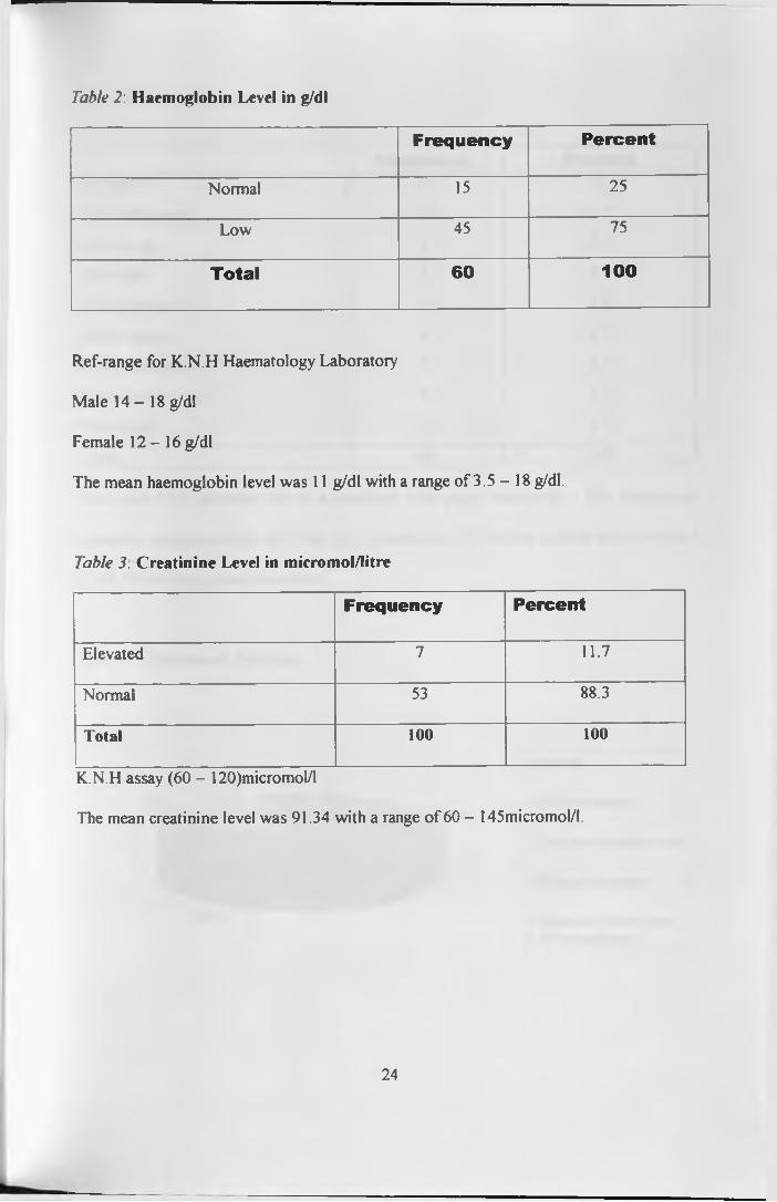
Table 2: Haemoglobin Level in g/dl
Frequency Percent
Normal 15 25
Low 45 75
Total 60 100
Ref-range for K.N.H Haematology Laboratory
Male 1 4 - 1 8 g/dl
Female 1 2 - 1 6 g/dl
The mean haemoglobin level was 11 g/dl with a range of 3.5 - 18 g/dl.
Table 3. Creatinine Level in micromol/litre
Frequency Percent
Elevated 7 11.7
Normal 53 88.3
Total 100 100
K.N.H assay (60 - 120)micromol/l
The mean creatinine level was 91.34 with a range o f 60 - 145micromol/l.
24

Table 4. Urine Culture
Frequency PercentNo growth 21 35
Escherichia coli 19 31.7
Proteus spp 5 8.3
Klebsiella 5 8.3
Pseudomonas 2 3.3
Staph aureus 4 6.7
Staph epidemidis 1 1.7
Enterococcus spp 1 1.7
Citrobacter 2 3.3
Total 60 100
There was 65% infection rate in association with gross heamaturia. The commonest
organism was Eschenchia colli, this was isolated in 31.7% o f the patients and accounted
for 48.7% of the organisms isolated.
Figure 7: Ultrasound Findings
7% 3%
37%
□ Normal
■ Bladder tumour
□ Thickened bladder walls
□ Enlarged prostate
■ Enlarged prostate and Hydronephrosis
25

Ultrasound examination was performed in all patients and was diagnostic in 60% of the
cases. Bladder tumours were detected in 36.7% of the cases.
Figure 8 Imaging of the Upper Urinary Tract by Intravenous Urography (IVU)
a Normal findings
■ Hydronephrosis
□ Renal cyst
D Filling defect in distal Ureter
■ Not done
IVU was performed in 19% of the patients and normal upper urinary tract was seen in
26.7% o f all cases.
Figure 9 Cystoscopic Features
□ Percent
26

Bladder tumours were the commonest lesions identified 41.7% followed by features of
chronic cystitis. In 5% of the patients there was normal cystoscopic appearance of the
urethra and bladder. Cystoscopic evaluation was diagnostic in 95% of the patients.
Three tumours missed on sonography were detected by cystoscopic evaluation.
Table 5: Distribution by Site of the Bladder Tumours
Frequency PercentTrigone 7 28
Lateral walls 10 40
Lateral wall and trigone 2 8
Posterior wall 2 8
Multifocal 3 12
Posterior wall and Trigone 1 4
Total 25 100
Most o f the tumours were located on the lateral wall, (40%) and the trigone 28% No
tumour was seen in the dome and anterior walls.
27

Figure 10. Histological Findings
45
40
35
30
25
20
15
10
5
0
□ Normal
■ Chronic cystitis
□ Transitional cell carcinoma
□ Metastatic squamous cell carcinoma (Primary - cervix)
■ Benign prostatic hyperplasia
□ Adenocarcinoma of the prostate
■ IC C and BPH
Percent
Figure 77: Smokers diagnosed with transitional cell carcinoma (TCC)
□ D iagnosed T C C
■ T C C not diagnosed
28

Figure 12: Non smokers with transitional cell carcinoma
Transitional cell carcinoma was diagnosed in only 25% o f non smokers while 65% of
smokers were diagnosed with transitional cell carcinoma.
Table 6: Co-relation between histological diagnosis and the findings ofcytological analysis of urine
Malignantcondition
DiagnosedHistologically
Malignant cells seen on cytology
Frequency Percent Frequency Percent
TCC 24 85.7 l l 40.8
Adenocarcinoma o f
the prostate
2 7.15 0 0
Metastatic squamous
cell carcinoma o f the
cervix
2 7.15 l 1.9
Total 28 100 12 42.9
Chronic cystitis was the most frequent histological finding 41.7% followed by
transitional cell carcinoma 38.3%. The histological findings were reported as normal in
29

3.3% of the cases. The overall malignancy rate was 46.6%. There was no case o f false
positive or false negative cytology, this gives a sensitivity o f 42.9% and a specificity of
100%. There is however a significant co-relation between malignancy detection by
cytology and histology.(Pearsons St2 8.1, P< 0.01) .The association between smoking and
malignancy was significant. (Pearsons 9C1 17.7, P< 0.001). There was also a significant
co-relation between the cystoscopic features and the histological findings (Persons T2
18.8, P< 0.001.
Figure J3\ Distribution of Diagnosed Malignant Condition by Age Group
Age Group (yrs)
Most malignancies were detected above this age of 40 with a peak at 51 - 60 Age group.
No malignancy was detected below the age of 31 years.
30

Figure 14 Histological Grading of the Transitional Cell Carcinoma
1 7 %
Table 7. Surgical Treatment Offered
6.
Frequency PercentNo surgical treatment 35 58.3
TURP 2 3.3
TURBT 13 21.7
Open stone removal 4 6.7
Open prostatectomy 3 5
TURP and TURBT 1 1.7
Channel TURP 2 3.3
To ta l 60 100
The most frequent surgical treatment modality offered was Transurethral resection o f the
bladder tumour (TURBT). No surgical treatment was offered to 58.3% of the patients.
One patient had both superficial bladder tumour and BPE and he underwent TURBT and
TURP.
31

Table 8: Non surgical treatment offered
T reatment Frequency Percent
Radiotherapy 10 25
Convectional antibiotics 25 62.6
Anti tuberculous drugs 2 5
Anti schistosomal drugs 2 5
Hormonal 1 2.5
All patients with unresectable bladder tumours were offered radiotherapy. Two patients
had features of chronic cystitis suggestive o f tuberculous infection and were put on anti
tuberculous drugs. Anti schistosomal drugs were offered to two patients. The rest of the
patients with chronic cystitis (25) were put on conventional antibiotics. One patient with
cancer o f the prostate declined orchidectomy and was put on hormonal treatment.
32

DISCUSSION
The male to female ratio in this study was 2:1 in keeping with the findings o f other
studies23,31’38. Male to female ratio o f 3:1 is mentioned in several studies27'48,54 The
mean age at presentation was 43.4 years with a range of 18 - 83 years which reflects the
figure quoted in other studies done in some parts of Africa 27‘30. The mean age at
presentation is however lower than that quoted in the European series (50 - 60) ’
Inflammatory cystitis due to schistosoma haematobium was diagnosed in only 3.3% of
the patients, higher figures are quoted in studies done in Sudan and West Africa ‘
The overall urine infection rate in this study was 65%, Escherichia colli being the most
prevalent organism isolated. Dawan and associates reported 66% infection rate in
association with gross haematuria 27 while Turner and colleagues found 55% infection
rate in association with gross haematuria and a 39% rate in association with
malignancy.26
Several researchers have strongly contested the role o f urine cytology in the detection of
genitourinary malignancy. 31,36,38 There is also lack of uniform consistency between
studies with regards to the sensitivity o f cytological analysis of urine samples in the
detection o f genitourinary malignancy 31,36,38 Several researchers have reported
sensitivity rates o f 70% and above. Aziz and Ndaguatha found a rate o f 93.1% 53
while Mbonu, Turner and Colleagues found rates varying between 70- 90% 26 28 On the
contrary, some researchers have reported very low sensitivity rates.31,36,38
33

• • • • _Researchers at the Dundee Royal infirmary reported cytologic sensitivity o f 25%,
investigators in Freeman Hospital (U K) quoted 26% sensitivity rate31, while Chahol and
Colleagues found 42% sensitivity rate.36 In this study urine cytology was positive in 12
out o f 28 patients diagnosed with malignancy. There were no false negative or false
positive findings, this gives a cytologic sensitivity of 42.7%.
There are possible explanations for the variation in sensitivity o f urine cytology between
different studies.
i) Cytologic sensitivity is generally poor for low grade tumours '
ii) Factors related to experience o f the cytologist.
iii) Method o f specimen collection - barbotaged urine samples obtained at
cystoscopy have high cytologic sensitivity5.
In this study however there was a significant correlation between malignancy detection
by cytology and histological analysis (Pearsons it2 8.1 p = 0.01).
The role o f transabdominal sonography in the detection o f bladder tumours is well
established.12 The sensitivity o f transabdominal sonography in the detection o f bladder
tumours ranges from 60 to 80%.12 As pointed by other investigators the main drawback
of sonography is the fact that it is subject to inter observer variability .12'32 48 In this study
sonography was performed in all patients and bladder tumours were detected 36.7% of
the cases. Sonography missed 12% o f the tumours which were subsequently picked up
on cystoscopy, overall sonography was diagnostic in 60% o f the patients.
34

Intravenous urography was performed in only 31.79% of the patients due to financial
constraints on the part o f the subjects. Some investigators have argued that it is not
necessary to perform intravenous urography in all patients presenting with gross
haematuria since ultrasonography can give equivalent or superior diagnostic
information.12 Transitional cell carcinoma is a pan urothelial tumour and therefore it
appears logical to perform intravenous urography whenever bladder cancer is suspected
so as to rule out synchronous upper urinary tract tumours.
Cystoscopic evaluation was diagnostic in 95% of the patients. Some researchers have
reported diagnostic sensitivity o f cystoscopy to be greater than 90% in the detection of
bladder tumours." Cystoscopy picked 12% of the bladder tumours which were missed on
ultrasonography. Five percent o f the patients had normal cystoscopic appearance one of
which was reported on histologic examination to have features of carcinoma insitu. The
most frequent cystoscopic feature was tumour of the urinary bladder 41.7%, followed by
chronic cystitis (35%). Researchers in Khartoum detected bladder tumours in 45% of the
patients.30 Investigators at the Ahmed Bello University Teaching Hospital in Nigeria
detected bladder tumours in 31% of the patients followed by cystitis 22% and benign
prostatic hypertrophy 14%. 27 Most studies have shown that prostatic carcinoma is not a
frequent cause of gross haematuria,23 27 30 in this study cystoscopic evaluation yielded
bleeding from prostatic carcinoma in 3.3% of the patients.
Most of the bladder tumours were located on the lateral walls (40%) and Trigone (28%).
A previous study conducted locally had shown most tumours to be located in the trigone
35

(51% )?4 All primary malignancies of the urinary bladder in this study were o f the
transitional cell type. In Europe and North America transitional cell carcinoma is the
most frequent type o f bladder malignancy accounting for over 95% and this is confirmed
by consistent unity between different studies.31'34'37,38'46’47 In the bilharzial endemic zones
o f Egypt, Sudan and West Africa, squamous cell carcinoma accounts for between 20 to
50% o f urinary bladder malignancies23"27’30. Two patients had normal cystoscopic
appearance and histological features this gives a failed diagnosis rate of 3.3%. The failed
diagnosis rate 3.3% is similar to the finding of other studies done in Africa 20 23
Cystoscopic evaluation yielded the diagnosis o f a malignancy in 46.6% of the patients.
The malignancy rate o f 46.6% is higher than the rate o f 23% mentioned in most
studies.3* 46 A possible explanation for the high malignancy rate could be due to the fact
that most patients with gross haematuria due to Benign prostatic hypertrophy do not
routinely undergo cystocopic evaluation in KNH. Two patients (3.3%) were known to
have squamous cell carcinoma o f the cervix and had developed metastasis to the bladder
at the time they presented with gross haematuria. This study showed that there was a
significant co-relation between the cystoscopic impression and the eventual histological
diagnosis (Pearsons 3T2 18.8, p = 0.001). No malignant condition was diagnosed below
the age of 35 years. Most malignancies (53.6%) were detected between the age o f 50 -
60 years. This is similar to the findings o f studies done in Asia 48 50, UK 31’37’39 and other
parts o f Africa. 27‘30 Alisahi and associates detected 90% o f bladder malignancies in
those aged above 60 years38
36

In this study there was a strong association between smoking and malignancy (Pearsons
f 17.7, p = 0.001). Several studies have confirmed reduced risk o f detecting urological
malignancy in those below 40 years o f age.31,38'47 In this study 55% o f the males who
presented with gross haematuria were eventually diagnosed w ith a malignancy a lower
rate of 30% was seen in females. Most bladder malignancies w ere o f histological grade I
and II in keeping with findings in other studies.38 54
In general only 41.7% o f the patients who presented with gross haematuria were offered
surgical treatment Transurethral resection o f the bladder tum our was the most frequent
surgical treatment modality offered.
37

CO N CLUSIO N
Cystocopy and Trans abdominal ultrasound of the kidney ureter and bladder
are the most valuable investigations in the evaluation o f gross haematuria.
Urine cytology for malignant cells has got a supportive role in the evaluation
o f gross haematuria
Gross haematuria in patient aged above 40 years should be regarded with
suspicion as the chances o f detecting a malignancy are high.
Transitional cell carcinoma is the most frequent type o f urological malignancy
associated with gross haematuria.
There is a high malignancy rate in association with gross haematuria.
38

R ECO M M EN D ATIO N S
Cystoscopy and Abdominal Ultrasound should be mandatory in all patients
presenting with gross haematuria.
A wide scale study should be carried out in the bilharzial endemic zones of
coastal region and shores o f Lake Victoria to see if histological pattern of
bladder tumours is changing.
A large multicentre study similar to this would be a valuable scientific
resource to be compared against studies done in other parts o f the world.
39
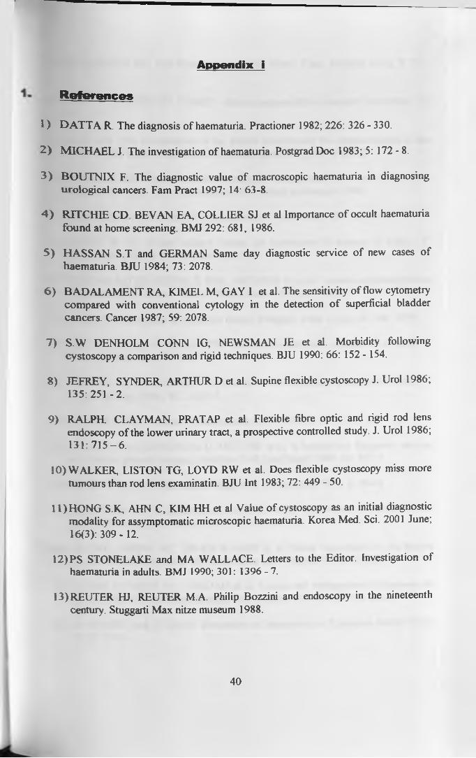
A p p e n d ix i
References
1) D A T T A R. The diagnosis o f haematuria. Practioner 1982; 226: 326 - 330.
2 ) M ICHAEL J. The investigation o f haematuria. Postgrad Doc 1983; 5: 172 - 8.
3 ) BOUTNIX F. The diagnostic value o f macroscopic haematuria in diagnosing urological cancers. Fam Pract 1997; 14: 63-8.
4 ) RITCHIE CD BEVAN E A COLLIER SJ et al Importance o f occult haematuria found at home screening. BMJ 292: 681, 1986.
5 ) HASSAN S.T and GERMAN Same day diagnostic service o f new cases o f haematuria. BJU 1984; 73: 2078.
6 ) BADALAMENT RA, KIMEL M, GAY I et al. The sensitivity o f flow cytometry compared with conventional cytology in the detection o f superficial bladder cancers. Cancer 1987; 59: 2078.
7 ) S.W DENHOLM CONN IG, NEWSMAN JE et al. Morbidity following cystoscopy a comparison and rigid techniques. BJU 1990: 66: 152 - 154.
8) JEFREY, SYNDER, ARTHUR D et al. Supine flexible cystoscopy J. Urol 1986; 135: 251 -2 .
9) RALPH CLAYMAN, PRATAP et al. Flexible fibre optic and rigid rod lens endoscopy o f the lower urinary tract, a prospective controlled study. J. Urol 1986; 131: 7 1 5 -6 .
10) WALKER, LISTON TG, LOYD RW et al. Does flexible cystoscopy miss more tumours than rod lens examinatin. BJU Int 1983; 72: 449 - 50.
11) HONG S.K, AHN C, KIM HH et al Value o f cystoscopy as an initial diagnostic modality for assymptomatic microscopic haematuria. Korea Med. Sci. 2001 June; 16(3): 309-12.
12) PS STONELAKE and MA WALLACE. Letters to the Editor. Investigation of haematuria in adults. BMJ 1990; 301: 1396 - 7.
13) REUTER HJ, REUTER M.A. Philip Bozzini and endoscopy in the nineteenth century. Stuggarti Max nitze museum 1988.
40

1 4 ) R U T E N B E R G D C . E in B lasansp iegel B eim . D tsch. Pract. M edizin 1976; 7: 73 - 7.
1 5 ) PAW LIK K. BER1CHT VOMXI - Intern congress Rom Zentrabl Chir 18904; 18: 4 1 7 - 2 1 .
16) K ELL HA. The examination of the female bladder and the catheterisation of the ureters under direct inspection. Johns Hop Hosp Bull 1893; 4:101-2
1 7 ) W ARPLER RH. Electromedical and surgical instruments No. 1 New York, 1909 (Reprint: Stuttgart: Max nize museum for medical endoscopy 1995.
1 8 ) BURCHARD P. The flexible Pan endoscope. J. urol 1982, 127: 479-81.
1 9 ) DAVEY W. W. Genito urinary system. In companion to surgery in Africa. I s' E dn London: David Livingstone 1965:318
20) YEBOAH ED QUARTERLY JKM, ARTHUR SA et al. causes and management o f haematuria in Accra - Ghana. Ghana Med J 1975; 299-306.
21) O ’RILLY Haematuria o f unknown origin. Postgrad. Med. J Urol 50: 746, 1974.
22) GILLAY DA and O REILLY PH. haematuria analysed, a prospective study. J. Urol Soc Med 1984; 8 7 :1 1 5 -7 .
2 3 ) OSEGBE and D.N. AMAKU EO. Haematuria in Nigerians a prospective study. J Trop Med Hyg. 1984; 87: 1115 -7
2 4 ) BITTON JP Effectiveness of haematuria clinics BJ Urol 1993; 71: 247 - 52.
25) TALBOT R W BENNISTER JJ. HITS NH et al A haematuria diagnostic service in a district general hospital. Ann Roy Coll Sug (Engl) 1989; 66: 343-5
26) TURNER AG et al. Haematuria diagnostic service BMJ 1977; 2: 29-31
27) D DAW AN G.D KALAY1, J.A OSUIDE. et al. Haematuria in Africa. Is the pattern changing: BJU March 2001; 87 (4): 326-30 .
28) MBONU AMENE PC, NWAFOR AME et al Gross haematuria in the Negro population. An analysis o f 100 adult cases: urol Nephrol 1991; 23: 261-3
29) DAFUR JS UGWO BT, YEBOAH et al. Causes and management of haematuria in Jos University Teaching Hospital, Jos, Nigeria JUMED 1997; 2: 62-9.
30) AR SHARFI and HASS AN. Evaluation o f haematuria in Khartoum EAMJ 1994; 71(1): 29-31
41

3 1 ) K H ADRA M.H, PICKARD RS CHARLTON M et al. a prospective study o f 1930 patients with haematuria to evaluate the current diagnostic practice J Urol Feb 2000; 162: 5 2 4 -7 .
32) S U T T O N , J.M. Evaluation of in adults JAMA, 263; 2475, 1990.
3 3 ) M O S L E Y D H S C H A T Z IJ, B R E N E M A N GM e t al lo n g te rm an ticoagu lation th e ra p y : c o m p lic a tio n s and con tro l in a rev iew o f 978 case s JAMA 1963; 186: 9 1 4 - 6 .
3 4 ) B O M AN H , H E D E L IN H, H O L M A N G S et al. T h e resu lts o f evaluation o f ad u lt h a e m a tu r ia an a ly sed acco rd in g to re ferra l in fo rm ation fo llo w up. Scand J. U rol N e p h ro n 2001 D ec 359 (6). 497 - 50.
3 5 ) JOHNSTON SR, KEANE PF et al. the haematuria clinic referral pattern in Northern Ireland. Ulster Medical Journal 67 (1): 25-8, 1998 May.
36 ) CHAHAL GOGOI NK, SUNDARAM SK et al - Is it necessary to perform urine cytology in screening patients with haematuria. European urology. 39(3) 283 - 286. 2001.
37) GANT A A 3 year prospective review of suspected cancer referrals to a haematuria clinic BJU Int 2004 June S 93 (4): 87-8.
38) S.A ALISAHI D BYRNE CM GOODMAN et al: Haematuria investigation based on Standard protocol with emphasis on diagnosis o f urological malignancy. JR coll Surg Edinb Feb 2002; 47: 422-7.
39) BROWN F.M; Urine Cytology: Is it Still the gold standard for screening. Urol clin North Arc 27:25, 2000.
40) V. KUMAR, E.COSSAR, HR PATEL et al. Flexible cyctoscopy are we using it Judiciously. BJU 2004; 593 (4): 48.
41) FIELDING et al: Tumour detection by virtual cystoscopy with color mapping of bladder wall thickness. J Urol 2002 Feb; 167: 559-62.
42) A.G TURNER. WF HENDRY, GB WILLIAMS et al A haematuria diagnostic service BMJ, 1977; 2: 29-31
43) CUTTINO JT CLARK RL FEASTERSH et al. The evaluation o f gross haematuria in anticoagulated patients; efficacy of I V Urogrophy and cystoscopy. AJR 1987 Sept; 149(3): 527-8.
42

4 4 ) ANTLOAK SJ AND MELLINGER GT. Urologic evaluation of haematuria occurring during anticoagulation therapy. Jurol 1969; 101: 111 -3.
^ 5 ) SCHUSTA G.A and LEWIS G.A. Clinical significance of haematuria in patients o n anticoagulation therapy JUROL 1987; 137: 923.
4 6 ) CARTER WC and ROUS S.W Gross haematuria in 110 adult urologic patients. U rol 1981; 18: 342-4
4 7 ) MARIANI MARITA, CATHERINE et al. the significance o f adult haematuria in 1000 haematuria evaluation, including risk benefit and cost effective analysis. Jurol 1989: 141: 3 5 0 -3 .
4 8 ) SIDNEY KH YIP H, WILFRED C et al. Day case diagnostic service use o f ultrasonography and flexible cytoscopy urology 1998; 52:762-6
4 9 ) PK TAN and HC CHANG, YY SITOH et al. a haematuria clinic - a preliminary audit and consideration for a one stop clinic, Singapore Med J. 1992 vol 39(11) 501 -3.
5 0 ) GOONEWARDEWA and ABEY GUNASEKERA AM Haematuria as a presenting symptom in a tertiary Hospital in Sri Lanka. Ceylon Med J. 43(3) 156 - 8 . 1998
51) NASHIKAWA MIURA Y, SUZUKI K et al. Clinical assessment of patients with pointed out by mass screening: Acta urologic Japonica 1992 June: 38 (6) 647-51
52) IWATA S. OGAWA Y, SUGIYAMA Y et al. statistical Study of outpatients with Haematuria. Acta urologica Japonica Nov 1985 31 (11); 1988 - 94.
53) NDAGUATHA PLW. A review of urinary bladder cancers at KNH (dissertation M. Med) 1985
54) ABDEL Aziz. Urinary cytology in the detection of urinary bladder tumour, in K.N.H. Med dissertation 1988.
55) AWORI N. Carcinoma of the urinary bladder in Kenya, Proc. Assoc. E.A. 6: 207 -2 0 8 1983.
43

D A T A C O L L E C TIO N FORM
Ap p e n d ix ii
C v s t o s c o n ic Findings In Th e Initial EvaluationO *Gross
H a o m a tu ria .
Study Code __
In patient number ______________ ZZ1A ) P E R S O N A L AN D DEM OGRAPHIC D A TA
Nam e ......................................................
Age in years ...................................................
District of usual Residence ..................................................
Occupation ...............................................
Smoking \ \ Yes No \ _j
I f yes to the above state duration and average cigarettes smoked per day
B ) P R E-O P ER A TIV E D A TA
Laboratory Investigations
i) U rine based tests
Urinalysis................................................................................
Urine cytology...............................................................................
44

Microscopy culture and sensitivity
ii) Heamatological
Hb .........................................
Platelets .........................................
PTI ..........................................
iii) Biochemical
Serum Creatinine...........................
iv) Others (Specify)
Imaging
1) KUB X-Ray ............
2) KUB ULTRA SOUND............
3) I.V.U. ............
4) C.T. Scan ............
5) Others (Specify) ............
C ) IN TR A-O PER ATIVE D A TA
1) Type of cystoscopy.................
2) C ystoscopy find ings...................
3) Cystoscopic impression.........
4) Surgical treatment offered ...
5) Other treatment modalities....
Post Operative Data
1) Histology report.....................
45

KENYATTA NATIONAL HOSPITALHospital Rd. along, Ngong Rd.
P.O. Box 20723, Nairobi.
Tel: 726300-9 Fax: 725272
Telegrams: "MEDSUP", Nairobi.Email: [email protected]
Ref: KNH-ERC/01/2714 Date: 31t!l May 2005
Dr. Otieno Simba Raymond Dept, of Surgery Faculty of Medicine University of Nairobi
Dear Dr. Otieno
RESEARCH PROPOSAL: "A PROSPECTIVE STUDY ON THECYSTOSCOPIC FINDINGS IN THE INITIAL EVALUATION OF GROSS HAEMATURIA AT KENYATTA N. HOSPITAL" (P45/03/2005)
This is to inform you that.Kenyatta National Hospital Ethics and Research Committee has reviewed and approved revised version your above cited research proposal for the period 31st May 2005 to 30th May 2006. You will be required to request for a renewal of the approval if you intend to continue with the study beyond the deadline given.
On behalf of the Committee, I wish you fruitful research and look foiward to receiving a summary of the research findings upon completion of the study.
This information will form part of database that will be consulted in future when processing related research study so as to minimize chances of study duplication.
Yours sincerely,
Prof. A. N. GUANTAI SECRETARY - KNH-ERCCc: Prof. K. M Bhatt, Chairperson, and KNH-ERC
The Deputy Director (C/S), KNH The Dean, Faculty of Medicine, UON The Chairman, Dept, of Surgery, UON The HOD, Medical Records, KNH Supervisors: ;Mr<Francis Owilla, Dept, of Surgery, UON
Mr. P. Mungai Ngugi, Dept, of Surgery, UON