A prokaryotic viral sequence is expressed and conserved in ... › lab › manuelidis › 17...
6
A prokaryotic viral sequence is expressed and conserved in mammalian brain Yang-Hui Yeh a , Vignesh Gunasekharan a , and Laura Manuelidis a,1 a Section of Neuropathology, Yale University Medical School, New Haven, CT 06510 Edited by Günter Blobel, HHMI at the Rockefeller University, New York, NY, and approved May 25, 2017 (received for review April 12, 2017) A natural and permanent transfer of prokaryotic viral sequences to mammals has not been reported by others. Circular “SPHINX” DNAs <5 kb were previously isolated from nuclease-protected cy- toplasmic particles in rodent neuronal cell lines and brain. Two of these DNAs were sequenced after Φ29 polymerase amplification, and they revealed significant but imperfect homology to segments of commensal Acinetobacter phage viruses. These findings were surprising because the brain is isolated from environmental micro- organisms. The 1.76-kb DNA sequence (SPHINX 1.8), with an iteron before its ORF, was evaluated here for its expression in neural cells and brain. A rabbit affinity purified antibody generated against a peptide without homology to mammalian sequences labeled a nonglycosylated ∼41-kDa protein (spx1) on Western blots, and the signal was efficiently blocked by the competing peptide. Spx1 was resistant to limited proteinase K digestion, but was unrelated to the expression of host prion protein or its pathologic amyloid form. Re- markably, spx1 concentrated in selected brain synapses, such as those on anterior motor horn neurons that integrate many complex neural inputs. SPHINX 1.8 appears to be involved in tissue-specific differen- tiation, including essential functions that preserve its propagation during mammalian evolution, possibly via maternal inheritance. The data here indicate that mammals can share and exchange a larger world of prokaryotic viruses than previously envisioned. circular viral DNA | maternal inheritance | microbiome | synaptic boutons | evolution T he transfer of highly conserved nucleic acid sequences be- tween elementary life-forms and mammalian species has only begun to be explored at the molecular level. The first examples of permanently incorporated lower life-forms in mammals are mitochondria. These complex organelles reside in the cytoplasm and are maternally inherited. Mitochondria derive from engulfed prokaryotes (1), an endosymbiotic concept subsequently solidified by sequencing homologies (2). Mitochondria contain a protected circular DNA genome of 16 kb, the only known cytoplasmic en- dosymbiont inherited through generations. They provide essential cellular functions such as the production of energy via ATP, and they perform in concert with, and are codependent on, nuclear genomic transcripts. Although some prokaryotes and many viruses can penetrate and be maintained within the cytoplasm of mam- malian cells and tissues for a lifetime, they are not normally trans- mitted from the oocyte or sperm to their progeny. The majority are infectious pathogens, although some can be maintained in a latent or hidden state without inducing obvious disease. Examples include quiescent tubercle bacilli arrested in lymphatic macrophages, DNA viruses such as the Epstein-Barr and papova viruses, and RNA myxo-paramyxoviruses. In humans, both the JC papova viral DNA and measles viral structures have been detected in- cidentally in normal brain (3, 4). Nevertheless, the rare transfer of these infectious agents to germline cells, especially with pro- gressive dissemination to normal offspring, stands in sharp con- trast to the conserved cytoplasmic perpetuation of mitochondrial DNA. Indeed, the intergenerational transmission of other envi- ronmental DNAs or RNAs by maternal cytoplasmic inheritance has not been reported in mammals. The data here demonstrate that a commensal circular cytoplasmic DNA is propagated and expressed in various mammalian cells, including neurons and pancreatic insulin-secreting cells. This implicates archaic symbi- otic functions. Even in the nucleus, where episomal DNA viruses replicate and can reside for years, integration into nuclear DNA in the germline is rare. RNA retroviruses that integrate their cDNA via reverse transcriptase are the major exception. Numerous copies of long interspersed repeat elements (known as LINES) were initially isolated from human and mouse nuclear DNA, shown to be ho- mologous with each other, and organized together in megabase Giemsa-band domains on chromosome arms (5– 7). Their long ORFs suggested transcriptional and functional potentials that became clear only with the subsequent sequencing of contemporary epidemic retroviral isolates. Although the function of LINES remains incom- plete, truncated to full-length transcripts are highly expressed in mammalian brain. Such elements, including lower copy human endogenous retroviruses, once established in the genome, can re- integrate into nuclear DNA and spread with increasing represen- tation to define and organize large cohesive tissue-specific and other selected gene families (8–10). On an evolutionary scale, some xenotropic retroviruses may be selected because they are not pathogenic for their host, but can kill closely related competitive species in their local environment (8). The incorporation of ret- roviruses in Drosophila and plants (11) further emphasizes ancient molecular exchanges with environmental viruses. The recent demonstration that the ubiquitous human herpesvirus 6 can in- tegrate into germline chromosomal telomeres in ∼1% of children also reveals ongoing nascent viral transfers, with reactivation and transmission to others via transfusion and transplantation (12, 13). We proposed that commensal and/or environmental viruses might initiate or be involved in late-onset neurodegenerative diseases (14). Data in transmissible encephalopathies (TSEs), Significance Circular SPHINX DNAs with significant homology to segments of Acinetobacter phage viruses were previously isolated from cytoplasmic particles in mammalian cells and brain. Acinetobacter are widespread opportunistic pathogens, and permanent transfer of prokaryotic viruses to mammals has not been reported. To find whether 324-aa ORF of SPHINX is translated into its cognate protein (spx1), an antibody was raised against an internal peptide with no homology to any known mammalian cellular or viral se- quences. Western blots of cells and brains (rodent and human) displayed antigen-specific spx1 protein in cultured cells where spx1 was perinuclear. In brain, the protein concentrated in terminal synaptic boutons. Because brain is environmentally isolated, this SPHINX element is an antediluvian symbiont probably involved in sophisticated neural functions. Author contributions: Y.-H.Y., V.G., and L.M. designed research; Y.-H.Y., V.G., and L.M. performed research; Y.-H.Y., V.G., and L.M. analyzed data; and L.M. wrote the paper. The authors declare no conflict of interest. This article is a PNAS Direct Submission. 1 To whom correspondence should be addressed. Email: [email protected]. This article contains supporting information online at www.pnas.org/lookup/suppl/doi:10. 1073/pnas.1706110114/-/DCSupplemental. www.pnas.org/cgi/doi/10.1073/pnas.1706110114 PNAS Early Edition | 1 of 6 MICROBIOLOGY
Transcript of A prokaryotic viral sequence is expressed and conserved in ... › lab › manuelidis › 17...

A prokaryotic viral sequence is expressed andconserved in mammalian brainYang-Hui Yeha, Vignesh Gunasekharana, and Laura Manuelidisa,1
aSection of Neuropathology, Yale University Medical School, New Haven, CT 06510
Edited by Günter Blobel, HHMI at the Rockefeller University, New York, NY, and approved May 25, 2017 (received for review April 12, 2017)
A natural and permanent transfer of prokaryotic viral sequencesto mammals has not been reported by others. Circular “SPHINX”DNAs <5 kb were previously isolated from nuclease-protected cy-toplasmic particles in rodent neuronal cell lines and brain. Two ofthese DNAs were sequenced after Φ29 polymerase amplification,and they revealed significant but imperfect homology to segmentsof commensal Acinetobacter phage viruses. These findings weresurprising because the brain is isolated from environmental micro-organisms. The 1.76-kb DNA sequence (SPHINX 1.8), with an iteronbefore its ORF, was evaluated here for its expression in neural cellsand brain. A rabbit affinity purified antibody generated against apeptide without homology to mammalian sequences labeled anonglycosylated ∼41-kDa protein (spx1) on Western blots, and thesignal was efficiently blocked by the competing peptide. Spx1 wasresistant to limited proteinase K digestion, but was unrelated to theexpression of host prion protein or its pathologic amyloid form. Re-markably, spx1 concentrated in selected brain synapses, such as thoseon anterior motor horn neurons that integrate many complex neuralinputs. SPHINX 1.8 appears to be involved in tissue-specific differen-tiation, including essential functions that preserve its propagationduring mammalian evolution, possibly via maternal inheritance. Thedata here indicate that mammals can share and exchange a largerworld of prokaryotic viruses than previously envisioned.
circular viral DNA | maternal inheritance | microbiome | synaptic boutons |evolution
The transfer of highly conserved nucleic acid sequences be-tween elementary life-forms and mammalian species has only
begun to be explored at the molecular level. The first examplesof permanently incorporated lower life-forms in mammals aremitochondria. These complex organelles reside in the cytoplasmand are maternally inherited. Mitochondria derive from engulfedprokaryotes (1), an endosymbiotic concept subsequently solidifiedby sequencing homologies (2). Mitochondria contain a protectedcircular DNA genome of 16 kb, the only known cytoplasmic en-dosymbiont inherited through generations. They provide essentialcellular functions such as the production of energy via ATP, andthey perform in concert with, and are codependent on, nucleargenomic transcripts. Although some prokaryotes and many virusescan penetrate and be maintained within the cytoplasm of mam-malian cells and tissues for a lifetime, they are not normally trans-mitted from the oocyte or sperm to their progeny. The majority areinfectious pathogens, although some can be maintained in a latentor hidden state without inducing obvious disease. Examples includequiescent tubercle bacilli arrested in lymphatic macrophages,DNA viruses such as the Epstein-Barr and papova viruses, andRNA myxo-paramyxoviruses. In humans, both the JC papovaviral DNA and measles viral structures have been detected in-cidentally in normal brain (3, 4). Nevertheless, the rare transferof these infectious agents to germline cells, especially with pro-gressive dissemination to normal offspring, stands in sharp con-trast to the conserved cytoplasmic perpetuation of mitochondrialDNA. Indeed, the intergenerational transmission of other envi-ronmental DNAs or RNAs by maternal cytoplasmic inheritancehas not been reported in mammals. The data here demonstratethat a commensal circular cytoplasmic DNA is propagated and
expressed in various mammalian cells, including neurons andpancreatic insulin-secreting cells. This implicates archaic symbi-otic functions.Even in the nucleus, where episomal DNA viruses replicate
and can reside for years, integration into nuclear DNA in thegermline is rare. RNA retroviruses that integrate their cDNA viareverse transcriptase are the major exception. Numerous copiesof long interspersed repeat elements (known as LINES) were initiallyisolated from human and mouse nuclear DNA, shown to be ho-mologous with each other, and organized together in megabaseGiemsa-band domains on chromosome arms (5–7). Their long ORFssuggested transcriptional and functional potentials that became clearonly with the subsequent sequencing of contemporary epidemicretroviral isolates. Although the function of LINES remains incom-plete, truncated to full-length transcripts are highly expressed inmammalian brain. Such elements, including lower copy humanendogenous retroviruses, once established in the genome, can re-integrate into nuclear DNA and spread with increasing represen-tation to define and organize large cohesive tissue-specific andother selected gene families (8–10). On an evolutionary scale,some xenotropic retroviruses may be selected because they are notpathogenic for their host, but can kill closely related competitivespecies in their local environment (8). The incorporation of ret-roviruses in Drosophila and plants (11) further emphasizes ancientmolecular exchanges with environmental viruses. The recentdemonstration that the ubiquitous human herpesvirus 6 can in-tegrate into germline chromosomal telomeres in ∼1% of childrenalso reveals ongoing nascent viral transfers, with reactivation andtransmission to others via transfusion and transplantation (12, 13).We proposed that commensal and/or environmental viruses
might initiate or be involved in late-onset neurodegenerativediseases (14). Data in transmissible encephalopathies (TSEs),
Significance
Circular SPHINX DNAs with significant homology to segmentsof Acinetobacter phage viruses were previously isolated fromcytoplasmic particles in mammalian cells and brain. Acinetobacterare widespread opportunistic pathogens, and permanent transferof prokaryotic viruses to mammals has not been reported. To findwhether 324-aa ORF of SPHINX is translated into its cognateprotein (spx1), an antibody was raised against an internal peptidewith no homology to any known mammalian cellular or viral se-quences. Western blots of cells and brains (rodent and human)displayed antigen-specific spx1 protein in cultured cells wherespx1 was perinuclear. In brain, the protein concentrated interminal synaptic boutons. Because brain is environmentallyisolated, this SPHINX element is an antediluvian symbiontprobably involved in sophisticated neural functions.
Author contributions: Y.-H.Y., V.G., and L.M. designed research; Y.-H.Y., V.G., and L.M.performed research; Y.-H.Y., V.G., and L.M. analyzed data; and L.M. wrote the paper.
The authors declare no conflict of interest.
This article is a PNAS Direct Submission.1To whom correspondence should be addressed. Email: [email protected].
This article contains supporting information online at www.pnas.org/lookup/suppl/doi:10.1073/pnas.1706110114/-/DCSupplemental.
www.pnas.org/cgi/doi/10.1073/pnas.1706110114 PNAS Early Edition | 1 of 6
MICRO
BIOLO
GY

such as human Creutzfeldt-Jakob disease (CJD) and sheepscrapie, strongly suggest that they are caused by an infectiousagent that induces neurodegeneration and host prion protein(PrP) amyloid after a prolonged infectious, but clinically silentperiod (14). A subset of amyloid and other protein misfoldingdiseases, such as postencephalitic Parkinson’s disease, have aviral history, and measles infections can also induce neurofi-brillary tangles that are indistinguishable from those seen inAlzheimer’s disease brain (e.g., ref. 15). The eradication of hu-man kuru and the dramatic reduction of epidemic U.K. cow TSEwere achieved by removing infectious material. In addition, sub-stantial agent strain, PrP recombinant, and recent nucleic aciddata (14, 16, 17) together indicate a still-unresolved causalprotected environmental virus. The cytoplasm of CJD- andscrapie-infected cells, but not control cells, also contains virus-like particle arrays (18), and because we were able to isolatethese nuclease-protected particles with quantitative recovery ofinfectivity, but with little or no detectable PrP (17, 19), we beganto analyze protected nucleic acids. Using Φ29 rolling circle am-plification, several circular DNA sequences of <5 kb with ORFswere thereby discovered in brain and cultured neuronal cell lines.These circular DNA sequences were named SPHINX ele-ments for their initial association with slow progressive hiddeninfections of X origin (20). They had no homology to mam-malian nuclear or mitochondrial DNA, but had significant nu-cleotide homology (<10−105, 70%) to replication initiation viralregions of Acinetobacter, a wound pathogen that is common insoil and water (20). The bacterial virus is typically ∼12 kb, andbacterial DNA was not detectable, nor were SPHINX DNAsfound in any reagents used. Subsequently, the SPHINX DNAswere independently isolated in Europe from human multiplesclerosis brains, cow milk, and serum (21, 22), and additionalPCR studies here revealed SPHINX DNAs were present inuninfected samples, and thus may not be the sole causal TSEagent (17). Their unique cytoplasmic residence, strong bacte-riophage homology, and potential mammalian functions, espe-cially in brain, impelled further study.We here focus on the SPHINX 1.8 DNA translation into
protein and its representation in cultured neuronal GT1 cells,and in control versus TSE brain samples with severe end-stageneurodegeneration (e.g., refs. 14 and 23). To find whether theSPHINX 1.8 DNA was processed into its 324-aa cognate protein(spx1), rabbit antibodies were raised against selected ORFpeptides that had no homology with any mouse, human, or otherknownmammalian nuclear or cytoplasmic sequence in the database.Two rabbit antisera, as well as an affinity-purified antibody, cleanlybound a blotted 41-kDa protein band close to its predicted 37.3-kDamass; antibody binding was completely blocked by its immunogenicpeptide. This 41-kDa spx1 band is expressed not only in culturedcells exposed to bovine serum, but also is obvious in environ-mentally isolated brain samples removed sterilely from differentmammalian species that included wild-type (wt) and transgenicmice, Syrian hamsters, and human brain-glioblastoma samples.Most remarkably, in situ studies revealed a very high signal in-tensity of spx1 in synaptic boutons innervating anterior motorhorn neurons, as well as in pancreatic islet, but not exocrine, cells.Together, the results here indicate spx1 can have a fundamentalrole in cellular differentiation, neoplasia, and synaptic neuralfunction. Protected cytoplasmic SPHINX DNA particles areprobably the tip of the iceberg of DNAs originating from symbi-otic prokaryotic viral sequences in the environment that are con-served in mammalian evolution.
ResultsCytoplasmic SPHINX DNAs were originally detected only afterremoving >99.5% of nuclear DNA followed by powerful nucleasedigestions that remove unprotected nucleic acids (17, 20), astrategy used to purify viruses. The level of SPHINX 1.8 DNA
was still low compared with residual protected mitochondrialDNA, even with high PCR cycles (17, 20). Because we were notexpecting an obvious spx1 protein signal in whole-cell homoge-nates, we tested whole-cell homogenates and subcellular fractions.To our surprise, the predicted spx1 band was readily detected inWestern blots of GT1 cells, rodent brain, and human glioblasto-mas lysed in 1% Triton-PBS; to avoid off-target antibodies thatare often generated by full-length proteins, we chose two pep-tides without any homology to mammalian sequences in thedatabase for rabbit immunizations (SI Materials and Methods).Both peptides produced antibodies to the same 41-kDa band,and this band was blocked by each peptide independentlyconfirming the identity of a correct single target. One rabbit’ssera was affinity purified to label the 41-kDa band without anycross-talk. Fig. 1 shows the various whole-cell samples includingcontrol normal (NL) and GT1 cells infected with the FU-CJDagent strain isolate (lanes 1–2), NL and FU-CJD mouse brains(lanes 3–4), NL and 263K scrapie-infected hamster brains (lanes5–6), and a frozen human glioblastoma section. Fig. 1A showsthe many gold-stained proteins present in each lane, and Fig.1B shows that the spx1 antibody highlights a major 41-kDaband in all samples. Replicate blot of samples 1–6 in Fig. 1Cshows the spx1 blocking peptide prevented antibody binding. Asecond human glioblastoma (GB2) expressing the astrocyte-specific protein GFAP also showed the same 41-kDa spx1 bandas shown in Fig. 1D. Thus, spx1 is highly conserved acrossmammalian species.The spx1 band in SDS gels is ∼3.7 kDa larger than its pre-
dicted 37.3-kDa mass. It is not unusual for a protein to display aMr on SDS-page that is different from its predicted mass, and large(10 kDa) discrepancies are often a result of glycosylation orsumoylation. Spx1 has three potential N-glycosylation sequences(N-X-S/T), but glycosidases showed it has neither N- norO-glycosylation. In contrast, PNGase F treatment of PrP removedN-glycosylation residues but was unaffected by O-glycosidase, aspredicted (Fig. 2A and SI Materials and Methods). Thus, spx1shows no evidence of glycosylation that might account for itsretarded migration. Phosphorylation typically yields multiplebands, unlike the single band detected here. There are more than200 different posttranslational modifications that may affect gelmigration, in addition to shape, other tightly bound cell compo-nents such as small peptides and lipids, and charge character-istics. Notably, spx1 has a very high percentage of strongly acidic Dand E residues (20%) in its terminal 80-aa protein that could ac-count for its gel shift (24). The presence of an iteron (replicationinitiation binding site) at the beginning of the 1.8 ORF might alsoretard its migration.In addition to the 41-kDa band, a second minor 19-kDa band
is also positive in the brain lanes loaded with more protein thanthe GT1 lanes. This 19-kDa band was also detected in the GT1samples, as in the longer exposures (Fig. 3A, lane 1*), and the19-kDa band was also blocked by its centrally placed peptide1 immunogen ELDEFRKRIGVLDTEYTR (Fig. 1C), as well asby its more terminal C-peptide 2 RNRLSDRFKQGDESA (Fig.S1). However, whereas the 19-kDa band in mouse and hamsterbrains represented 4% (±0.6% SEM) of the total spx1 protein,GT1 cells consistently had a much lower percentage of this band(1%±0.4% SEM; P < 0.005). In sum, the spx1 antibody ishighly specific for its cognate protein, and the 19-kDa band is a41-kDa spx1 product that is significantly higher in brain thanin GT1 cells.We wanted to find out whether the spx1 protein might be
differentially expressed in TSE agent-infected brains with severeneurodegeneration. To test whether spx1 was dependent on, orrelated to, host PrP expression and pathology, we analyzed bothproteins in brains of wt mice, Tga20 mice with 8–10-fold thenormal copies of PrP, and PrP null mice that are not susceptibleto infection (14, 23). In addition, because limited short proteinase
2 of 6 | www.pnas.org/cgi/doi/10.1073/pnas.1706110114 Yeh et al.

K (PK) digestions of whole-cell Triton lysates reveal PrP amyloidresistance (PrP-res) not seen in uninfected controls (e.g., ref. 17),it was of interest to find whether spx1 was partially resistant underthe same standard digestion conditions (SI Materials and Meth-ods). Parallel blots of each control and PK-treated lysates weredenatured in SDS and then analyzed on blots for detection withspx1 antibody (Fig. 3A) and PrP antibody (Fig. 3B). Under thesestandard conditions, every NL and infected brain showed compa-rable amounts of PK-resistant full-length spx1, equal to 8% ofundigested sample. The 19-kDa band in PK digests (PK+ lanes inFig. 3A) was significantly higher (11% ±1.2% SEM; P < 0.0001)than the 4% in the undigested control brains, further verifying itderives from the full-length 41-kDa band. This blot was sub-sequently probed with tubulin antibody as a loading control, andto confirm that unlike spx1, even at 40× loads, tubulin wascompletely digested (no Tu in the PK+ lanes). Spx1 resistancewas comparable in brains from the different mouse genotypesincluding wt (CD-1 lanes 1–4), high PrP (Tga20 lanes 5–8), andPrP null mice (lanes 9–10). In contrast, the blot in Fig. 3B,loaded with the same digests, shows PrP-res bands are all re-duced in size and present only in infected brain. No PrP-ressignal is seen in uninfected CD-1 mice (lane 2’), high PrPTga20 mice (lane 6’), uninfected hamster (lane 12’), or PrP nullmice (lanes 9’–10’). All infected brain samples (FU-CJD andscrapie-263K) showed the characteristic lower digested PrP-resband pattern. GT1 neuronal cells, as brain, consistently showedcomparable resistance of the 41-kDa band (10%), but the PKdigests again showed a far weaker 19-kDa signal in GT1 cellscompared with brain (1% vs. 4%), as seen with its PrP counterpartin Fig. 2B. In sum, spx1 resistance was readily apparent and showedno relation to host PrP expression or PrP-res pathology. The rea-sons for spx1 resistance, and the significantly lower amounts of the19-kDa band in GT1 cells, are not known, but one possibility is thata ligand in brain may limit digestion of some molecules.To find out whether spx1 is compartmentalized differently in
monotypic GT1 cells versus complex brain samples, we firstevaluated the proportion of spx1 in crude and more purified
subcellular fractions from previously characterized frozen NLand infected samples (19, 25). The only meaningful finding fromthese studies was that the vast majority of the spx1 protein (83%)partitioned with the 10,000 × g cytosol, whereas mouse andhamster brains contained 18% of the spx1 in the membrane-myelinated axon fraction that also contains many synapses anddendrites. To delineate more precisely the cellular location and
Fig. 1. Western blots of GT1 cells and brain homogenates. (A) Many gold-stained proteins in samples analyzed are shown. This blot was stained for gold afterfirst detecting spx1 antibodies, as shown in (B), as detailed (SI Materials and Methods). (B and C) Replicate samples 1–6 from the same gel that was split in halffor antibody (B) and antibody blocking (C) performed and detected in parallel. A major 41-kDa band is present in all control (NL) and infected GT1 cells (lanes1 and 2), mouse brain (Mo-CNS, lanes 3 and 4), hamster brain (Ha-CNS, lanes 5 and 6) and a human glioblastoma brain sample (Hu-GB, lane 7). An additional19-kDa spx1-positive band is seen in the central nervous system samples. The GT1 cells had much less 19-kDa band. Another gel blot of the same sample inlane 1* confirms the lower band in GT1 cells in much longer exposures. The replicate half blot of samples 1–6 (C) shows both bands are virtually abolishedwhen antibody is incubated with its cognate peptide. Another glioblastoma (HuGB2; D) also shows the same spx1 band in addition to more abundant GlialFibrillary Acidic protein (GFAP) in two parallel lanes separated for binding to each respective antibody. FU indicates infection with the FU-CJD agent, and SC isscrapie infection with the 263K agent strain. Marker kDa (kd) are indicated.
Fig. 2. Glycosylation and PK resistance of spx1. Replicate blots of mousebrain homogenates (A) were digested with PNGase F to remove N-linkedsugars (N), or neuraminidase+O-glycosidase for O-linked sugars (O). Spx1detection shown in lanes 1–3 and PrP in lanes 4–6. No decrease in Mr fromdeglycosylation is seen in spx1 digests, whereas PrP, known to have N-linkedsugars, is markedly reduced in size by PNGase F (lane 5), but not O-linkeddigestion (lane 6), as expected. (B) Shows resistant spx1 in GT1 control (NL)and FU-CJD infected cells. In NL and FU-CJD samples (lanes 2 and 4, respec-tively), 10% of spx1 was PK resistant, whereas tubulin (Tu) showed no re-sistance. Parallel samples show no PrP-res in NL, whereas 25% PrP-res is seenin the FU-CJD cells (compare lanes 6 and 8).
Yeh et al. PNAS Early Edition | 3 of 6
MICRO
BIOLO
GY

potential cell type-specific expression of spx1, we pursued in situexperiments. Lightly fixed GT1 cells were permeabilized withSaponin (18; SI Materials and Methods). Because spx1 showedsignificant PK resistance, and its genomic DNA was protected,we also exposed cells to more disruptive GdnHCl treatments (SIMaterials and Methods) that unmask hidden antigens; 2 and 3 MGdnHCl opens and inactivates many viruses, including CJD andscrapie (25). In GT1 cells, the spx1 (red signal) preferentiallyconcentrated in the perinuclear RER-Golgi region and showedrelatively low plasma membrane staining (Fig. 4). Only a faintpositive signal is seen in the Saponin-only cells (Fig. 4A), and astronger signal is achieved after 2 and 3 M GdnHCl unmasking(Fig. 4 B and C). Inclusion of the spx1 peptide (Fig. 4D) com-pletely blocks the antibody signal, as in Western blots. No re-producible difference was seen between NL and FU-CJD-infectedcells, and Fig. 4E at higher magnification demonstrates the intenseperinuclear spx1 signal after 2 M GdnHCl unmasking.Because rodent brains and other tissues had been fixed for
several days in formalin, we used autoclaving in citrate buffer toexpose buried antigens and PrP amyloid (26). This treatment alsointensifies spx1 staining, consistent with a protected or maskedform. Fig. 4F shows that spx1 is produced in a highly cell type- andtissue-specific pattern. The exocrine pancreas cells lack detectablespx1 protein, whereas the islet insulin-producing cells containabundant spx1 protein (red). Fig. 4 G–I are parallel sections ofrepresentative coronal brain slices and spleen. There is negligiblesignal when the primary spx1 antibody is omitted (Fig. 4G), bothbrain and spleen are positive when spx1 antibody is included (Fig.4H), and the spx1 signal in these tissues is completely blocked bythe peptide (Fig. 4I). Remarkably, synaptic boutons that in-nervate anterior motor horn neurons are intensely stained (ar-rows). There is much weaker staining of RER-Golgi region ofneuronal perikarya compared with cytoplasmic GT1 cell staining.The nuclei also show no spx1 signal in brain cross-sections. Notethat a vessel receives no positive bouton-like structures, and thata myelinated axon is also not stained. In sum, spx1 expression iscell type- and tissue-specific, and its presence in terminal syn-aptic structures contrasts with its diffuse cytoplasmic pattern inthe more primitive GT1 neuronal cells and specialized islet cells.These data indicate differentiation-specific processing. Additional
cell and tissue-specific spx1 expression patterns in various differ-entiated and neoplastic cells will be reported separately, and theyfurther support a more global role for spx1 in differentiation.
DiscussionThe current experiments establish the normal mammalian ex-pression of a 41-kDa protein that derives from a bacterial viralreplication initiation homolog, rather than from any previouslydescribed mammalian nuclear, cytoplasmic, or viral sequence.SPHINX 1.8 DNA and its spx1 cognate protein do not match anyknown nuclear or mitochondrial sequence, nor any mammalianvirus. Indeed, the possibility of new symbiotic genomes in pro-tected cytoplasmic particles has not been widely pursued. TheSPHINX 1.8 DNA showed numerous differences from repre-sentative contemporary Acinetobacter viruses in the database,unlike the 16-kb mitochondrial DNA extracted from particles atthe same time; the latter showed 100% homology to the consensusdatabase entry (20). Furthermore, a protein from another viralreplication-initiation region of Acinetobacter was independentlyidentified by deep proteomic sequencing of highly infectiousCJD brain particles that lacked PrP (27). This adds to and broadensthe earlier results that verify the specificity of a spx1 antibody for areplication initiation viral protein. The 41-kDa peptide migration(Mr) was close to its predicted mass, was not glycosylated, and wasspecifically blocked by both chosen antigenic peptides in Westernblots and cytological preparations that included nontransfectedneuroectodermal cells, various rodent brains, spleen and pan-creatic tissues, and human glioblastoma samples. In sum, theSPHINX 1.8 DNA is clearly replicated, transcribed, and trans-lated in selected tissues and cell types including mammalianbrain, an environmentally isolated organ.An antiserum raised elsewhere against an environmental Aci-
netobacter virus with homology to our second 2.4-kb SPHINXDNA, also isolated here from mammalian cytoplasmic particles,similarly revealed its predicted 27-kDa ORF protein in brain (17,20). Together, these findings support the concept that otherSPHINX-like circular DNAs can lurk in protected cytoplasmicparticles of mammals. The finding of several different SPHINXDNAs has suggested action of more than one of these elements inconcert, as well as potential helper functions in other pathologic
Fig. 3. Replicate blots of brain homogenates tested for PK resistance of spx1 (A) and PrP (B) on replicate blots separated for binding to antibody. Variousgenotypes showed ∼8% resistance of full-length spx1 (PK+ lanes) under standard digestions (SI Materials and Methods) in wt mice, and those with 10× PrP(Tga20), and null PrP (PrP−). Hamster showed the same pattern, although the NL (lane 12) in this blot was a partial digest. The same samples showed PrP-resbands of reduced size only in infected brain (lanes 3, 8, and 11), and no PrP is detected in PrP null mice (lane 10). Tubulin bands in these lanes (Tu), shown atthe bottom of both blots, were detected after spx1 and PrP staining and displayed no PK resistant bands. Undigested samples were loaded at 10× in the PrPblot to bring out multiple PrP bands.
4 of 6 | www.pnas.org/cgi/doi/10.1073/pnas.1706110114 Yeh et al.
infections (20, 22). Because isolated neuronal cell lines and normalbrain produce spx1, other latent circular DNA elements, includingthe 2.4-kb DNA, need to be further interrogated for their
conservation and expression in mammalian brain and other dif-ferentiated tissues. These data also lead to the question of howthis type of sequence originated, and how it is maintained.
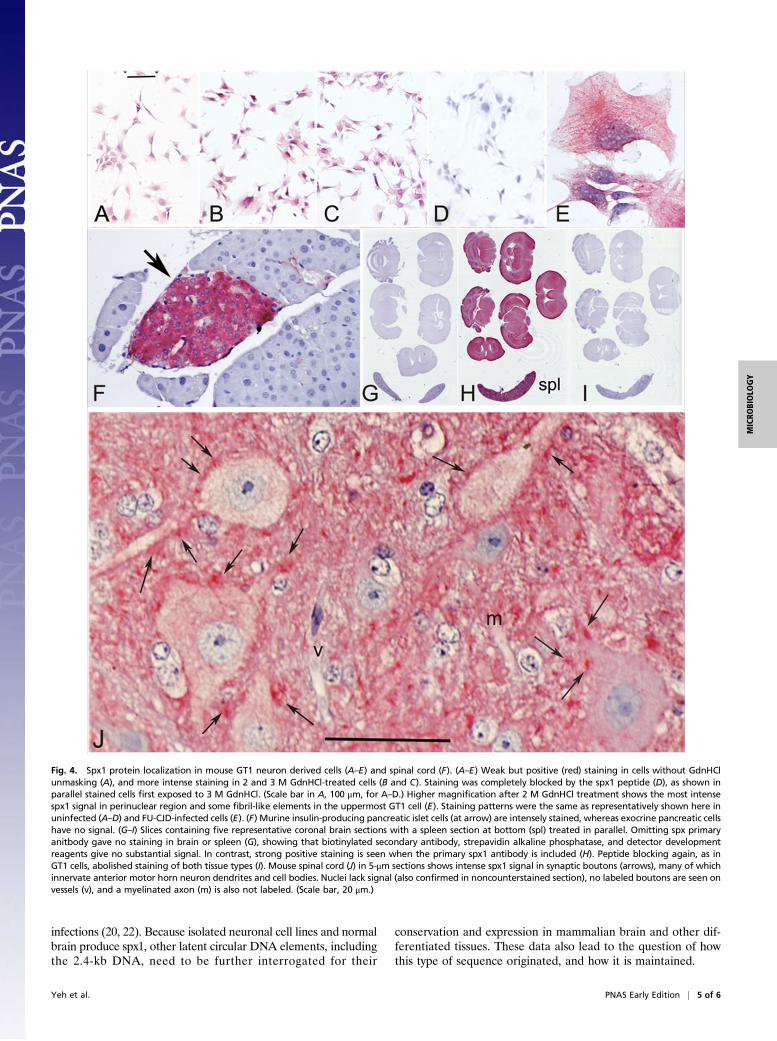
Fig. 4. Spx1 protein localization in mouse GT1 neuron derived cells (A–E) and spinal cord (F). (A–E) Weak but positive (red) staining in cells without GdnHClunmasking (A), and more intense staining in 2 and 3 M GdnHCl-treated cells (B and C). Staining was completely blocked by the spx1 peptide (D), as shown inparallel stained cells first exposed to 3 M GdnHCl. (Scale bar in A, 100 μm, for A–D.) Higher magnification after 2 M GdnHCl treatment shows the most intensespx1 signal in perinuclear region and some fibril-like elements in the uppermost GT1 cell (E). Staining patterns were the same as representatively shown here inuninfected (A–D) and FU-CJD-infected cells (E). (F) Murine insulin-producing pancreatic islet cells (at arrow) are intensely stained, whereas exocrine pancreatic cellshave no signal. (G–I) Slices containing five representative coronal brain sections with a spleen section at bottom (spl) treated in parallel. Omitting spx primaryanitbody gave no staining in brain or spleen (G), showing that biotinylated secondary antibody, strepavidin alkaline phosphatase, and detector developmentreagents give no substantial signal. In contrast, strong positive staining is seen when the primary spx1 antibody is included (H). Peptide blocking again, as inGT1 cells, abolished staining of both tissue types (I). Mouse spinal cord (J) in 5-μm sections shows intense spx1 signal in synaptic boutons (arrows), many of whichinnervate anterior motor horn neuron dendrites and cell bodies. Nuclei lack signal (also confirmed in noncounterstained section), no labeled boutons are seen onvessels (v), and a myelinated axon (m) is also not labeled. (Scale bar, 20 μm.)
Yeh et al. PNAS Early Edition | 5 of 6
MICRO
BIOLO
GY

The 1.8 (and the 2.4-kb) SPHINX DNAs both contain non-coding intervening sequences that are not in the environmentalhomolog, implicating an evolutionary divergence from their cur-rent environmental counterparts. The remarkable representationand concentration of spx1 in such critical structures as synapsessuggests they were probably conserved on the basis of a functionaladvantage. Detailed tissue-specific expression and conservation ofspecific synaptic patterns of spx1, including ongoing studies ofhuman brain, in addition to the current results comparing moreprimitive neuronal cells, brain, and pancreatic tissue, strongly in-dicate that spx1 is fundamentally involved in differentiation.Cellular processing of spx1 might contribute to the tissue-specificexpression, and neural-derived cells showed significantly loweramounts of the 19-kDa spx1 band in GT1 cells versus brain. Thiswas apparent in both undigested and PK+-resistant preparations.A higher proportion of the cleaved 19-kDa product in brain,possibly protected by a brain-specific ligand or a conformationalchange, might direct spx1 to collect under terminal synaptic mem-branes in neurons, but not in more primitive neuronal GT1 cells.Although we have no evidence that spx1 itself is a causal factor inneurodegeneration, its location on anterior motor horn neuronsthat are the targets of amyotrophic lateral sclerosis encourage ad-ditional antisense strategies and clinical CSF marker studies forearly diagnosis of neurodegeneration.
We propose that protected SPHINX viral elements in theenvironment originally invaded via wounds, or were taken up bycells in the intestinal tract after their bacterial hosts weredigested by pancreatic enzymes. These viral DNAs probablypersisted and spread in animals as symbiotic elements that de-veloped into functional and essential mammalian collaboratorsduring evolution. Because SPHINX DNAs were initially isolatedfrom cytoplasmic particles, they probably reside and replicate inthe cytoplasm, although we cannot rule out a nuclear episomalresidence. Such data implicate maternal cytoplasmic inheritance,as used for the propagation of protected mitochondrial DNA.
Materials and MethodsStandard Western blots were made as previously described, and bound an-tibodies were developed using chemiluminescence with quantitation, aspreviously seen in refs. 18, 26, and 28. Archival tissues were fixed in formalin,and after unmasking, antibodies bound were developed with alkaline phos-phatase detectors (23, 26, 29) (SI Materials and Methods). Antibodies tospx1 peptides were generated in rabbits and affinity purified by New EnglandPeptides, as described in detail (SI Materials and Methods), and brain and cellfractions were made as previously (19, 25).
ACKNOWLEDGMENTS. We thank Alexander Vortmeyer for the frozenglioblastoma sections and Patrick Melich for his excellent help with theimmunocytochemistry.
1. Sagan L (1967) On the origin of mitosing cells. J Theor Biol 14:255–274.2. Embley TM (2006) Multiple secondary origins of the anaerobic lifestyle in eukaryotes.
Philos Trans R Soc Lond B Biol Sci 361:1055–1067.3. White FA, 3rd, Ishaq M, Stoner GL, Frisque RJ (1992) JC virus DNA is present in many
human brain samples from patients without progressive multifocal leukoencephal-opathy. J Virol 66:5726–5734.
4. Elsner C, Dörries K (1992) Evidence of human polyomavirus BK and JC infection innormal brain tissue. Virology 191:72–80.
5. Manuelidis L (1982) Nucleotide sequence definition of a major human repeated DNA,the Hind III 1.9 kb family. Nucleic Acids Res 10:3211–3219.
6. Manuelidis L, Biro PA (1982) Genomic representation of the Hind II 1.9 kb repeatedDNA. Nucleic Acids Res 10:3221–3239.
7. Manuelidis L (1982) Repeated DNA sequences and nuclear structure. Genome Evo-lution, ed Flavell GDaR (Academic Press, London), Special Vol 20, pp 263–285.
8. Taruscio D, Manuelidis L (1991) Integration site preferences of endogenous retro-viruses. Chromosoma 101:141–156.
9. Manuelidis L (1990) A view of interphase chromosomes. Science 250:1533–1540.10. Chen TL, Manuelidis L (1989) SINEs and LINEs cluster in distinct DNA fragments of
Giemsa band size. Chromosoma 98:309–316.11. Kumar A (1998) The evolution of plant retroviruses: Moving to green pastures. Trends
Plant Sci 3:371–374.12. Tweedy J, et al. (2016) Complete genome sequence of germline chromosomally in-
tegrated human herpesvirus 6A and analyses integration sites define a new humanendogenous virus with potential to reactivate as an emerging infection. Viruses8:E19.
13. Clark DA (2016) Clinical and laboratory features of human herpesvirus 6 chromosomalintegration. Clin Microbiol Infect 22:333–339.
14. Manuelidis L (2013) Infectious particles, stress, and induced prion amyloids: A unifyingperspective. Virulence 4:373–383.
15. McQuaid S, Allen IV, McMahon J, Kirk J (1994) Association of measles virus withneurofibrillary tangles in subacute sclerosing panencephalitis: A combined in situhybridization and immunocytochemical investigation. Neuropathol Appl Neurobiol20:103–110.
16. Schmidt C, et al. (2015) A systematic investigation of production of synthetic prionsfrom recombinant prion protein. Open Biol 5:150165.
17. Botsios S, Manuelidis L (2016) CJD and scrapie require agent-associated nucleic acidsfor infection. J Cell Biochem 117:1947–1958.
18. Manuelidis L, Yu Z-X, Barquero N, Mullins B (2007) Cells infected with scrapie andCreutzfeldt-Jakob disease agents produce intracellular 25-nm virus-like particles. ProcNatl Acad Sci USA 104:1965–1970.
19. Kipkorir T, Colangelo CM, Manuelidis L (2015) Proteomic analysis of host braincomponents that bind to infectious particles in Creutzfeldt-Jakob disease. Proteomics15:2983–2998.
20. Manuelidis L (2011) Nuclease resistant circular DNAs copurify with infectivity inscrapie and CJD. J Neurovirol 17:131–145.
21. Whitley C, et al. (2014) Novel replication-competent circular DNA molecules fromhealthy cattle serum and milk and multiple sclerosis-affected human brain tissue.Genome Announc 2:e00849-14.
22. Zur Hausen H, Bund T, de Villiers EM (March 28, 2017) Infectious agents in bovine redmeat and milk and their potential role in cancer and other chronic diseases. Curr TopMicrobiol Immunol, 10.1007/82_2017_3.
23. Arjona A, Simarro L, Islinger F, Nishida N, Manuelidis L (2004) Two Creutzfeldt-Jakobdisease agents reproduce prion protein-independent identities in cell cultures. ProcNatl Acad Sci USA 101:8768–8773.
24. Guan Y, et al. (2015) An equation to estimate the difference between theoreticallypredicted and SDS PAGE-displayed molecular weights for an acidic peptide. Sci Rep5:13370.
25. Botsios S, Tittman S, Manuelidis L (2015) Rapid chemical decontamination of in-fectious CJD and scrapie particles parallels treatments known to disrupt microbes andbiofilms. Virulence 6:787–801.
26. Manuelidis L, Fritch W, Xi YG (1997) Evolution of a strain of CJD that induces BSE-likeplaques. Science 277:94–98.
27. Kipkorir T, Tittman S, Botsios S, Manuelidis L (2014) Highly infectious CJD particleslack prion protein but contain many viral-linked peptides by LC-MS/MS. J Cell Biochem115:2012–2021.
28. Manuelidis L, Valley S, Manuelidis EE (1985) Specific proteins associated withCreutzfeldt-Jakob disease and scrapie share antigenic and carbohydrate determi-nants. Proc Natl Acad Sci USA 82:4263–4267.
29. Manuelidis L, Chakrabarty T, Miyazawa K, Nduom NA, Emmerling K (2009) The kuruinfectious agent is a unique geographic isolate distinct from Creutzfeldt-Jakob dis-ease and scrapie agents. Proc Natl Acad Sci USA 106:13529–13534.
6 of 6 | www.pnas.org/cgi/doi/10.1073/pnas.1706110114 Yeh et al.