
A Primary Care Provider’s Guide to Prevention and ...
9
177 Top Spinal Cord Inj Rehabil 2020;26(3):177-185 © 2020 American Spinal Injury Association www.asia-spinalinjury.org doi: 10.46292/sci2603-177 Corresponding author: Mary Kristina Henzel, MD, PhD, Spinal Cord Injuries and Disorders Service, Louis Stokes VA Medical Center, 10700 East Blvd Spinal Injury Unit, 128(W), Cleveland, OH 44106; phone: 216-791-3800, ext. 63658; email: [email protected] A Primary Care Provider’s Guide to Prevention and Management of Pressure Injury and Skin Breakdown in People With Spinal Cord Injury Nicole R. Rosin, APNP, CRRN, WCC, 1 Robyn S. Tabibi, MD, 2 John D. Trimbath, PA-C, MPAS, 3 and Mary Kristina Henzel, MD, PhD 4,5 1 Spinal Cord Injury Primary Care, Clement J. Zablocki VA Medical Center, Milwaukee, Wisconsin; 2 HealthEast Clinic, Roselawn, St. Paul, Minnesota; 3 Crossroads Health, Mentor, Ohio; 4 Spinal Cord Injuries and Disorders Center, Louis Stokes Cleveland VA Medical Center, Cleveland, Ohio; 5 Case Western Reserve University/MetroHealth System, Department of Physical Medicine & Rehabilitation, Cleveland, Ohio Abstract: Skin breakdown, including burns and pressure injuries (PrIs), is a devastating complication of spinal cord injury (SCI). Chronic wounds place the person with SCI at high risk of infections, sepsis, and death. Skin health and breakdown is individual and multifactorial, thus prevention requires individualized education focused on patient preferences and goals. Assessment requires an accurate description of wound type/PrI stage, location, size, wound bed, wound margin, epithelialization, exudate, and peri-wound condition. PrIs should be staged using the National Pressure Injury Advisory Panel (NPIAP) staging system. Successful treatment requires optimal wound bed preparation, pressure off-loading, and access to surgical specialists if needed. Mattress and seating systems, pressure relief, skin microclimate, nutrition, and home supports should be optimized. To promote wound healing and aid prevention, identifiable causes need to be removed, risk factors improved, and wound care provided. Infection should be treated with input from infectious disease specialists. Consideration for specialized surgical management including flaps and primary closures should be coordinated with the interdisciplinary team to optimize outcomes. If comorbid conditions promote wound chronicity, a palliative rather than curative treatment plan may be needed. Key words: paraplegia, pressure ulcer, primary care health, quadriplegia, spinal cord injuries, wounds and injuries Health Maintenance Checklist 1. Perform a full skin exam on initial evaluation and annually. 2. Identify key interdisciplinary team members (seating specialist, occupational therapist, physical therapist, wound center, wound nurse, social worker) for patients with impaired mobility and sensation who are at high risk of skin breakdown. 3. Assess risk factors for skin breakdown, including durable medical equipment (wheelchair cushions, mattresses, commode chairs, etc.). 4. Assess sufficiency of home care support. Episodic Care Key Points 1. Assess cause of wound with input from interdisciplinary team for deep-tissue pressure injury or partial/full thickness wounds. 2. Assess and document wound location, size, appearance of wound bed, wound edges and peri-wound, and pressure injury stage using National Pressure Injury Advisory Panel Guidelines. 3. Develop wound treatment plan focused on control of infection, removal of necrotic or nonviable tissue, moisture management of the wound environment, and frequency of treatment. 4. Obtain additional home care support services (home care nursing, home health aides) or extended care facility placement if needed. Case Report A 38-year-old male with a 14-year history of C6 complete tetraplegia returns to your clinic to report that, during his last bowel care, his home care nurse found a dark purple spot on his coccyx that he wants you to look at. He had a history of a small sacral “bed sore” at the time of his initial spinal cord injury (SCI), but it healed during his rehab. He had no skin breakdown from then until now. Nothing has changed in his health or daily routine that he can think of, and he has no idea what caused the wound.
Transcript of A Primary Care Provider’s Guide to Prevention and ...

177
Top Spinal Cord Inj Rehabil 2020;26(3):177-185 © 2020 American Spinal Injury Associationwww.asia-spinalinjury.orgdoi: 10.46292/sci2603-177
Corresponding author: Mary Kristina Henzel, MD, PhD, Spinal Cord Injuries and Disorders Service, Louis Stokes VA Medical Center, 10700 East Blvd Spinal Injury Unit, 128(W), Cleveland, OH 44106; phone: 216-791-3800, ext. 63658; email: [email protected]
A Primary Care Provider’s Guide to Prevention and Management of
Pressure Injury and Skin Breakdown in People With Spinal Cord Injury
Nicole R. Rosin, APNP, CRRN, WCC,1 Robyn S. Tabibi, MD,2 John D. Trimbath, PA-C, MPAS,3 and Mary Kristina Henzel, MD, PhD4,5
1Spinal Cord Injury Primary Care, Clement J. Zablocki VA Medical Center, Milwaukee, Wisconsin; 2HealthEast Clinic, Roselawn, St. Paul, Minnesota; 3Crossroads Health, Mentor, Ohio; 4Spinal Cord Injuries and Disorders Center, Louis Stokes Cleveland VA Medical Center, Cleveland, Ohio; 5Case Western Reserve University/MetroHealth System, Department of Physical Medicine &
Rehabilitation, Cleveland, Ohio
Abstract: Skin breakdown, including burns and pressure injuries (PrIs), is a devastating complication of spinal cord injury (SCI). Chronic wounds place the person with SCI at high risk of infections, sepsis, and death. Skin health and breakdown is individual and multifactorial, thus prevention requires individualized education focused on patient preferences and goals. Assessment requires an accurate description of wound type/PrI stage, location, size, wound bed, wound margin, epithelialization, exudate, and peri-wound condition. PrIs should be staged using the National Pressure Injury Advisory Panel (NPIAP) staging system. Successful treatment requires optimal wound bed preparation, pressure off-loading, and access to surgical specialists if needed. Mattress and seating systems, pressure relief, skin microclimate, nutrition, and home supports should be optimized. To promote wound healing and aid prevention, identifiable causes need to be removed, risk factors improved, and wound care provided. Infection should be treated with input from infectious disease specialists. Consideration for specialized surgical management including flaps and primary closures should be coordinated with the interdisciplinary team to optimize outcomes. If comorbid conditions promote wound chronicity, a palliative rather than curative treatment plan may be needed. Key words: paraplegia, pressure ulcer, primary care health, quadriplegia, spinal cord injuries, wounds and injuries
Health Maintenance Checklist
1. Perform a full skin exam on initial evaluation and annually.2. Identify key interdisciplinary team members (seating specialist,
occupational therapist, physical therapist, wound center, wound nurse, social worker) for patients with impaired mobility and sensation who are at high risk of skin breakdown.
3. Assess risk factors for skin breakdown, including durable medical equipment (wheelchair cushions, mattresses, commode chairs, etc.).
4. Assess sufficiency of home care support.
Episodic Care Key Points
1. Assess cause of wound with input from interdisciplinary team for deep-tissue pressure injury or partial/full thickness wounds.
2. Assess and document wound location, size, appearance of wound bed, wound edges and peri-wound, and pressure injury stage using National Pressure Injury Advisory Panel Guidelines.
3. Develop wound treatment plan focused on control of infection, removal of necrotic or nonviable tissue, moisture management of the wound environment, and frequency of treatment.
4. Obtain additional home care support services (home care nursing, home health aides) or extended care facility placement if needed.
Case Report
A 38-year-old male with a 14-year history of C6 complete tetraplegia returns to your clinic to report that, during his last bowel care, his home care nurse found a dark purple spot on his coccyx that he wants you to look at. He had a history of
a small sacral “bed sore” at the time of his initial spinal cord injury (SCI), but it healed during his rehab. He had no skin breakdown from then until now. Nothing has changed in his health or daily routine that he can think of, and he has no idea what caused the wound.

178 Topics in spinal cord injury rehabiliTaTion/summer 2020
Introduction
Skin breakdown, including burns and pressure injuries (PrIs), are devastating complications of SCI. It decreases community integration and quality of life while increasing risk of complications, including soft tissue infections, osteomyelitis, sepsis, and death. Managing the multiple factors promoting the development and chronicity of PrIs requires a multidisciplinary approach.1,2 A treatment plan for wound care should be developed with the person with SCI, which—with contributing factors in mind—may include palliative wound care.3 The development of an interdisciplinary team for persons with SCI who live in rural areas or lack access to transportation for medical appointments may require significant effort by the primary care provider (PCP). In addition, lack of access to specialized SCI care increases risk of PrI development,1 and long-distance travel via wheelchair or gurney can create or worsen PrIs.2
Initial Assessment
Assessment of skin problems in individuals with SCI requires adequate time for examination. Skin breakdown is often caused by multiple factors and requires PCPs to familiarize themselves with the individual’s overall health, mobility, and social support. PCPs should plan for a comprehensive history and full body evaluation whether or not the patient has wounds. Before the patient’s visit, PCPs should try to confirm the patient’s mobility needs to permit planning for transfers in the clinic. Also, the patient should bring in a list of any durable medical equipment used at home including the name, model, and vendor, if available.
The ultimate management of skin breakdown in people with SCI is prevention. This requires familiarity with the individual’s level of SCI, functional ability, and burn and pressure injury risk factors. An exploration of the patient’s personal goals of care regarding their skin and their understanding of their overall health as well as the identification of barriers to implementing health care plans will optimize patient care.3
Although there are risk assessment tools4 to help identify a person’s likelihood of developing PrIs,
more than 200 risk factors are known to contribute to PrI development.5 See Table 1 for factors contributing to overall health and PrI prevention.6,7
Prevention
The cornerstone for skin breakdown prevention in persons with SCI is education that takes into account the individual’s preferences and life goals. The team should regularly assess the person’s overall perception of quality of life and self-efficacy; this perception may be an indication of the patient’s ability and willingness to implement preventive measures (Table 2), such as performing regular pressure relief and skin checks, checking equipment and wheelchair cushions, maintaining nutrition, achieving moisture control, and avoiding smoking.8-10
Identifying and Assessing a Pressure Injury
PrIs occur over bony prominences or surfaces that experience pressure from equipment, body parts, or decreased mobility. This includes but is not limited to the ischial tuberosities, sacrum, heels, hips, knees, ankles, spine, back of the head, and scapulae. PrIs can occur due to prolonged pressure, high pressure for short periods (e.g., falls), or pressure in combination with shearing. They should be staged according to the National Pressure Injury Advisory Panel (NPIAP) staging system11 to document their severity. Refer to NPIAP for full definitions of the different stages of PrI (https://www.npiap.com/). Table 3 provides abridged descriptions of the stages.
Wound Assessment
Wound assessment should begin with the identification of the type of wound: PrI, bacterial or fungal infections, venous stasis, arterial insufficiency, diabetic, incontinence dermatitis, burns, or other lesions (e.g., melanoma). Wounds must be visually inspected, palpated, and assessed for malodor. Wound assessment should include location, size, stage, presence of eschar, slough, granulation tissue, epithelialization, appearance of wound margins, exudate type and amount, and state of peri-wound tissue.

Pressure Injury and Skin Breakdown 179
Table 1. Key risk factors for skin breakdown in people with spinal cord injury (SCI)
Risk category Key factors
Skin breakdown history BurnsSkin and soft tissue infectionsPressure injuries, stage 3 or 4Prior wound surgerySkin managementa
Factors related to neurologic impairment Neurological level of SCIDuration of SCIProtective sensationSkin protection behaviors (i.e., weight shifts)Muscle atrophySpasticity and contracture managementUse of mechanical medical devices (compression socks, splints, and bracesa)
Functional status Level of independence Ability to perform transfersAbility to perform pressure reliefAssistance required
Comorbidities adversely impacting tissue nutrition and perfusion Tobacco useImpaired renal, cardiovascular, or pulmonary functionSepsisPoor nutritional statusExtremes of weighta
Pregnancy
Moisturea Bowel and bladder managementa
Frequency of incontinencea
Nutritiona Ability to prepare mealsa
Ability to feed selfa
Durable medical equipment for everyday mobility and self-care (including state of repaira)
WheelchairsSeat cushionsBathroom equipmentTransfer equipment Beds and mattresses
Psychosocial assessment Male genderUnmarried statusa
Substance useSocial supportsa
Homecare servicesa
Daily activitiesa
Incomea
Education levelVocational statusDepression, posttraumatic stress disorder, or severe anxiety
aAssessments that can be performed during the nursing intake.
Wounds should be photographed and measured in a consistent manner at each visit.6 Linear wound measurements of length and width may be obtained using cotton-tipped swabs to determine the greatest vertical distance and the greatest horizontal distance. When describing specific characteristics, such as tunneling or undermining, a clock is a useful reference. For example, “there
is undermining from 1 o’clock until 7 o’clock, deepest is 3 cm at 4 o’clock.”
Burn Assessment
Burns should be classified as superficial, superficial partial thickness, deep partial thickness, or full thickness. Fourth degree burns extend into

180 Topics in spinal cord injury rehabiliTaTion/summer 2020
Table 2. Recommended pressure injury prevention interventions
Use pressure redistributing devices including specialty mattresses, specialty cushions for wheelchairs, shower commode chairs, and heel protective boots for use in bed.
Avoid multiple layers of pillows, “donut”-shaped cushions, and bed pans that impair tissue perfusion.
Perform skin checks every morning and evening in bed and 30 to 60 minutes after initiation of any new device or seating system.
Perform wheelchair pressure relief and/or weight shifting every 15 to 30 minutes for at least 2 minutes. Individuals who reposition often in the wheelchair are less likely to have a pressure injury history.
Perform bed repositioning every 2 hours increasing time as tolerated, alternating between left side, right side, and back.
Table 3. Abridged pressure injury descriptions
Stage Description
Stage 1 Non-blanchable erythema of intact skin that does not resolve with pressure relief
Stage 2(Not used to describe moisture-associated skin damage, dermatitis, adhesive-related or traumatic wounds)
Partial thickness skin loss with exposed dermis Fluid-filled blister or shallow open ulcer
Stage 3 Full thickness skin loss Fatty tissue and some slough can be present. Bloody or serosanguinous drainage is common. Deeper tissues are not exposed.
Stage 4 Full thickness skin and tissue loss with exposed bone, tendon, fascia, muscle, ligament, cartilage, or hardware Slough or eschar is often present but does not obscure wound depth. Undermining and tunneling often occur.
Unstageable Full thickness skin and tissue loss obscured by either slough or eschar (tough fibrous brown or black substance) that covers >50% of the wound bed.
Deep-tissue pressure injury Persistent non-blanchable deep red, maroon, or purple discoloration in a localized area. May or may not progress to full thickness injury.
deep tissue below the dermis including muscle, adipose, tendon, and bone.12
Describing the Wound Bed
The wound bed needs to be cleaned prior to assessment. Assessment can be methodized using the TIME framework (Tissue, Infection/Inflammation control, Moisture imbalance, and Epithelial advancement)13 (see Table 4). This method helps define active wound issues, aids with developing a wound care plan,14 and tracks wound healing or worsening when combined with wound measurements. Note that wound margins are considered normal if the free edges gradually meet across the wound bed.
Wound Treatment
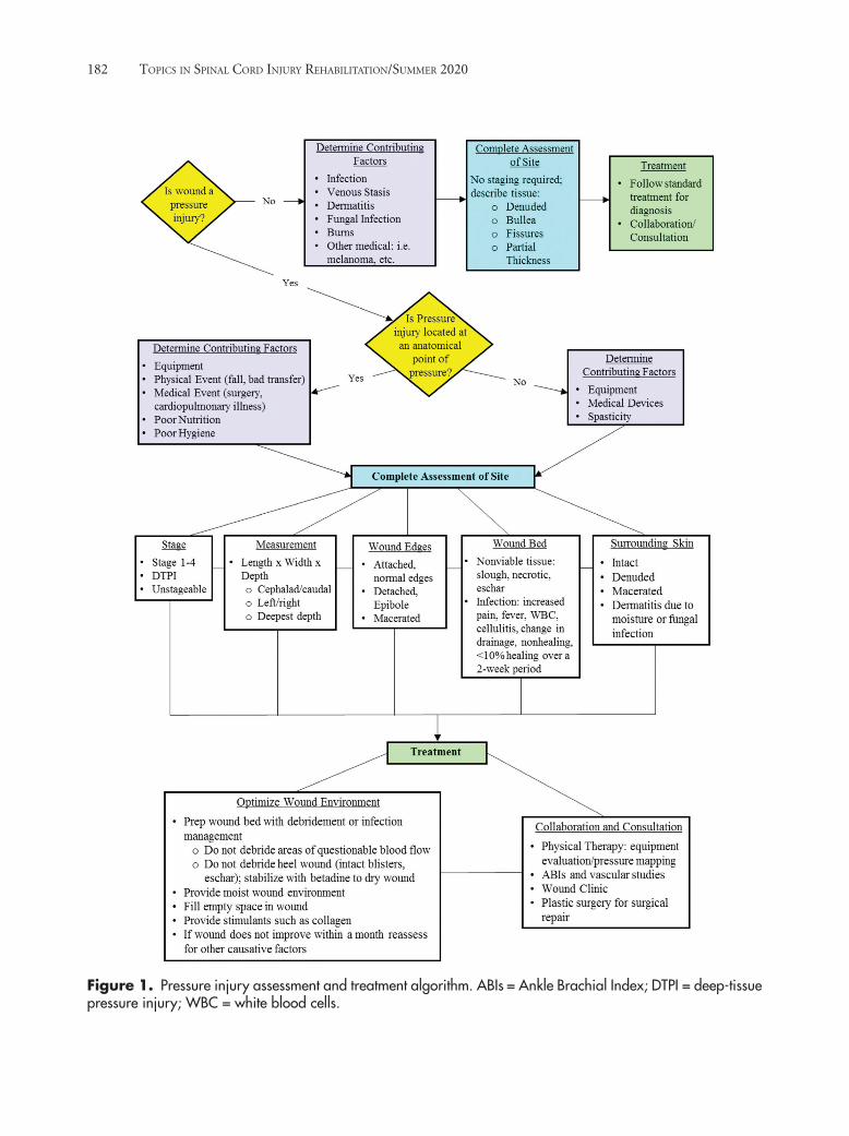
Successful treatment of skin breakdown requires optimal wound bed preparation, pressure off-loading, nutrition, durable medical equipment, and access to surgical specialists, if needed. See Figure 1 for a suggested wound management algorithm.
To optimally treat a new or chronic wound, the primary care team must engage persons with SCI and their support network to investigate the factors (Table 1) driving development and persistence of the wound—particularly noting positioning on support surfaces and in wheelchair seating systems—and provide them with appropriate wound care to promote wound healing and aid

Pressure Injury and Skin Breakdown 181
Table 4. TIME framework for wound bed description and management
Framework component Common descriptors Wound management options
Tissue: Description and general appearance
EscharSloughFibrinous sloughBiofilmGranulation tissue EpithelializationIdentifiable structures seen or palpatedMacerationDeep-tissue pressure injury
DebridementWound cleansingNegative pressure wound therapy (NPWT)
Infectious/Inflammatory signs: Local Regional Systemic
Non-healing or worseningWarmthErythemaSwelling or indurationIncreased painFriable red tissueFoul odorIncreased drainagePurulent drainagePalpable or visible boneNew satellite wounds, cellulitis, or rashSepsis
DebridementWound cleansingTopical anti-biofilm productsTopical antimicrobialsSystemic antibiotics
Moisture: Exudate type and amount
SerousSerosanguinousSanguineousPurulentDressing saturationMacerationDry wound bed
Adjust dressing absorbency & hydrating properties Manage infectionProtect peri-wound Manage edemaNPWT
Edges & Epithelialization:AdvancingNonadvancing
AttachedDetachedEpithelializationRolled wound edgesDried callus and debrisMacerated
DebridementSkin graftingBiologic productsNPWT
prevention. When comorbid conditions promote wound chronicity (diabetes, tobacco use, arterial or venous insufficiency, and poor perfusion due to unrelieved pressure), a palliative rather than a curative wound treatment plan may be needed.3,6,7
The wound stage or severity is used to develop the wound treatment plan. Healing full-thickness wounds can be challenging and may require spe cialty consultation. Unmanaged biofilm amplifies wound inflammation, leading to breakdown of newly formed extracellular matrix and promoting wound chronicity. Sharp debridement is used to remove the necrotic debris when eschar is present. Debridement should be avoided for stable, dry, nonfluctuant eschar on the heels or areas with little tissue coverage
over bony prominences. These areas should be left intact, painted with povidone iodine, and kept dry. Cautious wound debridement may be performed in the clinic in people with SCI who lack pain sensation, taking care to avoid triggering autonomic dysreflexia. If the wound bed is covered with loose slough or biofilm, a wound specialist should be consulted regarding enzymatic or autolytic debriding agents and anti-biofilm surfactant cleansers. Once the wound bed is optimized, wound healing occurs if the wound bed is kept clean, moist, and at body temperature.
Selection of a dressing for the wound is guided by wound environment needs, support for wound care treatments, and cost (see Table 5). Wound dressings need to be cautiously removed to avoid
182 Topics in spinal cord injury rehabiliTaTion/summer 2020
Figure 1. Pressure injury assessment and treatment algorithm. ABIs = Ankle Brachial Index; DTPI = deep-tissue pressure injury; WBC = white blood cells.

Pressure Injury and Skin Breakdown 183
Table 5. Commonly available wound products and their use10,17
Category (Brand name) Uses Application, frequency, and contraindications
Gauze • Partial and full thickness• Mild-moderate drainage
• Outer dressing • Daily • Avoid overpacking
Alginates & hydrofibers(Restore Calcium Alginate, Tegaderm Alginate, Aquacel)
• Partial and full thickness • Moderate-heavy drainage • Filling tunnels or undermining • Available with silver
• Directly onto wound bed• Secure with top dressing.• Every 1-2 days• Not for dry eschar or heavy bleeding
Transparent films(Hypafix, Opsite, Tegaderm)
• Superficial • Little drainage • Intact skin protection• Dressing securement
• Over bare skin or skin-prep gel• Weekly or as needed • Can cause maceration• Remove gently from fragile skin
Hydrocolloids(DuoDerm, Tegaderm hydrocolloid, Comfeel)
• Partial and shallow full thickness • Moderate drainage• Intact skin protection• Dressing securement• Autolytic debridement of mild necrotic tissue
• Over bare skin or skin-prep gel • As a top dressing• Every 3-7 days• Not for heavy exudate, fragile skin, or primary dressing for
deep wounds
Hydrogels(Curafil, DuoDerm Hydroactive Gel, Restore Hydrogel, Tegaderm Hydrogel)
• Partial and full thickness • Dry wound bed• Infected/necrotic wounds• Autolytic debridement of mild necrotic tissue• Available with antimicrobial
• Directly to wound bed• Top dressing to avoid desiccation• Cover with an occlusive film for autolytic debridement• Every 1-3 days• Avoid with heavy exudates • Can cause maceration
Foams(Allevyn, Hydrofera Blue, Hydrocell, Mepilex, PolyMem, Ultra Foam with AFM)
• Partial and full thickness • Heavy drainage• Infected/necrotic wounds • Filling tunnels and undermining• Available with antimicrobial
• Directly to wound bed • May need top dressing • Every 3-7 days• Not for dry eschar or nondraining wounds • Can cause maceration
Composite dressings (Xtrasorb, Mesorb, CombiDERM, Tegaderm+Pad)
• Partial and shallow full thickness • Little drainage • Infected/necrotic wounds • Hypergranulation tissue
• Directly to wound bed• Used as a top dressing• Every 3-7 days• Remove gently from fragile skin
Cadexomer iodine(Inadine, Iodoflex, Iodosorb)
• Partial and full thickness• Biofilm or necrotic slough
• Directly to wound bed • Cover with absorptive top dressing• Every 1-5 days• Reassess in 7-14 days • Use <50 g/application and <150 g/week • Avoid if iodine sensitivity
Collagenase(Santyl)
• Enzymatic debridement for slough and eschar
• Critically colonized and infected wounds
• Directly to wound bed• Requires absorptive dressing • No more often than daily• Avoid using with silver products
Medical grade Leptospermum or manuka honey (Medi-Honey, thera-Honey)
• Full thickness • Infected/necrotic wounds • Biofilm reduction• Autolytic debridement of slough and eschar
• Directly to wound bed • Every 1-7 days• Cover with absorptive dressing• Can cause maceration
Negative pressure wound therapy
• Partial and full thickness with depth • Moderate-heavy drainage• Postoperatively for incisional, graft, or dehiscence
• Trained provider/nurse must apply• Every 48-72 hours• Avoid if malignancy, infection, or untreated osteomyelitis

184 Topics in spinal cord injury rehabiliTaTion/summer 2020
further injuries from pressure or adhesive stripping of fragile epidermis. Wet-to-dry gauze dressings are rarely recommended for people with SCI due to the risk of mechanically debriding both necrotic and healthy tissue as well as creating a nidus of a pressure when the gauze compresses. Frequency of wound care is based on product recommendation, amount of drainage or infection, and availability of wound care dressing assistance. For deep but uninfected wounds, wound specialists may order negative pressure wound therapy to remove biofilm and control moisture, stimulate granulation tissue formation, and promote wound contraction. The wound should be assessed and measured every 1 to 2 weeks. If wound healing is not improved after 1 month of treatment, it should be re-evaluated and the treatment adjusted. If the wound is worsening, infection or poor adherence to treatment recommendation may be the cause. After re-evaluation, a referral to a wound care specialist should be considered.3,6,7
Depending on the location and cause of a PrI, evaluation of the appropriateness of the person’s durable medical equipment may be needed. This will likely require the provider to find community support with a physical medicine and rehabilitation physician, physical or occupational therapist, or wheelchair seating specialist. Because the care needs of people with SCI for PrI prevention and treatment can be complex, a multidisciplinary team is the standard of care.
Osteomyelitis
Acute and chronic osteomyelitis may contribute to nonhealing of stage 4 PrIs and has been found in 25%-47%15,16 of nonhealing severe PrIs. It can also complicate the plan of care if flap closure is desired. Imaging with radiographs, three-phase bone scan with SPECT, MRI with contrast, or tagged white blood cell (WBC) scan with SPECT are useful diagnostic tools. Definitive diagnosis relies on bone biopsy and culture. Treatment of osteomyelitis should include debridement of infected bone and 6 weeks of appropriate antibiotics.3,6,7
Surgical Management
Consideration for surgical management including flaps and primary closures should be coordinated with an interdisciplinary team to ensure that the person’s condition is optimized before surgery including tobacco cessation, durable medical equipment including seating check for dysfunction, stable psychological state, and ability to complete bed rest after flap. The postoperative care plan will likely require an extended care facility stay for supported bed rest followed by a progressive seating protocol to minimize the risk of flap failure.6
Complications
Chronic wounds can lead to multiple complications including bacteremia, sinus tracts and abscesses, septic arthritis, endocarditis, fistulas, and joint contractures. Squamous cell carcinoma, or Marjolin ulcers, can occur in long-term (years) chronic wounds and can be identified by increased pain, discharge, and bleeding with verrucous hyperplasia.6
Case Resolution
Your clinic nurse used your floor-based lift to transfer the patient to an exam table, and you identified a coccygeal deep-tissue pressure injury. You could not find a cause on initial assessment and referred him emergently to a wound care clinic. He returned to see you after 2 weeks and reported trying to do some bed rest, but he continued to work at his desk job about 6 hours per day with frequent pressure relief. The wound evolved to an unstageable PrI, which the wound care clinic treated with a topical enzymatic debriding agent. After a month, the unstageable injury did not improve. The patient was referred to a wheelchair seating clinic, where the contoured hybrid gel and foam cushion was found to be positioned backwards inside the cushion cover. In this orientation, the ridge that should separate his thighs pressed up against his coccyx. Further

Pressure Injury and Skin Breakdown 185
questioning revealed that a new home health aide started working for him about 2 months ago, around the time the wound appeared. After correcting the wheelchair cushion orientation and surgical debridement, the wound was found to be a shallow stage 4 PrI, and it healed after 3 months of negative pressure wound therapy.
Conclusion
The successful prevention and treatment of wounds in people with SCI requires an interdisciplinary approach in order to minimize
risk factors, remove identifiable causes, and promote wound healing. Infection should be treated with input from infectious disease specialists. Consideration for specialized surgical management should be coordinated with the interdisciplinary team. Unmanageable comorbid conditions may promote wound chronicity requiring a palliative treatment approach.
Acknowledgments
The authors have no conflicts of interest to report.
REFERENCES
1. van Loo MA, Post MW, Bloemen JH, et al. Care needs of persons with long-term spinal cord injury living at home in the Netherlands. Spinal Cord. 2010;48(5):423–428. doi:10.1038/sc.2009.142
2. Sabharwal S, Mezaros M, Duafenbach L. Telerehabilitation across the continuum of care for individuals with spinal cord injury. Paper presented at the Proceedings of the State of the Science Conference on Telerehabilitation. 2001.
3. Norton L, Parslow N, Johnston D, et al. Best practice recommendations for the prevention and management of pressure injuries. In: Foundations of Best Practice for Skin and Wound Management. A supplement of Wound Care Canada. Canadian Association of Wound Care; 2017. https://www.woundscanada.ca/docman/public/health-care-professional/bpr-workshop/172-bpr-prevention-and-management-of-pressure-injuries-2/file
4. Mortenson WB, Miller WC. A review of scales for assessing the risk of developing a pressure ulcer in individuals with SCI. Spinal Cord. 2008;46(3):168-175. doi:10.1038/sj.sc.3102129
5. Byrne DW, Salzberg CA. Major risk factors for pressure ulcers in the spinal cord disabled: A literature review. Spinal Cord. 1996;34(5):255-263. doi:10.1038/sc.1996.46
6. Henzel MK, Bogie KM. Medical management of pressure injuries in patients with spinal cord disorders. In: Kirshblum S, Lin V (eds.), Spinal Cord Medicine. 3rd ed. New York, NY: Demos Medical; 2019: 516-543.
7. Consortium for Spinal Cord Medicine Clinical Practice Guidelines. Pressure Injury Prevention and Treatment Following Spinal Cord Injury: A Clinical Practice Guideline for Health-Care Professionals. 2nd ed. Washington, DC: Paralyzed Veterans of America; 2014.
8. Groah SL, Schladen M, Pineda CG, et al. Prevention of pressure ulcers among people with spinal cord injury: A systematic review. PM R. 2015;7(6):613-636. doi:10.1016/j.pmrj.2014.11.014
9. McInnes E, Jammali-Blasi A, Bell-Syer SE, et al. Support surfaces for pressure ulcer prevention. Cochrane Database Syst Rev. 2015;(9):CD001735. doi:10.1002/14651858.CD001735.pub5
10. Sonenblum SE, Sprigle SH. Some people move it, move it . . . for pressure injury prevention. J Spinal Cord Med. 2018;41(1):106-110. doi:10.1080/10790268.2016.1245806
11. Edsberg LE, Black JM, Goldberg M, et al. Revised national pressure ulcer advisory panel pressure injury staging system: Revised pressure injury staging system. J Wound Ostomy Continence Nurs. 2016;43(6):585-597. doi:10.1097/WON.0000000000000281
12. Toussaint J, Singer AJ. The evaluation and management of thermal injuries: 2014 update. Clin Exp Emerg Med. 2014 Sep 30;1(1):8-18. doi:10.15441/ceem.14.029
13. European Wound Management Association (EMWA). Wound Preparation in Practice. London, UK: MEP Ltd; 2004. http://ewma.org/fileadmin/user_upload/EWMA.org/Position_documents_2002-2008/pos_doc_English_final_04.pdf
14. Harries RL, Bosanquet DC, Harding KG. Wound bed preparation: TIME for an update. Int Wound J. 2016;13(suppl 3):8–14. doi:10.1111/iwj.12662
15. Sapico FL, Ginunas VJ, Thornhill-Joynes M, et al. Quantitative microbiology of pressure sores in different stages of healing. Diagn Microbiol Infect Dis. 1986;5(1):31-38. doi:10.1016/0732-8893(86)90089-1
16. Bates-Jensen BM, Guihan M, Garber SL, et al. Character-istics of recurrent pressure ulcers in veterans with spinal cord injury. J Spinal Cord Med. 2009;32(1):34-42. doi:10.1080/10790268.2009.11760750
17. Government of Canada. Wound care products list (by category with examples of brand names). 2019-06-25. https://www.canada.ca/en/indigenous-services-canada/services/first-nations-inuit-health/non-insured-health-benefits/health-provider-information/medical-supplies-equipment-information/benefits-criteria/dressing-list.html