A potential energy-saving heat treatment for re-circulated ... · both methods than centrifugation...
174
A potential energy-saving heat treatment for re-circulated irrigation water and its biological mechanisms Wei Hao Dissertation submitted to the faculty of the Virginia Polytechnic Institute and State University in partial fulfillment of the requirements for the degree of Doctor of Philosophy In Plant Pathology, Physiology, and Weed Science Chuanxue Hong, Chair Boris A. Vinatzer Antonius B. Baudoin Erik L. Stromberg D. Michael Benson October 29, 2012 Blacksburg, VA Keywords: 16S rRNA, α-proteobacteria, γ-proteobacteria, bacteria, biocontrol, chlamydospore, colony PCR-SSCP, culture-dependent plating, Firmicutes, greenhouse crops, oospore, pathogen-suppressing, PCR-DGGE, re-circulated irrigation water, zoospore Copyright 2012, Wei Hao
Transcript of A potential energy-saving heat treatment for re-circulated ... · both methods than centrifugation...

A potential energy-saving heat treatment for re-circulated irrigation
water and its biological mechanisms
Wei Hao
Dissertation submitted to the faculty of the Virginia Polytechnic Institute
and State University in partial fulfillment of the requirements for the degree
of
Doctor of Philosophy
In
Plant Pathology, Physiology, and Weed Science
Chuanxue Hong, Chair
Boris A. Vinatzer
Antonius B. Baudoin
Erik L. Stromberg
D. Michael Benson
October 29, 2012
Blacksburg, VA
Keywords: 16S rRNA, α-proteobacteria, γ-proteobacteria, bacteria,
biocontrol, chlamydospore, colony PCR-SSCP, culture-dependent plating,
Firmicutes, greenhouse crops, oospore, pathogen-suppressing, PCR-DGGE,
re-circulated irrigation water, zoospore
Copyright 2012, Wei Hao
A potential energy-saving heat treatment for re-circulated irrigation water and its
biological mechanisms
Wei Hao
Abstract
Heat pasteurization is an effective water treatment to address the emerging plant
pathogen issue associated with increased water recycling practices in the ornamental
horticulture industry. The current protocol that recommends treating water at 95°C for 30
s, however, faces two major challenges: its energy cost and environmental footprint. We
hypothesized that temperature required to inactivate major pathogens in re-circulated
water may be substantially lowered from 95°C with extended exposure time. The goal of
this study was to test this hypothesis and make this water decontamination technology
economically more attractive while reducing its environmental impact. Specific
objectives were to (1) examine the effect of water temperature on the survival of
Phytophthora and bacterial species, two major groups of plant pathogens in water
recycling systems, and (2) elucidate the underlying biological mechanisms by which
plant pathogens are killed at those temperatures. Lab assays were performed to determine
the survival of zoospores and chlamydospores of P. nicotianae, and oospores of P. pini as
well as seven bacterial species after heat treatments at given periods of time. Greenhouse
experiments were conducted to determine the applicability of the lab assay data to the
real world using annual vinca (Catharanthus roseus) and P. nicotianae as a model
system. The results of these studies indicated that the water temperature required to
eliminate Phytophthora and bacterial species can be lowered to 48°C from 95°C if
treatment time extends to 24 h. Two major steps were taken to elucidate the underlying
biological mechanisms. Firstly, a scheme based on the DNA fingerprint and sequence
analysis was developed for characterizing bacterial species in irrigation water, after
comparing two typing strategies, three sample concentration methods, and evaluating
conditions in denaturing gradient gel electrophoresis (DGGE) profiling. Bacterial species
detected by culture-dependent and -independent strategies were rather different. The

iii
greater bacterial diversity was detected when water samples were concentrated by using
both methods than centrifugation or filtration alone. As for DGGE profiling, 40 to 60%
denaturant concentrations at 70 V for 16 h revealed the highest bacterial diversity.
Secondly, water samples were taken from an irrigation reservoir in a local nursery and
analyzed for bacterial diversity following heat treatments at 42 and 48°C. After these heat
treatments α-proteobacteria, γ-proteobacteria, and Firmicutes became dominant which
presents a substantial shift of bacterial community structure compared to those in the
control water at 25°C. Among the dominant in treated water were Bacillus,
Pseudomonas, Paenibacillus, Brevibacillus, and Lysobacter species, which may have
potential biocontrol activities against plant pathogens. This study provided the scientific
basis for developing a more energy-efficient and environmentally sound heat
pasteurization protocol for water decontamination.

iv
Acknowledgements
I would like to express my sincere gratitude to my advisor Dr. Chuan Hong for his
guidance, advice, and encouragement throughout the four years of my research projects.
I also would like to thank the members of my graduate committee, Drs. Boris Vinatzer,
Anton Baudoin, Erik Stromberg, and Michael Benson for their support and valuable
advice throughout my doctoral program. Without their help, this work would not have
been possible.
I would like to thank the financial support provided by the Fred C. Gloeckner
Foundation, Inc and a grant from the USDA National Institute of Food and Agriculture -
Specialty Crop Research Initiative (Agreement #: 2010-51181-21140) for my research
work and other expenses.
I greatly appreciate all my colleagues and friends in the Department of Plant
Pathology, Physiology, and Weed Science, and Hampton Roads Agricultural Research
and Extension Center at Virginia Tech, whose advice, support, assistance, and
encouragement have contributed to the completion of my research. My special thanks go
to Dr. Ping Kong, Dr. Giovanni Cafa, Patricia Richardson, Xiao Yang, Dr. Venkatesan
Parkunan, Dr. Zhihan Xu, Dr. Heather Olson, Lynn Rallos, Lauren Achtemeier, Michael
Pistininzi, Andrew Rotzin, and Haijie Liu. I am also very thankful to the PPWS and
HRAREC staff members including Donna Ford, Patsy Niece, Judy Fielder, Cris
Thompson, and Ava Borden for all their help during my time in graduate school.
I am forever grateful to my parents for their love, encouragement, and support.
v
Attributions
Several colleagues have contributed to this dissertation.
Chuanxue Hong, Ph.D. is a professor in Department of Plant Pathology, Physiology and
Weed Science and Hampton Roads Agricultural Research and Extension Center at
Virginia Tech. He is the primary advisor and committee chair for this project. He advised
each and every major step of this project from research planning to execution, data
analyses and interpretation, and edited this dissertation.
Chapter 2. Inactivation of Phytophthora and bacterial species in water by a potential
energy-saving heat treatment
Boris A. Vinatzer, Ph.D., an associate professor in Department of Plant Pathology,
Physiology and Weed Science at Virginia Tech, serves on my graduate committee. He
supported and guided the bacterial assays. He is listed as a co-author in a publication of
work described in Chapter 2.
Monday O. Ahonsi, Ph.D., a former post-doctoral research associate in Dr. Hong's lab,
performed the assays investigating zoosporic survival of Phytophthora nicotianae (Figure
2-1). He is listed as a co-author in a publication of the work described in Chapter 2.
vi
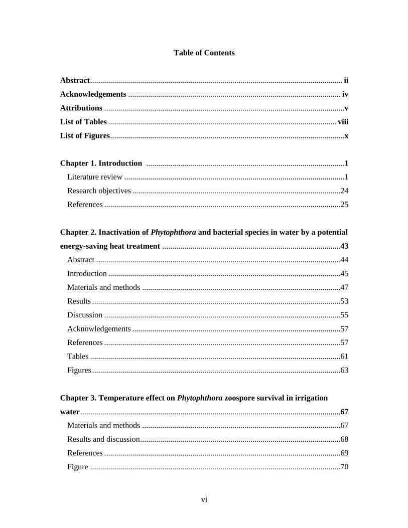
Table of Contents
Abstract .............................................................................................................................. ii
Acknowledgements .......................................................................................................... iv
Attributions ........................................................................................................................v
List of Tables .................................................................................................................. viii
List of Figures .....................................................................................................................x
Chapter 1. Introduction ...................................................................................................1
Literature review ..............................................................................................................1
Research objectives ........................................................................................................24
References ......................................................................................................................25
Chapter 2. Inactivation of Phytophthora and bacterial species in water by a potential
energy-saving heat treatment .........................................................................................43
Abstract ..........................................................................................................................44
Introduction ....................................................................................................................45
Materials and methods ...................................................................................................47
Results ............................................................................................................................53
Discussion ......................................................................................................................55
Acknowledgements ........................................................................................................57
References ......................................................................................................................57
Tables .............................................................................................................................61
Figures ............................................................................................................................63
Chapter 3. Temperature effect on Phytophthora zoospore survival in irrigation
water ..................................................................................................................................67
Materials and methods ...................................................................................................67
Results and discussion ....................................................................................................68
References ......................................................................................................................69
Figure .............................................................................................................................70
vii
Chapter 4. Characterization of microbial diversity in irrigation water: sample
concentration and processing strategies ........................................................................71
Abstract ..........................................................................................................................71
Introduction ....................................................................................................................72
Materials and methods ...................................................................................................74
Results ............................................................................................................................79
Discussion ......................................................................................................................82
References ......................................................................................................................84
Tables .............................................................................................................................91
Figures ............................................................................................................................97
Chapter 5. Bacterial community structure shifts in irrigation water under heat
treatment ........................................................................................................................105
Abstract ........................................................................................................................105
Introduction ..................................................................................................................105
Materials and methods .................................................................................................106
Results ..........................................................................................................................112
Discussion ....................................................................................................................115
References ....................................................................................................................118
Tables ...........................................................................................................................124
Figures ..........................................................................................................................130
Chapter 6. Summary and future directions ................................................................137
References ....................................................................................................................140
Appendix A: Bacterial isolates recovered from irrigation water in Chapter 4 ........142
Appendix B: Bacterial isolates recovered from irrigation water in Chapter 5 ........148
viii
List of Tables
Chapter 2
Table 2.1 Growth of six bacterial plant pathogens and a human commensal bacterium
after heat treatment at different temperatures for 24 h ......................................................61
Table 2.2 Growth of Xanthomonas campestris and Escherichia coli after heat treatment
at 48°C for 24 h ..................................................................................................................62
Chapter 4
Table 4.1 Water quality measurements in September 2011 and May 2012 ......................91
Table 4.2 Effects of sample concentration method, DGGE denaturant concentration and
running conditions on the number of resultant bands and Shannon index (H) of bacterial
diversity in irrigation water sampled in September 2011 and May 2012 ..........................92
Table 4.3 Alignment of 16S rRNA gene sequences obtained by colony PCR and excised
DGGE bands from irrigation water sampled in September 2011 and May 2012 ..............94
Chapter 5
Table 5.1 Water quality measurements in February and May 2012 ...............................124
Table 5.2 Total colony-forming units per plate on nutrient agar as affected by
temperature ......................................................................................................................125
Table 5.3 The number of resultant bands and Shannon diversity index (H) of bacterial
diversity at temperature treatments of irrigation water sampled in February and May 2012
..........................................................................................................................................126
Table 5.4 Bacteria in heat-treated water at 42 and 48°C and control (25°C) as identified
by partial sequences of 16S rDNA genes via culture-dependent and -independent
strategies .........................................................................................................................127
ix
Appendix
Table A.1 Bacterial isolates obtained via culture-dependent strategy from irrigation water
sample in Chapter 4. ........................................................................................................142
Table A.2 Bacterial isolates obtained via culture-independent strategy (PCR-DGGE)
from irrigation water sample in Chapter 4 .......................................................................146
Table B.1 Bacterial isolates recovered from irrigation water sample at control (25°C), 42
and 48°C via culture-dependent strategy in Chapter 5 ....................................................148
Table B.2 Bacterial isolates recovered from irrigation water sample at control (25°C), 42
and 48°C via culture-independent strategy (PCR-DGGE) in Chapter 5..........................160
x
List of Figures
Chapter 2
Figure 2.1 Effect of heat treatments on survival of (a) motile zoospores and (b) encysted
zoospores of Phytophthora nicotianae in laboratory .........................................................63
Figure 2.2 Effect of heat treatments on survival of zoospores of Phytophthora nicotianae
as indicated in the (a) percentage diseased plants and (b) percentage blighted shoots of
annual vinca 7 d after inoculation in the greenhouse .........................................................64
Figure 2.3 Germination of chlamydospores of Phytophthora nicotianae as affected by
heat treatments ...................................................................................................................65
Figure 2.4 Germination of oospores of Phytophthora pini as affected by heat treatments
............................................................................................................................................66
Chapter 3
Figure 3.1 Effect of heat treatments (25°C as control, 42 and 48°C) on survival of
zoospore of Phytophthora nicotianae in sterile de-ionized water (SDW) and irrigation
water (IW) at (a) 6 h, (b) 12 h, (c) 24 h, and (d) 48 h ........................................................70
Chapter 4
Figure 4.1 SSCP banding patterns of bacterial colonies recovered from irrigation water
sampled in (a) September 2011 and (b) May 2012 ............................................................97
Figure 4.2 Bacterial diversity in irrigation water samples as revealed through culture
dependent strategy based on partial sequences of 16S rDNA genes .................................98
Figure 4.3 Comparison of DGGE fingerprints of bacterial 16S rRNA amplicons from
irrigation water samples via three concentration methods: centrifugation (P), 0.2 µm
filtration (F), and a combination of centrifugation and filtration (C), with a 40 to 60%
DGGE gel denaturant concentration at 70 V for 16 h .....................................................100
xi
Figure 4.4 DGGE fingerprints of bacterial 16S rRNA fragments amplified from
irrigation water as affected by gel denaturant concentrations (a) 30 to 60%, (b) 40 to
60%, and (c) 40 to 70% under three conditions (70 V for 16 h, 90 V for 16 h, and 120 V
for 12 h)............................................................................................................................101
Figure 4.5 Bacterial diversity in irrigation water samples as revealed through culture-
independent strategy (PCR-DGGE) from partial sequences of 16S rDNA amplicons ...102
Figure 4.6 Flow chart illustrating steps in characterization of bacterial species in
irrigation water ................................................................................................................104
Chapter 5
Figure 5.1 Phylogenetic analysis of partial 16S rDNA sequences of representative
bacterial isolates from irrigation water samples collected in (A) February and (B) May
2012 after heat treatments at 25 (as control, black), 42 (orange), and 48°C (red) ...........130
Figure 5.2 . DGGE fingerprinting of bacterial 16S rRNA fragments in irrigation water
sampled in A) February and B) May 2012 after heat treatments at 25 (control), 42, and
48°C .................................................................................................................................133
Figure 5.3 Dendrogram derived from a distance analysis of DGGE bands and the Ward's
Hierarchical Method of clustering to show relationships between DGGE patterns for
different temperature treatments (25 as control, 42, and 48°C) in irrigation water sampled
in A) February and B) May 2012 .....................................................................................134
Figure 5.4 Phylogenetic tree of partial sequences of 16S rDNA from representative
bacteria in irrigation water samples collected in (A) February and (B) May 2012 after
heat treatments at 25 (as control, black), 42 (orange), and 48°C (red) via culture-
independent strategy (PCR-DGGE) .................................................................................135
1
Chapter 1
Introduction
Literature review
Plant pathogens in irrigation systems
Numerous plant pathogens, including zoosporic oomycetes, bacteria, viruses, fungi and
nematodes, have been found in irrigation water (Thomson and Allen 1974; Geldreich 1996;
Hong and Moorman 2005; Cayanan et al. 2009). These plant pathogens can enter the irrigation
system in several ways. They may inhabit the water source, the soil, nearby diseased resident
plants. Alternatively, they may be brought in from other places via infected plants or other
contaminated materials. Irrigation water sources, including ponds, lakes, rivers, streams, and
manmade reservoirs, may be contaminated by underlying or surrounding soil and plant debris,
thus affecting the entire irrigation system. Plant pathogens may also enter the irrigation system at
several points along the distribution path. Any irrigation method in which the water comes in
contact with soil or plant debris may acquire plant pathogens, for example, overhead sprinkling,
furrow flooding, field flooding, and flooded floor or bench. Runoff from flooding or other
methods of irrigation can re-circulate pathogens throughout the system. In outdoor production
facilities, when the excess water is captured and recycled, soil, which is possibly contaminated,
may be a significant source of pathogens. Also, pathogen-contaminated soil may fall or be blown
into water holding tanks, and then contaminate the whole irrigation system (Hong and Moorman
2005).
2
Irrigation systems is a powerful means for pathogen dissemination from a single infection
spot to the entire production facility and from an infested farm to all the other farms sharing the
same water resources (Harrison et al. 1987; Krczal et al. 1995; Hong and Moorman 2005).
Irrigation water facilitates pathogen infection by inoculating plants at every irrigation event and
providing a wet environment required for pathogens to germinate, colonize, and reproduce
themselves. Recycling irrigation also may allow monocyclic soilborne pathogens to complete
more than one disease cycle per growing season.
Zoosporic oomycetes, particularly those belonging to the genera Pythium Pringsh. and
Phytophthora de Bary, are the most common and destructive plant pathogens found in water
(Baker and Matkin 1978; Stanghellini and Rasmussen 1994; Stanghellini et al. 1996a;
Stanghellini et al. 1996b; Bush et al. 2003; Hong and Moorman 2005). Many species of
Phytophthora, Pythium, and other plant pathogens have been reported from irrigation water
(Bush et al., 2003; Neher and Duniway, 1992; Hong et al., 2003; Hong and Moorman, 2005;
Thomson and Allen, 1974). Specifically, over 17 species of Phytophthora have been reported in
irrigation reservoirs and natural waterways. These include P. alni, P. cactorum, P. cambivora, P.
capsici, P. cinnamomi, P. citricola, P. citrophthora, P. cryptogea, P. drechsleri, P.
gonapodyides, P. heveae, P. insolita, P. megasperma, P. nicotianae, P. pseudosyringae, P.
ramorum, P. syringae, and numerous undescribed Phytophthora species (Ho et al. 2002; Hong
and Moorman 2005; Ivors and Greene 2008). Twenty six Pythium species also have been
reported in ponds, rivers, canals, streams, lakes, wells, and sewage (Hong and Moorman 2005).
Bewley and Buddin (1921) detected P. cryptogea and P. parasitica (syn. P. nicotianae)
in greenhouse irrigation water in the early 1920’s. Many species of Phytophthora carried in
water have been reported to cause many diseases in fruit crops, vegetables, ornamentals, and
3
forest trees (Dukes et al. 1977; Neher and Duniway 1992; von Broembsen et al. 2001; Yamak et
al. 2002; Bush et al. 2003; Watanabe et al. 2008). In Germany, ten known Phytophthora species
and at least twelve new taxa were isolated from drains in reservoirs and wells at four commercial
nurseries with water recirculation systems (Themann et al. 2002). In a water re-circulation
irrigation system at a perennial container nursery in southwestern Virginia, seven Phytophthora
species and many Pythium species were found (Bush et al. 2003). In Japan, P. helicoides, P.
aphanidermatum, and P. myriotylum were recovered in ebb-and-flow and hydroponic culture
systems, causing root rot of miniature rose (Rosa hybrida L.), kalanchoe (Kalanchoe
blossfeldiana Poelln.), cucumber, tomato, tobacco, bell pepper, floral crops, lettuce, artichoke
and many other crops (Watanabe et al. 2008).
Zoospores are a major dispersal and infective propagule in the life cycle of Phytophthora
species. They may be dispersed by their own locomotion using flagella, or passively by water
current. The latter is particularly important in greenhouse industries that employ recirculation of
the irrigation water, including hydroponic and ebb-and-flow cultural systems (Neher and
Duniway 1992; Stanghellini et al. 1996a). Thomson and Allen (1974) indicated that zoospores
were the only propagules of Phytophthora spp. present in the irrigation water from 20 sites in
citrus areas near Phoenix, AZ. Stanghellini et al. (1996b) concluded that zoospores of Pythium
aphanidermatum were the propagule responsible for pathogen dissemination in hydroponic
systems after efficacy trials of surfactants. Surfactants could rapidly kill fungal structures lacking
a cell wall (i.e. zoospores), but not affect fungal structures having a cell wall (i.e. hyphae,
encysted zoospores, sporangia, oospores, or chlamydospores). Stanghellini et al. (1996a) made
the same observation with Phytophthora capsici. Thinggaard and Andersen (1995) also
suggested that zoospores of P. cryptogea were strongly involved in spread of root rot in ebb-and-

4
flow systems. In re-circulated irrigation at a large ornamental nursery, zoospores comprised more
than 94% of Phytophthora propagules in runoff water entering the retention basin (Charlton and
von Broembsen 2000). Usually, oospores or chlamydospores are found in sediment or plant
debris at the bottom of the reservoir (Pittis and Colhoun 1984).
Bacteria are also common pathogens found in irrigation water (Toze 1999, 2006b). A
total of eight species of bacteria has been reported in irrigation reservoirs and natural waterways
as reviewed by Hong and Moorman (2005). Erwinia species are among the most frequently
detected bacterial pathogens in water (Harrison et al. 1987; Cappaert et al. 1988; Eayre et al.
1995). The soft rot bacterium Erwinia carotovora subsp. carotovora was detected in irrigation
water from drains, ditches, streams, rivers, and lakes in southern Scotland and in Colorado,
United States (McCarter-Zorner et al. 1984). Ralstonia solanacearum biovar 2, which causes
brown rot in potato and other plants has been observed in different soil and water systems, and it
may spread to and survive in local waterways following disease in potato fields (Stevens and van
Elsas 2010). Pseudomonas syringae strains have been detected in rain, snow, alpine streams,
lakes, epilithic biofilms, and irrigation water (Morris et al. 2008). Xanthomonas species have
also been found in irrigation water (Steadman et al. 1979; Hoitink and Boehm 1999).
Twenty-seven genera of fungi have been found in irrigation water based on a review by
Hong and Moorman (2005). Species in the genera Alternaria, Botrytis, Ascochyta, Rhizoctonia,
and Verticillium are present in water because of their natural abundance but they may not survive
well in water (Bewley and Buddin 1921; Shokes and McCarter 1979). Shokes and McCarter
(1979) recovered thirteen species of Fusarium, Rhizoctonia solani, R. zeae, and Macrophomina
phaseolina from sediment of irrigation ponds. Fusarium oxysporum has been reported to spread
through irrigation water and cause plant diseases (Jenkins and Averre 1983; Wick and Haviland
5
1992), but it may not always be able to live long enough in water to reach and infect plants
(Rattink 1990). Fusarium oxysporum f. sp. basilici spread via a re-circulating water system and
caused fusarium wilt on basil in greenhouses (Wick and Haviland 1992). Colletotrichum spread
via water in hydroponically grown tomatoes (Jenkins and Averre 1983).
At least ten viruses have been reported in river, lake and irrigation systems (Hong and
Moorman 2005). Pelargonium flower break virus (PFBV) was transmitted via a re-circulating
nutrient system from infected plants to healthy plants in 6 weeks (Krczal et al. 1995). And
tomato mosaic virus (ToMV) also spread rapidly in re-circulating nutrient solution from
inoculated plants and remained infective for at least 6 months (Pares et al. 1992).
Thirteen species of plant parasitic nematodes have also been reported in irrigation water
(Hong and Moorman 2005). Because plant parasitic nematodes are pathogenic roundworms that
live in water films in soil and plant tissues, they are essentially aquatic organisms and can be
washed from soil and enter the irrigation systems. Faulkner and Bolander (1967) demonstrated
that irrigation canals were sources of large numbers of plant parasitic nematodes. Simmons et al.
(2008) indicated that Ditylenchus dipsaci and foliar nematode Aphelenchoides ritzemabosi may
be transported across alfalfa fields via irrigation water.
Capture and reuse of surface water for irrigation is of critical importance to ensure an
adequate supply of quality water in light of growing global water scarcity (Exall 2004; Hong and
Moorman 2005; Toze 2006a). Many nurseries and greenhouses have implemented collection,
recovery, and recirculation of irrigation water. These practices reduce water consumption and
release of fertilizer and pesticide pollutants into the environment, and provide important nutrients
for crops (Bush et al. 2003; Hong et al. 2003; Steele and Odumeru 2004; Hong and Moorman
2005; Cayanan et al. 2009). Re-circulation of irrigation water generally involves collecting
6
nursery effluent into holding tanks or outdoor reservoirs through soil lined channels then mixing
with fresh water from nearby rivers or wells for subsequent irrigation (Bush et al., 2003).
However, re-circulation of water may accumulate plant pathogens in irrigation systems and
spread to new crops. Van Voorst et al. (1987) demonstrated that when Phytophthora nicotianae
inoculum was added to the roots of tomatoes grown in a greenhouse by nutrient film technique in
which the nutrient solution was re-circulated, the pathogen also freely circulated within the
system and caused polycyclic infection to the hosts. Once pathogens enter an irrigation system,
the repeated pathogen exposure due to water re-circulation increases the occurrence of diseases
(Hong and Epelman 2001). Werres et al. (2007) indicated that Phytophthora ramorum can
survive and infect Rhododendron species through contaminated re-circulated irrigation water
Decontamination of irrigation water
One principle of plant disease management in the ornamental industry is to ensure the
health of the plant material entering production (Daughtrey and Benson 2005). Use of a clean
water source and preventing plant pathogens from entering irrigation systems are the first and
important steps to ensure irrigation water health. Water treatment prior to delivering to crops
should be used as the last resort but it is necessary in most situations for crop health risk
avoidance and mitigation (Hong and Moorman 2005).
Currently, physical, chemical, and biological means are utilized for controlling plant
pathogens in water. These include filtration, heat treatment, chlorination, use of surfactants, and
ultraviolet (UV) radiation. All these control methods aim at reducing microbial levels, or
inactivating microorganisms (Evans 1994; Hong and Moorman 2005; Schumann and D'Arcy
2006). Several factors must be considered when selecting water treatments for a particular

7
production facility. For example, it may be most cost-effective to use high quality and not re-
circulated water for sensitive crops, such as young plants, and use recycled water only for those
less susceptible plant species. In making a decision, it is useful to first identify the target
pathogens for treatment (classes, life stages) in order to design the parameters for the treatment
system. Some important considerations include: 1. quantity of water to be treated and therefore
the installation and operation cost; 2. water quality conditions, such as pH, turbidity, electrical
conductivity, etc, and the water quality changes allowed during treatment; 3. training, education
and safety of the workers; and 4. residual which may enter the environment or have potential
phytotoxicity to the crops (Hong and Moorman 2005).
Each of these water decontamination technologies has their advantages and
disadvantages. UV radiation, chlorination, and heat are generally effective, but may have a
negative effect on benefical microorganisms in irrigation water. Slow sand filtration is less
disruptive to the beneficial microorganisms, but may be less effective on plant pathogens (Ehret
et al. 2001). Most of the water treatments are expensive, and a certain level of education/training
is needed for the growers to actually employ these control methods. Moreover, none of the
practices are simple or one-time treatments but require monitoring and repeated treatments (von
Broembsen et al. 2001; Hong and Moorman 2005).
Filtration is a separation process by the flow of a liquid through a porous material, such
as a bed of fine filter sand, to retain the solids and allow the liquid (filtrate) to pass through (Ellis
1985). Slow sand filtration is efficient against many plant pathogens spread in irrigation water
(Ellis 1985; Wohanka 1995; Runia et al. 1997; van Os 1999). It's a low energy input technology
and also the construction and operation are simple (McPherson et al. 1995). Use of slow
8
filtration is limited due to its limited capacity and clogging caused by high loads of particulate
matter in water (Hong and Moorman 2005).
Chlorination is widely used in horticultural production to disinfest irrigation water. It is
highly effective against a number of pathogens, especially the oomycetes Pythium and
Phytophthora, with a dose of free residual chlorine lower than 2 ppm (Hong et al. 2003; Cayanan
et al. 2009; Granke and Hausbeck 2010). This technology only needs a relatively low initial and
operation cost, and the installation, operation, and maintenance is relatively simple. But chlorine
efficacy depends largely upon water quality parameters and may also produce byproducts
harmful to human health and the environment (Hong and Moorman 2005).
UV radiation is an effective choice for irrigation water in a nursery or greenhouse to
eliminate plant pathogens and algae. Exposure to germicidal UV radiation kills microorganisms
by damage to DNA in the form of strand breaks, cross links, and dimerization of adjacent
pyrimidine bases in DNA, which usually is lethal or mutagenic to cells (Setlow and Setlow
1962). UV radiation adds no residual chemistry to the water after treatment, therefore, there is no
residual disinfestation effect. It also destroys the beneficial microorganisms in water (Ehret et al.
2001). It is typically used in combination with filtration to remove organic matter, which
increases light transmission and efficacy (Fynn et al. 2011).
Surfactants disinfest the irrigation water by disrupting the membrane of microorganisms
due to their amphilic character, and rapidly killing the plant pathogens. Certain surfactants,
mainly cationic, are commonly used as disinfestants in water (Prescott et al. 2005). Surfactants
are highly efficient against zoosporic pathogens, such as Phytophthora and Pythium species,
because the primary inoculum zoospores, which lack cell walls, are very sensitive to surfactants
(Stanghellini and Miller 1997). Limitations of surfactants include the toxic residue to the
9
environment due to slow degradation, and potential phytotoxicity when using in irrigation
systems (Uhlig and Wissemeier 2000; Irish et al. 2002).
Heat is one of the most reliable methods for treating water to eliminate many types of
plant pathogens, including fungi, bacteria, and viruses. Research on the thermal inactivation of
microorganisms in diverse media started as early as 1920 (Bigelow and Esty 1920; Coelho et al.
2000). For example, Bigelow and Esty (1920) showed the effect of different temperature and
exposure time combinations necessary to inactivate thermophilic bacteria and Grooshevoy et al.
(1941) used natural heat sources to disinfest seed bed soil.
Heat treatment is commonly used by greenhouse growers, especially in the Netherlands
and the United Kingdom. This practice is one of the most commonly used water treatments in the
closed soilless growing system and has been used for several years (Runia et al. 1988; Runia
1995; van Os 1999). However, this technology is used to a lesser extent in other countries, due to
energy efficiency issues and high investment costs (Hong and Moorman 2005).
Extensive studies have been conducted to investigate thermal inactivation of plant
pathogens in infested plants, soil, and culture media. Half of mycelium of two isolates of
Phytophthora cinnamomi were inactivated by hot water at 39°C for 26.3 and 51.7 min or 43°C
for 2.7 and 3.3 min on agar disks, respectively. And Fraser fir seedlings inoculated with P.
cinnamomi in growing medium within 14 d were disinfested by water therapy at 45°C for 15 min
(Benson 1978). Hyphae and oospores of Phytophthora cactorum and P. cinnamomi survived
only 30 min at 45°C in inoculated walnut twigs or artificially infested Reiff silty clay loam soil
(Juarezpalacios et al. 1991). The population of chlamydospores of P. nicotianae in soil, as baited
with tomato seedlings, declined to very low levels after heat treatment at 47°C for 2 h, or 50 and
53°C for only 5 min (Coelho et al. 2000). Pullman et al. (1981) found that temperatures between
10
37 to 50°C for different time periods can kill mycelia, spores, and resting spores of Verticillium
dahliae, Pythium ultimum, and Thielaviopsis basicola on agar media. Twenty-three, 27, 33, and
68 min of heat treatment at 50°C were required to cause 90% mortality of V. dahliae (strains T9
and SS4), P. ultimum, and T. basicola, respectively. There are differences in temperature
response between different types of fungal propagules within the same species. For example, to
inactivate Phytophthora cinnamomi on agar discs within 1 to 2 h, the required temperature was
38°C for mycelium, and 40°C for chlamydospores (Gallo et al. 2007). Mycelium of P. capsici
was more sensitive than oospores with the former being eliminated with a heat treatment at 42.5
to 45°C for 30 min and the latter still surviving a heat treatment at 50°C for 30 min in soil
(Bollen 1985).
Propagule suspensions have been used by researchers to study the effect of heat on
inactivation of plant pathogen spores by heating to a certain temperature for a certain period of
time, which provides the basis for estimating the temperature and time required to control many
types of plant pathogens in irrigation water (Runia et al. 1988; van Os et al. 1988; McPherson et
al. 1995; Runia 1995; Poncet et al. 2001). Dyer et al. (2007) demonstrated that oospores of the
oomycete Aphanomyces cochlioides, the primary inoculum during growing seasons, were
susceptible to moderately high temperatures. The viability of oospores in water declined to zero
after exposure to 45°C for 72 h or 50°C for 6 h. The authors also suggested that the heat
sensitivity of Phytophthora spp. in the laboratory closely corresponded that in soil. Oospore
suspensions of P. infestans, which may be a long-term source of inoculum, did not germinate
after treatment at or above 46°C for 2 h, or at 40°C for 12 h (Fay and Fry 1997).
The development of current protocols for heat treatment started with Runia et al. (1988)
who studied the disinfection of recirculated water by heat treatment using tobacco mosaic virus
11
(TMV), Verticillium dahliae, and Fusarium oxysporum f. sp. melongenae in the laboratory and
greenhouse. TMV was one of the main root-infecting viruses in crops in soil-less cultures in the
Netherlands, and F. oxysporum f. sp. melongenae was one of the most resistant soilborne fungal
pathogens (Bollen 1985). Fungal spore suspensions were dilution-plated and host plants were
inoculated by dipping root systems into fungal suspensions or by rubbing leaves with TMV
suspension after heat treatments. Results indicated that TMV can be inactivated after heat
treatment at 95°C for 10 s, and spores of V. dahliae were killed at 90°C for 10 s. The propagules
of F. oxysporum declined significantly after being heat-treated at 94°C for 10 s. McPherson et al.
(1995) studied the effect of heat treatment on Phytophthora cryptogea and Pythium
aphanidermatum inoculated in re-circulated hydroponic nutrient solutions on tomato and
cucumber crops. They reported that the inoculated pathogen did not spread through the re-
circulating system after heat treatment at 95°C for 30 s compared to non-treated nutrient solution,
which transmitted diseases to most plants. Poncet et al. (2001) reported the disinfestation of
drainage water in soilless cultures of roses. They counted total bacteria and Agrobacterium
tumefaciens in drainage water before and after heat treatment. Bacteria, including A. tumefaciens,
with an initial concentration of 108 cfu per liter, in the re-circulated irrigation water were
eliminated after heat treatment at 95°C for 30 s. Therefore, based primarily on the studies
described above that were performed in the Netherlands (Runia et al. 1988) and the United
Kingdom (McPherson et al. 1995), current protocols recommend raising and maintaining water
temperature to 95°C for 30 s.
Several studies in regard to heat disinfestation of nutrient solution systems were
performed using heat exchangers. In collaboration with the Institute of Agricultural and
Environmental Engineering at Wageningen, Runia et al. (1988) constructed a heat-exchange
12
system to treat drainwater reused for soilless cultures in the Netherlands. Basically, the
drainwater returning from the plants was collected in a recatchment tank. After filtering out
organic particles and leaves the drainwater was pumped into the first heat exchanger to preheat
to about 80 to 90°C. Water was then further heated to the disinfestation temperature above 95°C
by a second heat exchanger. This temperature was kept for a certain exposure time. After heat
treatment the disinfected water flowed back to the first heat exchanger to be cooled down and be
stored in a holding tank to cool further. The heat recovered from the hot disinfested water was
used in the first heat exchanger, and an external heat source was used for the second heat
exchanger. A switch valve was used to control water returning to the recatchment tank if the
water was not properly disinfested. McPherson et al. (1995) used commercially available heat
exchangers for milk heat treatment. Poncet et al. (2001) used the “L’ien thermodisinfection” co
heat-exchange system. Their disinfestation system was similar to that described by Runia et al.
(1988) as the drainage water was treated successively through two heat exchangers.
Re-circulated irrigation water in commercial greenhouse circulation systems can be heat
treated similarly to commercial heat treatment of other fluids, such as milk and drinking water
(Evans 1994). The basic principles are the same as in the disinfestation system built by Runia et
al. (1988). Two heat exchangers with one being linked to an external heat source are used for
heat treatment of the irrigation water with an initial filtering of water through a rapid sand filter
to remove plant parts and other debris (Ehret et al. 2001). It is important that the quality of the
water to be treated is high to prevent elevated levels of sodium and chloride that would result in
phytotoxicity. The holding tanks used to store disinfested water must be in an area where the
water can be protected from new contamination prior to use. Corrosion-free materials, such as
stainless steel and synthetic materials should be used to build the equipment, also copper- and
13
zinc- based materials should be avoided due to their phytotoxicity (Runia et al. 1988). Because
high temperatures cause calcium salts to precipitate inside the equipment thus leading to a lower
efficiency of the disinfestation system over time, washing the equipment with an acid solution
(pH 2 to 3) for a few minutes before each disinfestation cycle is advised to remove accumulated
salts (van Os et al. 1988). The use of heat exchangers increased the efficiency of energy use and
reduced the energy input to a relatively low level (Runia et al. 1988; Runia 1995). Overall, heat
treatment systems are not difficult to install and they are easy to monitor with a thermostat (Ehret
et al. 2001).
Heat treatment involves no chemicals and thus presents no health hazard to workers
(Gurol 2005). It also presents minimal environmental hazards compared to chemical treatments.
Some chemical disinfestants such as copper will not degrade in water and will easily accumulate.
Also, excessive or accidental applications associated with recirculation of water treated with
these disinfestants can easily lead to damaging chemical concentrations in water, which can be
taken up by the plants and soil, causing phytotoxicity (Gurol 2005; Blom 2008). Heat treatment
on the other hand will not have secondary effects on the environment.
The major limitations of current heat treatment protocols for re-circulated irrigation water
are energy inefficiency, the cost of required equipment, the volume of the storage tank needed to
hold treated water, and the killing of beneficial microorganisms (Runia et al. 1988; van Os et al.
1988; Ehret et al. 2001; Hong and Moorman 2005).
The cost of heat treatment is relatively high due to the initial investment in the equipment
and continuous consumption of energy to produce heat. The investment for construction of the
heat disinfestation system is substantial (Poncet et al. 2001), so it is not quite affordable for
smaller floriculture farms. Runia et al. (1988) indicated that their disinfestation system needed
14
about 1.5 m3 natural gas per 1 m
3 drainwater, resulting in usage of about 2700 m
3 gas per
hectare. Blom (2008) also reported that as high as 20.2 to 39.6 m3
of fuel was needed to heat
every 1 m3 of water, and resulted in an estimated cost of 7.7 to 15.2 US dollar per 1 m
3 water
heated according to the current price average 10.87 dollar per 1000 ft3 of natural gas in 2012
(EIA 2012). Also the burning of fossil fuels emits a large amount of greenhouse gases, especially
carbon dioxide (CO2). These greenhouse gases are major contributors to global warming, which
has become a worldwide concern (De Oliveira et al. 2005). In addition to the continuously
increasing cost of energy, the natural gas consumption for heating the contaminated water can be
energy inefficient and cost prohibitive when there are no other sustainable energy sources.
Application of the described heat-exchangers to recover some energy for preheating water will
reduce the cost and it may also be feasible to use heat-treated water for only the critical stages of
crop production such as during the propagation of highly susceptible plants (Bewley and Buddin
1921).
The worsening energy crisis is further hurting the economics of this highly effective
water treatment until a more efficient heat treatment system is developed. The possibility of
substituting heat with treatment time and alternative energy sources have been studied.
The efficacy of heat treatment depends on both temperature and treatment time. Sublethal
temperatures, usually below 50°C, can be lethal to soilborne plant pathogens if maintained for
long periods (Smith 1923; Munnecke et al. 1976; Vanuden and Vidalleiria 1976; Gallo et al.
2007). Sublethal heating of infected roots in soil reduced survival of Armillaria mellea. The
fungus was killed in the roots of citrus seedlings at 40°C for 2 to 3 h (Munnecke et al. 1976).
Runia et al. (1988) also reported that the viability of spores of Fusarium oxysporum f. sp.
melongenae was reduced with increasing exposure time at the same heat treatment temperature.

15
It is thus likely that reducing treatment temperature while increasing exposure time will still have
the same efficacy as the current heat treatment protocol. By using a heating system developed by
a commercial company and laboratory setups, Runia and Amsing (2001) indicated that heating at
85°C for 3 min had the same efficacy as heating at 95°C for 30 s for both Tomato mosaic virus
and F. oxysporum f. sp. lycopersici. The burrowing nematode Radopholus similis was eliminated
at 48, 50, and 52.5°C for 5, 2 min, and 30 s, respectively. Also temperature-exposure times lethal
to pathogens were well established for soil steam sterilization where exposure time ranges from
0.5 to 12 h (Bollen 1985; Evans 1994), so inactivation of plant pathogens in irrigation water
might be achieved at a lower temperature when extending treatment time from seconds to hours.
The energy consumption and cost of water disinfestation will be substantially reduced if
new heat practices can be developed for horticulture. Also, new sustainable energy sources such
as solar irradiation and geothermal energy have great potential to be applied in heat treatment of
re-circulated irrigation water, once the appropriate disinfestation systems have been developed.
Solar water pasteurization has been used to disinfest drinking water, but solar radiation is
seldom used in treating re-circulated irrigation water (Jorgensen et al. 1998; Duff and Hodgson
2005; Kang et al. 2006). As early as 1984, solar cookers were used to decontaminate relatively
small amounts of drinking water (Ciochetti and Metcalf 1984). Conroy et al. (1996) employed
drinking water contained in transparent plastic bottles exposed to the sun for a few hours, which
reduced the rate of childhood diarrhea by 10%. There are two main types of solar water
pasteurization systems: batch and continuous flow. In batch systems, refillable vessels are used,
and it usually takes a full day of sun for a batch system to disinfest water (Ciochetti and Metcalf
1984; Andreatta et al. 1994; Conroy et al. 1996). In a continuous flow-through system, water
from a supply reservoir flows through a heat exchanger at first, then through a solar collector,

16
such as copper pipes, which heats the water up to a desired temperature. Heat exchangers here
are also used to preheat untreated water and a thermostatic valve is used to control temperature
and flow (Jorgensen et al. 1998; Duff and Hodgson 2005). Jorgensen et al. (1998) used a water
decontaminator device (HS 231545; Roerslev Smedie, Roerslev, Denmark) for heating drinking
water by solar radiation in a flow-through system of copper pipes. The water temperature could
be raised up to 85°C. When the required temperature was reached and detected by a sensor, the
water flows out through an open valve. The heating temperature was set at 65°C. Daily
production was about 50 L of decontaminated water per m2 of solar panel, and it was possible to
double the amount by using a heat exchanger to recycle the heat. Since the water
decontamination device can be used for years and the running cost is low, the cost of pasteurized
water can be estimated at less than US dollar 0.008 per liter.
Water disinfestation systems using solar panels are energy-efficient, cost-effective, and
easy to install and transport (Duff and Hodgson 2005; Kang et al. 2006). However, these systems
may not be able to hold water at a high temperature for a relatively long period, and can only
treat small amounts of water. Therefore, current solar water disinfestation systems which have
been developed probably are not very useful for commercial soilless culturing greenhouses,
where as much as 175 to 275 m3 per hectare per day of runoff water needs to be disinfested
(Blom 2008). New advanced solar water heating systems with a much larger water-treating
capacity need to be developed for common usage in agriculture.
Geothermal energy, which is the energy contained as heat within the earth’s interior,
could be used to replace fossil fuels to some extent (Lund et al. 2005; Ozgener et al. 2005).
Geothermal energy has been commercially utilized for over 80 years and was used in more than
72 countries in 2005 (Lund et al. 2005; Ozgener et al. 2005). In the United States, geothermal

17
energy has been directly used in heating of pools and spas, greenhouse and aquaculture facilities,
space and district heating, snow melting, agricultural drying, ground-source heat pumps; and
generating electricity (Ozgener et al. 2005). Most of the applications have experienced continual
increase over the years; however, there is no record of geothermal energy being directly used to
disinfest water. Because using geothermal energy instead of fuels could significantly reduce the
emission of pollutants, such as greenhouse gases, and since only a small fraction of the
geothermal potential has been developed so far, there is plenty of space for an increased use of
geothermal energy (Fridleifsson 2001; Lund et al. 2005). Geothermal energy thus has great
potential in heat treatment of contaminated water.
Characterization of microbial diversity in irrigation water
To evaluate and choose the most appropriate and efficient disinfestation treatments in
irrigation water, knowledge of the microbial diversity within the system is an important
precondition (Hong and Moorman 2005). Knowledge of the microbial diversity and how it
changes would facilitate understanding of the dynamics of plant pathogens in irrigation water.
Specifically, these data would help identify naturally occurring biological control agents against
plant pathogenic species. It could also support the physicochemical assessment of water quality
and health in irrigation systems (Liu et al. 2011).
Microbial communities can be characterized using different methods. Traditional
morphological and culture-dependent technologies, such as plating, and culture-independent
molecular technologies, such as polymerase chain reaction (PCR) and molecular fingerprinting,
give a picture of the structural composition of the microbial community in water (Alsanius and
Jung 2003). However, morphological and culture-dependent approaches, especially for bacterial
18
species, can only detect a very small proportion of the total microbial community due to the
difficulties of discriminating morphologically similar organisms, and the limitations of a
majority of microbes being unculturable in field samples (Amann et al. 1995; Vartoukian et al.
2010). Molecular techniques have been shown to be very powerful to investigate the composition
and structure of environmental microbial communities besides the traditional culture-dependent
approaches (Case et al. 2007).
Microbial communities can be regarded as a mixture of microbial genomes. Genomic
DNA sequences and their numbers indicate the community structure, which has been defined as
the amount and distribution of (genomic) information in a particular habitat (Xie et al. 2009).
Phylogenetically meaningful sequences, such as randomly amplified genome fragments, small
subunit ribosomal RNA (rRNA) genes and conserved functional genes, have been used as
molecular markers to analyze microbial communities by different technical strategies, including
molecular hybridization, clone library profiling, and genetic fingerprinting by denaturing
gradient gel electrophoresis (DGGE) (Wikstroim et al. 1999; Xie et al. 2009). Since the 1980s,
rRNA-PCR allows microorganisms in a sample to be phylogenetically typed and quantified
based on the sequence of their rRNA genes (Eisen 2007).
Since 1990, the 16S rRNA gene has been used as a molecular tool to investigate
environmental microbial communities (Giovannoni et al. 1990). The 16S rRNA gene is highly
conserved between different bacteria and archaea species, but the nine hypervariable regions (V1
to V9) with different sequence diversity among different species can be used as phylogenetic
markers to detect bacterial community composition (Van de Peer et al. 1996). In addition,
Chakravorty et al. (2007) demonstrated that none of the single regions can distinguish all
bacteria species, but the V6 region can be used to identify most bacterial species except
19
enterobacteriaceae. Genomic DNAs are extracted directly from an environmental sample, such
as water, soil or biofilm, and the 16s rRNA genes of the microbial community are amplified from
the mixture by PCR. Molecular tools utilizing the16S rRNA gene to study microbial
communities include cloning libraries, in-situ hybridization, and fingerprinting methods, such as
DGGE (Tringe and Rubin 2005; Case et al. 2007).
PCR-DGGE has been used as a rapid and efficient approach to characterize microbial
communities of many environmental samples, such as soil, different water sources, eg,
groundwater, river, lake, sea water, drinking water; biofilms, and even the symbiotic microbial
community of other organisms, eg. sponges (Kozdroj and van Elsas 2001; Kawai et al. 2002;
Cho et al. 2003; Feris et al. 2003; Thoms et al. 2003; Brakstad and Lodeng 2005; Lyautey et al.
2005; Yan et al. 2007). Muyzer et al. (1993) was the first to report on the use of DGGE to
separate PCR amplified fragments of genes coding for 16S rRNA, in order to analyze the genetic
diversity of complex microbial populations. In DGGE, PCR products of similar size but different
base-pair sequence are separated based on the different electrophoretic mobility of partially
melted double-stranded fragments in polyacrylamide gels (Fischer and Lerman 1979).
Theoretically, numbers and intensity of bands represent phylotype number and abundance in a
microbial community (Muyzer and Smalla 1998; Nubel et al. 1999; Sigler et al. 2004). The
separation of PCR products with optimum resolution is very critical to a good DGGE-based
community structure analysis (Sheffield et al. 1989). This technique was particularly useful in
the detection and identification of unculturable microbial populations and those present in low
abundance (Holben et al. 2004). Cherif et al. (2008) demonstrated that PCR-DGGE was more
accurate for bacterial identification in soil samples than other molecular methods. Lyautey et al.
20
(2005) also indicated that PCR-DGGE is useful for assessing the bacterial diversity of epilithic
biofilms in rivers.
However, for irrigation water samples, the reliability and stability of PCR-DGGE to
investigate the microbial community diversity compared with traditional culture-dependent
approaches have not been determined. Brakstad and Lodeng (2005) revealed more than thirty
different phylotypes in seawater from the North Sea by using only PCR-DGGE fingerprinting of
16S rRNA. Yan et al. (2007) investigated the microbial diversity of a plankton community in
lakes by PCR-DGGE fingerprinting and concluded that PCR-DGGE fingerprinting might be
more sensitive and effective to characterize the microbial diversity within an aquatic ecosystem
than traditional methods. Liu et al. (2011) studied the microbial planktonic community diversity
in a subtropical river by using PCR-DGGE fingerprinting. On the other hand, some researchers
demonstrated that traditional culture-dependent methods were needed along with the molecular
fingerprinting techniques to investigate the microbial communities (Kawai et al. 2002; Cho et al.
2003). Both cultured colonies growing on nutrient agar media after plating water samples and
direct analysis of genomic bacterial DNAs from these samples by PCR-DGGE were utilized to
study the bacterial communities in groundwater (Cho et al. 2003). Kawai et al. (2002) also
indicated the importance of culture-independent methods to investigate the bacterial diversity in
partially purified water, and conventional plating methods on media complemented the study.
Previous PCR-DGGE based microbial diversity studies have used different conditions,
including the denaturant concentration ranges, electrophoresis voltage and time. Sigler et al.
(2004) demonstrated that the DGGE profile of a bacterial community changed as the
electrophoresis running time increased, and they recommended minimizing running time for
optimum band resolution. In practice, various DGGE denaturant concentrations running under
21
different electrophoretic conditions have been used to study different environmental samples.
DGGE with the denaturant concentration of 30 to 55% (a 100% denaturant concentration is
defined as 40% [v/v] formamide and 7 M urea) running at 200 V for 3 h was used to study the
microbial diversity of groundwater samples (Cho et al. 2003). Bacterial communities in partially
purified water were analyzed by PCR-DGGE with a 40 to 60% denaturant concentration running
for 12 h at 100 V (Kawai et al. 2002). The diversity of planktonic communities in lakes was
determined by DGGE having denaturant concentrations of 40 to 60% running at 120 V for 16 h
(Yan et al. 2007). The bacterial diversity of the epilithic biofilm in a river was assessed by PCR-
DGGE with denaturant concentrations of 35 to 70% running at 100 V for 18 h (Lyautey et al.
2005). Thoms et al. (2003) investigated the bacterial community of the sponge Aplysina
cavernicola by DGGE with 30 to 70% denaturant concentrations at 150 V for 6 h. Kozdroj and
van Elsas (2001) characterized different bacterial richness in soil by using PCR-DGGE with a 45
to 65% denaturant concentration running at 100 V for 16 h. Cherif et al. (2008) also investigated
microbial communities in soils by PCR-DGGE, but they used 40 to 60% denaturant
concentration and electrophoretic conditions of 99 V for 17 h. But very few studies have been
conducted to characterize the bacterial community diversity in irrigation water systems.
A better knowledge of dynamics of microorganisms in water will enable a more rapid and
detailed assessment of the naturally occurring biological control agents against plant pathogenic
species (Mazzola 2004; Liu et al. 2011). By manipulating and enhancing the beneficial
microorganisms that exist in water, a pathogen-suppressing biological control system in
irrigation water with minimum negative impacts may be obtained in greenhouses (Weller et al.
2002; Welbaum et al. 2004).

22
Numerous studies have described and characterized the diversity of soil microorganisms,
and tested them as biocontrol agents of diseases caused by soilborne plant pathogens. The most
extensively studied bacterial organisms, including Pseudomonas spp., Bacillus subtilis, and
Enterobacter cloacae, have been reported to control many seedling diseases and root rots on
several crops (Punja 1997). One of the major mechanisms is that bacteria produce substances
that have a direct and deleterious effect on plant pathogens (Whipps 2001; Kobayashi and
Crouch 2009). Other interactions between bacterial species and fungal pathogens range from
parasitism and competition, to antibiosis, all of which have been exploited in the area of
biological control of plant pathogenic (Whipps 2001; Duffy et al. 2003).
Parasitic interaction is characterized by the intimate association of two dissimilar
organisms where one partner increases its fitness at the expense of the second partner, and it may
be considered as pathogenic if a parasitic association with a bacterium leads to disease in the
plant pathogen (Kobayashi and Crouch 2009). Bacteria can parasitize and degrade fungal plant
pathogens, thus take off nutrients to inhibit the fungal growth. Parasitism could range from
simple attachment of cells to hyphae, such as Enterobacter cloacae inhibiting Pythium ultimum
(Nelson et al. 1986), to complete lysis and degradation of hyphae, such as Pseudomonas putida
strain 06909 suppressing Phytophthora nicotianae (Yang et al. 1994).
Competition between bacterial and fungal plant pathogens for space, nutrients, or iron is
another biocontrol mechanism (Whipps 2001). Scher and Baker (1982) indicated that
Pseudomonas putida competing for iron in the infection court induced the soil suppressiveness to
Fusarium wilt pathogens. Duijff et al. (1993) also demonstrated that the mechanism of
Pseudomonas putida WCS358r suppressing Fusarium wilt of carnation caused by Fusarium
oxysporum f. sp. dianthi depended on siderophore-mediated competition for iron.
23
Antibiosis is the antagonistic relationship between the bacterium and plant pathogenic
species by the metabolic substances produced by bacteria. It may be nonspecific and conducted
at a distance (Whipps 2001). For example, bacteria produce antifungal compounds to cause host
cell leakage or lysis, and then use the released cellular contents as a nutrient source (Kobayashi
and Crouch 2009). Pseudomonas fluorescens strain Pf-5 provides highly effective biocontrol
activity across a broad range of fungal plant pathogens through the production of several
secondary metabolites, including antifungal compounds such as pyoluteorin, pyrrolnitrin, and
2,4- diacylphloroglucinol, as well as several rhizoxin derivatives (Loper et al. 2007; Loper et al.
2008).
Bacillus subtilis Strain QST713 have a spectrum of antagonistic activity against over 40
plant diseases including, gray mold, damping-off, and powdery mildews through a mixed
mechanisms of competition, parasitism, antibiosis, and induction of systemic acquired resistance
(SAR) (Paulitz and Belanger 2001). And Lysobacter enzymogenes is antagonistic to a broad
range of plant pathogens by infecting the pathogens intracellularly (Christensen and Cook 1978;
Kobayashi and Crouch 2009).
Most biocontrol interactions are provided by single agents, but using mixtures or a
combination of biocontrol agents would improve the biocontrol effect. That's because the mixed
agents may have different or complementary mechanisms against plant pathogens. These
multiple interactions are very common in the rhizosphere, for example, in suppressive soil or
compost (Whipps 2001). Raupach and Kloepper (1998) proved that seed treatments using
mixture of Bacillus pumilus strains INR7, Bacillus subtilis strain GB03, and Curtobacterium
flaccumfaciens strain ME1 showed a greater control on the cucumber pathogens Colletotrichum
24
orbiculare, Pseudomonas syringae pv. lachrymans, and Erwinia tracheiphila than any single
application.
A number of studies have investigated the microbial diversity in water; however, there
are only very few attempts that effectively link these descriptive studies to the function of
microbial communities and their influence on controlling plant pathogens in water. Brevibacillus
laterosporus BPM3 isolated from a natural hot water spring strongly inhibited growth of plant
pathogenic fungi, including Fusarium oxysporum f. sp. ciceri, F. semitectum, Magnaporthe
grisea and Rhizoctonia oryzae) (Saikia et al. 2011). Biocontrol agents have been used to control
plant pathogens in a hydroponic system for decades in Canada. Pseudomonas fluorescens and P.
corrugata strains are used in a greenhouse in Canada to control Pythium diseases by reducing
zoospore germination and chemotaxis directly or by producing an antifungal compound (Paulitz
and Belanger 2001).
Research objectives:
The ultimate goal of this dissertation was to reinvigorate heat treatment for irrigation
water by improving its energy efficiency. Specific objectives are described below.
Objective 1: To test the hypothesis that water temperature required to inactivate major pathogens
in water may be lowered substantially from 95°C with extended exposure time to improve
energy efficiency and reduce environmental footprint.
To evaluate effect of water temperature on survival of zoospores of P. nicotianae in the
laboratory and on annual vinca (Catharanthus roseus cv. Little Bright Eye) in the
greenhouse;
25
To determine how life stages of Phytophthora species other than zoospores respond to
water temperatures;
To test how bacterial species respond to water temperatures.
Objective 2: To develop a scheme for collection, detection, and identification of bacterial species
present in irrigation water.
To compare the culture-dependent and –independent strategies;
To determine the most efficient method for bacteria concentration in water samples;
To identify the most appropriate conditions for PCR-DGGE profiling the 16S rRNA
sequences of bacterial diversity.
Objective 3. To test the hypothesis that the bacterial community structure shifts under heat
treatments at 42 and 48°C in irrigation water which enhances antagonistic activities against plant
pathogens.
To use both culture-dependent and -independent strategies to characterize the bacterial
community structures in irrigation water samples treated at 42 and 48°C and compare
with those at 25°C (control);
To identify potential pathogen-suppressing bacteria in the irrigation reservoir of study.
References
Alsanius, B. W. & Jung, V. (2003). Dynamics in functional diversity of heterotrophic
microorganisms in a closed hydroponic commercial nursery stock plant production
system. European Journal of Horticultural Science, 68 (2), 67-76
26
Amann, A. I., Ludwig, W. & Schleifer, A.-H. (1995). Phylogenetic identification and in situ
detection of individual microbial cells without cultivation. Microbiological Reviews, 59
(1), 143-169
Andreatta, D., Yegian, D. T., Connelly, L. & Metcalf, R. H. (1994) Recent advances in devices
for the heat pasteurization of drinking water in the developing world. Paper presented at
the 29th Intersociety Energy Conversion Engineering Conference, Monterey, CA, 7-11
August
Baker, K. F. & Matkin, O. A. (1978). Dectection and control of pathogens in water. Ornamentals
Northwest, Apr-May, 12-13
Benson, D. M. (1978). Thermal inactivation of Phytophthora cinnamomi for control of Fraser fir
root rot. Phytopathology, 68 (9), 1373-1376
Bewley, W. F. & Buddin, W. (1921). On the fungus flora of a glasshouse water supplies in
relation to plant disease. The Annuals of Applied Biology, 8, 10-19
Bigelow, G. J. & Esty, J. R. (1920). The thermal death point in relation to time of typical
thermophilic organisms. The Journal of Infectious Diseases, 27, 602-617
Blom, T. J. (2008). The Science of Growing: Sterilization of recirculating irrigation water.
http://wwwprivaca/newsletter/news-science-sterilizationhtml
Bollen, G. J. (1985). Lethal temperatures of soil fungi. (In C. A. Parker, A. D. Rovira, K. J.
Moore & P. T. W. Wong (Eds.), Ecology and Management of Soilborne Plant Pathogens.
(pp. 191-193). St Paul: American Phytopathological Society)
Brakstad, O. G. & Lodeng, A. G. G. (2005). Microbial diversity during biodegradation of crude
oil in seawater from the North Sea. Microbial Ecology, 49 (1), 94-103
27
Bush, E. A., Hong, C. X. & Stromberg, E. L. (2003). Fluctuations of Phytophthora and Pythium
spp. in components of a recycling irrigation system. Plant Disease, 87 (12), 1500-1506
Cappaert, M. R., Powelson, M. L., Franc, G. D. & Harrison, M. D. (1988). Irrigation water as a
source of inoculum of soft rot Erwinias for aerial stem rot of potatoes. Phytopathology,
78 (12 PART 2), 1668-1672
Case, R. J., Boucher, Y., Dahllof, I., Holmstrom, C., Doolittle, W. F. & Kjellebery, S. (2007).
Use of 16S rRNA and rpoB genes as molecular markers for microbial ecology studies.
Applied and Environmental Microbiology, 73 (1), 278-288
Cayanan, D. F., Dixon, M., Zheng, Y. B. & Llewellyn, J. (2009). Response of container-grown
nursery plants to chlorine used to disinfest irrigation water. Hortscience, 44 (1), 164-167
Chakravorty, S., Helb, D., Burday, M., Connell, N. & Alland, D. (2007). A detailed analysis of
16S ribosomal RNA gene segments for the diagnosis of pathogenic bacteria. Journal of
Microbiological Methods, 69, 330-339
Charlton, N. D. & von Broembsen, S. L. (2000). Survival, settling and lateral dispersal of
encysted zoospores of Phytophthora spp. in captured irrigation runoff. Phytopathology,
90, S13
Cherif, H., Ouzari, H., Marzorati, M., Brusetti, L., Jedidi, N., Hassen, A. & Daffonchio, D.
(2008). Bacterial community diversity assessment in municipal solid waste compost
amended soil using DGGE and ARISA fingerprinting methods. World Journal of
Microbiology & Biotechnology, 24 (7), 1159-1167
Cho, H. B., Lee, J. K. & Choi, Y. K. (2003). The genetic diversity analysis of the bacterial
community in groundwater by denaturing gradient gel electrophoresis (DGGE). Journal
of Microbiology, 41 (4), 327-334
28
Christensen, P. & Cook, F. D. (1978). Lysobacter new genus of nonfruiting gliding bacteria with
a high base ratio. International Journal of Systematic Bacteriology, 28 (3), 367-393
Ciochetti, D. A. & Metcalf, R. H. (1984). Pasteurization of naturally contaminated water with
solar-energy. Applied and Environmental Microbiology, 47 (2), 223-228
Coelho, L., Mitchell, D. J. & Chellemi, D. O. (2000). Thermal inactivation of Phytophthora
nicotianae. Phytopathology, 90 (10), 1089-1097
Conroy, R. M., Elmore-Meegan, M., Joyce, T., McGuigan, K. G. & Barnes, J. (1996). Solar
disinfection of drinking water and diarrhoea in Maasai children: A controlled field trial.
Lancet, 348 (9043), 1695-1697
Daughtrey, M. L. & Benson, D. M. (2005). Principles of plant health management for
ornamental plants. Annual Review of Phytopathology, 43, 141-169
De Oliveira, M. E. D., Vaughan, B. E. & Rykiel, E. J. (2005). Ethanol as fuels: Energy, carbon
dioxide balances, and ecological footprint. Bioscience, 55 (7), 593-602
Duff, W. S. & Hodgson, D. A. (2005). A simple high efficiency solar water purification system.
Solar Energy, 79 (1), 25-32
Duffy, B., Schouten, A. & Raaijmakers, J. M. (2003). Pathogen self-defense: Mechanisms to
counteract microbial antagonism. Annual Review of Phytopathology, 41, 501-538
Duijff, B. J., Meijer, J. W., Bakker, P. A. H. M. & Schippers, B. (1993). Siderophore-mediated
competition for iron and induced resistance in the suppression of Fusarium wilt of
carnation by fluorescent Pseudomonas spp. Netherlands Journal of Plant Pathology, 99
(5-6), 277-289

29
Dukes, P. D., Jenkins, S. F. & Thompson, S. S. (1977). Detection and some observations of
Phytophthora parasitica var. nicotianae in ponds used for irrigation of tobacco. Tobacco
Science, 21, 97-100
Dyer, A. T., Windels, C. E., Cook, R. D. & Leonard, K. J. (2007). Survival dynamics of
Aphanomyces cochlioides oospores exposed to heat stress. Phytopathology, 97 (4), 484-
491
Eayre, C. G., Bartz, J. A. & Concelmo, D. E. (1995). Bacteriophages of Erwinia carotovora and
Erwinia ananas isolated from freshwater lakes. Plant Disease, 79 (8), 801-804
Ehret, D. L., Alsanius, B., Wohanka, W., Menzies, J. G. & Utkhede, R. (2001). Disinfestation of
recirculating nutrient solutions in greenhouse horticulture. Agronomie, 21 (4), 323-339
Eisen, J. A. (2007). Environmental shotgun sequencing: Its potential and challenges for studying
the hidden world of microbes. Plos Biology, 5 (3), 384-388
Ellis, K. V. (1985). Slow sand filtration. CRC Critical Reviews in Environmental Control, 315-
354
Energy Information Administration, E. I. A.(2012). Short-Term Energy and Winter Fuels
Outlook U.S. Energy Information Administration. Retrieved October, 2012, 2012, from
http://www.eia.gov/forecasts/steo/index.cfm
Evans, R. D. (1994). Control of microorganisms in flowing nutrient solutions (In R. D.
MacElroy, C. A. Mitchell, M. Andre et al. (Eds.), Life Sciences and Space Research Xxv
vol 14. Advances in Space Research. (pp. 367-375). Oxford: Pergamon Press Ltd)
Exall, K. (2004). A review of water reuse and recycling, with reference to Canadian practice and
potential: 2. Applications. Water Quality Research Journal of Canada, 39 (1), 13-28
30
Faulkner, L. R. & Bolander, W. J. (1967). Occurrence of large nematode populations in
irrigation canals of south central Washington. Nematologica, 12, 591-600
Fay, J. C. & Fry, W. E. (1997). Effects of hot and cold temperatures on the survival of oospores
produced by United States strains of Phytophthora infestans. American Potato Journal,
74 (5), 315-323
Feris, K. P., Ramsey, P. W., Frazar, C., Rillig, M. C., Gannon, J. E. & Holben, W. E. (2003).
Structure and seasonal dynamics of hyporheic zone microbial communities in free-stone
rivers of the western United States. Microbial Ecology, 46 (2), 200-215
Fischer, S. G. & Lerman, L. S. (1979). Length independent separation of DNA restriction
fragments in 2 dimensional gel electrophoresis. Cell, 16 (1), 191-200
Fridleifsson, I. B. (2001). Geothermal energy for the benefit of the people. Renewable &
Sustainable Energy Reviews, 5 (3), 299-312
Fynn, R. P., Fisher, P., Frances, A. & Argo, B. (eds) (2011) Using ultraviolet (UV) light for
water treatment. A grower's guide for nursery and greenhouse irrigation. University of
Florida Institute of Food and Agriculture Extension,
Gallo, L., Siverio, F. & Rodriguez-Perez, A. M. (2007). Thermal sensitivity of Phytophthora
cinnamomi and long-term effectiveness of soil solarisation to control avocado root rot.
Annals of Applied Biology, 150 (1), 65-73
Geldreich, E. E. (1996). Pathogenic agents in freshwater resources. Hydrological Processes, 10
(2), 315-333
Giovannoni, S. J., Britschgi, T. B., Moyer, C. L. & Field, K. G. (1990). Genetic diversity in
Sargasso Sea bacterioplankton. Nature, 345 (6270), 60-63
31
Granke, L. L. & Hausbeck, M. K. (2010). Effects of temperature, concentration, age, and
algaecides on Phytophthora capsici zoospore infectivity. Plant Disease, 94 (1), 54-60
Grooshevoy, S. E., Levyky, P. M. & Malbeiva, E. I. (1941). Methods of disinfecting seed-bed
soil by natural sources of heat. Reviews of Applied Mycology, 20, 87-88
Gurol, M. D. (2005) Facts and Myths about Irrigation Water. 2011 (2011.01.25)
Harrison, M. D., Franc, G. D., Maddox, D. A., Michaud, J. E. & McCarterzorner, N. J. (1987).
Presence of Erwinia carotovora in surface water in North America. Journal of Applied
Bacteriology, 62 (6), 565-570
Ho, H. H., Zeng, H. C. & Zheng, F. C. (2002). Phytophthora insolita on Hainan Island. Botanical
Bulletin of Academia Sinica (Taipei), 43 (3), 227-230
Hoitink, H. A. J. & Boehm, M. J. (1999). Biocontrol within the context of soil microbial
communities: A substrate-dependent phenomenon. Annual Review of Phytopathology,
37, 427-446
Holben, W. E., Feris, K. P., Kettunen, A. & Apajalahti, J. H. A. (2004). GC Fractionation
enhances microbial community diversity assessment and detection of minority
populations of bacteria by denaturing gradient gel electrophoresis. Applied and
Environmental Microbiology, 70 (4), 2263-2270
Hong, C. X. & Epelman, G. (2001). Effect of pathogen concentration and exposure frequency on
Phytophthora blight of annual vinca under drip irrigation systems. Phytopathology, 91,
S40
Hong, C. X. & Moorman, G. W. (2005). Plant pathogens in irrigation water: Challenges and
opportunities. Critical Reviews in Plant Sciences, 24 (3), 189-208
32
Hong, C. X., Richardson, P. A., Kong, P. & Bush, E. A. (2003). Efficacy of chlorine on multiple
species of Phytophthora in recycled nursery irrigation water. Plant Disease, 87 (10),
1183-1189
Irish, B. M., Correll, J. C. & Morelock, T. E. (2002). The effect of synthetic surfactants on
disease severity of white rust on spinach. Plant Disease, 86 (7), 791-796
Ivors, K. L. & Greene, M. D. (2008). Identifying Phytophthora species isolated from stream
baits in North Carolina. Phytopathology, 98, S72
Jenkins, S. F. J. & Averre, C. W. (1983). Root diseases of vegetable in hydroponic culture
system in North Carolina USA greenhouses. Plant Disease, 67 (9), 968-970
Jorgensen, A. J. F., Nohr, K., Sorensen, H. & Boisen, F. (1998). Decontamination of drinking
water by direct heating in solar panels. Journal of Applied Microbiology, 85 (3), 441-447
Juarez-Palacios, C., Felix-Gastelum, R., Wakeman, R. J., Paplomatas, E. J. & Devay, J. E.
(1991). Thermal sensitivity of three species of Phytophthora and the effect of soil
solarization on their survival Plant Disease, 75 (11), 1160-1164
Kang, G., Roy, S. & Balraj, V. (2006). Appropriate technology for rural India - solar
decontamination of water for emergency settings and small communities. Transactions of
the Royal Society of Tropical Medicine and Hygiene, 100 (9), 863-866
Kawai, M., Matsutera, E., Kanda, H., Yamaguchi, N., Tani, K. & Nasu, M. (2002). 16S
ribosomal DNA-based analysis of bacterial diversity in purified water used in
pharmaceutical manufacturing processes by PCR and denaturing gradient gel
electrophoresis. Applied and Environmental Microbiology, 68 (2), 699-704
Kobayashi, D. Y. & Crouch, J. A. (2009). Bacterial/fungal interactions: from pathogens to
mutualistic endosymbionts. Annual Review of Phytopathology, 47, 63-82
33
Kozdroj, J. & van Elsas, J. D. (2001). Structural diversity of microbial communities in arable
soils of a heavily industrialised area determined by PCR-DGGE fingerprinting and
FAME profiling. Applied Soil Ecology, 17 (1), 31-42
Krczal, G., Albouy, J., Damy, I., Kusiak, C., Deogratias, J. M., Moreau, J. P., Berkelmann, B. &
Wohanka, W. (1995). Transmission of pelargonium flower break virus (PFBV) in
irrigation systems and by thrips. Plant Disease, 79 (2), 163-166
Liu, L., Yang, J. & Zhang, Y. (2011). Genetic diversity patterns of microbial communities in a
subtropical riverine ecosystem (Jiulong River, southeast China). Hydrobiologia, 678 (1),
113-125
Loper, J. E., Henkels, M. D., Shaffer, B. T., Valeriote, F. A. & Gross, H. (2008). Isolation and
identification of rhizoxin analogs from Pseudomonas fluorescens Pf-5 by using a
genomic mining strategy. Applied and Environmental Microbiology, 74 (10), 3085-3093
Loper, J. E., Kobayashi, D. Y. & Paulsen, I. T. (2007). The genomic sequence of Pseudomonas
fluorescens Pf-5: Insights into biological control. Phytopathology, 97 (2), 233-238
Lund, J. W., Freeston, D. H. & Boyd, T. L. (2005). Direct application of geothermal energy:
2005 Worldwide review. Geothermics, 34 (6), 691-727
Lyautey, E., Lacoste, B., Ten-Hage, L., Rols, J.-L. & Garabetian, F. (2005). Analysis of bacterial
diversity in river biofilms using 16S rDNA PCR-DGGE: methodological settings and
fingerprints interpretation. Water Research, 39 (2-3), 380-388
Mazzola, M. (2004). Assessment and management of soil microbial community structure for
disease suppression. Annual Review of Phytopathology, 42, 35-59
McCarter-Zorner, N. J., Franc, G. D., Harrison, M. D., Michaud, J. E., Quinn, C. E., Sells, I. A.
& Graham, D. C. (1984). Soft rot Erwinia bacteria in surface and underground waters in
34
southern Scotland and in Colorado, United States. Journal of Applied Bacteriology, 57
(1), 95-105
McPherson, G. M., Harriman, M. R. & Pattison, D. (1995). The potential for spread of root
diseases in recirculating hydroponic systems and their control with disinfection.
Mededelingen Faculteit Landbouwkundige en Toegepaste Biologische Wetenschappen
Universiteit Gent, 60 (28), 371-379
Morris, C. E., Sands, D. C., Vinatzer, B. A., Glaux, C., Guilbaud, C., Buffiere, A., Yan, S. C.,
Dominguez, H. & Thompson, B. M. (2008). The life history of the plant pathogen
Pseudomonas syringae is linked to the water cycle. Isme Journal, 2 (3), 321-334
Munnecke, D. E., Wilbur, W. & Darley, E. F. (1976). Effect of heating or drying on Armillaria
mellea or Trichoderma viride and relation to survival of Armillaria mellea in soil.
Phytopathology, 66 (11), 1363-1368
Muyzer, G., Dewaal, E. C. & Uitterlinden, A. G. (1993). Profiling of complex microbial-
population by denaturing gradient gel-electrophoresis analysis of polymerase chain
reaction-amplified genes-coding for 16S rebosomal-RNA. Applied and Environmental
Microbiology, 59 (3), 695-700
Muyzer, G. & Smalla, K. (1998). Application of denaturing gradient gel electrophoresis (DGGE)
and temperature gradient gel electrophoresis (TGGE) in microbial ecology. Antonie Van
Leeuwenhoek International Journal of General and Molecular Microbiology, 73 (1), 127-
141
Neher, D. & Duniway, J. M. (1992). Dispersal of Phytophthora parasitica in tomato fields by
furrow irrigation. Plant Disease, 76 (6), 582-586
35
Nelson, E. B., Chao, W. L., Norton, J. M., Nash, G. T. & Harman, G. E. (1986). Attament of
Enterobacter cloacae of hyphae of Pythium ultimum possible role in the bological control
of Pythium preemergence damping off. Phytopathology, 76 (3), 327-335
Nubel, U., Garcia-Pichel, F., Kuhl, M. & Muyzer, G. (1999). Quantifying microbial diversity:
Morphotypes, 16S rRNA genes, and carotenoids of oxygenic phototrophs in microbial
mats. Applied and Environmental Microbiology, 65 (2), 422-430
Ozgener, L., Hepbasli, A. & Dincer, I. (2005). Energy and exergy analysis of geothermal district
heating systems: an application. Building and Environment, 40 (10), 1309-1322
Pares, R. D., Gunn, L. V. & Cresswell, G. C. (1992). Tomato Mosaic Virus infection in a
recirculating nutrient solution. Journal of Phytopathology-Phytopathologische Zeitschrift,
135 (3), 192-198
Paulitz, T. C. & Belanger, R. R. (2001). Biological control in greenhouse system. Annual
Review of Phytopathology, 39, 103-133
Pittis, J. E. & Colhoun, J. (1984). Isolation and identification of Phythiaceous fungi from
irrigation water and their pathogenicity to Antirrhinum, tomato and Chamaecyparis-
Lawsoniana. Phytopathologische Zeitschrift-Journal of Phytopathology, 110 (4), 301-318
Poncet, C., Offroy, M. & Bonnet, G. (2001). Disinfection of recycling water in rose cultures.
Acta Horticulturae, 547, 121-126
Prescott, L. M., Harley, J. P. & Klein, D. A. (2005). Microbiology. 6th edn. (Columbus, OH The
McGraw-Hill Companies)
Pullman, G. S., Devay, J. E. & Garber, R. H. (1981). Soil solarization and thermal death - A
logarithmic relationship between time and temperature for 4 soilborne plant pathogens.
Phytopathology, 71 (9), 959-964
36
Punja, Z. K. (1997). Comparative efficacy of bacteria, fungi, and yeasts as biological control
agents for diseases of vegetable crops. Canadian Journal of Plant Pathology, 19 (3), 315-
323
Rattink, H. (1990). Epidemiology of Fusarium wilt in Cyclamen in an ebb and flow system.
Netherlands Journal of Plant Pathology, 96 (3), 171-178
Raupach, G. S. & Kloepper, J. W. (1998). Mixtures of plant growth-promoting rhizobacteria
enhance biological control of multiple cucumber pathogens. Phytopathology, 88, 1158-
1164
Runia, W. T. (1995). A review of possibilities for disinfection of recirculation water from
soilless cultures. Acta Horticulturae, 382, 221-228
Runia, W. T. & Amsing, J. J. (2001). Disinfection of recirculation water from closed cultivation
systems by heat treatment. Acta Horticulturae, 548, 215-222
Runia, W. T., Michielsen, J. M. G. P., Van Kuik, A. J. & Van Os, E. A. Elimination of root
infecting pathogens in recirculation water by slow sand filtration. In: Proceedings 9th of
international congress on Soilless culture, Jersey, 1997. pp 395-408
Runia, W. T., Vanos, E. A. & Bollen, G. J. (1988). Disinfection of drainwater from soilless
cultures by heat-treatment. Netherlands Journal of Agricultural Science, 36 (3), 231-238
Saikia, R., Gogoi, D. K., Mazumder, S., Yadav, A., Sarma, R. K., Bora, T. C. & Gogoi, B. K.
(2011). Brevibacillus laterosporus strain BPM3, a potential biocontrol agent isolated
from a natural hot water spring of Assam, India. Microbiological Research, 166 (3), 216-
225
37
Scher, F. M. & Baker, R. (1982). Effect of Pseudomonas putida and a synthetic iron chelator on
induction of soil suppressiveness to Fusarium wilt pathogens Phytopathology, 72 (12),
1567-1574
Schumann, G. L. & D'Arcy, C. J. (2006). Essential Plant Pathology. First edn. (St. Paul,
Minnesota: APS Press)
Setlow, R. B. & Setlow, J. K. (1962). Evidence that ultraviolet-induced thymine dimers in DNA
cause biological damage. Proceedings National Academy of Science - Biochemistry, 48,
1250-1257
Sheffield, V. C., Cox, D. R., Lerman, L. S. & Myers, R. M. (1989). Attachment of a 40-base-pair
G+C-rich sequence (GC-Clamp) to genomic DNA fragments by the polymerase chain
reaction results in improved detection of single-base changes. Proceedings of the
National Academy of Sciences of the United States of America, 86 (1), 232-236
Shokes, F. M. & McCarter, S. M. (1979). Occurrence dissemination and survival of plant
pathogens in surface irrigation ponds in Southern Georgia USA. Phytopathology, 69 (5),
510-516
Sigler, W. V., Miniaci, C. & Zeyer, J. (2004). Electrophoresis time impacts the denaturing
gradient gel electrophoresis-based assessment of bacterial community structure. Journal
of Microbiological Methods, 57 (1), 17-22
Simmons, B. L., Niles, R. K. & Wall, D. H. (2008). Distribution and abundance of alfalfa-field
nematodes at various spatial scales. Applied Soil Ecology, 38 (3), 211-222
Smith, J. H. (1923). The killing of Botrytis cinerea by heat, with a note on the determination of
temperature coefficients. Annals of Applied Biology, 10, 335-347
38
Stanghellini, M. E., Kim, D. H., Rasmussen, S. L. & Rorabaugh, P. A. (1996a). Control of root
rot of peppers caused by Phytophthora capsici with a nonionic surfactant. Plant Disease,
80 (10), 1113-1116
Stanghellini, M. E. & Miller, R. M. (1997). Biosurfactants: their identity and potential in the
biological control of zoosporic plant pathogens. Plant Disease, 81, 4-12
Stanghellini, M. E. & Rasmussen, S. L. (1994). Hydroponics - A solution for zoosporic
pathogens. Plant Disease, 78 (12), 1129-1138
Stanghellini, M. E., Rasmussen, S. L., Kim, D. H. & Rorabaugh, P. A. (1996b). Efficacy of
nonionic surfactants in the control of zoospore spread of Pythium aphanidermatum in a
recirculating hydroponic system. Plant Disease, 80 (4), 422-428
Steadman, J. R., Bay, R. W. & Hammer, M. J. (1979). Plant pathogen contamination in reused
irrigation waste water. Proceedings of Water Reuse Symposium, 3, 2038-2045
Steele, M. & Odumeru, J. (2004). Irrigation water as source of foodborne pathogens on fruit and
vegetables. Journal of Food Protection, 67 (12), 2839-2849
Stevens, P. & van Elsas, J. D. (2010). Genetic and phenotypic diversity of Ralstonia
solanacearum biovar 2 strains obtained from Dutch waterways. Antonie Van
Leeuwenhoek International Journal of General and Molecular Microbiology, 97 (2), 171-
188
Themann, K., Werres, S., Luettmann, R. & Diener, H. A. (2002). Observations of Phytophthora
spp. in water recirculation systems in commercial hardy ornamental nursery stock.
European Journal of Plant Pathology, 108 (4), 337-343
39
Thinggaard, K. & Andersen, H. (1995). Influence of watering frequency and electrical-
conductivity of the nutrient solution on Phytophthora root-rot in pot plants of Gerbera
Plant Disease, 79 (3), 259-263
Thoms, C., Horn, M., Wagner, M., Hentschel, U. & Proksch, P. (2003). Monitoring microbial
diversity and natural product profiles of the sponge Aplysina cavernicola following
transplantation. Marine Biology (Berlin), 142 (4), 685-692
Thomson, S. V. & Allen, R. M. (1974). Occurrence of Phytophthora species and other potential
plant pathogens in recycled irrigation water. Plant Disease Reporter, 58, 945-949
Toze, S. (1999). PCR and the detection of microbial pathogens in water and wastewater. Water
Research, 33 (17), 3545-3556
Toze, S. (2006a). Reuse of effluent water - benefits and risks. Agricultural Water Management,
80 (1-3), 147-159
Toze, S. (2006b). Water reuse and health risks - real vs. perceived. Desalination, 187 (1-3), 41-
51
Tringe, S. G. & Rubin, E. M. (2005). Metagenomics: DNA sequencing of environmental
samples. Nature Reviews Genetics, 6 (11), 805-814
Uhlig, B. A. & Wissemeier, A. H. (2000). Reduction of non-ionic surfactant phytotoxicity by
divalent cations. Crop Protection, 19 (1), 13-19
Van de Peer, Y., Chapelle, S. & De Wachter, R. (1996). A quantitative map of nucleotide
substitution rates in bacterial rRNA. Nucleic Acids Research, 24 (17), 3381-3391
van Os, E. A. (1999). Closed soilless growing systems: A sustainable solution for Dutch
greenhouse horticulture. Water Science and Technology, 39 (5), 105-112
40
van Os, E. A., van de Braak, N. J. & Klomp, G. Heat treatment for disinfecting drainwater,
Technical and economic aspects. In: Proceedings of the Seventh International Congress
on Soilless Culture, Flevohof, The Netherlands, 1988. pp 353-359
Van Voorst, G., Van Os, E. A. & Zadoks, J. C. (1987). Dispersal of Phytophthora nicotianae on
tomatoes grown by nutrient film technique in a greenhouse. Netherlands Journal of Plant
Pathology, 93 (4), 195-199
Vanuden, N. & Vidalleiria, M. M. (1976). Thermodynamic compensation in microbial thermal
death - Studies with yeasts. Archives of Microbiology, 108 (3), 293-298
Vartoukian, S. R., Palmer, R. M. & Wade, W. G. (2010). Strategies for culture of 'unculturable'
bacteria. FEMS Microbiology Letters, 309 (1), 1-7
von Broembsen, S. L., MacDonald, J. D. & Pscheidt, J. W. (2001). Disease management for
nurseries using recycling irrigation systems. (In R. K. Jones & D. M. Benson (Eds.),
Diseases of Woody Ornamentals and Trees in Nurseries. (pp. 423-432). St. Paul, MN:
APS Press)
Watanabe, H., Kageyama, K., Taguchi, Y., Horinouchi, H. & Hyakumachi, M. (2008). Bait
method to detect Pythium species that grow at high temperatures in hydroponic solutions.
Journal of General Plant Pathology, 74 (6), 417-424
Welbaum, G. E., Sturz, A. V., Dong, Z. M. & Nowak, J. (2004). Managing soil microorganisms
to improve productivity of agro-ecosystems. Critical Reviews in Plant Sciences, 23 (2),
175-193
Weller, D. M., Raaijmakers, J. M., Gardener, B. B. M. & Thomashow, L. S. (2002). Microbial
populations responsible for specific soil suppressiveness to plant pathogens. Annual
Review of Phytopathology, 40, 309-348
41
Werres, S., Wagner, S., Brand, T., Kaminski, K. & Seipp, D. (2007). Survival of Phytophthora
ramorum in recirculating irrigation water and subsequent infection of Rhododendron and
Viburnum. Plant Disease, 91 (8), 1034-1044
Whipps, J. M. (2001). Microbial interactions and biocontrol in the rhizosphere. Journal of
Experimental Botany, 52 (Special Issue), 487-511
Wick, R. L. & Haviland, P. (1992). Occurrence of Fusarium wilt of basil in the United States.
Plant Disease, 76, 323
Wikstroim, P., Andersson, A. & Forsman, M. (1999). Biomonitored complex microbial
communities using random amplified polymorphic DNA and principal component
analysis. Fems Microbiology Ecology, 29, 131-139
Wohanka, W. (1995). Disinfection of recirculating nutrient solutions by slow sand filtration.
Acta Horticulturae, 382, 246-255
Xie, B., Cui, Y. X., Yuan, Q. & Li, J. J. (2009). Pollutants removal and distribution of
microorganisms in a reed wetland of Shanghai Mengqing Park. Environmental Progress
& Sustainable Energy, 28 (2), 240-248
Yamak, F., Peever, T. L., Grove, G. G. & Boal, R. J. (2002). Occurrence and identification of
Phytophthora spp. pathogenic to pear fruit in irrigation water in the Wenatchee River
Valley of Washington State. Phytopathology, 92 (11), 1210-1217
Yan, Q. Y., Yu, Y. H., Feng, W. S., Deng, W. N. & Song, X. H. (2007). Genetic diversity of
plankton community as depicted by PCR-DGGE fingerprinting and its relation to
morphological composition and environmental factors in lake donghu. Microbial
Ecology, 54 (2), 290-297

42
Yang, C.-H., Menge, J. A. & Cooksey, D. A. (1994). Mutations affecting hyphal colonization
and pyoverdine production in Pseudomonads antagonistic toward Phytophthora
parasitica. Applied and Environmental Microbiology, 60 (2), 473-481
43
Chapter 2
Inactivation of Phytophthora and bacterial species in water by a potential energy-saving
heat treatment
This chapter was published on European Journal of Plant Pathology, 2012, 134(2):357-365 and
used with permission of European Journal of Plant Pathology, 2012.
W. Hao1,3
* , M. O. Ahonsi1,2
, B. A. Vinatzer3, and C. Hong
1,3
(1) Hampton Roads Agricultural Research and Extension Center, Virginia Tech Virginia Beach,
Virginia 23455, USA
(2) Energy Biosciences Institute, Institute for Genomic Biology, University of Illinois, Urbana,
Illinois 61801, USA
(3) Department of Plant Pathology, Physiology, and Weed Science, Virginia Tech, Blacksburg,
Virginia 24061, USA
*Corresponding author. Phone: 1-757-363-3941. Fax: 1-757-363-3950. Email: [email protected]
44
Keywords: chlamydospore, nursery and greenhouse crops, oospore, re-circulated irrigation
water, zoospore
Abstract
Plant pathogens in re-circulated irrigation water, especially Phytophthora and bacterial
species, present a significant health risk to nursery and greenhouse crops. Heat treatment at 95°C
for 30 s is one of the most reliable technologies for irrigation water decontamination. The
primary objective here was to examine whether the water temperature required to inactivate
major pathogens in re-circulated irrigation water can be lowered from 95°C to conserve energy
and improve horticultural profitability while reducing environmental footprint. Specifically, we
investigated the effect of water temperature on Phytophthora nicotianae zoospore survival in the
laboratory and on annual vinca under greenhouse conditions. We also assessed the effect of
water temperature on survival of chlamydospores of P. nicotianae, oospores of P. pini, six plant
pathogenic bacterial species and Escherichia coli. The zoospores of P. nicotianae did not survive
or cause any disease on annual vinca when exposed to 42°C for 12 h or 48°C for 6 h. No
chlamydospores of P. nicotianae survived at 42°C for 24 h or 48°C for 6 h, nor did the oospores
of P. pini at 42°C for 12 h or 48°C for 6 h. In addition, none of the seven bacterial species
survived 48°C for 24 h. These results indicate that the water temperature required to eliminate
Phytophthora and bacterial species may be lowered to 48°C from 95°C by longer exposure time,
improving the economics and reducing environmental footprint.
45
Introduction
Many horticultural enterprises have implemented collection, recovery, and reuse of
irrigation water to reduce water consumption and the release of pollutants, such as fertilizers and
pesticides, into the environment (Bush et al. 2003; Hong et al. 2003; Hong and Moorman 2005;
Cayanan et al. 2009). However, plant pathogens may be recycled and disseminated onto crops
through the irrigation system. A large number of studies have revealed that contaminated
irrigation water is one of the principal sources of disease inoculum in numerous crops (Thomson
and Allen 1974; van Kuik 1992; McPherson et al. 1995; Pettitt et al. 1998; Bush et al. 2003;
Hong and Moorman 2005; Cayanan et al. 2009). As reviewed by Hong and Moorman (2005), 17
Phytophthora species, 26 Pythium species, 27 genera of fungi, 8 species of bacteria, 10 viruses,
and 13 species of plant parasitic nematodes have been found in irrigation water sources all over
the world, including ponds, rivers, canals, streams, lakes, watersheds, reservoirs, wells, holding
tanks, and runoff water.
Zoosporic pathogens are the most common and destructive plant pathogens carried in
water, particularly those belonging to the genera Pythium Pringsh. and Phytophthora de Bary
(Baker and Matkin 1978; Stanghellini et al. 1996a; Stanghellini et al. 1996b; Bush et al. 2003;
Hong and Moorman 2005). Phytophthora species release zoospores as the primary dispersal and
infective propagules. They also produce oospores as adaptive spores, and along with
chlamydospores as survival structures (Erwin and Ribeiro 1996). Bacterial species including
Erwinia species, Ralstonia solanacearum, and Xanthomonas campestris, are also destructive
plant pathogens detected in water which attack a wide range of host plants (Hong and Moorman
2005). Water decontamination has become a priority for the floriculture industry to reduce the
spread of plant pathogens in re-circulated irrigation systems (Hong and Moorman 2005).
46
Heat is one of the most reliable water treatments (Hong and Moorman 2005). It also
poses minimal human health and environmental hazards. Current heat treatment protocols
recommend raising and maintaining water temperature at 95°C for 10 to 30 s, based primarily on
the studies performed in the Netherlands (Runia et al. 1988) and the United Kingdom
(McPherson et al. 1995). This water treatment is widely used in Europe. However, it is used to a
lesser extent in the United States due to concerns in regard to low energy efficiency and high
cost (Hong and Moorman 2005).
Previous studies have shown that the efficacy of heat treatment against plant pathogens
depends on both temperature and exposure time. Exposing plant pathogens to sublethal
temperatures, usually below 50°C, can inactivate them if maintained for long periods (Smith
1923; Munnecke et al. 1976; Runia et al. 1988; Gallo et al. 2007). Temperature-time exposures
lethal to plant pathogens have been well established for soil steam sterilization where exposure
time ranges from 0.5 to 12 h (Bollen 1985; Evans 1994). Heat treatment of infected roots with a
continuous flow of steam-air mix passing over the infected root pieces of Citrus species in sand
reduced survival of Armillaria mellea (Munnecke et al. 1976). The fungus was not viable on agar
after treatment at 49°C for 0.5 h, 41°C for 4 to 7 h, and 38°C for 24 to 38 h. Heat treatment in a
water bath at 40°C for 1 to 2 h inhibited the growth of mycelium and chlamydospores of
Phytophthora cinnamomi on cornmeal agar (Gallo et al. 2007). Runia et al. (1988) reported that
the viability of macroconidia of Fusarium oxysporum f. sp. melongenae in sterilized drain water
was reduced with increasing exposure time at the same heat treatment temperature. Runia and
Amsing (2001) indicated that heating of drain water to 85°C for 3 min had the same efficacy as
heating to 95°C for 30 s for both tomato mosaic virus and F. oxysporum f. sp. lycopersici.
47
Similarly, the burrowing nematode (Radopholus similis) was eliminated at 48, 50, and 52.5°C for
5, 2 min, and 30 s, respectively.
Based on the above studies and our own preliminary observations we hypothesized that
water temperature required to inactivate major pathogens in recycled irrigation water may be
lowered substantially from 95°C. The present study was to test this hypothesis and substantiate
the findings for energy-saving heat treatment. The specific objectives of this study were to (1)
evaluate effect of water temperature on survival of zoospores of P. nicotianae in the laboratory
and on annual vinca (Catharanthus roseus cv. Little Bright Eye) in the greenhouse; (2)
determine how life stages of Phytophthora species other than zoospores respond to water
temperatures; and (3) test how bacterial species respond to water temperatures.
Materials and methods
Temperature treatments of Phytophthora zoospores
P. nicotianae isolate 31A3, recovered from Petunia species in 2004 in Virginia, and
isolate 38E5, recovered from annual vinca (Catharanthus roseus cv. Pacific Red) in 2006 in
Virginia, were used in the zoospore survival tests. The "Wet-plate" method (Ahonsi et al. 2007)
with minor modifications was used to produce zoospores. In brief, three to four 3 mm diameter
V8-agar culture mycelial plugs (three to four weeks old) were incubated in 10 mL 20% V8 broth
in a 10 cm diameter Petri dish at 25°C in dark. The broth was washed off after one week with
growing mycelia attached to the bottom of the Petri dish. The Petri dishes with washed mycelia
were further incubated in the dark at 25°C for two to three weeks. Two hours before heat
treatment, a volume of 20 mL of pre-chilled (4°C) sterile de-ionized water (SDW) was added to
each Petri dish, which was then incubated at room temperature under fluorescent light (about
48
10000 lux) for zoospore release. The zoospore suspension was then collected in glass flasks for
later use.
Zoospore survival was first tested in the laboratory. An estimated 150 motile zoospores
or an estimated 200 forcefully encysted (by vortexing in 50 ml sterile Falcon tube) zoospores
were added to 8 mL of PARP-V8 broth (Erwin and Ribeiro 1996) in a 10 cm diameter Petri dish.
Three replicate plates were incubated at the respective combinations of treatment temperatures
and exposure times described below in temperature-controlled incubators (Intellus
Environmental Controllers, Percival Scientific, Inc.). The incubators were calibrated and
monitored before and during the temperature experiment using a HOBO Pendant®
temp/light
data logger (Onset Computer Corporation). Temperature treatments included 25 (as control), 34,
36, 38 and 40°C combined with an exposure time of 4, 6, 8, 10, 12, 14, and 16 h for motile
zoospores, and 25 (as control), 34, 36, 38, 40, and 42°C treated for 3, 6, 12, 24, 30, 36, and 48 h
for encysted zoospores. Immediately after temperature treatments, the plates were moved to
room temperature (25°C) for further incubation. Emerging colonies were counted 48 h after the
start of the experiments.
Additional tests were performed in the greenhouse. Annual vinca (Catharanthus roseus
cv. Little Bright Eye) were seeded in Metro Mix 360 (Sun Gro Horticulture) in multi-cell flats.
After germination, each seedling plug was potted into a 10 cm square plastic pot with pine bark
medium. Two hundred plants were maintained in a greenhouse (day hour/night hour = 12/12, 35
± 3°C at day time, 26 ± 3°C at night). Slow release fertilizer Osmocote PLUS (N-P-K = 15-9-12)
was applied when the seedlings were transplanted from flats into pots. Plants were watered as
needed. Plants which were fifty to sixty days old were used for inoculation experiments.
49
Zoospore suspensions at 5000 spores/mL were heat-treated at different temperatures for
different durations before inoculation. Eight treatments were included in the experiment: four
different temperatures (25 as control, 40, 42, and 48°C) combined with two different time
intervals (6 and 12 h). Temperature-controlled incubators were used for the temperature
treatments and one hour was needed to raise this volume of zoospore suspension from room
temperature to 48°C in the incubator in the preliminary experiments. Glass flasks (250 mL) were
used to hold the zoospore suspensions in incubators. The temperature treatments were started
early in the morning so that inoculations could be done in the evening. Plants were watered
manually before inoculation. Sixteen plants were used for each treatment. Once the temperature
treatments were completed, a volume of 7.5 mL zoospore suspension was sprayed onto the
foliage of each plant using a hand sprayer. Sixteen plants were sprayed with SDW as a negative
control. Inoculated plants for each treatment were placed under a plastic tent at room temperature
in the dark overnight to maintain moisture and facilitate infection. Plants were then moved to the
greenhouse for further symptom development. Numbers of total and diseased plants and shoots
in each treatment were counted. Subsequently, the percentage of diseased plants and shoots were
calculated as estimates of zoospore survival for each heat treatment.
The experiments were conducted three times. Because the results from the two isolates
were similar, only data from isolate 38E5 are presented here.
Production of chlamydospores and oospores by Phytophthora species
In preliminary experiments with four isolates of P. nicotianae, the isolate 26E7 produced
the most chlamydospores. Subsequently, this isolate was used in the chlamydospore tests. This
isolate was recovered from tobacco by Dr. D. Shew, North Carolina State University.
50
Chlamydospores were produced using the protocol described by Tsao (1971) with minor
modifications. In brief, three 7 mm diameter one-week-old V8 agar culture plugs were cut and
incubated in 36 mL 10 % V8 broth in a 500 mL glass bottle in the dark at 25°C for a week for
the mycelium mat to develop. Then 100 mL SDW was added to the bottle to deeply submerge
the mycelium mat. Bottles with mycelium mats were further incubated at 18°C in the dark for
another two to three weeks to produce chlamydospores. For chlamydospore separation, the
mycelium mat was blended at low speed using a variable speed laboratory blender (Waring
Laboratory) for 3 min in 100 mL SDW. Resultant chlamydospores and mycelium fragments in
the suspension were separated by centrifugation at 1500 g twice for 1 min. Chlamydospores in
the pellet were re-suspended in 10 mL SDW and diluted to a concentration of 250 spores/mL for
temperature treatments.
The homothallic species P. pini isolate 43J6, which always produced a large number of
oospores, was used for the oospore heat treatment study. This isolate was recovered from
Rhododendron catawbiense cv. Boursault in 2007 in Virginia. Oospores were produced using a
protocol adapted from Ann and Ko (1988). One-month-old V8-agar culture (in a 10 cm diameter
Petri dish), which had a large amount of oospores already, was blended using a variable speed
laboratory blender at high speed for 2 min in 100 mL SDW. The resultant oospore and mycelium
fragment mixtures were sieved successively through 125, 38, and 20 µm sieves. The 125 and 38
µm sieves were used to remove agar particles and the 20 µm sieve retained the oospores with an
average diameter of 28 µm. The oospores were collected in SDW and mixed with an equal
volume of 0.5% KMnO4 for 20 min to stimulate germination by breaking dormancy (Ann and
Ko 1988). Then, the oospores were washed and re-suspended in 10 mL SDW and diluted to 250
spores/mL for temperature treatments.
51
Temperature treatments of Phytophthora chlamydospores and oospores
Chlamydospore and oospore suspensions were incubated at different temperatures for
different durations as designated. Specifically, chlamydospore suspensions were treated at five
temperatures: 25 (as control), 38, 40, 42, or 48°C each for five durations (6, 12, 24, 48, and 72
h). Oospore suspensions were treated at the same temperatures for three durations (6, 12, and 24
h). Temperature-controlled incubators were used for the temperature treatments. One milliliter of
spore suspension in a 1.5 ml microtube was used as a replicate. Three replicates were included
for each treatment. Microtubes were placed into a tube rack in the incubator for temperature
treatments.
After heat treatment, an aliquot of 200 µl of chlamydospore suspension (approximately
50 spores) was plated onto 10% V8 agar medium (10 cm diameter Petri dish) for each replicate
and incubated at 25°C in the dark for 1 to 3 d. The germinated spores in each plate were counted
under a dissecting microscope (Model SMZ800, Nikon Instruments Inc.) and the germination
rate was calculated. Similarly, a 200 µl aliquot of heat-treated oospore suspension was plated on
S+L salt medium plate (Ann and Ko 1988). Germinated oospores were counted under a
dissecting microscope after incubating at 25°C under light for 2 to 5 d.
The water temperature treatment experiments for chlamydospores and oospores were
repeated three times.
Temperature treatments of bacterial species
Six common bacterial plant pathogens Agrobacterium tumefaciens, Erwinia carotovora,
E. amylovora, Pseudomonas syringae pv. tomato, Ralstonia solanacearum, Xanthomonas
52
campestris, and a laboratory strain of the human commensal Escherichia coli DH52 were tested
in this experiment. Three bacteria strains, A. tumefaciens, E. carotovora, and R. solanacearum
were kindly provided by Dr. A. Baudoin, and E. amylovora was provided by Plant Disease
Clinic, Department of Plant Pathology, Physiology, and Weed Science, Virginia Tech. The other
three bacterial strains were from the lab collection. The six plant bacteria strains were isolated
from plant tissues of different hosts. A. tumefaciens, E. amylovora, E. carotovora, R.
solanacearum, X. campestris and E. coli were grown on Luria-Bertani agar (LB) medium (10 cm
diameter Petri dish), and P. syringae on King's B (KB) medium. Bacteria were streaked out on
media plates from stocks, incubated at 28°C (37°C for E. coli) overnight in the dark, then
transferred to fresh media plates and grown for one more day. Each bacterial strain was
transferred to a sterile tube containing 4 ml SDW using a transfer loop. Bacterial clusters were
re-suspended and diluted to an optical density at 600 nm (OD600) of 0.01 with SDW.
Bacterial suspensions were heat-treated at different temperatures for 24 h. Six
temperature treatments were included: 28°C as control (with exception of 37°C for E. coli), 40,
42, 44, 46, and 48°C. Four replicates of 2 mL bacterial suspensions were used for each treatment.
After temperature treatments, bacterial suspensions were diluted to 1:1000, and the diluted
suspensions were streaked out onto media plates. Emerged colonies were counted for 1 to 3 d.
The experiment was performed three times.
Because X. campestris and E. coli did not grow well in the initial assays including the
controls (few colonies emerged at 28°C for X. campestris and 37°C for E. coli), an additional
experiment was conducted for these species. In brief, bacterial suspensions at OD600 of 0.01
were treated at 28/37°C (as control) and 48°C for 24 h. Three concentrations of the bacterial
53
suspension: undiluted, 1:10, and 1:100 dilutions, were streaked out onto LB media plates to
check for survival. The experiment was performed three times.
Statistical analysis
A completely randomized design was used in the greenhouse experiments. Data obtained
from repeated experiments were evaluated for homogeneity using R statistical software (R
Development Core Team, R Foundation for Statistical Computing. 2011. ISBN 3-900051-07-0),
and then pooled together. Student’s t test and analysis of variance (ANOVA) were conducted
using SAS (version 9.2, SAS Institute) to determine the effect of treatment temperatures and
exposure time on the survival of zoospores, on the germination rate of chlamydospores and
oospores, and on colony forming units of bacterial species.
Results
Water temperature effect on Phytophthora zoospores survival
Plating motile zoospore suspensions of P. nicotianae incubated at 36°C or above for 10 h
or longer did not result in any colonies whereas those at 25°C (control) produced 18 to 20
colonies per Petri dish (Fig. 2.1a). Similarly, plating encysted zoospore suspensions treated at
36°C or above for 12 h or longer resulted in zero colony development whereas those held at
25°C generated over a hundred colonies per Petri dish (Fig. 2.1b).
For the greenhouse zoospore survival tests, no disease developed on plants sprayed with
unheated SDW (negative control) while all plants inoculated with zoospore suspension held at
25°C for 6 and 12 h (positive controls) developed severe foliage blight (Fig. 2.2). The percentage
of diseased plants and blighted shoots declined significantly as the temperature increased. For 6-
54
h heat treatments, the percentage of diseased plants declined to 16% at 42°C, and to 0% at 48°C.
Likewise, the percentage of blighted shoots decreased from 83% in the control (25°C) to 2% at
42°C and to 0% at 48°C. After 12 h of heat treatment, the percentage of diseased plants dropped
to 34% at 40°C and 0% at 42°C. Similarly, the percentage of diseased shoots declined to less
than 10% at 40°C and 0% at 42°C (Fig. 2.2).
Water temperature effect on Phytophthora chlamydospore and oospore survival
Chlamydospore germination of P. nicotianae declined as the temperature and treatment
time increased (Fig. 2.3). Nearly 99% of chlamydospores did not germinate after heat treatment
at 40°C for 48 h, and 100% inhibition of germination occurred at 40°C for 72 h, at 42°C for 24 h,
or at 48°C for 6 h. Chlamydospore germination also decreased with time even at 25°C.
Oospore germination of P. pini also declined with increasing treatment temperature and
time (Fig. 2.4). Heat treatments at 40°C for 24 h, at 42°C for 12 h, or at 48°C for 6 h stopped the
germination of more than 98% oospores. At 25°C (control), oospores of P. pini had a relatively
high germination rate no matter how long they were exposed (Fig. 2.4).
Water temperature effect on bacterial species
Among the seven bacterial species, P. syringae was the most heat-sensitive and did not
survive at a water temperature of 40°C. A. tumefaciens and E. amylovora were slightly more
tolerant but did not survive at temperatures higher than 42°C. E. carotovora survived at 44°C at
a very low rate, but did not survive at 46°C. R. solanacearum survived at 46°C but was
completely killed when the water temperature was raised to 48°C (Table 2.1). Both X. campestris
55
and E. coli survived at 46°C, but neither survived at 48°C (Tables 2.1 and 2.2). Overall, none of
the seven species survived at 48°C for 24 h.
Discussion
This study demonstrated that heat treatments at 42°C for 24 h or at 48°C for 6 h
inactivated the major life stages of Phytophthora species, and heat treatments at 48°C for 24 h
inactivated all seven tested bacterial species. The results indicate that raising and maintaining
water temperature at 48°C for 24 h may be an alternative to the current heat treatment protocol
which uses 95°C for 30 s. This new temperature and exposure time combination could reduce
energy consumption and thus will have practical implications.
Phytophthora nicotianae represents high-temperature tolerant oomycetes, which are
known as water molds and the most frequently isolated plant pathogens in irrigation water (Hong
and Moorman 2005; Gallegly and Hong 2008). Therefore, it is expected that the heat and
exposure time combination that worked for P. nicotianae would work for a vast majority, if not
all, oomycete pathogens in irrigation water. Specifically, zoospores are the primary dispersal
propagules of Phytophthora and Pythium species in water (Thomson and Allen 1974;
Stanghellini et al. 1996a; Stanghellini et al. 1996b). Heating the infested water at 48°C for 6 h
provided 100% control of the aerial blight on annual vinca caused by zoospores of P. nicotianae
in the greenhouse. The required temperature to kill motile and encysted zoospores in the
laboratory was even lower due to the smaller volume of zoospore suspensions. Heat treatment at
48°C for 6 h also completely inhibited the viability of chlamydospores of P. nicotianae and
oospores of P. pini. The inactivation of the main life stages of P. nicotianae and P. pini were all
achieved at 48°C within 6 h instead of 24 h. Considering the size and wall thickness of

56
chlamydospores of P. nicotianae and oospores of P. pini, 48°C and 6 hour may suffice to kill
most oomycete pathogens and extending treatment time certainly will further improve the heat
treatment efficacy.
Heat treatment at 48°C for 24 h eliminated the growth of all seven bacterial species, each
of which showed different water temperature sensitivities. Hong and Moorman (2005) pointed
out that Erwinia species, P. syringae, R. solanacearum, and X. campestris are the most common
bacterial plant pathogens found in irrigation water. Our results suggest that heat treatment of
water at 48°C for 24 h could inactivate these bacterial species and Phytophthora species, both of
which make up the two of the major plant pathogen groups in water. More remarkably, this study
has shown that there is no risk of sacrificing the efficacy of heat treatment by lowering treatment
temperature significantly from 95°C in the current protocol.
Compared to current heat treatment (95°C for 30 s), the new recommendation (48°C for
24 h) could save energy consumption by 50% or more depending on the temperature of returning
water and the insulation of the holding tank. Assuming that the temperature of returning water is
20°C, only 55,000 Btu is needed to heat 500 L water to 48°C whereas 148,500 Btu is required to
heat the same quantity of water to 95°C. At the same time, modern insulation technology has
reduced heat loss to a minimum. According to the minimum energy performance standard
(MEPS and Labelling Requirements 2005) levels, the maximum allowable standing heat loss
from 500 L hot water (~50°C) of mains pressure electric water heaters is 3.15 kWh or 10,748
Btu within 24 h. The heat loss within 30 s for the current protocol is negligible. Therefore, the
energy required to raise and maintain the water temperature at 48°C for 24 h is approximately
65,748 Btu for the new heat treatment protocol, which is less than one half of what is needed for
the current protocol (148,500 Btu). Additional investigations are warranted to assess these
57
potential energy savings at production facilities, identify alternative energy generation and
retention sources and develop strategies to further improve the economics and environmental
footprint of this new heat treatment protocol.
Acknowledgements
This work was partly supported by the Fred C. Gloeckner Foundation, Inc and a grant
from the USDA National Institute of Food and Agriculture - Specialty Crop Research Initiative
(Agreement #: 2010-51181-21140). I would like to thank Dr. Anton Baudoin, Dr. Erik
Stromberg, and Dr. Michael Benson for their valuable advice on this study, and we also would
like to thank Patricia Richardson, Xiao Yang, and Lauren Achtemeier for assisting with the
greenhouse experiments.
References
Ahonsi, M. O., Banko, T. J. & Hong, C. X. (2007). A simple in-vitro 'wet-plate' method for mass
production of Phytophthora nicotianae zoospores and factors influencing zoospore
production. Journal of Microbiological Methods, 70, 557-560
Ann, P. J. & Ko, W. H. (1988). Induction of oospore germination of Phytophthora parasitica.
Phytopathology, 78 (3), 335-338
Baker, K. F. & Matkin, O. A. (1978). Dectection and control of pathogens in water. Ornamentals
Northwest, Apr-May, 12-13
Bollen, G. J. (1985). Lethal temperatures of soil fungi. (In C. A. Parker, A. D. Rovira, K. J.
Moore & P. T. W. Wong (Eds.), Ecology and Management of Soilborne Plant Pathogens.
(pp. 191-193). St Paul: American Phytopathological Society)
58
Bush, E. A., Hong, C. X. & Stromberg, E. L. (2003). Fluctuations of Phytophthora and Pythium
spp. in components of a recycling irrigation system. Plant Disease, 87 (12), 1500-1506
Cayanan, D. F., Dixon, M., Zheng, Y. B. & Llewellyn, J. (2009). Response of container-grown
nursery plants to chlorine used to disinfest irrigation water. Hortscience, 44 (1), 164-167
Erwin, D. C. & Ribeiro, O. K. (1996). Phytophthora Diseases Worldwide. (St. Paul: APS Press)
Evans, R. D. (1994). Control of microorganisms in flowing nutrient solutions (In R. D.
MacElroy, C. A. Mitchell, M. Andre et al. (Eds.), Life Sciences and Space Research Xxv
vol 14. Advances in Space Research. (pp. 367-375). Oxford: Pergamon Press Ltd)
Gallegly, M. E. & Hong, C. X. (2008). Phytophthora: Identifying Species by Morphology and
DNA Fingerprints. (St. Paul, MN, USA: APS Press)
Gallo, L., Siverio, F. & Rodriguez-Perez, A. M. (2007). Thermal sensitivity of Phytophthora
cinnamomi and long-term effectiveness of soil solarisation to control avocado root rot.
Annals of Applied Biology, 150 (1), 65-73
Hong, C. X. & Moorman, G. W. (2005). Plant pathogens in irrigation water: Challenges and
opportunities. Critical Reviews in Plant Sciences, 24 (3), 189-208
Hong, C. X., Richardson, P. A., Kong, P. & Bush, E. A. (2003). Efficacy of chlorine on multiple
species of Phytophthora in recycled nursery irrigation water. Plant Disease, 87 (10),
1183-1189
McPherson, G. M., Harriman, M. R. & Pattison, D. (1995). The potential for spread of root
diseases in recirculating hydroponic systems and their control with disinfection.
Mededelingen Faculteit Landbouwkundige en Toegepaste Biologische Wetenschappen
Universiteit Gent, 60 (28), 371-379
59
MEPS and Labelling Requirements. (2005). Equipment Energy Efficiency Program of
Australian, State and Territory and New Zealand Governments. Retrieved April 2, 2012,
from http://www.energyrating.gov.au/products-themes/water-heating/electric-water-
heaters/meps-and-labelling/
Munnecke, D. E., Wilbur, W. & Darley, E. F. (1976). Effect of heating or drying on Armillaria
mellea or Trichoderma viride and relation to survival of Armillaria mellea in soil.
Phytopathology, 66 (11), 1363-1368
Pettitt, T. R., Finlay, A. R., Scott, M. A. & Davies, E. M. (1998). Development of a system
simulating commercial production conditions for assessing the potential spread of
Phytophthora cryptogea root rot of hardy nursery stock in recirculating irrigation water.
Annals of Applied Biology, 132 (1), 61-75
Runia, W. T. & Amsing, J. J. (2001). Disinfection of recirculation water from closed cultivation
systems by heat treatment. Acta Horticulturae, 548, 215-222
Runia, W. T., Vanos, E. A. & Bollen, G. J. (1988). Disinfection of drainwater from soilless
cultures by heat-treatment. Netherlands Journal of Agricultural Science, 36 (3), 231-238
Smith, J. H. (1923). The killing of Botrytis cinerea by heat, with a note on the determination of
temperature coefficients. Annals of Applied Biology, 10, 335-347
Stanghellini, M. E., Kim, D. H., Rasmussen, S. L. & Rorabaugh, P. A. (1996a). Control of root
rot of peppers caused by Phytophthora capsici with a nonionic surfactant. Plant Disease,
80 (10), 1113-1116
Stanghellini, M. E., Rasmussen, S. L., Kim, D. H. & Rorabaugh, P. A. (1996b). Efficacy of
nonionic surfactants in the control of zoospore spread of Pythium aphanidermatum in a
recirculating hydroponic system. Plant Disease, 80 (4), 422-428
60
Thomson, S. V. & Allen, R. M. (1974). Occurrence of Phytophthora species and other potential
plant pathogens in recycled irrigation water. Plant Disease Reporter, 58, 945-949
Tsao, P. H. (1971). Chlamydospore formation in sporangium-free liquid cultures of
Phytophothora parasitica. Phytopathology, 61, 1412-1413
van Kuik, A. J. (1992). Spread of Phytophthora cinnamomi Rands in a recycling system.
Mededelingen Faculteit Landbouwkundige en Toegepaste Biologische Wetenschappen
Universiteit Gent, 57, 139-143
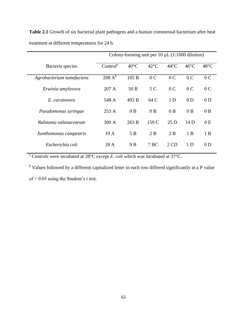
61
Table 2.1 Growth of six bacterial plant pathogens and a human commensal bacterium after heat
treatment at different temperatures for 24 h.
Bacteria species
Colony-forming unit per 10 µL (1:1000 dilution)
Controla 40°C 42°C 44°C 46°C 48°C
Agrobacterium tumefaciens 208 Ab 105 B 0 C 0 C 0 C 0 C
Erwinia amylovora 207 A 50 B 5 C 0 C 0 C 0 C
E. carotovora 548 A 492 B 64 C 1 D 0 D 0 D
Pseudomonas syringae 253 A 0 B 0 B 0 B 0 B 0 B
Ralstonia solanacearum 300 A 263 B 159 C 25 D 14 D 0 E
Xanthomonas campestris 19 A 5 B 2 B 2 B 1 B 1 B
Escherichia coli 18 A 9 B 7 BC 2 CD 1 D 0 D
a Controls were incubated at 28°C except E. coli which was incubated at 37°C.
b Values followed by a different capitalized letter in each row differed significantly at a P value
of < 0.05 using the Student’s t test.
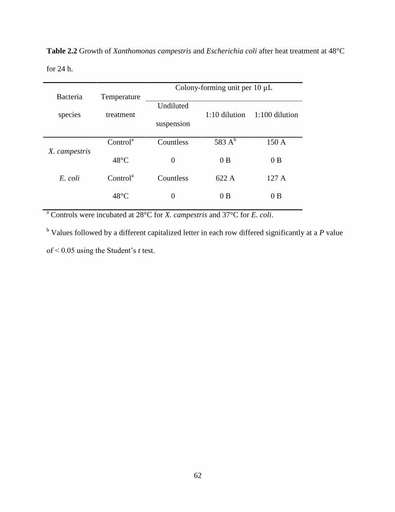
62
Table 2.2 Growth of Xanthomonas campestris and Escherichia coli after heat treatment at 48°C
for 24 h.
Bacteria
species
Temperature
treatment
Colony-forming unit per 10 µL
Undiluted
suspension
1:10 dilution 1:100 dilution
X. campestris
Controla Countless 583 A
b 150 A
48°C 0 0 B 0 B
E. coli Controla Countless 622 A 127 A
48°C 0 0 B 0 B
a Controls were incubated at 28°C for X. campestris and 37°C for E. coli.
b Values followed by a different capitalized letter in each row differed significantly at a P value
of < 0.05 using the Student’s t test.
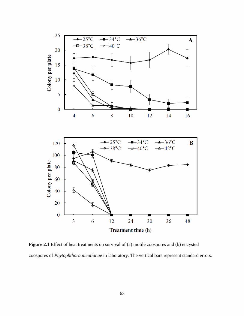
63
Figure 2.1 Effect of heat treatments on survival of (a) motile zoospores and (b) encysted
zoospores of Phytophthora nicotianae in laboratory. The vertical bars represent standard errors.
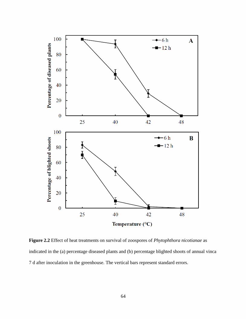
64
Figure 2.2 Effect of heat treatments on survival of zoospores of Phytophthora nicotianae as
indicated in the (a) percentage diseased plants and (b) percentage blighted shoots of annual vinca
7 d after inoculation in the greenhouse. The vertical bars represent standard errors.
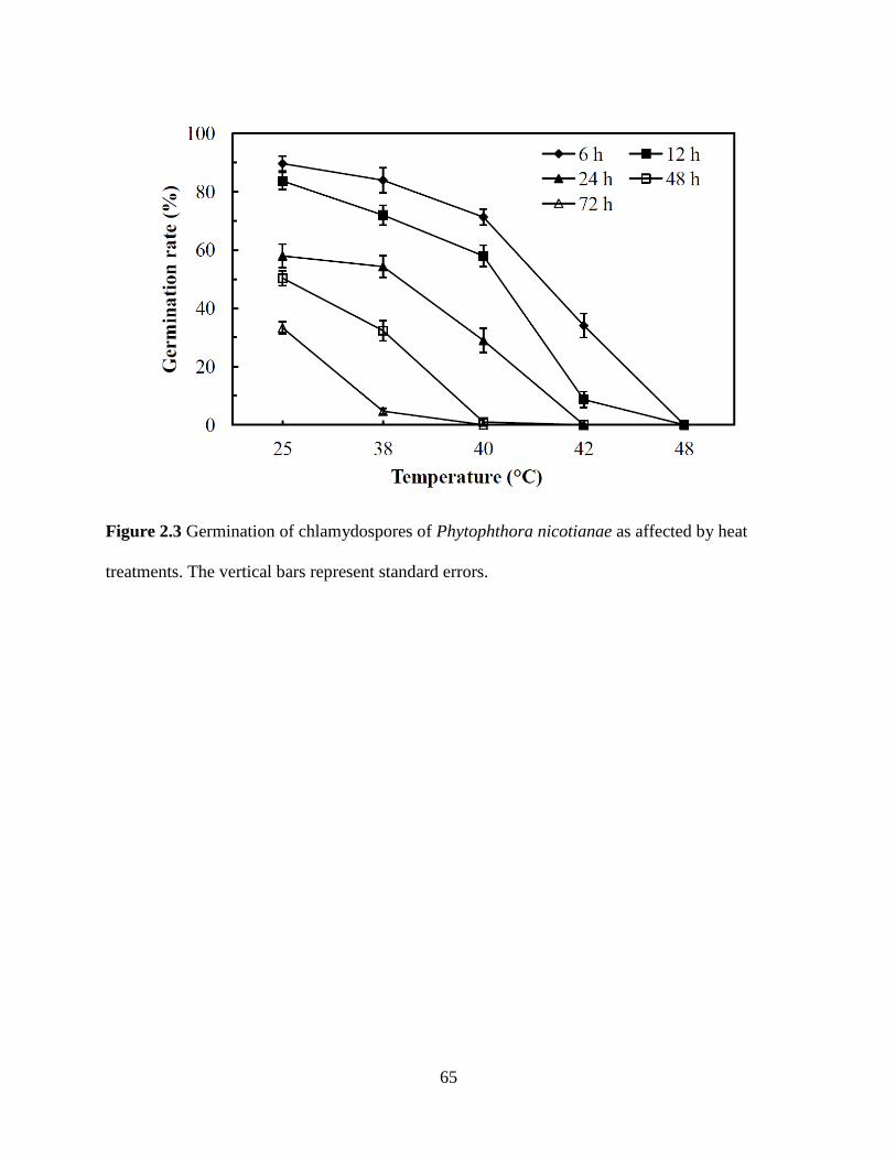
65
Figure 2.3 Germination of chlamydospores of Phytophthora nicotianae as affected by heat
treatments. The vertical bars represent standard errors.
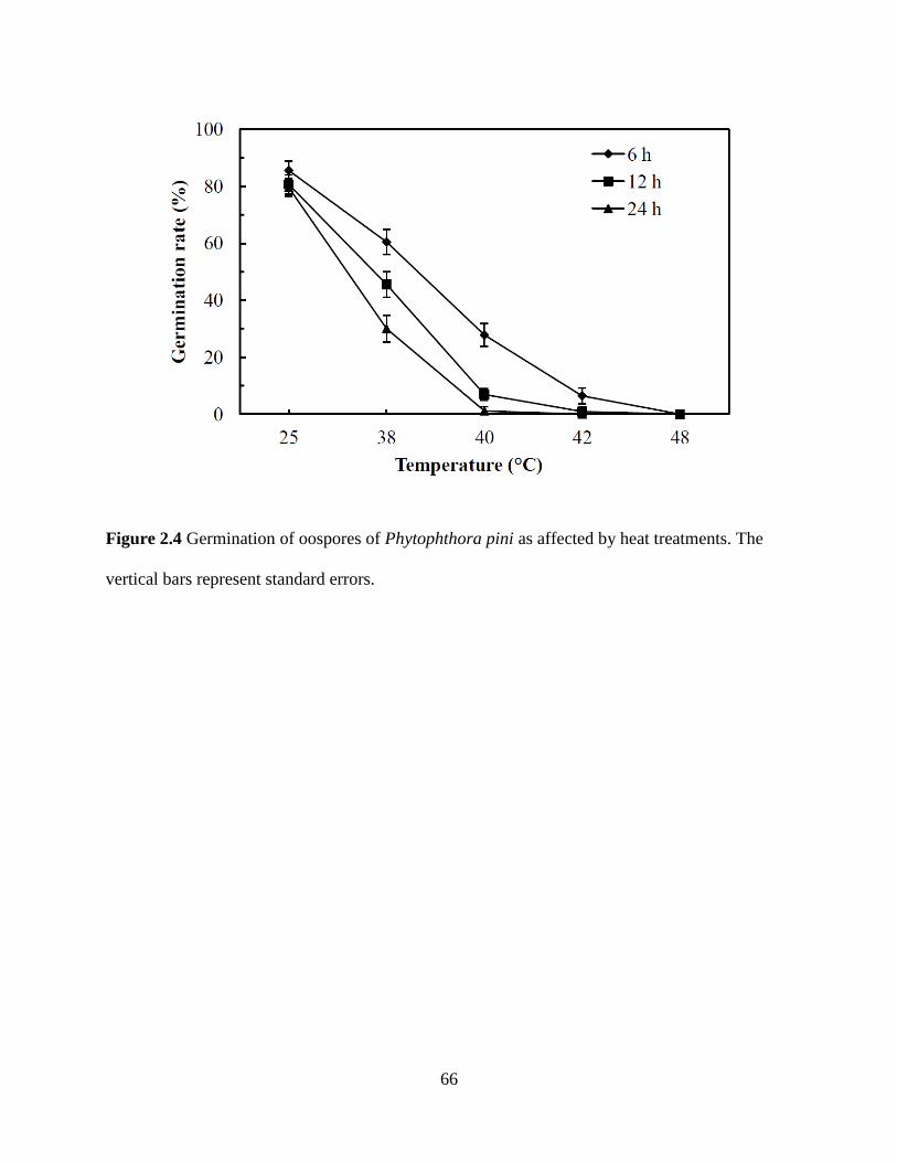
66
Figure 2.4 Germination of oospores of Phytophthora pini as affected by heat treatments. The
vertical bars represent standard errors.
67
Chapter 3
Temperature effect on Phytophthora zoospore survival in irrigation water
Our previous study in Chapter 2 (Hao et al. 2012) revealed the required temperature of
water heat treatment to inactivate major groups of plant pathogens can be lowered to 48°C for 24
h from the currently recommendation of 95°C for 30 s without efficacy sacrifice. Sterile
deionized water was consistently used in these experiments. However, what is going on in
irrigation water during the heat treatments at 42 and 48°C have not been studied. The objective
of this study was to compare the zoospore survival of Phytophthora nicotianae in irrigation
water (IW) and sterile deionized water (SDW) at heat treatment temperatures 42 and 48°C.
Materials and Methods
Water samples were collected from an irrigation reservoir at a local nursery in eastern
Virginia in June 2012. All samples were collected from the surface water column in the center of
the reservoir in sterile bottles. Three replicates of 1 L each were collected. Water samples were
then transported to the lab and immediately subjected to following experiments.
P. nicotianae isolate 38E5, recovered from annual vinca (Catharanthus roseus cv. Pacific
Red) in Virginia in 2006, were used in the zoospore survival tests. The "Wet-plate" method
(Ahonsi et al. 2007) with minor modifications was used to produce zoospores. In brief, three to
four 3 mm-diameter V8-agar culture mycelial plugs (three to four weeks old) were incubated in
10 mL 20% V8 broth in a 10-cm diameter Petri dish at 25°C in dark for a week. The broth was
washed off with growing mycelia attached to the bottom of the Petri dish. The Petri dishes with

68
washed mycelia were further incubated in dark at 25°C for 2 to 3 week. Two hours before heat
treatment, a volume of 20 mL of pre-chilled (4°C) SDW was added to each Petri dish, which was
then incubated at room temperature under fluorescent light (about 10,000 lux) for zoospore
release. The zoospore suspension was then collected in glass flasks.
Zoospore suspensions were diluted to 5,000 spore/mL with IW or SDW and heat-treated
at three different temperatures (25 as control, 42, and 48°C) for four different durations (6, 12,
24, and 48 h). A volume of 10 mL zoospore suspension was used as a replicate, and three
replicates were included each treatment. Temperature-controlled incubators (Intellus
Environmental Controllers, Percival Scientific, Inc.) were used for the temperature treatments.
The incubators were calibrated and monitored before and during the temperature experiment
using a HOBO®
temp/light pendant data logger (Onset Computer Corporation). Immediately
after temperature treatments, an aliquot of 100 µl of zoospore suspension (approximately 500
spores) was plated onto a 10-cm diameter Petri dish with 10% V8 agar medium plate. Three
plates each replicate were incubated at 25°C in the dark. Emerging colonies were counted after
48 h. The experiment was performed three times.
Data obtained from repeated experiments were evaluated for homogeneity and pooled
together. Student’s t test with a P value of 0.05 and analysis of variance (ANOVA) were
conducted to determine the significance of differences among treatment means. R statistical
software (R Development Core Team, R Foundation for Statistical Computing. 2011. ISBN 3-
900051-07-0) was used for all statistical analyses.
Results and discussion
69
Zoospores of P. nicotianae survived longer and at greater rates in SDW than IW (Fig.
3.1). This was consistently at 25°C across all the exposure times from 6 to 48 h, as indicated by
the number of cfu per plate. A large portion of zoospores in SDW survived but none of those in
IW did after a 6-h heat treatment at 42°C. No colony was observed in any dishes plated with
zoospores treated at the same temperature (42°C) for longer periods of time or at higher
temperature (48°C) in this study.
These results implicate that there were agents in irrigation water that worked against the
Phytophthora zoospore when compared to SDW. These agents may be microbes including
bacterial species and/or their metabolites. Additional studies are warranted to investigate what
microorganisms produce these substances, whether and how they can be integrated into the heat
treatment to further lower the treatment temperature, improving its economics and reducing
environmental footprint which was a central themes of Chapter 4 and 5.
References
Ahonsi, M. O., Banko, T. J. & Hong, C. X. (2007). A simple in-vitro 'wet-plate' method for mass
production of Phytophthora nicotianae zoospores and factors influencing zoospore
production. Journal of Microbiological Methods, 70, 557-560
Hao, W., Ahonsi, M. O., Vinatzer, B. A. & Hong, C. X. (2012). Inactivation of Phytophthora and
bacterial species in water by a potential energy-saving heat treatment. European Journal
of Plant Pathology, 134 (2), 357-365
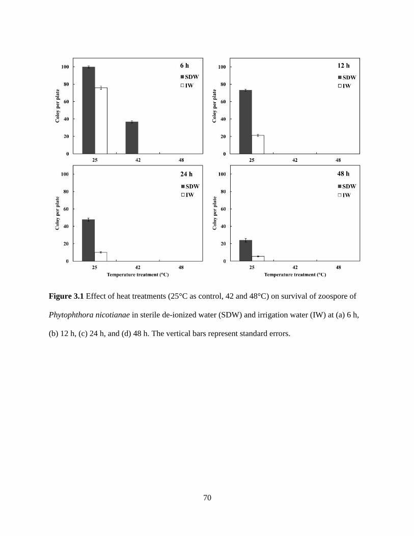
70
Figure 3.1 Effect of heat treatments (25°C as control, 42 and 48°C) on survival of zoospore of
Phytophthora nicotianae in sterile de-ionized water (SDW) and irrigation water (IW) at (a) 6 h,
(b) 12 h, (c) 24 h, and (d) 48 h. The vertical bars represent standard errors.

71
Chapter 4
Characterization of microbial diversity in irrigation water: sample concentration and
processing strategies
Abstract
Understanding the microbial diversity in irrigation reservoirs is essential to manage
pathogens originating and spreading through water and to identify naturally occurring biological
control agents against plant pathogens. The objective of this study was to develop an optimized
methodology for collection, detection, and identification of bacterial species present in irrigation
water. Specifically, (1) culture-dependent and -independent methods, (2) three water sample
concentration methods, and (3) different DGGE denaturant concentrations and different
electrophoresis conditions were compared for recovery of bacterial diversity from irrigation
water. Species composition determined by culture-dependent and -independent strategies were
substantially different. In the culture-independent strategy, more bacteria were detected in water
samples via a combination of centrifugation and filtration than when using each alone. PCR-
DGGE with a 40 to 60% denaturant concentration running at 70 V for 16 h revealed the highest
bacterial diversity. In conclusion, a step-wise methodology to characterize bacterial diversity in
irrigation water has been developed. In this scheme, both culture-dependent and -independent
strategies are employed. Centrifugation is used along with filtration to concentrate water
samples, and DGGE with 40 to 60% concentration at 70 V for 16 h is performed in the culture-
independent strategy.
72
Introduction
Understanding the microbial diversity in irrigation reservoirs is essential to assess and
manage crop health risk incited by plant pathogens originating and spreading through irrigation
systems (Hong & Moorman, 2005). Such studies help identify naturally occurring biological
control agents against plant pathogenic species, and also improve the physicochemical
assessment of water quality and health in irrigation systems (Liu et al. 2011). Of particular
interest to this project is whether there is bacterial community structure shift in irrigation water
after heat treatments and whether and how such shifts may have contributed to the pathogen
suppression at reduced heat treatment temperatures of 42 and 48°C.
Both culture-dependent and -independent approaches have been utilized for investigation
into microbial diversity in the environment. The culture-dependent approach consists of growing
microbes on general or selective media followed by morphological, biochemical, or molecular
identification. The main limitation of this approach is that the majority of environmental
microbes are unculturable (Amann et al. 1995; Vartoukian et al. 2010). The culture-independent
approach employs molecular techniques including clone libraries, in-situ hybridization,
molecular fingerprinting methods, and metagenomics (Case et al. 2007; Shokralla et al. 2012).
Denaturing gradient gel electrophoresis (DGGE) is a rapid and efficient DNA
fingerprinting technique for characterizing microbial communities (Kozdroj and van Elsas 2001;
Kawai et al. 2002; Cho et al. 2003; Feris et al. 2003; Thoms et al. 2003; Brakstad and Lodeng
2005; Lyautey et al. 2005; Yan et al. 2007). Muyzer et al. (1993) first used DGGE to separate
polymerase chain reaction (PCR) amplified 16S rRNA gene fragments in order to analyze the
genetic diversity of complex microbial populations. In DGGE, PCR products of similar size but
different base-pair sequences are separated based on their electrophoretic mobility (Fischer and
73
Lerman 1979). The DNA fragments encounter increasing concentrations of chemical denaturant
as they migrate through a polyacrylamide gel. Upon reaching a threshold denaturant
concentration, the weaker melting domains, which are the stretches of base pairs in the PCR
amplicon with an identical melting temperature, begin to denature. The DNA molecules
transition from helical to partially melted, at which time migration slows dramatically. The
different concentrations of denaturant and sequence variation within the DNA fragments result in
a pattern of bands as the DNA migrates through the gel. The separation of PCR products with
optimum resolution is very critical for a good DGGE-based community structure analysis
(Sheffield et al. 1989). Theoretically, the number and intensity of bands represents phylotype
number and abundance within a sample (Muyzer and Smalla 1998; Nubel et al. 1999; Sigler et
al. 2004). Cherif et al. (2008) demonstrated that PCR-DGGE was more accurate for bacterial
identification in soil samples than other molecular methods. Lyautey et al. (2005) also indicated
that PCR-DGGE was useful for assessing bacterial diversity of epilithic biofilms in rivers.
The efficiency of PCR-DGGE as opposed to traditional culture-dependent approaches for
characterization of microbial community diversity in aquatic environments has not been
determined. Some researchers used only PCR-DGGE to investigate microbial diversity in the
North Sea (Brakstad and Lodeng 2005) and planktonic community diversity in rivers and lakes
(Yan et al. 2007; Liu et al. 2011). Yan et al. (2007) concluded that PCR-DGGE fingerprinting
might be sensitive and effective to characterize the microbial diversity within an aquatic
ecosystem. On the other hand, other studies used traditional culture-dependent approaches along
with culture-independent molecular fingerprinting techniques to investigate microbial
communities, for example, in groundwater and partially purified water (Kawai et al. 2002; Cho
et al. 2003).

74
Various DGGE conditions, including different ranges of denaturant concentration,
electrophoresis voltage, and time have been used in previous PCR-DGGE based microbial
diversity studies of environmental samples (Kozdroj and van Elsas 2001; Kawai et al. 2002; Cho
et al. 2003; Thoms et al. 2003; Lyautey et al. 2005; Yan et al. 2007; Cherif et al. 2008).
However, how different DGGE conditions influence the results of a DGGE analysis has not been
investigated.
The aim of this study was to develop an optimized and standardized methodology to
collect, detect, and identify bacterial species present in greenhouse and nursery irrigation water.
The specific objectives were to (1) compare the culture-dependent strategy with -independent
strategy for investigation of bacterial diversity, (2) determine the most efficient method to
concentrate water samples, and (3) identify the most appropriate conditions for DGGE to analyze
bacterial species in irrigation water.
Materials and methods
Water samples
Water samples were collected from an irrigation reservoir at a local nursery in eastern
Virginia in September 2011 and May 2012. All samples were collected in sterile bottles from the
surface of a water column in the center of the reservoir. Three replicates of 1 L each were
collected. Water temperature and other water quality parameters were recorded on-site using a
Hydrolab DS5X multiparameter water quality meter (Hach®, Loveland, CO, USA). Water
samples were then transported to the lab and immediately passed through nylon membrane filters
(pore size 10 µm) to remove larger debris and algae. The filtrate was then used for further
processing.
75
Culture-dependent strategy
Plating
One hundred microliters of the filtrate was evenly spread in a 10-cm diameter Petri dish
with Nutrient Agar (NA) (DifcoTM
, Detroit, MI) using a sterile glass spreader. Three replicate
plates were used for each sample and incubated at 28°C for 2 d.
Colony PCR & SSCP analysis
Colonies on NA plates were grouped based on color and size. The total numbers of
colonies in each group were recorded. About 20 to 30% of the colonies from each group were
sub-cultured into 48 well plates containing NA medium, and incubated at 28°C for 2 d. Colony
PCRs were performed to amplify the hypervariable regions V6 to V8 of 16S rDNA with primers
16S f968 and r1401 (Nubel et al. 1996; Peixoto et al. 2002). Reagents supplied with Takara Taq
(Takara Bio Inc., Japan) were used in the reactions. The PCR mixture (25 μl) contained 0.1 μl
Takara Taq (5 units/ μl), 2.5 μl 10× PCR buffer (containing 1.5 mM MgCl2), 2 μl dNTP mixture
(2.5 mM each), 1 μl of each primer (10 μM), and 18.4 μl DNA-free nanopure water. Bacterial
colonies were picked directly from culture plates and added to the mixture. Cycling conditions
consisted of initial denaturation at 96°C for 2 min, followed by 40 cycles of denaturation
at 94°C
for 0.5 min, annealing at 55°C for 0.5 min, and an extension at 72°C for 1 min. For the last cycle,
the extension time was increased to 10 min.
Single-strand conformation polymorphism (SSCP) analyses were performed on the PCR
products as described by Kong et al. (2003) with minor modifications. In brief, the amplicons
were mixed with loading dye (1:4, v:v) and denatured by heating at 96°C for 10 min then chilled
76
on ice. Two microliters were loaded into each well on an 8% polyacrylamide gel
(acrylamide/bisacrylamide = 29:1) running at 220 V for 2.5 h in chilled 1× TBE buffer. After
electrophoresis, the gels were stained with silver nitrate (Beidler et al. 1982). SSCP banding
patterns of individual colonies were determined with the aid of a 100 bp DNA ladder (Promega
Corp., Madison, WI). PCR products with the same SSCP fingerprint were grouped and stored for
sequence analysis.
Culture-independent strategy
Irrigation water sample concentration
Three methods were evaluated for concentrating irrigation water samples after filtration
through a 10-µm membrane. In the first method (P), the filtrate was centrifuged at 12,200 g for
50 min, and the supernatant was discarded. The pellet was re-suspended in 1.5 mL SDW and
centrifuged at 11,750 g for 5 min. Genomic DNA was extracted from the final pellet. For the
second method (F), the filtrate was passed through nylon membrane filters (pore size 0.2 µm),
and the filters trapping bacterial cells were used for genomic DNA extraction. The third method
(C), involved centrifugation of the filtrate at 12,200 g for 50 min, the pellet was saved, and the
supernatant was then filtered through 0.2 µm filters. Total genomic DNA was extracted from the
pellet and the 0.2 µm filters and combined for analysis.
Genomic DNA extraction
The UltraClean Microbial DNA Isolation Kit (MO BIO Laboratories, Inc, Carlsbad, CA)
was used to extract bacterial genomic DNAs from the pellets following the instruction manual.
77
The PowerWater DNA Isolation Kit (MO BIO Laboratories, Inc, Carlsbad, CA) was used to
extract bacterial genomic DNA from the 0.2-µm filters.
PCR amplification and evaluation of DGGE conditions
For DGGE analysis, 16S rRNA fragments were amplified with primers GC-clamp-EUB
f933 and EUB r1387 covering the three hypervariable regions V6 to V8 (Kawai et al. 2002).
PCR mixture (50 μl) contained 0.2 μl Takara Taq (5 units/ μl), 5 μl 10× PCR buffer (containing
MgCl2), 7.5 μl BSA (0.3 mg/ml), 4 μl dNTP mixture (2.5 mM each), 2 μl of each primer (10
μM), 19.3 μl DNA-free nanopure water, and 10 μl DNA extract. Hot-start PCR was performed at
94°C for 4 min, followed by touchdown PCR with the following parameters: denaturation at
94°C for 1 min, annealing at 65°C decreasing by 1°C every cycle (1.5 min) to 55°C. Twenty
additional cycles were carried out at 55°C. Primer extension was carried out at 72°C for 2.5 min.
The final extension step was 7 min at 72°C.
DGGE was performed using a Bio-Rad DcodeTM
Universal Mutation Detection System
(Bio-Rad, Hercules, CA, USA) according to the manufacturer's instructions. PCR products were
electrophoresed in 1 mm thick vertical 8% polyacrylamide gels (acrylamide/bisacrylamide =
37.5:1) in 1× TAE buffer. Gels were prepared with three denaturant concentrations: 30 to 60%,
40 to 60%, and 40 to 70%. A 100% denaturant concentration is defined as 40% [v/v] formamide
and 7 M urea. Electrophoresis at 60°C was performed under three conditions: at 70 V for 16 h, at
90 V for 16 h, and at 120 V for 12 h. After electrophoresis, the gels were stained with silver
nitrate (Beidler et al. 1982).
Statistical analysis
78
DGGE gels were digitally documented using Epi Chemi II darkroom and then analyzed
using LabWorks software (UVP Laboratory, Upland, CA, USA). Each lane was detected
automatically with the width and length adjusted manually. Bands in each lane were detected
automatically and corrected manually. The Shannon index of diversity (H) was used to determine
the diversity of the bacterial community. It was calculated as H = - Σ PilnPi, the relative intensity
Pi of each band in the lanes was calculated as Pi = ni/N, ni is the maximum optical density (MOD)
of the band i, and N is the sum of the MOD for all the bands within the lane (Shannon and
Weaver 1963; Eichner et al. 1999). Bands with MOD greater than 3% of the sum of MODs were
considered. H values and band numbers of each replicate were analyzed by analysis of variance
(ANOVA) using R statistical software (R Development Core Team, R Foundation for Statistical
Computing. 2011. ISBN 3-900051-07-0) to determine the effects of the employed water
concentration method and DGGE conditions on recovery of bacterial diversity.
Re-isolation of DNA from gel and Cloning
Selected bands were excised from the DGGE gel using a “crush and soak” method
(Sambrook and Russell 2001) with minor modifications. In brief, bands were excised from the
gel using sterile gel cutting tips (BioExpress, Kaysville, UT, USA), transferred to 1.7 ml tubes,
and washed briefly with 0.5 mL TE buffer. The gel slice was then crushed using a sterile pestle
in 400 µl elution buffer (0.5 M NH4O(C2H3O), 10 mM EDTA), incubated at 37°C overnight in
an incubator shaker to allow DNA diffusion from the gel, then centrifuged for 5 min at 12,000
rpm to pellet acrylamide gel fragments. The supernatant was transferred and DNA precipitated in
1 mL 100% ethanol with 16 µl 5M NaCl at -20°C overnight, then centrifuged at 12,000 rpm for
20 min. Pellets were vacuum dried and re-suspended in 20 µl TE buffer. The extracted DNA was
79
re-amplified with 16S rRNA primers EUB f933 and EUB r1387 (Kawai et al. 2002) using the
same cycling conditions as colony PCR. These PCR amplicons were cloned using the pGEM-T
Easy Vector System (Promega Biosciences, LLC. San Luis Obispo, CA, USA). Randomly
picked white colonies were PCR amplified with the plasmid primers T7f (5' - TAA TAC GAC
TCA CTA TAG GG - 3') and SP6r (5' - ATT TAG GTG ACA CTA TAG AA - 3') under the
same cycling conditions used for 16S rRNA colony PCR. The PCR products were screened on
SSCP gels and stored for sequencing analysis.
Sequence analysis
About 20 to 30% of the colony PCR products of each SSCP pattern and all representative
PCR products from the culture-independent strategy were sequenced by Advanced Genetic
Technologies Center, University of Kentucky. Sequences were edited using the BioEdit
Sequence Alignment Editor (version 7.1.3.0) (Hall 1999), and subjected to a BLAST search
(http://blast.ncbi.nlm.nih.gov/Blast.cgi) to identify the closest matches in GenBank. The
phylogenetic trees for the partial bacterial 16S rDNA sequences cut into the same size were built
using the maximum likelihood method based on the Tamura-Nei model (Felsenstein 1981) in
MEGA 5.05 (Build#: 5110426) (Tamura and Nei 1993; Tamura et al. 2011) and iTOL
(Interactive tree of life, http://itol.embl.de/index.shtml) (Letunic and Bork 2007, 2011).
A total of 155 partial 16S rRNA sequences have been deposited in the GenBank sequence
database under accession numbers JX628632 to JX628749 and JX657295 to JX657331.
Results
Water quality
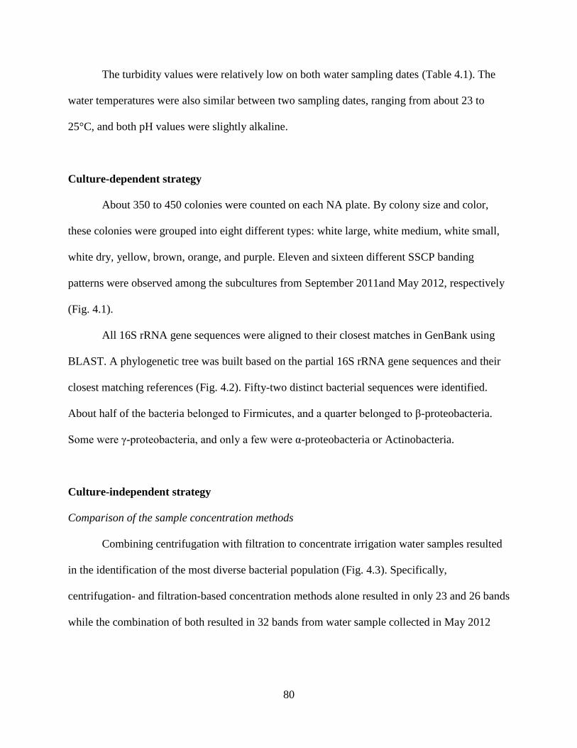
80
The turbidity values were relatively low on both water sampling dates (Table 4.1). The
water temperatures were also similar between two sampling dates, ranging from about 23 to
25°C, and both pH values were slightly alkaline.
Culture-dependent strategy
About 350 to 450 colonies were counted on each NA plate. By colony size and color,
these colonies were grouped into eight different types: white large, white medium, white small,
white dry, yellow, brown, orange, and purple. Eleven and sixteen different SSCP banding
patterns were observed among the subcultures from September 2011and May 2012, respectively
(Fig. 4.1).
All 16S rRNA gene sequences were aligned to their closest matches in GenBank using
BLAST. A phylogenetic tree was built based on the partial 16S rRNA gene sequences and their
closest matching references (Fig. 4.2). Fifty-two distinct bacterial sequences were identified.
About half of the bacteria belonged to Firmicutes, and a quarter belonged to β-proteobacteria.
Some were γ-proteobacteria, and only a few were α-proteobacteria or Actinobacteria.
Culture-independent strategy
Comparison of the sample concentration methods
Combining centrifugation with filtration to concentrate irrigation water samples resulted
in the identification of the most diverse bacterial population (Fig. 4.3). Specifically,
centrifugation- and filtration-based concentration methods alone resulted in only 23 and 26 bands
while the combination of both resulted in 32 bands from water sample collected in May 2012
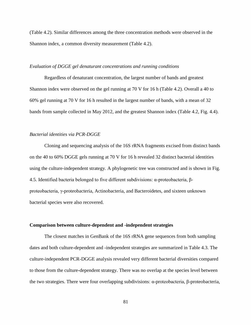
81
(Table 4.2). Similar differences among the three concentration methods were observed in the
Shannon index, a common diversity measurement (Table 4.2).
Evaluation of DGGE gel denaturant concentrations and running conditions
Regardless of denaturant concentration, the largest number of bands and greatest
Shannon index were observed on the gel running at 70 V for 16 h (Table 4.2). Overall a 40 to
60% gel running at 70 V for 16 h resulted in the largest number of bands, with a mean of 32
bands from sample collected in May 2012, and the greatest Shannon index (Table 4.2, Fig. 4.4).
Bacterial identities via PCR-DGGE
Cloning and sequencing analysis of the 16S rRNA fragments excised from distinct bands
on the 40 to 60% DGGE gels running at 70 V for 16 h revealed 32 distinct bacterial identities
using the culture-independent strategy. A phylogenetic tree was constructed and is shown in Fig.
4.5. Identified bacteria belonged to five different subdivisions: α-proteobacteria, β-
proteobacteria, γ-proteobacteria, Actinobacteria, and Bacteroidetes, and sixteen unknown
bacterial species were also recovered.
Comparison between culture-dependent and -independent strategies
The closest matches in GenBank of the 16S rRNA gene sequences from both sampling
dates and both culture-dependent and -independent strategies are summarized in Table 4.3. The
culture-independent PCR-DGGE analysis revealed very different bacterial diversities compared
to those from the culture-dependent strategy. There was no overlap at the species level between
the two strategies. There were four overlapping subdivisions: α-proteobacteria, β-proteobacteria,
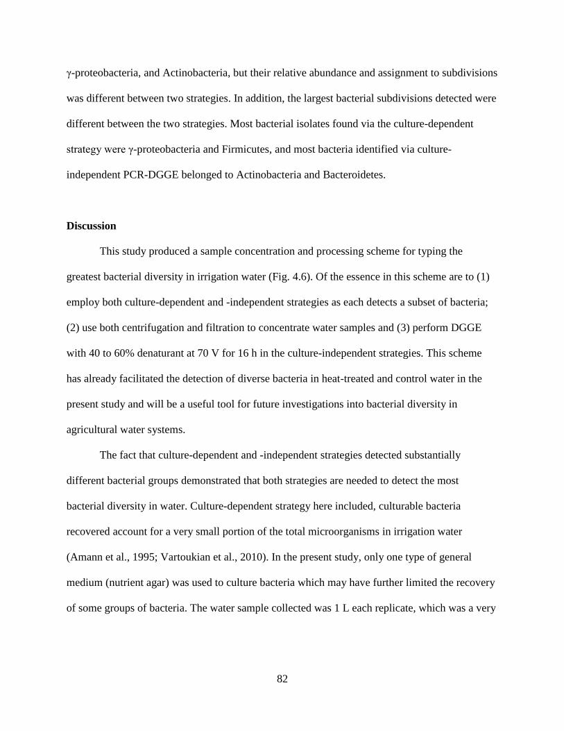
82
γ-proteobacteria, and Actinobacteria, but their relative abundance and assignment to subdivisions
was different between two strategies. In addition, the largest bacterial subdivisions detected were
different between the two strategies. Most bacterial isolates found via the culture-dependent
strategy were γ-proteobacteria and Firmicutes, and most bacteria identified via culture-
independent PCR-DGGE belonged to Actinobacteria and Bacteroidetes.
Discussion
This study produced a sample concentration and processing scheme for typing the
greatest bacterial diversity in irrigation water (Fig. 4.6). Of the essence in this scheme are to (1)
employ both culture-dependent and -independent strategies as each detects a subset of bacteria;
(2) use both centrifugation and filtration to concentrate water samples and (3) perform DGGE
with 40 to 60% denaturant at 70 V for 16 h in the culture-independent strategies. This scheme
has already facilitated the detection of diverse bacteria in heat-treated and control water in the
present study and will be a useful tool for future investigations into bacterial diversity in
agricultural water systems.
The fact that culture-dependent and -independent strategies detected substantially
different bacterial groups demonstrated that both strategies are needed to detect the most
bacterial diversity in water. Culture-dependent strategy here included, culturable bacteria
recovered account for a very small portion of the total microorganisms in irrigation water
(Amann et al., 1995; Vartoukian et al., 2010). In the present study, only one type of general
medium (nutrient agar) was used to culture bacteria which may have further limited the recovery
of some groups of bacteria. The water sample collected was 1 L each replicate, which was a very
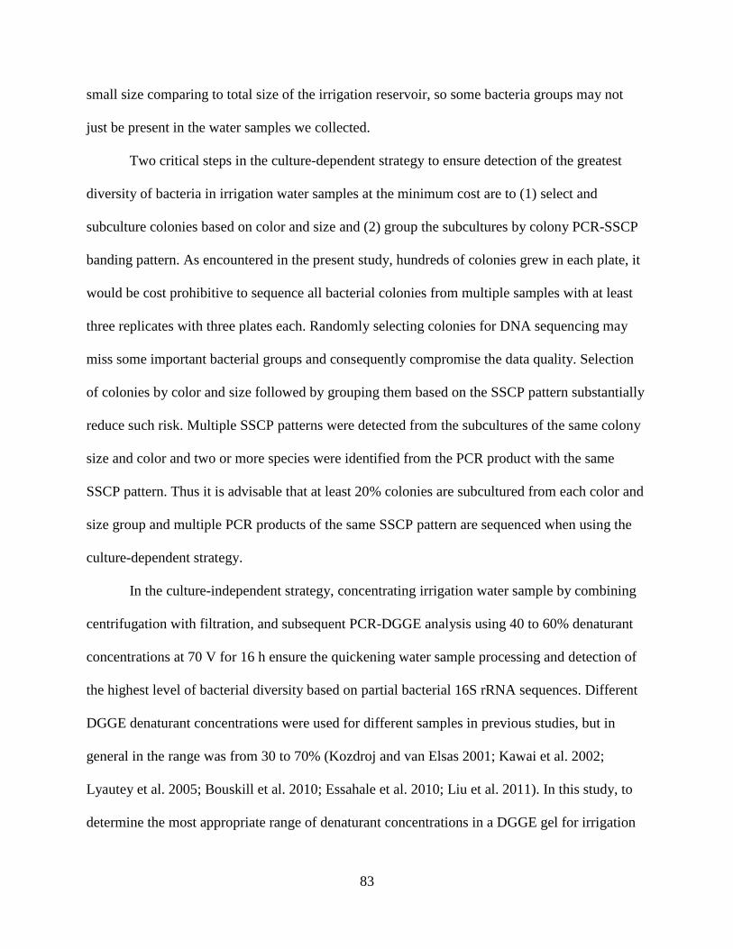
83
small size comparing to total size of the irrigation reservoir, so some bacteria groups may not
just be present in the water samples we collected.
Two critical steps in the culture-dependent strategy to ensure detection of the greatest
diversity of bacteria in irrigation water samples at the minimum cost are to (1) select and
subculture colonies based on color and size and (2) group the subcultures by colony PCR-SSCP
banding pattern. As encountered in the present study, hundreds of colonies grew in each plate, it
would be cost prohibitive to sequence all bacterial colonies from multiple samples with at least
three replicates with three plates each. Randomly selecting colonies for DNA sequencing may
miss some important bacterial groups and consequently compromise the data quality. Selection
of colonies by color and size followed by grouping them based on the SSCP pattern substantially
reduce such risk. Multiple SSCP patterns were detected from the subcultures of the same colony
size and color and two or more species were identified from the PCR product with the same
SSCP pattern. Thus it is advisable that at least 20% colonies are subcultured from each color and
size group and multiple PCR products of the same SSCP pattern are sequenced when using the
culture-dependent strategy.
In the culture-independent strategy, concentrating irrigation water sample by combining
centrifugation with filtration, and subsequent PCR-DGGE analysis using 40 to 60% denaturant
concentrations at 70 V for 16 h ensure the quickening water sample processing and detection of
the highest level of bacterial diversity based on partial bacterial 16S rRNA sequences. Different
DGGE denaturant concentrations were used for different samples in previous studies, but in
general in the range was from 30 to 70% (Kozdroj and van Elsas 2001; Kawai et al. 2002;
Lyautey et al. 2005; Bouskill et al. 2010; Essahale et al. 2010; Liu et al. 2011). In this study, to
determine the most appropriate range of denaturant concentrations in a DGGE gel for irrigation
84
water samples, a wider range of denaturant concentrations were initially tested (30 to 60% and
40 to 70%) then narrowed down to 40 to 60%, under three different electrophoretic conditions
(voltage × time). The optimum condition in terms of denaturant concentration, running voltage
and time for PCR-DGGE analysis of irrigation water was determined by comparing the bacterial
diversity among treatments.
The recovery of Bacillus, Brevibacillus, Enterobacter, Peanibacillus and Pseudomonas
species in the present study underscores the practical significance of investigations into the
microbial diversity in irrigation reservoirs. Strains of these species are known to have
antagonistic activities against plant pathogens. For example, Saikia et al. (2011) found that
Brevibacillus laterosporus BPM3 isolated from a natural hot spring strongly inhibited the growth
of Fusarium oxysporum f. sp. ciceri, F. semitectum, Magnaporthe grisea and Rhizoctonia
oryzae. Strains from Pseudomonas and Bacillus species have been reported to control many
seedling diseases and root rots on several crops (Yang et al. 1994; Punja 1997; Paulitz and
Belanger 2001; Loper et al. 2007). Other bacterial species belonging to genera of Enterobacter
and Peanibacillus also could have biocontrol activities as reported previously (Singh et al. 1999;
Saikia et al. 2011). Further studies are warranted to determine the dominant bacterial groups,
species and strains with the greatest biocontrol potential across and how these groups may be
utilized along with other pathogen avoidance and mitigation methods including heat treatment to
realize their full potential.
References
85
Amann, A. I., Ludwig, W. & Schleifer, A.-H. (1995). Phylogenetic identification and in situ
detection of individual microbial cells without cultivation. Microbiological Reviews, 59
(1), 143-169
Beidler, J. L., Hilliard, P. R. & Rill, R. L. (1982). Ultrasensitive staining of nucleic acids with
silver. Analytical Biochemistry, 126, 374-380
Bouskill, N. J., Barker-Finkel, J., Galloway, T. S., Handy, R. D. & Ford, T. E. (2010). Temporal
bacterial diversity associated with metal-contaminated river sediments. Ecotoxicology, 19
(2), 317-328
Brakstad, O. G. & Lodeng, A. G. G. (2005). Microbial diversity during biodegradation of crude
oil in seawater from the North Sea. Microbial Ecology, 49 (1), 94-103
Case, R. J., Boucher, Y., Dahllof, I., Holmstrom, C., Doolittle, W. F. & Kjellebery, S. (2007).
Use of 16S rRNA and rpoB genes as molecular markers for microbial ecology studies.
Applied and Environmental Microbiology, 73 (1), 278-288
Cherif, H., Ouzari, H., Marzorati, M., Brusetti, L., Jedidi, N., Hassen, A. & Daffonchio, D.
(2008). Bacterial community diversity assessment in municipal solid waste compost
amended soil using DGGE and ARISA fingerprinting methods. World Journal of
Microbiology & Biotechnology, 24 (7), 1159-1167
Cho, H. B., Lee, J. K. & Choi, Y. K. (2003). The genetic diversity analysis of the bacterial
community in groundwater by denaturing gradient gel electrophoresis (DGGE). Journal
of Microbiology, 41 (4), 327-334
Eichner, C. A., Erb, R. W., Timmis, K. N. & Wagner-Doebler, I. (1999). Thermal gradient gel
electrophoresis analysis of bioprotection from pollutant shocks in the activated sludge
microbial community. Applied and Environmental Microbiology, 65 (1), 102-109
86
Essahale, A., Malki, M., Marin, I. & Moumni, M. (2010). Bacterial diversity in Fez tanneries and
Morocco's Binlamdoune River, using 16S RNA gene based fingerprinting. Journal of
Environmental Sciences (China), 22 (12), 1944-1953
Felsenstein, J. (1981). Evolutionary trees from DNA sequences: a maximum likelihood
approach. Journal of Molecular Evolution, 17, 368-376
Feris, K. P., Ramsey, P. W., Frazar, C., Rillig, M. C., Gannon, J. E. & Holben, W. E. (2003).
Structure and seasonal dynamics of hyporheic zone microbial communities in free-stone
rivers of the western United States. Microbial Ecology, 46 (2), 200-215
Fischer, S. G. & Lerman, L. S. (1979). Length independent separation of DNA restriction
fragments in 2 dimensional gel electrophoresis. Cell, 16 (1), 191-200
Hall, T. A. (1999). BioEdit: a user-friendly biological sequence alignment editor and analysis
program for Windows 95/98/NT. Nucleic Acids Symposium Series, 41, 95-98
Kawai, M., Matsutera, E., Kanda, H., Yamaguchi, N., Tani, K. & Nasu, M. (2002). 16S
ribosomal DNA-based analysis of bacterial diversity in purified water used in
pharmaceutical manufacturing processes by PCR and denaturing gradient gel
electrophoresis. Applied and Environmental Microbiology, 68 (2), 699-704
Kong, P., Hong, C. X., Richardson, P. A. & Gallegly, M. E. (2003). Single-strand-conformation
polymorphism of ribosomal DNA for rapid species differentiation in genus
Phytophthora. Fungal Genetics and Biology, 39 (3), 238-249
Kozdroj, J. & van Elsas, J. D. (2001). Structural diversity of microbial communities in arable
soils of a heavily industrialised area determined by PCR-DGGE fingerprinting and
FAME profiling. Applied Soil Ecology, 17 (1), 31-42
87
Letunic, I. & Bork, P. (2007). Interactive Tree Of Life (iTOL): an online tool for phylogenetic
tree display and annotation. Bioinformatics (Oxford), 23 (1), 127-128
Letunic, I. & Bork, P. (2011). Interactive Tree Of Life v2: online annotation and display of
phylogenetic trees made easy. Nucleic Acids Research, 39 (Suppl. 2), W475-W478
Liu, L., Yang, J. & Zhang, Y. (2011). Genetic diversity patterns of microbial communities in a
subtropical riverine ecosystem (Jiulong River, southeast China). Hydrobiologia, 678 (1),
113-125
Loper, J. E., Kobayashi, D. Y. & Paulsen, I. T. (2007). The genomic sequence of Pseudomonas
fluorescens Pf-5: Insights into biological control. Phytopathology, 97 (2), 233-238
Lyautey, E., Lacoste, B., Ten-Hage, L., Rols, J.-L. & Garabetian, F. (2005). Analysis of bacterial
diversity in river biofilms using 16S rDNA PCR-DGGE: methodological settings and
fingerprints interpretation. Water Research, 39 (2-3), 380-388
Muyzer, G., Dewaal, E. C. & Uitterlinden, A. G. (1993). Profiling of complex microbial-
population by denaturing gradient gel-electrophoresis analysis of polymerase chain
reaction-amplified genes-coding for 16S rebosomal-RNA. Applied and Environmental
Microbiology, 59 (3), 695-700
Muyzer, G. & Smalla, K. (1998). Application of denaturing gradient gel electrophoresis (DGGE)
and temperature gradient gel electrophoresis (TGGE) in microbial ecology. Antonie Van
Leeuwenhoek International Journal of General and Molecular Microbiology, 73 (1), 127-
141
Nubel, U., Engelen, B., Felske, A., Snaidr, J., Wieshuber, A., Amann, R. I., Ludwig, W. &
Backhaus, H. (1996). Sequence heterogeneities of genes encoding 16S rRNAs in
88
Paenibacillus polymyxa detected by temperature gradient gel electrophoresis. Journal of
Bacteriology, 178 (19), 5636-5643
Nubel, U., Garcia-Pichel, F., Kuhl, M. & Muyzer, G. (1999). Quantifying microbial diversity:
Morphotypes, 16S rRNA genes, and carotenoids of oxygenic phototrophs in microbial
mats. Applied and Environmental Microbiology, 65 (2), 422-430
Paulitz, T. C. & Belanger, R. R. (2001). Biological control in greenhouse system. Annual
Review of Phytopathology, 39, 103-133
Peixoto, R. S., Coutinho, H. L. D., Rumjanek, N. G., Macrae, A. & Rosado, A. S. (2002). Use of
rpo B and 16S rRNA genes to analyse bacterial diversity of a tropical soil using PCR and
DGGE. Letters in Applied Microbiology, 35 (4), 316-320
Punja, Z. K. (1997). Comparative efficacy of bacteria, fungi, and yeasts as biological control
agents for diseases of vegetable crops. Canadian Journal of Plant Pathology, 19 (3), 315-
323
Saikia, R., Gogoi, D. K., Mazumder, S., Yadav, A., Sarma, R. K., Bora, T. C. & Gogoi, B. K.
(2011). Brevibacillus laterosporus strain BPM3, a potential biocontrol agent isolated
from a natural hot water spring of Assam, India. Microbiological Research, 166 (3), 216-
225
Sambrook, J. & Russell, D. W. (2001). Molecular Cloning: A Laboratory Manual. 3rd edn. (Cold
Spring Harbor, NY: Cold Spring Laboratory Press)
Shannon, C. & Weaver, W. (1963). The Mathematical Theory of Communication, Vol 3.
(Chicago, USA: Urbana University of Illinois Press)
Sheffield, V. C., Cox, D. R., Lerman, L. S. & Myers, R. M. (1989). Attachment of a 40-base-pair
G+C-rich sequence (GC-Clamp) to genomic DNA fragments by the polymerase chain
89
reaction results in improved detection of single-base changes. Proceedings of the
National Academy of Sciences of the United States of America, 86 (1), 232-236
Shokralla, S., Spall, J. L., Gibson, J. F. & Hajibabaei, M. (2012). Next-generation sequencing
technologies for environmental DNA research. Molecular Ecology, 21 (8, Sp. Iss. SI),
1794-1805
Sigler, W. V., Miniaci, C. & Zeyer, J. (2004). Electrophoresis time impacts the denaturing
gradient gel electrophoresis-based assessment of bacterial community structure. Journal
of Microbiological Methods, 57 (1), 17-22
Singh, P. P., Shin, Y. C., Park, C. S. & Chung, Y. R. (1999). Biological control of fusarium wilt
of cucumber by chitinolytic bacteria. Phytopathology, 89 (1), 92-99
Tamura, K. & Nei, M. (1993). Estimation of the number of nucleotide substitutions in the control
region of mitochondrial-DNA in humans and chimpanzees. Molecular Biology and
Evolution, 10 (3), 512-526
Tamura, K., Peterson, D., Peterson, N., Stecher, G., Nei, M. & Kumar, S. (2011). MEGA5:
molecular evolutionary genetics analysis using maximum likelihood, evolutionary
distance, and maximum parsimony methods. Molecular Biology and Evolution, 28
(2731-2739)
Thoms, C., Horn, M., Wagner, M., Hentschel, U. & Proksch, P. (2003). Monitoring microbial
diversity and natural product profiles of the sponge Aplysina cavernicola following
transplantation. Marine Biology (Berlin), 142 (4), 685-692
Vartoukian, S. R., Palmer, R. M. & Wade, W. G. (2010). Strategies for culture of 'unculturable'
bacteria. FEMS Microbiology Letters, 309 (1), 1-7
90
Yan, Q. Y., Yu, Y. H., Feng, W. S., Deng, W. N. & Song, X. H. (2007). Genetic diversity of
plankton community as depicted by PCR-DGGE fingerprinting and its relation to
morphological composition and environmental factors in lake donghu. Microbial
Ecology, 54 (2), 290-297
Yang, C.-H., Menge, J. A. & Cooksey, D. A. (1994). Mutations affecting hyphal colonization
and pyoverdine production in Pseudomonads antagonistic toward Phytophthora
parasitica. Applied and Environmental Microbiology, 60 (2), 473-481
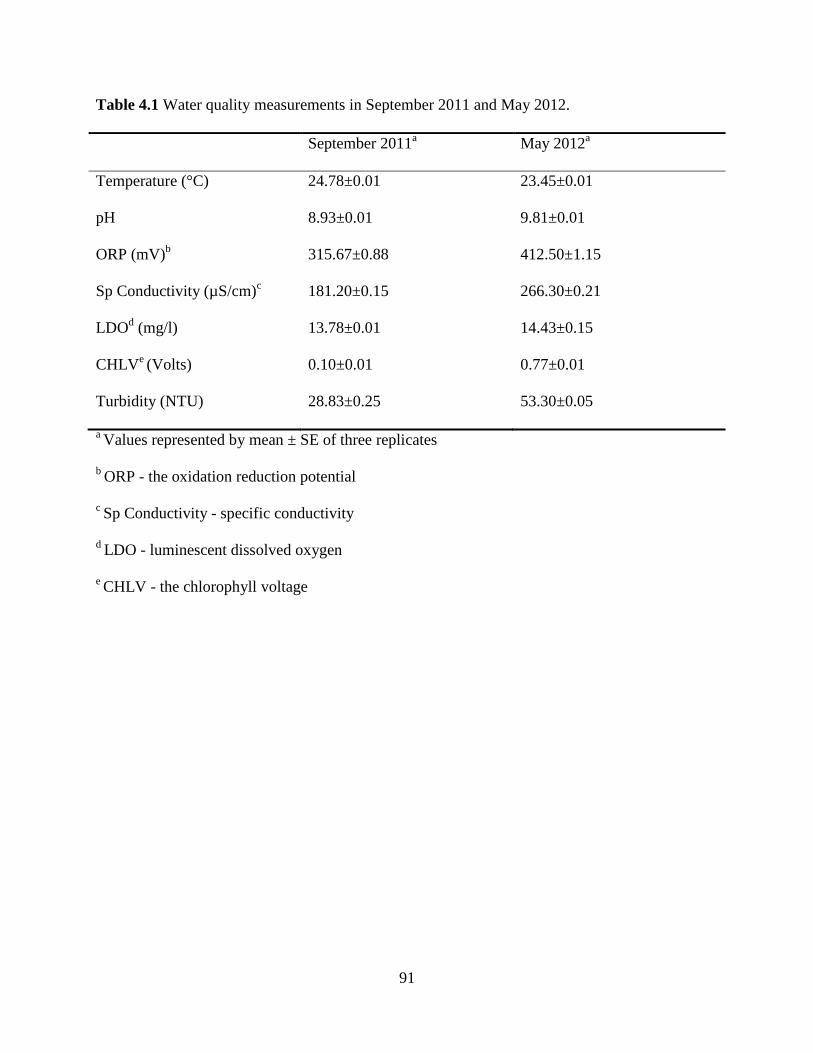
91
Table 4.1 Water quality measurements in September 2011 and May 2012.
September 2011a May 2012
a
Temperature (°C) 24.78±0.01 23.45±0.01
pH 8.93±0.01 9.81±0.01
ORP (mV)b 315.67±0.88 412.50±1.15
Sp Conductivity (µS/cm)c 181.20±0.15 266.30±0.21
LDOd (mg/l) 13.78±0.01 14.43±0.15
CHLVe (Volts) 0.10±0.01 0.77±0.01
Turbidity (NTU) 28.83±0.25 53.30±0.05
a Values represented by mean ± SE of three replicates
b ORP - the oxidation reduction potential
c Sp Conductivity - specific conductivity
d LDO - luminescent dissolved oxygen
e CHLV - the chlorophyll voltage
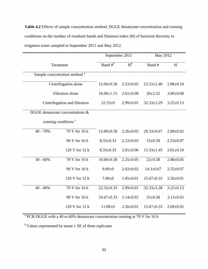
92
Table 4.2 Effects of sample concentration method, DGGE denaturant concentration and running
conditions on the number of resultant bands and Shannon index (H) of bacterial diversity in
irrigation water sampled in September 2011 and May 2012.
September 2011 May 2012
Treatment Band #b H
b Band # H
Sample concentration method a
Centrifugation alone 15.00±0.58 2.53±0.05 23.33±2.40 2.88±0.10
Filtration alone 16.00±1.15 2.61±0.08 26±2.52 3.00±0.08
Centrifugation and filtration 22.33±0 2.99±0.01 32.33±3.29 3.25±0.13
DGGE denaturant concentrations &
running conditions c
40 - 70% 70 V for 16 h 12.00±0.58 2.26±0.05 20.33±0.67 2.89±0.02
90 V for 16 h 8.33±0.33 2.12±0.03 15±0.58 2.53±0.07
120 V for 12 h 8.33±0.33 2.01±0.06 15.33±1.45 2.61±0.10
30 - 60% 70 V for 16 h 10.00±0.58 2.25±0.05 22±0.58 2.88±0.05
90 V for 16 h 9.00±0 2.02±0.02 14.3±0.67 2.55±0.07
120 V for 12 h 7.00±0 1.85±0.01 15.67±0.33 2.56±0.01
40 - 60% 70 V for 16 h 22.33±0.33 2.99±0.01 32.33±3.28 3.25±0.13
90 V for 16 h 10.67±0.33 2.14±0.02 25±0.58 3.13±0.02
120 V for 12 h 11.00±0 2.26±0.01 15.67±0.33 2.69±0.02
a PCR-DGGE with a 40 to 60% denaturant concentration running at 70 V for 16 h
b Values represented by mean ± SE of three replicates

93
c PCR-DGGE using bacterial cells collected via a combination of centrifugation and filtration
method
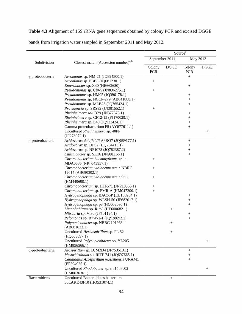
94
Table 4.3 Alignment of 16S rRNA gene sequences obtained by colony PCR and excised DGGE
bands from irrigation water sampled in September 2011 and May 2012.
Subdivision Closest match (Accession number)a,b
Sourcec
September 2011 May 2012
Colony
PCR
DGGE Colony
PCR
DGGE
γ-proteobacteria Aeromonas sp. NM-21 (JQ894500.1) +
Aeromonas sp. PBB3 (JQ681230.1) +
Enterobacter sp. X40 (HE662680) +
Pseudomonas sp. Cf0-5 (JN836275.1) +
Pseudomonas sp. HM05 (JQ396178.1) +
Pseudomonas sp. NCCP-279 (AB641888.1) +
Pseudomonas sp. MLB28 (JQ765424.1) +
Providencia sp. SRS82 (JN381552.1) +
Rheinheimera soli B29 (JN377675.1) +
Rheinheimera sp. CF12-15 (FJ170029.1) +
Rheinheimera sp. E49 (JQ922424.1) +
Gamma proteobacterium F8 (AY077611.1) +
Uncultured Rheinheimera sp. 48PP
(JF278072.1)
+
β-proteobacteria Acidovorax delafieldii A3RO7 (JQ689177.1) +
Acidovorax sp. DPS2 (HQ704415.1) +
Acidovorax sp. NF1078 (JQ782387.2) +
Chitinibacter sp. SK16 (JN981166.1) +
Chromobacterium haemolyticum strain
MDA0585 (NR_043957.1)
+
Chromobacterium violaceum strain NBRC
12614 (AB680302.1)
+
Chromobacterium violaceum strain 968
(HM449690.1)
+
Chromobacterium sp. IITR-71 (JN210566.1) +
Chromobacterium sp. PMR-A (HM047300.1) +
Hydrogenophaga sp. BAC55P (EU130964.1) +
Hydrogenophaga sp. WLSH-50 (JF682017.1) +
Hydrogenophaga sp. p3 (HQ652595.1) +
Limnohabitans sp. Rim8 (HE600682.1) +
Mitsuaria sp. Vi30 (JF501194.1) +
Pelomonas sp. R7W-1-1 (JQ928692.1) +
Polynucleobacter sp. NBRC 101963
(AB681633.1)
+
Uncultured Herbaspirillum sp. FL 52
(HQ008597.1)
+
Uncultured Polynucleobacter sp. YL205
(HM856566.1)
+
α-proteobacteria Azospirillum sp. DJM2D4 (JF753513.1) +
Mesorhizobium sp. RITF 741 (JQ697665.1) +
Candidatus Azospirillum massiliensis URAM1
(EF394925.1)
+
Uncultured Rhodobacter sp. rm15b3c02
(HM003636.1)
+
Bacteroidetes Uncultured Bacteroidetes bacterium
30LAKE43F10 (HQ531074.1)
+
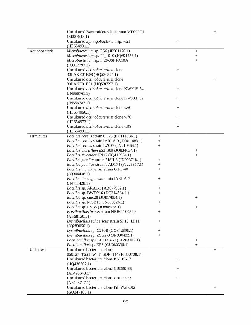
95
Uncultured Bacteroidetes bacterium ME002C1
(FJ827913.1)
+
Uncultured Sphingobacterium sp. w21
(HE654931.1)
+
Actinobacteria Microbacterium sp. E56 (JF501120.1) +
Microbacterium sp. FI_1010 (JQ691553.1) +
Microbacterium sp. I_29-J6NFA10A
(JQ917793.1)
+
Uncultured actinobacterium clone
30LAKE01B08 (HQ530574.1)
+
Uncultured actinobacterium clone
30LAKE01E01 (HQ530592.1)
+
Uncultured actinobacterium clone KWK1S.54
(JN656761.1)
+
Uncultured actinobacterium clone KWK6F.62
(JN656787.1)
+
Uncultured actinobacterium clone w60
(HE654966.1)
+
Uncultured actinobacterium clone w70
(HE654972.1)
+
Uncultured actinobacterium clone w98
(HE654991.1)
+
Firmicutes Bacillus cereus strain CT25 (EU111736.1) +
Bacillus cereus strain IARI-S-9 (JN411483.1) +
Bacillus cereus strain LZ027 (JN210566.1) +
Bacillus marisflavi p53 B09 (JQ834634.1) +
Bacillus mycoides TN12 (JQ415984.1) +
Bacillus pumilus strain MSII-6 (JN993718.1) +
Bacillus pumilus strain TAD174 (FJ225317.1) +
Bacillus thuringiensis strain GTG-40
(JQ004436.1)
+
Bacillus thuringiensis strain IARI-A-7
(JN411428.1)
+
Bacillus sp. ARA1-1 (AB677952.1) +
Bacillus sp. BWDY-6 (DQ314534.1 ) +
Bacillus sp. cmc28 (JQ917994.1) +
Bacillus sp. MGB13 (JN000926.1) +
Bacillus sp. PZ 35 (JQ808528.1) +
Brevibacillus brevis strain NBRC 100599
(AB681205.1)
+
Lysinibacillus sphaericus strain SP19_LP11
(JQ289050.1)
+
Lysinibacillus sp. C250R (GQ342695.1) +
Lysinibacillus sp. ZSG2-3 (JN990432.1) +
Paenibacillus sp.FSL H3-469 (EF203107.1) +
Paenibacillus sp. XP8 (GU080335.1) +
Unknown Uncultured bacterium clone
060127_T6S1_W_T_SDP_144 (FJ350708.1)
+
Uncultured bacterium clone BST15-17
(HQ436607.1)
+
Uncultured bacterium clone CRD99-65
(AF428643.1)
+
Uncultured bacterium clone CRP99-73
(AF428727.1)
+
Uncultured bacterium clone Filt WallC02
(GQ247163.1)
+
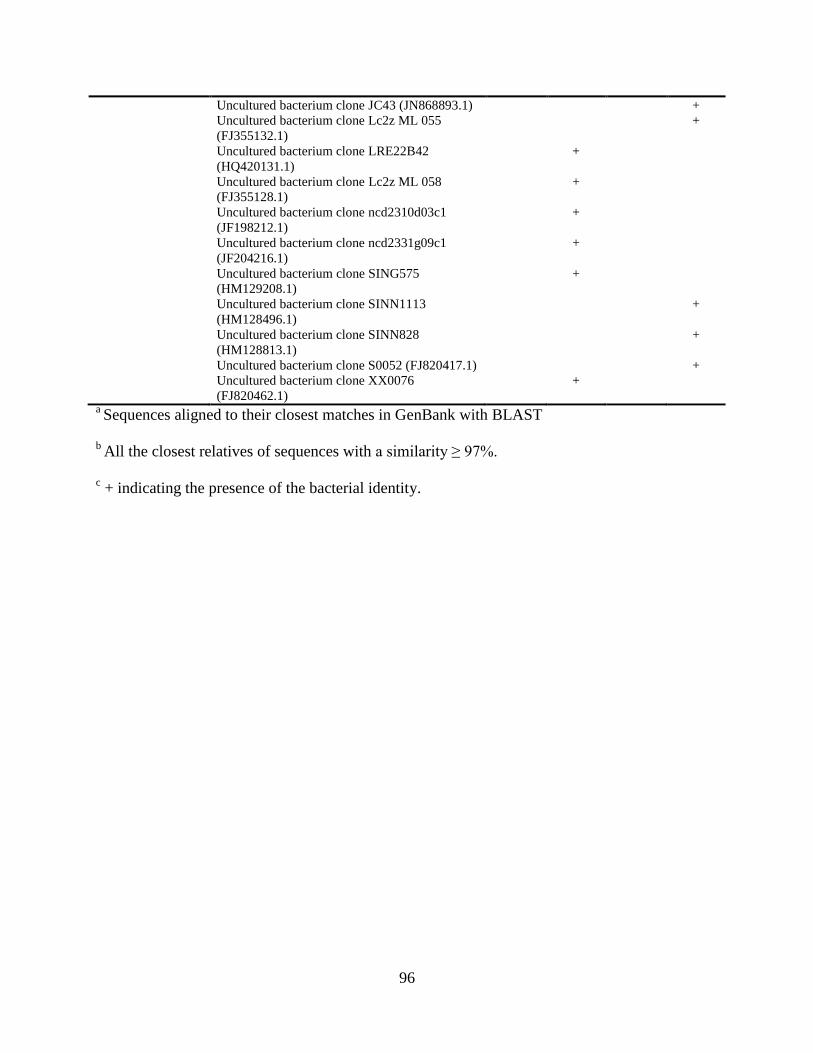
96
Uncultured bacterium clone JC43 (JN868893.1) +
Uncultured bacterium clone Lc2z ML 055
(FJ355132.1)
+
Uncultured bacterium clone LRE22B42
(HQ420131.1)
+
Uncultured bacterium clone Lc2z ML 058
(FJ355128.1)
+
Uncultured bacterium clone ncd2310d03c1
(JF198212.1)
+
Uncultured bacterium clone ncd2331g09c1
(JF204216.1)
+
Uncultured bacterium clone SING575
(HM129208.1)
+
Uncultured bacterium clone SINN1113
(HM128496.1)
+
Uncultured bacterium clone SINN828
(HM128813.1)
+
Uncultured bacterium clone S0052 (FJ820417.1) +
Uncultured bacterium clone XX0076
(FJ820462.1)
+
a Sequences aligned to their closest matches in GenBank with BLAST
b All the closest relatives of sequences with a similarity ≥ 97%.
c + indicating the presence of the bacterial identity.
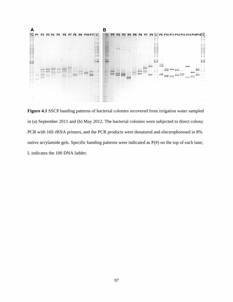
97
Figure 4.1 SSCP banding patterns of bacterial colonies recovered from irrigation water sampled
in (a) September 2011 and (b) May 2012. The bacterial colonies were subjected to direct colony
PCR with 16S rRNA primers, and the PCR products were denatured and electrophoresed in 8%
native acrylamide gels. Specific banding patterns were indicated as P(#) on the top of each lane;
L indicates the 100 DNA ladder.
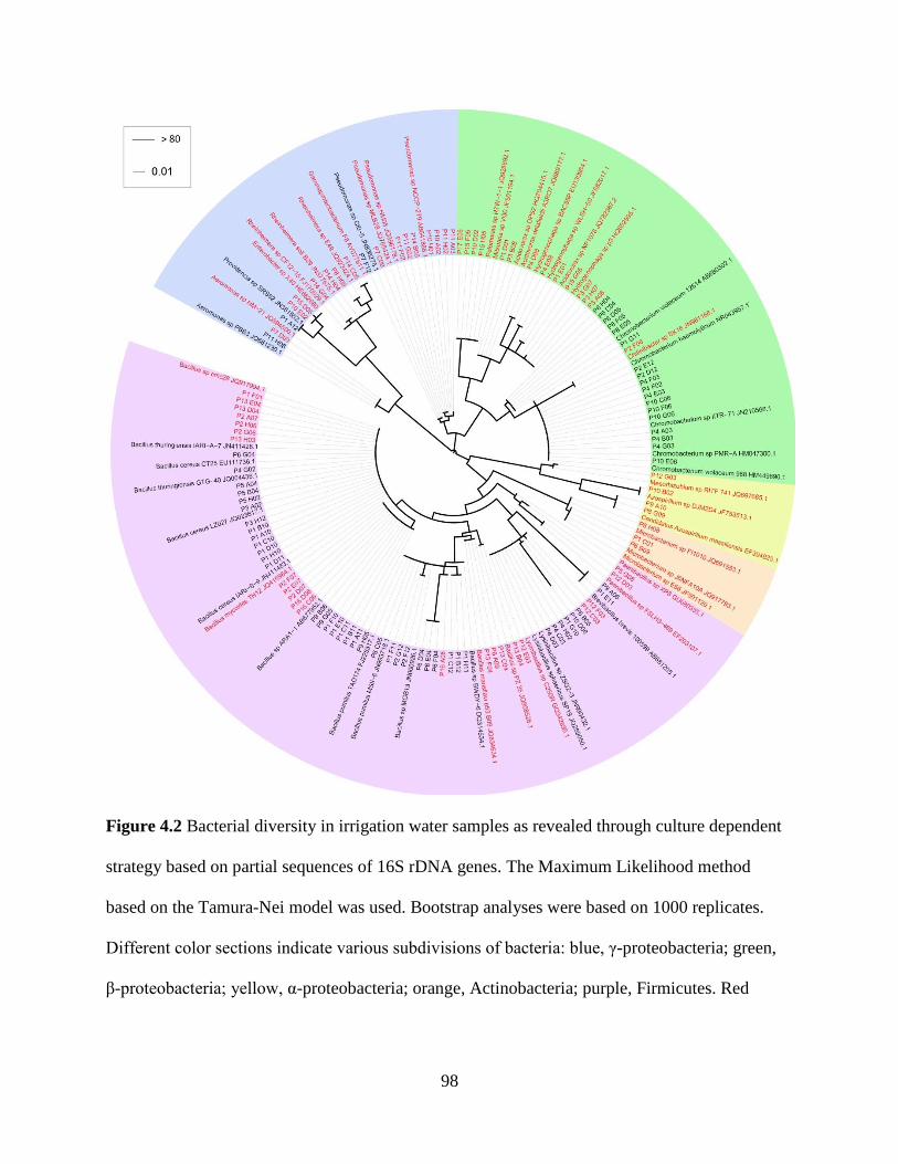
98
Figure 4.2 Bacterial diversity in irrigation water samples as revealed through culture dependent
strategy based on partial sequences of 16S rDNA genes. The Maximum Likelihood method
based on the Tamura-Nei model was used. Bootstrap analyses were based on 1000 replicates.
Different color sections indicate various subdivisions of bacteria: blue, γ-proteobacteria; green,
β-proteobacteria; yellow, α-proteobacteria; orange, Actinobacteria; purple, Firmicutes. Red

99
labels denote sequences from May 2012, and black labels denote sequences from September
2011.
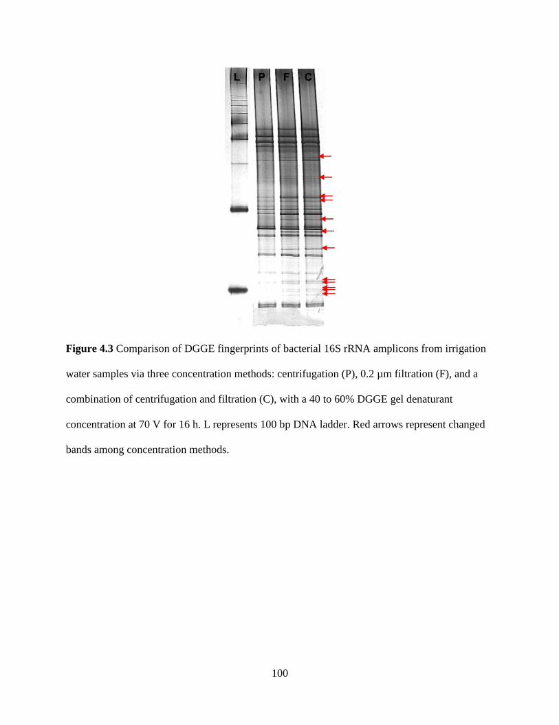
100
Figure 4.3 Comparison of DGGE fingerprints of bacterial 16S rRNA amplicons from irrigation
water samples via three concentration methods: centrifugation (P), 0.2 µm filtration (F), and a
combination of centrifugation and filtration (C), with a 40 to 60% DGGE gel denaturant
concentration at 70 V for 16 h. L represents 100 bp DNA ladder. Red arrows represent changed
bands among concentration methods.
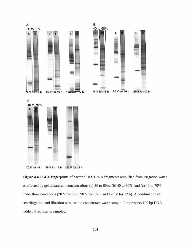
101
Figure 4.4 DGGE fingerprints of bacterial 16S rRNA fragments amplified from irrigation water
as affected by gel denaturant concentrations (a) 30 to 60%, (b) 40 to 60%, and (c) 40 to 70%
under three conditions (70 V for 16 h, 90 V for 16 h, and 120 V for 12 h). A combination of
centrifugation and filtration was used to concentrate water sample. L represents 100 bp DNA
ladder. S represents samples.
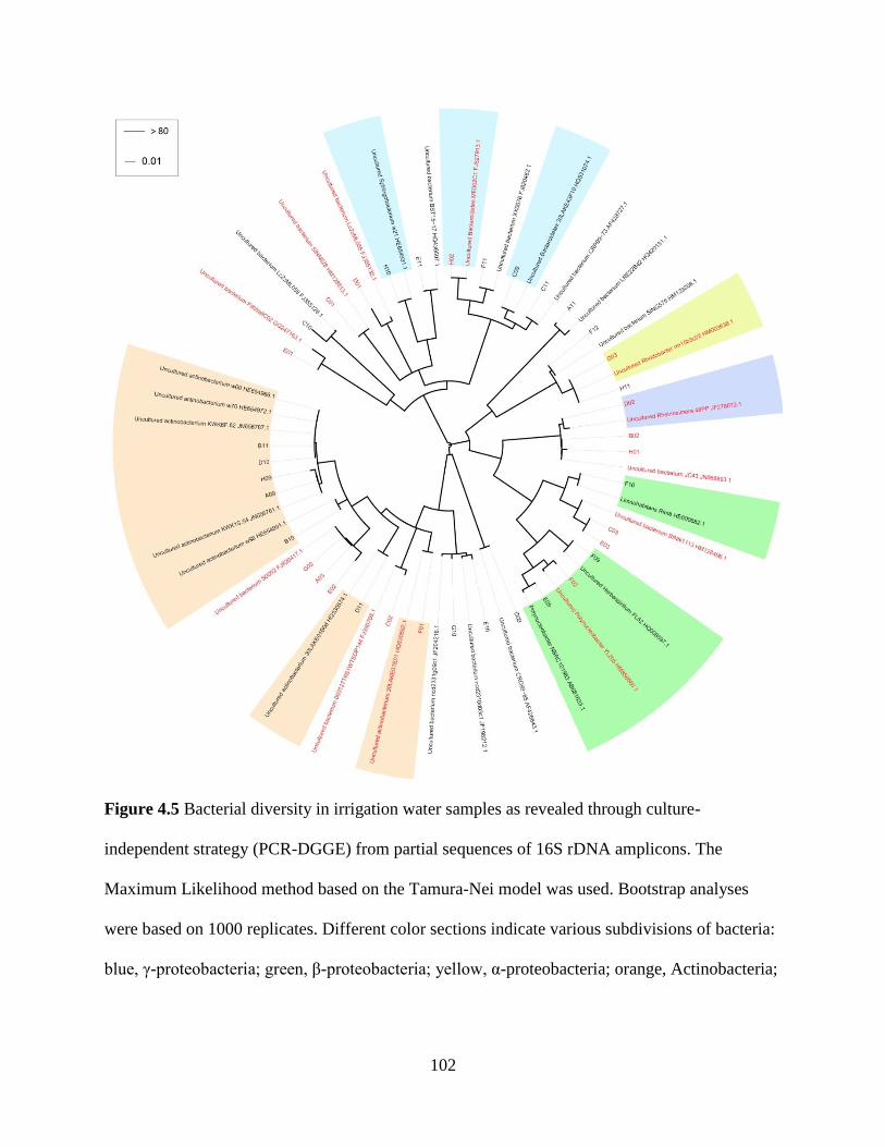
102
Figure 4.5 Bacterial diversity in irrigation water samples as revealed through culture-
independent strategy (PCR-DGGE) from partial sequences of 16S rDNA amplicons. The
Maximum Likelihood method based on the Tamura-Nei model was used. Bootstrap analyses
were based on 1000 replicates. Different color sections indicate various subdivisions of bacteria:
blue, γ-proteobacteria; green, β-proteobacteria; yellow, α-proteobacteria; orange, Actinobacteria;

103
light blue, Bacteroidetes; unshaded, unknown bacterial species. Red labels denote sequences
from May 2012, and black labels denote sequences from September 2011.
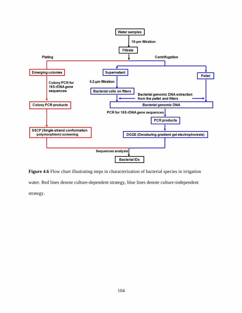
104
Figure 4.6 Flow chart illustrating steps in characterization of bacterial species in irrigation
water. Red lines denote culture-dependent strategy, blue lines denote culture-independent
strategy.
105
Chapter 5
Bacterial community structure shifts in irrigation water under heat treatment
Abstract
Our previous study revealed the required temperature of heat treatment can be lowered to
48°C for 24 h from the currently recommendation of 95°C for 30 s without sacrificing
decontamination efficacy. However, the mechanisms by which plant pathogens in irrigation
water are suppressed or killed at 42 and 48°C are yet to be elucidated. The objective of this study
was to determine whether such temperatures shift bacterial community structure in irrigation
water, enhancing antagonistic activities against plant pathogens. Both culture-dependent and -
independent strategies were employed. The structure of both culturable and uncultured bacterial
community in irrigation water substantially shifted after the heat treatments. In contrast to that at
25°C, the two largest bacterial groups detected at 42 and 48°C were γ-proteobacteria and
Firmicutes. Specifically, these included genera of Bacillus, Paenibacillus, Pseudomonas,
Brevibacillus, and Lysobacter. These bacteria are known to have biocontrol potential; their
increased abundance implicated bacterial contributions to heat treatment of irrigation water at 42
and 48°C. Further experiments are being conducted to evaluate those bacteria for biocontrol
activities against Phytophthora species.
Introduction
Heat pasteurization is one of the safest and most reliable methods for water
decontamination in horticulture (Runia et al. 1988; Runia 1995; van Os 1999). Our recent study
106
revealed that water treatment temperature can be lowered to 48°C for 24 h from the current
recommendation of 95°C for 30 s without sacrificing decontamination efficacy (Hao et al. 2012).
This is a major improvement in energy efficiency and economics, two major hurdles currently
affecting the adoption of this water treatment (Hong and Moorman 2005). With the current
protocol, all microorganisms in water are killed at 95°C, resulting in a biological vacuum
allowing invasion by plant pathogens through other avenues such as contaminated plants and
planting materials (Schumann and D'Arcy 2006).
The question asked in this part of the dissertation is whether and how the water
temperatures of 48 and 42°C may affect the bacteria especially those beneficial bacteria in
irrigation water. We hypothesized that these water temperatures shift the bacterial community
structure in irrigation water and enhance antagonistic activities against plant pathogens, thus
contributes to the suppression or killing of plant pathogens. The present study was performed to
test this hypothesis with irrigation water samples from a local nursery in eastern Virginia.
Specific objectives were to (1) to characterize the bacterial community structures in water
samples treated at 42 and 48°C and compare with those at 25°C (control) using the scheme
described in the previous chapter, and (2) identify potential pathogen-suppressing bacteria in the
irrigation reservoir of study.
Materials and Methods
Water samples
Water samples were collected from an irrigation reservoir at a local nursery in eastern
Virginia in February and May 2012. All samples were collected in sterile bottles from the
surface of water columns in the center of the reservoir. Three replicates of 10 L each were
107
collected. Water temperature and other water quality parameters were recorded on-site using a
Hydrolab DS5X multiparameter water quality meter (Hach®, Loveland, CO, USA). Water
samples were then transported to the lab and immediately used for following experiments.
Temperature treatments
Irrigation water samples were heat-treated at three temperatures of 25, 42, and 48°C for
48 h. For each treatment three 1-L volume replicates were transferred to 1-L Erlenmeyer flasks
and placed in temperature-controlled incubators (Intellus Environmental Controllers, Percival
Scientific, Inc.). The incubators were calibrated and monitored before and during the temperature
experiment using a HOBO Pendant®
temp/light data logger (Onset Computer Corporation). Once
the temperature treatments were completed, water was passed through nylon membrane filters
(pore size 10 µm) to remove larger debris and algae.
Culture-dependent strategy
Plating
One hundred microliters of the filtrate was evenly spread on Nutrient Agar (NA)
(DifcoTM
, Detroit, MI) in a 10-cm diameter Petri dish using a sterile glass spreader. Three NA
medium plates were used for each replicate per treatment and incubated at 28°C for 2 d.
Colony PCR & SSCP analysis
The emerging colonies on NA plates were grouped based on color, texture and size. The
total numbers of colonies in each group were recorded. About 20 to 30% of colonies from each
group were subcultured into 48-well plates containing NA medium, and incubated at 28°C for 2
108
d. Colony PCRs amplifying bacterial 16S rDNA were performed for each subculture with
primers 16S f968 and r1401 containing hypervariable regions V6 to V8 (Nubel et al. 1996;
Peixoto et al. 2002). Reagents supplied with Takara Taq (Takara Bio Inc., Japan) were used in 25
μl reactions containing 0.1 μl Takara Taq (5 units/μl), 2.5 μl 10× PCR buffer (containing 1.5 mM
MgCl2), 2 μl dNTP mixture (2.5 mM each), 1 μl of each primer (10 μM), and 18.4 μl DNA-free
nanopure water. The bacterial colonies were picked directly from the culture plate and added to
the mixture. Cycling conditions were an initial denaturation at 96°C for 2 min, followed by 40
cycles of denaturation at 94°C for 0.5 min, annealing at 55°C for 0.5 min, and extension at 72°C
for 1 min. For the last cycle, the extension time was increased to 10 min.
Single-strand conformation polymorphism (SSCP) analyses were performed for the
colony PCR products as described by Kong et al. (2003) with minor modifications. In brief, the
amplicons were mixed with loading dye (1:4, v:v) and denatured by heating at 96°C for 10 min
then chilled on ice. Two microliters of each sample was loaded on 8% polyacrylamide gels
(acrylamide/bisacrylamide = 29:1) running at 220 V for 2.5 h in chilled 1× TBE buffer. After
electrophoresis, the gels were stained with silver nitrate (Beidler et al. 1982). SSCP banding
patterns of individual colonies were categorized with the aid of a 100 bp DNA ladder (Promega
Corp., Madison, WI). Colony PCR products were grouped by SSCP fingerprint subsequently
selected for DNA sequencing.
Culture-independent strategy
Genomic DNA extraction
The filtrate passing through 10-µm filters was concentrated through a combination of
centrifugation and 0.2-µm filtration. In brief, the filtrate was centrifuged at 12,200 g for 50 min,
109
the pellet was saved, and the supernatant was filtered through 0.2-µm filters. The UltraClean
Microbial DNA Isolation Kit (MO BIO Laboratories, Inc, Carlsbad, CA) was used to extract
genomic DNA from the pellets following the instruction manual. The PowerWater DNA
Isolation Kit (MO BIO Laboratories, Inc, Carlsbad, CA) was used to extract genomic DNA from
the 0.2-µm filters. Total genomic DNA from both pellets and filters were combined for DGGE
analyses.
PCR amplification and DGGE
For DGGE analysis, 16S rRNA fragments containing three hypervariable regions V6 to
V8 were amplified with primers GC-clamp-EUB f933 and EUB r1387 (Kawai et al. 2002). The
PCR mixture (50 μl) was adapted to contain 0.2 μl Takara Taq (5 units/μl), 5 μl 10× PCR buffer
(containing 1.5 mM MgCl2), 7.5 μl BSA (0.3 mg/ml), 4 μl dNTP mixture (2.5 mM each), 2 μl of
each primer (10 μM), 19.3 μl DNA-free nanopure water and 10 μl DNA extract. A hot-start PCR
was performed at 94°C for 4 min, followed by touchdown PCR with the following parameters:
the denaturation was set at 94°C for 1 min, the annealing temperature was initially set at 65°C
and then decreased by 1°C every cycle (1.5 min) until it reached 55°C. Twenty additional cycles
were carried out at 55°C. Primer extension was carried out at 72°C for 2.5 min. with a final
extension step for 7 min at 72°C.
DGGE was performed using a Bio-Rad DcodeTM
Universal Mutation Detection System
(Bio-Rad, Hercules, CA, USA) according to the manufacturer's instructions. PCR products were
electrophoresed in 1 mm thick denaturing 8% polyacrylamide gel (acrylamide:bisacrylamide =
37.5:1) in 1× TAE buffer. Gels were prepared with a denaturant concentration of 40 to 60%.
110
Electrophoresis at 60°C was run at 70 V for 16. After electrophoresis, the gels were stained with
silver nitrate (Beidler et al. 1982).
DGGE gels were digitally documented using the Epi Chemi II darkroom and analyzed
using LabWorks software (UVP Laboratory, Upland, CA). Each lane was detected automatically
with the width and length adjusted manually. Bands in each lane were detected automatically and
corrected manually.
DNA reisolation and cloning
Unique bands among temperature treatments were selected from the DGGE gels. DNAs
were re-isolated from the acrylamide gel using a “crush and soak” method with minor
modifications (Sambrook and Russell 2001). In brief, bands were excised using sterile gel
cutting tips (BioExpress, Kaysville, UT), transferred to 1.7 ml tubes and washed briefly with 0.5
mL TE buffer. The gel fragment was then crushed using a sterile pestle in 400 µl elution buffer
(0.5 M NH4OAc, 10 mM EDTA), incubated at 37°C overnight in an incubator shaker to allow
DNA diffusion from the gel, then centrifuged for 5 minutes at 12,000 rpm to pellet the
acrylamide. The supernatant was transferred and DNA precipitated in 1 mL 100% ethanol with
16 µl 5M NaCl at -20°C overnight, then centrifuged at 12,000 rpm for 20 min. The pellets were
vacuum dried and re-suspended in 20 µl TE buffer. The extracted DNAs were re-amplified with
16S rRNA primers EUB f933 and EUB r1387 (Kawai et al. 2002) using the same cycling
conditions as colony PCR. These PCR amplicons were cloned using the pGEM-T Easy Vector
System (Promega). Randomly picked white (positive) colonies were PCR amplified with the
plasmid primers T7 (5' - TAA TAC GAC TCA CTA TAG GG - 3') and SP6 (5' - ATT TAG
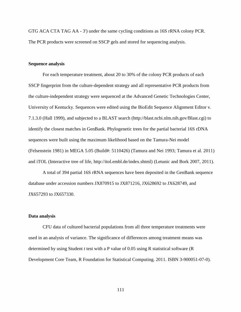
111
GTG ACA CTA TAG AA - 3') under the same cycling conditions as 16S rRNA colony PCR.
The PCR products were screened on SSCP gels and stored for sequencing analysis.
Sequence analysis
For each temperature treatment, about 20 to 30% of the colony PCR products of each
SSCP fingerprint from the culture-dependent strategy and all representative PCR products from
the culture-independent strategy were sequenced at the Advanced Genetic Technologies Center,
University of Kentucky. Sequences were edited using the BioEdit Sequence Alignment Editor v.
7.1.3.0 (Hall 1999), and subjected to a BLAST search (http://blast.ncbi.nlm.nih.gov/Blast.cgi) to
identify the closest matches in GenBank. Phylogenetic trees for the partial bacterial 16S rDNA
sequences were built using the maximum likelihood based on the Tamura-Nei model
(Felsenstein 1981) in MEGA 5.05 (Build#: 5110426) (Tamura and Nei 1993; Tamura et al. 2011)
and iTOL (Interactive tree of life, http://itol.embl.de/index.shtml) (Letunic and Bork 2007, 2011).
A total of 394 partial 16S rRNA sequences have been deposited in the GenBank sequence
database under accession numbers JX870915 to JX871216, JX628692 to JX628749, and
JX657293 to JX657330.
Data analysis
CFU data of cultured bacterial populations from all three temperature treatments were
used in an analysis of variance. The significance of differences among treatment means was
determined by using Student t test with a P value of 0.05 using R statistical software (R
Development Core Team, R Foundation for Statistical Computing. 2011. ISBN 3-900051-07-0).
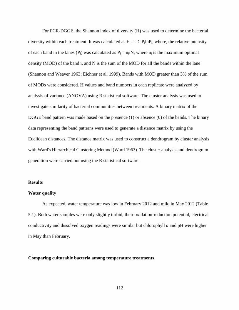
112
For PCR-DGGE, the Shannon index of diversity (H) was used to determine the bacterial
diversity within each treatment. It was calculated as H = - Σ PilnPi, where, the relative intensity
of each band in the lanes (Pi) was calculated as Pi = ni/N, where ni is the maximum optimal
density (MOD) of the band i, and N is the sum of the MOD for all the bands within the lane
(Shannon and Weaver 1963; Eichner et al. 1999). Bands with MOD greater than 3% of the sum
of MODs were considered. H values and band numbers in each replicate were analyzed by
analysis of variance (ANOVA) using R statistical software. The cluster analysis was used to
investigate similarity of bacterial communities between treatments. A binary matrix of the
DGGE band pattern was made based on the presence (1) or absence (0) of the bands. The binary
data representing the band patterns were used to generate a distance matrix by using the
Euclidean distances. The distance matrix was used to construct a dendrogram by cluster analysis
with Ward's Hierarchical Clustering Method (Ward 1963). The cluster analysis and dendrogram
generation were carried out using the R statistical software.
Results
Water quality
As expected, water temperature was low in February 2012 and mild in May 2012 (Table
5.1). Both water samples were only slightly turbid, their oxidation-reduction potential, electrical
conductivity and dissolved oxygen readings were similar but chlorophyll a and pH were higher
in May than February.
Comparing culturable bacteria among temperature treatments
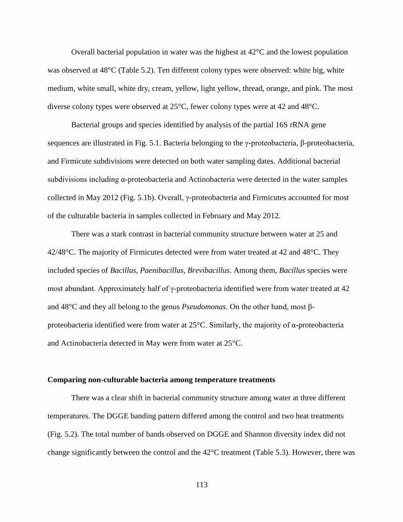
113
Overall bacterial population in water was the highest at 42°C and the lowest population
was observed at 48°C (Table 5.2). Ten different colony types were observed: white big, white
medium, white small, white dry, cream, yellow, light yellow, thread, orange, and pink. The most
diverse colony types were observed at 25°C, fewer colony types were at 42 and 48°C.
Bacterial groups and species identified by analysis of the partial 16S rRNA gene
sequences are illustrated in Fig. 5.1. Bacteria belonging to the γ-proteobacteria, β-proteobacteria,
and Firmicute subdivisions were detected on both water sampling dates. Additional bacterial
subdivisions including α-proteobacteria and Actinobacteria were detected in the water samples
collected in May 2012 (Fig. 5.1b). Overall, γ-proteobacteria and Firmicutes accounted for most
of the culturable bacteria in samples collected in February and May 2012.
There was a stark contrast in bacterial community structure between water at 25 and
42/48°C. The majority of Firmicutes detected were from water treated at 42 and 48°C. They
included species of Bacillus, Paenibacillus, Brevibacillus. Among them, Bacillus species were
most abundant. Approximately half of γ-proteobacteria identified were from water treated at 42
and 48°C and they all belong to the genus Pseudomonas. On the other hand, most β-
proteobacteria identified were from water at 25°C. Similarly, the majority of α-proteobacteria
and Actinobacteria detected in May were from water at 25°C.
Comparing non-culturable bacteria among temperature treatments
There was a clear shift in bacterial community structure among water at three different
temperatures. The DGGE banding pattern differed among the control and two heat treatments
(Fig. 5.2). The total number of bands observed on DGGE and Shannon diversity index did not
change significantly between the control and the 42°C treatment (Table 5.3). However, there was
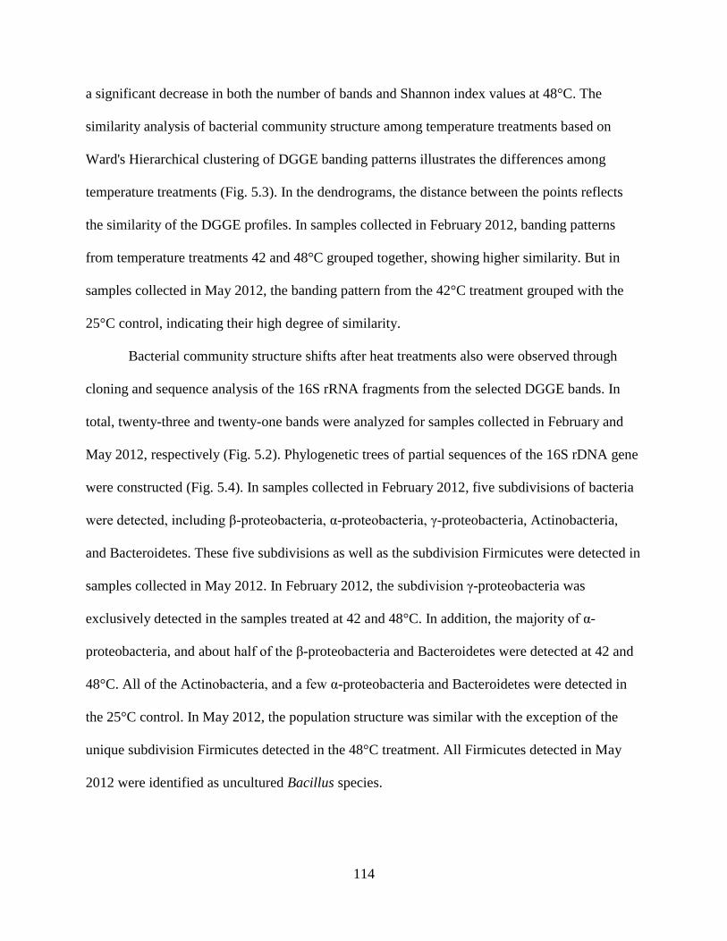
114
a significant decrease in both the number of bands and Shannon index values at 48°C. The
similarity analysis of bacterial community structure among temperature treatments based on
Ward's Hierarchical clustering of DGGE banding patterns illustrates the differences among
temperature treatments (Fig. 5.3). In the dendrograms, the distance between the points reflects
the similarity of the DGGE profiles. In samples collected in February 2012, banding patterns
from temperature treatments 42 and 48°C grouped together, showing higher similarity. But in
samples collected in May 2012, the banding pattern from the 42°C treatment grouped with the
25°C control, indicating their high degree of similarity.
Bacterial community structure shifts after heat treatments also were observed through
cloning and sequence analysis of the 16S rRNA fragments from the selected DGGE bands. In
total, twenty-three and twenty-one bands were analyzed for samples collected in February and
May 2012, respectively (Fig. 5.2). Phylogenetic trees of partial sequences of the 16S rDNA gene
were constructed (Fig. 5.4). In samples collected in February 2012, five subdivisions of bacteria
were detected, including β-proteobacteria, α-proteobacteria, γ-proteobacteria, Actinobacteria,
and Bacteroidetes. These five subdivisions as well as the subdivision Firmicutes were detected in
samples collected in May 2012. In February 2012, the subdivision γ-proteobacteria was
exclusively detected in the samples treated at 42 and 48°C. In addition, the majority of α-
proteobacteria, and about half of the β-proteobacteria and Bacteroidetes were detected at 42 and
48°C. All of the Actinobacteria, and a few α-proteobacteria and Bacteroidetes were detected in
the 25°C control. In May 2012, the population structure was similar with the exception of the
unique subdivision Firmicutes detected in the 48°C treatment. All Firmicutes detected in May
2012 were identified as uncultured Bacillus species.
115
Bacterial community structure shift with heat treatment
Culturable bacteria in the subdivisions Firmicutes and γ-proteobacteria increased in
abundance in the 42 and 48°C treated irrigation water (Table 5.4). The three major bacterial
groups identified were the genera Bacillus and Paenibacillus in Firmicutes, and Pseudomonas in
γ-proteobacteria. And other some minor groups found were Brevibacillus and Lysobacter
species. Bacteria identified via PCR-DGGE belonging to the subdivisions α-proteobacteria, γ-
proteobacteria, and Bacteroidetes were found only at 42 and 48°C treatments. Bacteria
similar/identical to Pseudomonas aeruginosa strain NTS6 were the most common detected on
two sampling dates by both culturing and PCR-DGGE at heat-treated irrigation water. The
majority of bacteria identified in the subdivisions β-proteobacteria and Actinobacteria were from
control water.
Discussion
This study demonstrated that the dominant bacterial populations in the water samples
shifted after heat treatments at 42 and 48°C when compared with those at the 25°C control. The
two largest groups of bacteria detected in heat-treated irrigation water were γ-proteobacteria and
Firmicutes. These findings may help understand the underlying biological mechanism by which
Phytophthora and bacterial pathogens are killed or suppressed under the new heat treatment.
Our previous study (Chapter 2) showed that water temperature required to inactivate
major groups of plant pathogens can be lowered to 48°C for 24 h in sterile deionized water
(SDW). In addition, the survival rate of Phytophthora zoospores at 42°C was lower in irrigation
water than SDW (Chapter 3). The enhanced inactivation of Phytophthora zoospores in irrigation
water as well as the killing of major groups of plant pathogens at heat temperatures of 42 and
116
48°C may be attribute partially to the bacterial community shift at such temperatures. Current
heat treatment protocol recommending a high temperature of 95°C destroys most
microorganisms in water. Thus, the aquatic ecosystem is off biological balance, because
microorganisms constitute the base of food webs and changes in composition and structure can
profoundly affect the stability and sustainability of the ecosystem (Fuhrman and Ouverney 1998;
Pomeroy et al. 2007). The proposed heat treatment temperature of 48°C would not break the
entire microbial community in irrigation water since it is much lower than the lethal temperature
of most microorganisms (Baker 1962; Schumann 1991). In fact, the heat temperatures of 42 and
48°C changed the composition and structure of bacterial communities in irrigation water, leading
to an increased abundance of several bacterial groups compared to the 25°C control. These
bacterial groups may be antagonistic and thus enhance the control of plant pathogens present in
irrigation water.
Bacteria in the subdivisions Firmicutes and γ-proteobacteria, including Bacillus,
Pseudomonas, Paenibacillus, Brevibacillus, and Lysobacter species, may contribute to
inactivation of plant pathogens in irrigation water after heat treatment by biocontrol activities.
Firmicutes and γ-proteobacteria were the two largest bacteria groups identified in heat-treated
irrigation water, and Bacillus, Paenibacillus, and Pseudomonas were the three major bacterial
genera. Pseudomonas species are the most extensively studied bacterial organisms utilized for
biocontrol of many seedling diseases and root rots on several crops (Punja 1997). Pseudomonas
putida strain 06909 suppresses Phytophthora nicotianae by complete lysis and degradation of
hyphae of P. nicotianae (Yang et al. 1994). Pseudomonas putida WCS358r suppresses fusarium
wilt of carnation caused by Fusarium oxysporum f. sp. dianthi via siderophore-mediated
competition for iron (Duijff et al. 1993). Pseudomonas fluorescens strain Pf-5 provides highly
117
effective biocontrol activity against a broad range of fungal plant pathogens through the
production of several secondary metabolites, including antifungal compounds such as
pyoluteorin, pyrrolnitrin, and 2,4- diacylphloroglucinol, as well as several rhizoxin derivatives
(Loper et al. 2007; Loper et al. 2008). This strain has been used in a Canadian greenhouse to
control pythium diseases by reducing zoospore germination and chemotaxis (Paulitz and
Belanger 2001). Bacillus species have also been investigated frequently as biocontrol agents.
Bacillus pumilus strains INR7 and Bacillus subtilis strain GB03 control the cucumber pathogens
Colletotrichum orbiculare, Pseudomonas syringae pv. 1achrymans, and Erwinia tracheiphila
(Raupach and Kloepper 1998). Bacillus subtilis Strain QST713 has a spectrum of antagonistic
activity against over 40 plant diseases, including gray mold, damping-off, and powdery mildews,
through a mixture of mechanisms consisted of competition, parasitism, antibiosis, and induction
of systemic acquired resistance (SAR) (Paulitz and Belanger 2001). Paenibacillus species are not
commonly tested as biocontrol agents against plant pathogens, but Paenibacillus sp. 300 did
suppress fusarium wilt of cucumber (Cucumis sativus) caused by Fusarium oxysporum f. sp.
cucumerinum in nonsterile, soilless potting medium (Singh et al. 1999). In addition, several other
bacterial species in the genera Brevibacillus and Lysobacter, which only detected in heat-treated
irrigation water, have been reported to biologically control a number of plant pathogens.
Brevibacillus laterosporus BPM3 isolated from a natural hot water spring strongly inhibited
growth of plant pathogenic fungi, including Fusarium oxysporum f. sp. ciceri, F. semitectum,
Magnaporthe grisea and Rhizoctonia oryzae) (Saikia et al. 2011). Lysobacter enzymogenes is
antagonistic to a broad range of plant pathogens by infecting the pathogens intracellularly
(Christensen and Cook 1978; Kobayashi and Crouch 2009). Therefore, the increase of these
118
bacterial groups after heat treatment at 42 and 48°C may result in more antagonistic activities
against plant pathogens in irrigation water, contributing to the efficacy of the new heat treatment.
There are probably more bacteria than those detected in this study with biocontrol effect
contributing to control of plant pathogens in irrigation water at 42 and 48°C. The culturable
bacteria recovered from irrigation water here just compose a very small portion of the total
microorganisms because the majority of environmental microbes are unculturable (Amann et al.
1995; Vartoukian et al. 2010). Moreover, most bacteria detected through culture-independent
PCR-DGGE were unknown species and could not be identified to the genus level. In addition,
the microbial community population in irrigation water varies with environmental conditions.
To further investigate the potential biological mechanisms of pathogen controlling at 42
and 48°C, in vitro and in vivo experiments are being conducted to test the biocontrol activities of
selected bacterial isolates. Once the nature-occurring bacteria with biological activity are
confirmed existing in the irrigation system, by manipulating and enhancing those beneficial
microorganisms would build a pathogen-suppressing biological control system with minimal
negative impact (Weller et al. 2002; Welbaum et al. 2004).
References
Amann, A. I., Ludwig, W. & Schleifer, A.-H. (1995). Phylogenetic identification and in situ
detection of individual microbial cells without cultivation. Microbiological Reviews, 59
(1), 143-169
Baker, K. F. (1962). Principles of heat treatment of soil and planting material. Jour Australian
Inst Agric Sci, 28 ((2)), 118-126
119
Beidler, J. L., Hilliard, P. R. & Rill, R. L. (1982). Ultrasensitive staining of nucleic acids with
silver. Analytical Biochemistry, 126, 374-380
Christensen, P. & Cook, F. D. (1978). Lysobacter new genus of nonfruiting gliding bacteria with
a high base ratio. International Journal of Systematic Bacteriology, 28 (3), 367-393
Duijff, B. J., Meijer, J. W., Bakker, P. A. H. M. & Schippers, B. (1993). Siderophore-mediated
competition for iron and induced resistance in the suppression of Fusarium wilt of
carnation by fluorescent Pseudomonas spp. Netherlands Journal of Plant Pathology, 99
(5-6), 277-289
Eichner, C. A., Erb, R. W., Timmis, K. N. & Wagner-Doebler, I. (1999). Thermal gradient gel
electrophoresis analysis of bioprotection from pollutant shocks in the activated sludge
microbial community. Applied and Environmental Microbiology, 65 (1), 102-109
Felsenstein, J. (1981). Evolutionary trees from DNA sequences: a maximum likelihood
approach. Journal of Molecular Evolution, 17, 368-376
Fuhrman, J. A. & Ouverney, C. C. (1998). Marine microbial diversity studied via 16S rRNA
sequences: Cloning results from coastal waters and counting of native archaea with
fluorescent single cell probes. Aquatic Ecology, 32 (1), 3-15
Hall, T. A. (1999). BioEdit: a user-friendly biological sequence alignment editor and analysis
program for Windows 95/98/NT. Nucleic Acids Symposium Series, 41, 95-98
Hao, W., Ahonsi, M. O., Vinatzer, B. A. & Hong, C. X. (2012). Inactivation of Phytophthora and
bacterial species in water by a potential energy-saving heat treatment. European Journal
of Plant Pathology, 134 (2), 357-365
Hong, C. X. & Moorman, G. W. (2005). Plant pathogens in irrigation water: Challenges and
opportunities. Critical Reviews in Plant Sciences, 24 (3), 189-208
120
Kawai, M., Matsutera, E., Kanda, H., Yamaguchi, N., Tani, K. & Nasu, M. (2002). 16S
ribosomal DNA-based analysis of bacterial diversity in purified water used in
pharmaceutical manufacturing processes by PCR and denaturing gradient gel
electrophoresis. Applied and Environmental Microbiology, 68 (2), 699-704
Kobayashi, D. Y. & Crouch, J. A. (2009). Bacterial/fungal interactions: from pathogens to
mutualistic endosymbionts. Annual Review of Phytopathology, 47, 63-82
Kong, P., Hong, C. X., Richardson, P. A. & Gallegly, M. E. (2003). Single-strand-conformation
polymorphism of ribosomal DNA for rapid species differentiation in genus
Phytophthora. Fungal Genetics and Biology, 39 (3), 238-249
Letunic, I. & Bork, P. (2007). Interactive Tree Of Life (iTOL): an online tool for phylogenetic
tree display and annotation. Bioinformatics (Oxford), 23 (1), 127-128
Letunic, I. & Bork, P. (2011). Interactive Tree Of Life v2: online annotation and display of
phylogenetic trees made easy. Nucleic Acids Research, 39 (Suppl. 2), W475-W478
Loper, J. E., Henkels, M. D., Shaffer, B. T., Valeriote, F. A. & Gross, H. (2008). Isolation and
identification of rhizoxin analogs from Pseudomonas fluorescens Pf-5 by using a
genomic mining strategy. Applied and Environmental Microbiology, 74 (10), 3085-3093
Loper, J. E., Kobayashi, D. Y. & Paulsen, I. T. (2007). The genomic sequence of Pseudomonas
fluorescens Pf-5: Insights into biological control. Phytopathology, 97 (2), 233-238
Nubel, U., Engelen, B., Felske, A., Snaidr, J., Wieshuber, A., Amann, R. I., Ludwig, W. &
Backhaus, H. (1996). Sequence heterogeneities of genes encoding 16S rRNAs in
Paenibacillus polymyxa detected by temperature gradient gel electrophoresis. Journal of
Bacteriology, 178 (19), 5636-5643
121
Paulitz, T. C. & Belanger, R. R. (2001). Biological control in greenhouse system. Annual
Review of Phytopathology, 39, 103-133
Peixoto, R. S., Coutinho, H. L. D., Rumjanek, N. G., Macrae, A. & Rosado, A. S. (2002). Use of
rpo B and 16S rRNA genes to analyse bacterial diversity of a tropical soil using PCR and
DGGE. Letters in Applied Microbiology, 35 (4), 316-320
Pomeroy, L. R., Williams, P. J. L. B., Azam, F. & Hobbie, J. E. (2007). The microbial loop.
Oceanography, 20 (2), 28-33
Punja, Z. K. (1997). Comparative efficacy of bacteria, fungi, and yeasts as biological control
agents for diseases of vegetable crops. Canadian Journal of Plant Pathology, 19 (3), 315-
323
Raupach, G. S. & Kloepper, J. W. (1998). Mixtures of plant growth-promoting rhizobacteria
enhance biological control of multiple cucumber pathogens. Phytopathology, 88, 1158-
1164
Runia, W. T. (1995). A review of possibilities for disinfection of recirculation water from
soilless cultures. Acta Horticulturae, 382, 221-228
Runia, W. T., Vanos, E. A. & Bollen, G. J. (1988). Disinfection of drainwater from soilless
cultures by heat-treatment. Netherlands Journal of Agricultural Science, 36 (3), 231-238
Saikia, R., Gogoi, D. K., Mazumder, S., Yadav, A., Sarma, R. K., Bora, T. C. & Gogoi, B. K.
(2011). Brevibacillus laterosporus strain BPM3, a potential biocontrol agent isolated
from a natural hot water spring of Assam, India. Microbiological Research, 166 (3), 216-
225
Sambrook, J. & Russell, D. W. (2001). Molecular Cloning: A Laboratory Manual. 3rd edn. (Cold
Spring Harbor, NY: Cold Spring Laboratory Press)
122
Schumann, G. L. (1991). Plant Diseases: Their Biology and Social Impact (St. Paul, MN, USA:
APS Press, American Phytopathological Society)
Schumann, G. L. & D'Arcy, C. J. (2006). Essential Plant Pathology. First edn. (St. Paul,
Minnesota: APS Press)
Shannon, C. & Weaver, W. (1963). The Mathematical Theory of Communication, Vol 3.
(Chicago, USA: Urbana University of Illinois Press)
Singh, P. P., Shin, Y. C., Park, C. S. & Chung, Y. R. (1999). Biological control of fusarium wilt
of cucumber by chitinolytic bacteria. Phytopathology, 89 (1), 92-99
Tamura, K. & Nei, M. (1993). Estimation of the number of nucleotide substitutions in the control
region of mitochondrial-DNA in humans and chimpanzees. Molecular Biology and
Evolution, 10 (3), 512-526
Tamura, K., Peterson, D., Peterson, N., Stecher, G., Nei, M. & Kumar, S. (2011). MEGA5:
molecular evolutionary genetics analysis using maximum likelihood, evolutionary
distance, and maximum parsimony methods. Molecular Biology and Evolution, 28
(2731-2739)
van Os, E. A. (1999). Closed soilless growing systems: A sustainable solution for Dutch
greenhouse horticulture. Water Science and Technology, 39 (5), 105-112
Vartoukian, S. R., Palmer, R. M. & Wade, W. G. (2010). Strategies for culture of 'unculturable'
bacteria. FEMS Microbiology Letters, 309 (1), 1-7
Ward, J. H. (1963). Hierarchical grouping to optimize an objective function. Journal of the
American Statistical Association, 58 (301), 236-244
123
Welbaum, G. E., Sturz, A. V., Dong, Z. M. & Nowak, J. (2004). Managing soil microorganisms
to improve productivity of agro-ecosystems. Critical Reviews in Plant Sciences, 23 (2),
175-193
Weller, D. M., Raaijmakers, J. M., Gardener, B. B. M. & Thomashow, L. S. (2002). Microbial
populations responsible for specific soil suppressiveness to plant pathogens. Annual
Review of Phytopathology, 40, 309-348
Yang, C.-H., Menge, J. A. & Cooksey, D. A. (1994). Mutations affecting hyphal colonization
and pyoverdine production in Pseudomonads antagonistic toward Phytophthora
parasitica. Applied and Environmental Microbiology, 60 (2), 473-481
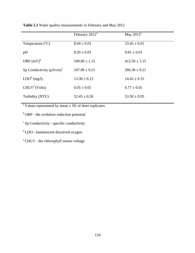
124
Table 5.1 Water quality measurements in February and May 2012
February 2012a May 2012
a
Temperature (°C) 8.04 ± 0.01 23.45 ± 0.01
pH 8.20 ± 0.01 9.81 ± 0.01
ORP (mV)b 549.00 ± 1.15 412.50 ± 1.15
Sp Conductivity (µS/cm)c 247.90 ± 0.21 266.30 ± 0.21
LDOd (mg/l) 13.38 ± 0.15 14.43 ± 0.15
CHLVe (Volts) 0.05 ± 0.01 0.77 ± 0.01
Turbidity (NTU) 52.43 ± 0.26 53.30 ± 0.05
a Values represented by mean ± SE of three replicates
b ORP - the oxidation reduction potential
c Sp Conductivity - specific conductivity
d LDO - luminescent dissolved oxygen
e CHLV - the chlorophyll sensor voltage

125
Table 5.2 Total colony-forming units per plate on nutrient agar as affected by temperature a, b
Treatment (°C) February 2012 May 2012
25 (control) 349.33 ± 26.72 347.67 ± 18.32
42 401 ± 5.57 471.33 ± 45.62
48 155 ± 2 127 ± 16.46
a Values represented by mean ± SE of three replicates
b A volume of 100 µl water plated on each nutrient agar plate
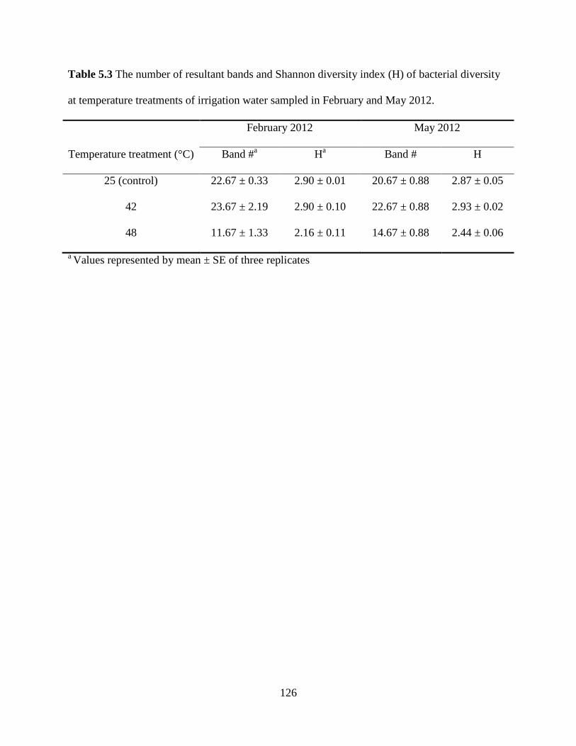
126
Table 5.3 The number of resultant bands and Shannon diversity index (H) of bacterial diversity
at temperature treatments of irrigation water sampled in February and May 2012.
February 2012 May 2012
Temperature treatment (°C) Band #a H
a Band # H
25 (control) 22.67 ± 0.33 2.90 ± 0.01 20.67 ± 0.88 2.87 ± 0.05
42 23.67 ± 2.19 2.90 ± 0.10 22.67 ± 0.88 2.93 ± 0.02
48 11.67 ± 1.33 2.16 ± 0.11 14.67 ± 0.88 2.44 ± 0.06
a Values represented by mean ± SE of three replicates
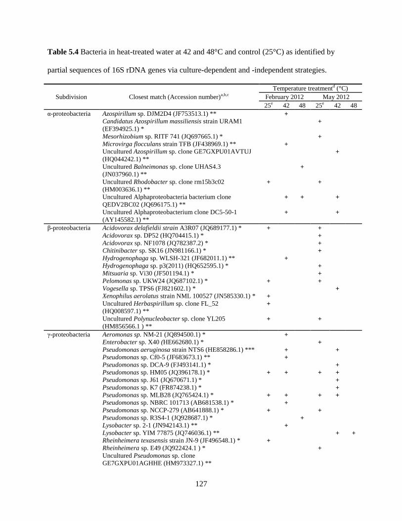
127
Table 5.4 Bacteria in heat-treated water at 42 and 48°C and control (25°C) as identified by
partial sequences of 16S rDNA genes via culture-dependent and -independent strategies.
Subdivision Closest match (Accession number)a,b,c
Temperature treatmentd (°C)
February 2012 May 2012
25e 42 48 25
e 42 48
α-proteobacteria Azospirillum sp. DJM2D4 (JF753513.1) ** +
Candidatus Azospirillum massiliensis strain URAM1
(EF394925.1) * +
Mesorhizobium sp. RITF 741 (JQ697665.1) * +
Microvirga flocculans strain TFB (JF438969.1) ** +
Uncultured Azospirillum sp. clone GE7GXPU01AVTUJ
(HQ044242.1) ** +
Uncultured Balneimonas sp. clone UHAS4.3
(JN037960.1) ** +
Uncultured Rhodobacter sp. clone rm15b3c02
(HM003636.1) ** + +
Uncultured Alphaproteobacteria bacterium clone
QEDV2BC02 (JQ696175.1) ** + + +
Uncultured Alphaproteobacterium clone DC5-50-1
(AY145582.1) ** + +
β-proteobacteria Acidovorax delafieldii strain A3R07 (JQ689177.1) * + +
Acidovorax sp. DP52 (HQ704415.1) * +
Acidovorax sp. NF1078 (JQ782387.2) * +
Chitinibacter sp. SK16 (JN981166.1) * +
Hydrogenophaga sp. WLSH-321 (JF682011.1) ** +
Hydrogenophaga sp. p3(2011) (HQ652595.1) * +
Mitsuaria sp. Vi30 (JF501194.1) * +
Pelomonas sp. UKW24 (JQ687102.1) * + +
Vogesella sp. TPS6 (FJ821602.1) * +
Xenophilus aerolatus strain NML 100527 (JN585330.1) * +
Uncultured Herbaspirillum sp. clone FL_52
(HQ008597.1) ** +
Uncultured Polynucleobacter sp. clone YL205
(HM856566.1 ) ** + +
γ-proteobacteria Aeromonas sp. NM-21 (JQ894500.1) * +
Enterobacter sp. X40 (HE662680.1) * +
Pseudomonas aeruginosa strain NTS6 (HE858286.1) *** + +
Pseudomonas sp. Cf0-5 (JF683673.1) ** +
Pseudomonas sp. DCA-9 (FJ493141.1) * +
Pseudomonas sp. HM05 (JQ396178.1) * + + + +
Pseudomonas sp. J61 (JQ670671.1) * +
Pseudomonas sp. K7 (FR874238.1) * +
Pseudomonas sp. MLB28 (JQ765424.1) * + + + +
Pseudomonas sp. NBRC 101713 (AB681538.1) * +
Pseudomonas sp. NCCP-279 (AB641888.1) * + +
Pseudomonas sp. R3S4-1 (JQ928687.1) * +
Lysobacter sp. 2-1 (JN942143.1) ** +
Lysobacter sp. YIM 77875 (JQ746036.1) ** + +
Rheinheimera texasensis strain JN-9 (JF496548.1) * +
Rheinheimera sp. E49 (JQ922424.1 ) * +
Uncultured Pseudomonas sp. clone
GE7GXPU01AGHHE (HM973327.1) **
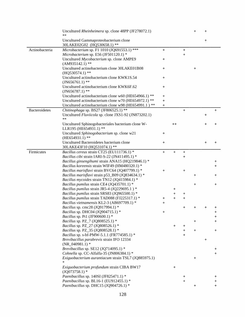
128
Uncultured Rheinheimera sp. clone 48PP (JF278072.1)
** + +
Uncultured Gammaproteobacterium clone
30LAKE02G02 (HQ530658.1) ** +
Actinobacteria Microbacterium sp. F1 1010 (JQ691553.1) *** + +
Microbacterium sp. E56 (JF501120.1) * +
Uncultured Mycobacterium sp. clone AMPE9
(AM935142.1) ** +
Uncultured actinobacterium clone 30LAKE01B08
(HQ530574.1) ** + +
Uncultured actinobacterium clone KWK1S.54
(JN656761.1) ** +
Uncultured actinobacterium clone KWK6F.62
(JN656787.1) ** +
Uncultured actinobacterium clone w60 (HE654966.1) ** +
Uncultured actinobacterium clone w70 (HE654972.1) ** +
Uncultured actinobacterium clone w98 (HE654991.1 ) ** +
Bacteroidetes Chitinophaga sp. BS27 (JF806525.1) ** + +
Uncultured Fluviicola sp. clone JXS1-92 (JN873202.1)
** +
Uncultured Sphinogobacteriales bacterium clone W-
LLR195 (HE654931.1) ** ++ + +
Uncultured Sphinogobacterium sp. clone w21
(HE654931.1) ** +
Uncultured Bacteroidetes bacterium clone
30LAKE43F10 (HQ531074.1) ** + + +
Firmicutes Bacillus cereus strain CT25 (EU111736.1) * + + +
Bacillus cibi strain IARI-S-22 (JN411495.1) * +
Bacillus ginsengihumi strain ANA15 (HQ219846.1) *
+
Bacillus koreensis strain WIF49 (HM480320.1) *
+
Bacillus marisflavi strain BVC64 (JQ407799.1) * + +
Bacillus marisflavi strain p53_B09 (JQ834634.1) * + +
Bacillus mycoides strain TN12 (JQ415984.1) *
Bacillus pumilus strain CE4 (JQ435701.1) * + +
Bacillus pumilus strain JR5-4 (JQ229695.1 ) * +
Bacillus pumilus strain SRS83 (JQ965500.1) * + +
Bacillus pumilus strain TAD088 (FJ225317.1) * + + +
Bacillus vietnamensis KL2-3 (AB697709.1) * +
Bacillus sp. cmc28 (JQ917994.1) * + + + +
Bacillus sp. DHC04 (JQ904715.1) * + +
Bacillus sp. Pt1 (JF900600.1) * +
Bacillus sp. PZ_7 (JQ808525.1) * + +
Bacillus sp. PZ_27 (JQ808526.1) * + +
Bacillus sp. PZ_35 (JQ808528.1) * + + +
Bacillus sp. s-bf-PMW-5.1.1 (FR774585.1) * +
Brevibacillus parabrevis strain IFO 12334
(NR_040981.1) * +
Brevibacillus sp. SE12 (JQ714095.1) * +
Cohnella sp. CC-Alfalfa-35 (JN806384.1) * +
Exiguobacterium aurantiacum strain TSL7 (JQ885975.1)
* +
Exiguobacterium profundum strain CIBA BW17
(JQ073758.1) * +
Paenibacillus sp. 140SI (JF825471.1) * + +
Paenibacillus sp. BL16-1 (EU912455.1) * + +
Paenibacillus sp. DHC15 (JQ904726.1) * + +

129
Paenibacillus sp. Dv34 (HQ728085.1) * +
Paenibacillus sp. KMC003 (EU219972.1) * +
Paenibacillus sp. MN2-8 (JQ396530.1) * +
Paenibacillus sp. WYT011 (JQ807858.1) * +
Paenibacillus terrigena strain B30 (JN252080.1) * + +
Paenibacillus timonensis strain 3584BRRJ (JF309265.1)
* +
a Sequences aligned to their closest matches in GenBank using BLAST.
b All the closest relatives of sequences with a similarity ≥ 96%.
c * indicating detection via culture-dependent strategy, ** indicating detection via culture-
independent strategy, *** indicating detection via both strategies
d + indicating the presence of the bacterial identity
e 25°C as control
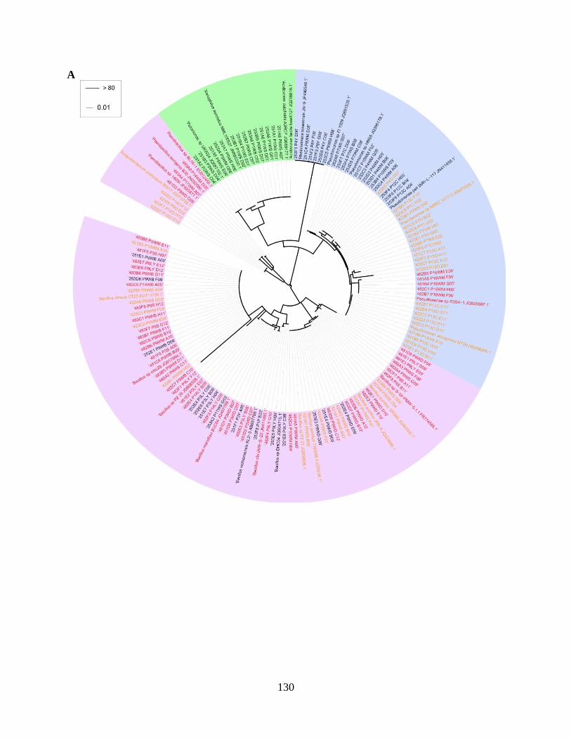
130
A

131
Figure 5.1 Phylogenetic analysis of partial 16S rDNA sequences of representative bacterial
isolates from irrigation water samples collected in (A) February and (B) May 2012 after heat
treatments at 25 (as control, black), 42 (orange), and 48°C (red). The Maximum Likelihood
method based on the Tamura-Nei model was used. Bootstrap analyses were based on 1000
replicates. Different color shadings indicate various subdivisions of bacteria: blue, γ-
B

132
proteobacteria; green, β-proteobacteria; yellow, α-proteobacteria, purple, Firmicutes; and orange,
Actinobacteria.

133
Figure 5.2 DGGE fingerprinting of bacterial 16S rRNA fragments in irrigation water sampled in
A) February and B) May 2012 after heat treatments at 25 (control), 42, and 48°C. The DGGE
was run at70 V for 16 h with a gel denaturant concentration of 40 to 60%. Red arrows represent
selected bands. L represents 100 bp DNA ladder.
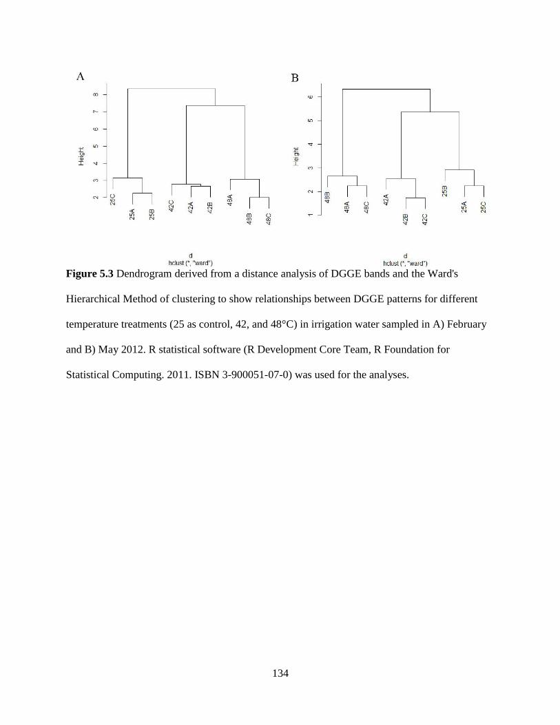
134
Figure 5.3 Dendrogram derived from a distance analysis of DGGE bands and the Ward's
Hierarchical Method of clustering to show relationships between DGGE patterns for different
temperature treatments (25 as control, 42, and 48°C) in irrigation water sampled in A) February
and B) May 2012. R statistical software (R Development Core Team, R Foundation for
Statistical Computing. 2011. ISBN 3-900051-07-0) was used for the analyses.
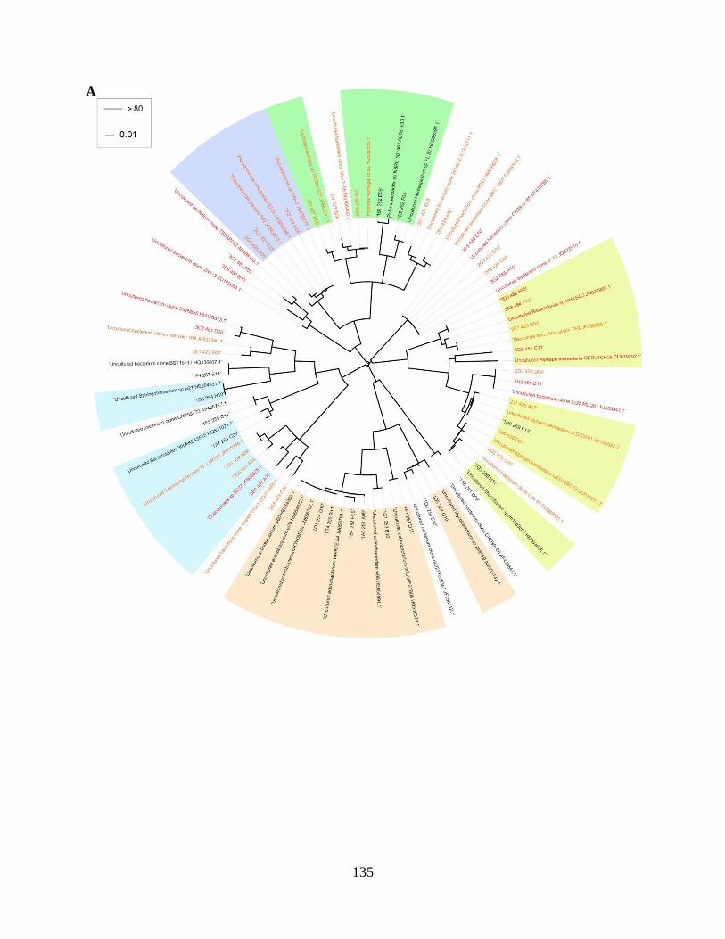
135
A
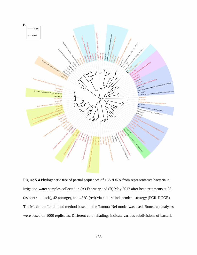
136
Figure 5.4 Phylogenetic tree of partial sequences of 16S rDNA from representative bacteria in
irrigation water samples collected in (A) February and (B) May 2012 after heat treatments at 25
(as control, black), 42 (orange), and 48°C (red) via culture-independent strategy (PCR-DGGE).
The Maximum Likelihood method based on the Tamura-Nei model was used. Bootstrap analyses
were based on 1000 replicates. Different color shadings indicate various subdivisions of bacteria:
B

137
blue, γ-proteobacteria; green, β-proteobacteria; yellow, α-proteobacteria; purple, Firmicutes;
orange, Actinobacteria; light blue, Bacteroidetes; unshaded, unknown bacterial species.
138
Chapter 6
Summary and future directions
Heat treatment is a reliable and effective decontamination technology for re-circulated
irrigation water in the ornamental horticulture industry (van Os et al. 1988; Hong and Moorman
2005; Hao et al. 2012). The current protocol recommends raising water temperature to 95°C and
maintaining it at that temperature for 30 s (Runia et al. 1988; McPherson et al. 1995). The major
limitations associated with this water treatment are its energy inefficiency, high costs, and
environmental footprint (Poncet et al. 2001; Hong and Moorman 2005). The overall goal of this
dissertation was to make this technology economically more attractive while reducing its
environmental impact. We hypothesized that the temperature required to inactivate major
pathogens in re-circulated water may be substantially lowered from 95°C. The specific
objectives of this dissertation were to test the above hypothesis, including investigation into
biological mechanisms by which plant pathogens are killed at the substantially lowered water
temperature.
The effect of water temperature on the survival of Phytophthora and bacterial species,
two major groups of plant pathogens in re-circulating water systems, was examined.
Phytophthora species, known as water molds, are the most frequently isolated plant pathogens in
irrigation water (Hong and Moorman 2005; Gallegly and Hong 2008). P. nicotianae, a high-
temperature tolerant species, was used as a major model system in this study. Heat treatments at
42°C for 24 h or at 48°C for 6 h inactivated the major life stages of Phytophthora species in the
laboratory and greenhouse tests. It is expected that the heat and exposure time combination that
139
worked for P. nicotianae would work for a vast majority, if not all, oomycete pathogens in
irrigation water. Heat treatment at 48°C for 24 h inactivated all seven tested bacterial species,
some of which are the most common bacterial plant pathogens found in irrigation water (Hong
and Moorman 2005). These results indicated that raising water temperature to 48°C and
maintaining it for 24 h could be an alternative to the current heat treatment protocol which uses
95°C for 30 s. Use of a lower temperature with a longer exposure time could substantially reduce
energy consumption, consequently improve the economics of heat treatment, and hence the
profitability of the ornamental horticulture industry while reducing its environmental footprint.
Two steps were taken to elucidate the underlying biological mechanisms of pathogen
removal at the substantially lowered water temperature. Firstly, a water sample concentration and
processing scheme was developed for typing bacterial species in irrigation water samples. The
key component of this scheme to detect the greatest diversity of bacteria in irrigation water
samples were to (1) employ both culture-dependent and -independent technologies as each
detects a subset of bacteria, (2) use both centrifugation and filtration to concentrate water
samples and (3) perform PCR-DGGE with 40 to 60% denaturant at 70 V for 16 h. Secondly,
water samples were taken from an irrigation reservoir in a local nursery and analyzed for
bacterial diversity following heat treatments at 42 and 48°C. A substantial shift in the dominant
bacterial populations were found at 42 and 48°C when compared with the control at 25°C. Of the
most significance were γ-proteobacteria and Firmicutes becoming dominant in heat-treated
water. These included Bacillus, Pseudomonas, Paenibacillus, Brevibacillus, and Lysobacter
species, which are known to have a great potential as biological control agents. For example,
Bacillus and Pseudomonas species have been investigated extensively as biocontrol agents
(Punja 1997). Some species and isolates in the genera Paenibacillus, Brevibacillus, and
140
Lysobacter have also been reported to biologically control a number of plant pathogens
(Christensen and Cook 1978; Singh et al. 1999; Kobayashi and Crouch 2009; Saikia et al. 2011).
These results indicated that bacterial community shifts may have contributed to the pathogen
removal under heat treatments at 42 and 48°C.
To substantiate the findings from the above studies, additional investigations are needed
to evaluate the new heat treatment temperatures at production facilities, identify alternative
energy generation and retention sources. To further elucidate its underlying biological
mechanisms additional experiments will need to determine the biological control activities of
selected bacterial species and isolates. Moreover, investigation into the abundance and frequency
of these bacterial species in irrigation water at multiple locations also are warranted.
In summary, this dissertation provided the scientific basis for developing a more energy-
efficient and environmentally sound heat treatment for re-circulated irrigation water.
References
Christensen, P. & Cook, F. D. (1978). Lysobacter new genus of nonfruiting gliding bacteria with
a high base ratio. International Journal of Systematic Bacteriology, 28 (3), 367-393
Gallegly, M. E. & Hong, C. X. (2008). Phytophthora: Identifying Species by Morphology and
DNA Fingerprints. (St. Paul, MN, USA: APS Press)
Hao, W., Ahonsi, M. O., Vinatzer, B. A. & Hong, C. X. (2012). Inactivation of Phytophthora and
bacterial species in water by a potential energy-saving heat treatment. European Journal
of Plant Pathology, 134 (2), 357-365
Hong, C. X. & Moorman, G. W. (2005). Plant pathogens in irrigation water: Challenges and
opportunities. Critical Reviews in Plant Sciences, 24 (3), 189-208
141
Kobayashi, D. Y. & Crouch, J. A. (2009). Bacterial/fungal interactions: from pathogens to
mutualistic endosymbionts. Annual Review of Phytopathology, 47, 63-82
McPherson, G. M., Harriman, M. R. & Pattison, D. (1995). The potential for spread of root
diseases in recirculating hydroponic systems and their control with disinfection.
Mededelingen Faculteit Landbouwkundige en Toegepaste Biologische Wetenschappen
Universiteit Gent, 60 (28), 371-379
Poncet, C., Offroy, M. & Bonnet, G. (2001). Disinfection of recycling water in rose cultures.
Acta Horticulturae, 547, 121-126
Punja, Z. K. (1997). Comparative efficacy of bacteria, fungi, and yeasts as biological control
agents for diseases of vegetable crops. Canadian Journal of Plant Pathology, 19 (3), 315-
323
Runia, W. T., Vanos, E. A. & Bollen, G. J. (1988). Disinfection of drainwater from soilless
cultures by heat-treatment. Netherlands Journal of Agricultural Science, 36 (3), 231-238
Saikia, R., Gogoi, D. K., Mazumder, S., Yadav, A., Sarma, R. K., Bora, T. C. & Gogoi, B. K.
(2011). Brevibacillus laterosporus strain BPM3, a potential biocontrol agent isolated
from a natural hot water spring of Assam, India. Microbiological Research, 166 (3), 216-
225
Singh, P. P., Shin, Y. C., Park, C. S. & Chung, Y. R. (1999). Biological control of fusarium wilt
of cucumber by chitinolytic bacteria. Phytopathology, 89 (1), 92-99
van Os, E. A., van de Braak, N. J. & Klomp, G. Heat treatment for disinfecting drainwater,
Technical and economic aspects. In: Proceedings of the Seventh International Congress
on Soilless Culture, Flevohof, The Netherlands, 1988. pp 353-359
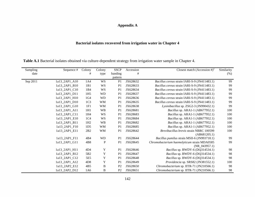
142
Appendix A
Bacterial isolates recovered from irrigation water in Chapter 4
Table A.1 Bacterial isolates obtained via culture-dependent strategy from irrigation water sample in Chapter 4.
Sampling
date
Sequence # Colony
#
Colony
type
SSCP
banding
pattern
Accession
#
Closest match (Accession #)a Similarity
(%)
Sep 2011 1a13_2AP1_A10 1A4 WS P1 JX628632 Bacillus cereus strain IARI-S-9 (JN411483.1) 99
1a13_2AP1_B10 1B1 WS P1 JX628633 Bacillus cereus strain IARI-S-9 (JN411483.1) 99
1a13_2AP1_C10 1B4 WS P1 JX628634 Bacillus cereus strain IARI-S-9 (JN411483.1) 99
1a13_2AP1_D11 1H5 WD P1 JX628637 Bacillus cereus strain IARI-S-9 (JN411483.1) 99
1a13_2AP1_H10 1G4 WD P1 JX628636 Bacillus cereus strain IARI-S-9 (JN411483.1) 99
1a13_2AP1_D10 1C3 WM P1 JX628635 Bacillus cereus strain IARI-S-9 (JN411483.1) 99
1a13_2AP1_G10 1F1 WM P1 JX628638 Lysinibacillus sp. ZSG2-3 (JN990432.1) 99
1a13_2AP1_A11 1H1 WB P1 JX628681 Bacillus sp. ARA1-1 (AB677952.1) 100
1a13_2AP1_C11 1H4 WS P1 JX628683 Bacillus sp. ARA1-1 (AB677952.1) 100
1a13_2AP1_E10 1C4 WS P1 JX628684 Bacillus sp. ARA1-1 (AB677952.1) 100
1a13_2AP1_B11 1H2 WB P1 JX628682 Bacillus sp. ARA1-1 (AB677952.1) 100
1a13_2AP1_F10 1D5 WM P1 JX628685 Bacillus sp. ARA1-1 (AB677952.1) 100
1a13_2AP1_E11 2B2 WM P1 JX628642 Brevibacillus brevis strain NBRC 100599
(AB681205.1)
100
1a13_2AP1_F11 4B4 WD P1 JX628644 Bacillus pumilus strain MSII-6 (JN993718.1) 99
1a13_2AP1_G11 4B8 P P1 JX628645 Chromobacterium haemolyticum strain MDA0585
(NR_043957.1)
99
1a13_2AP1_H11 4D4 Y P1 JX628646 Bacillus sp. BWDY-6 (DQ314534.1) 98
1a13_2AP1_B12 5B2 Y P1 JX628647 Bacillus sp. BWDY-6 (DQ314534.1) 98
1a13_2AP1_C12 5E5 Y P1 JX628648 Bacillus sp. BWDY-6 (DQ314534.1) 98
1a13_2AP1_A12 4D8 Y P1 JX628649 Providencia sp. SRS82 (JN381552.1) 100
1a13_2AP2_E12 4B5 B P2 JX628650 Chromobacterium sp. IITR-71 (JN210566.1) 98
1a13_2AP2_D12 1A6 B P2 JX628651 Chromobacterium sp. IITR-71 (JN210566.1) 98
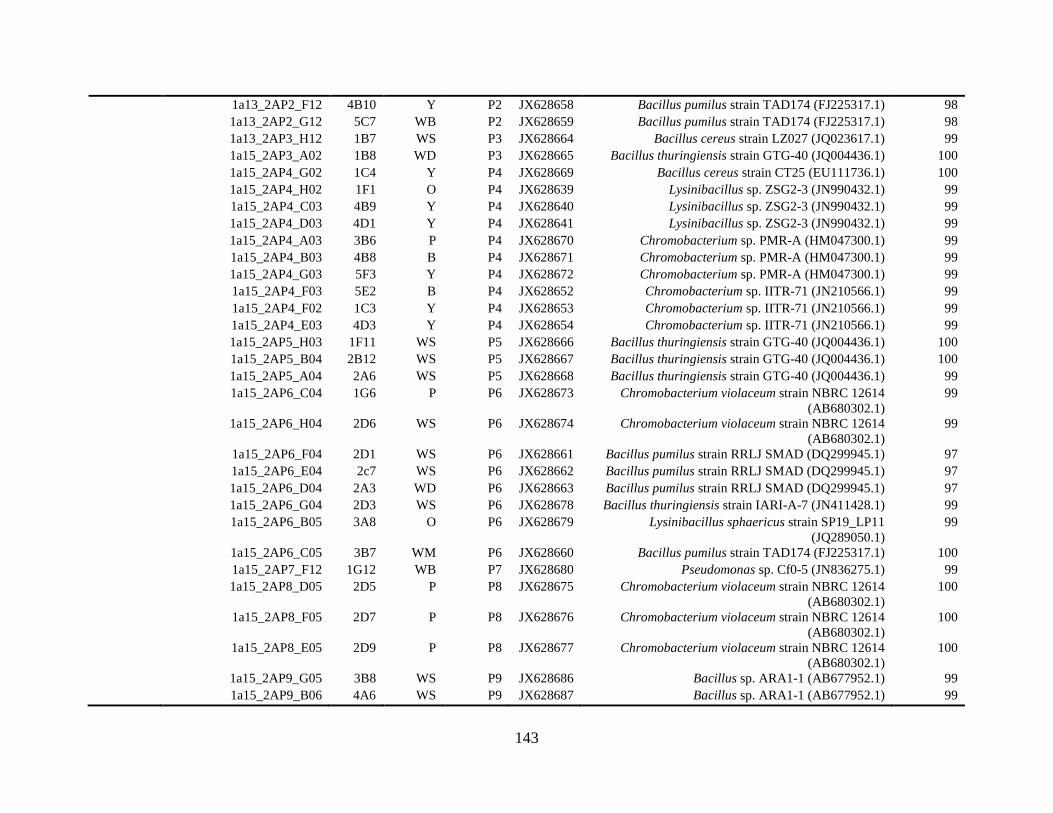
143
1a13_2AP2_F12 4B10 Y P2 JX628658 Bacillus pumilus strain TAD174 (FJ225317.1) 98
1a13_2AP2_G12 5C7 WB P2 JX628659 Bacillus pumilus strain TAD174 (FJ225317.1) 98
1a13_2AP3_H12 1B7 WS P3 JX628664 Bacillus cereus strain LZ027 (JQ023617.1) 99
1a15_2AP3_A02 1B8 WD P3 JX628665 Bacillus thuringiensis strain GTG-40 (JQ004436.1) 100
1a15_2AP4_G02 1C4 Y P4 JX628669 Bacillus cereus strain CT25 (EU111736.1) 100
1a15_2AP4_H02 1F1 O P4 JX628639 Lysinibacillus sp. ZSG2-3 (JN990432.1) 99
1a15_2AP4_C03 4B9 Y P4 JX628640 Lysinibacillus sp. ZSG2-3 (JN990432.1) 99
1a15_2AP4_D03 4D1 Y P4 JX628641 Lysinibacillus sp. ZSG2-3 (JN990432.1) 99
1a15_2AP4_A03 3B6 P P4 JX628670 Chromobacterium sp. PMR-A (HM047300.1) 99
1a15_2AP4_B03 4B8 B P4 JX628671 Chromobacterium sp. PMR-A (HM047300.1) 99
1a15_2AP4_G03 5F3 Y P4 JX628672 Chromobacterium sp. PMR-A (HM047300.1) 99
1a15_2AP4_F03 5E2 B P4 JX628652 Chromobacterium sp. IITR-71 (JN210566.1) 99
1a15_2AP4_F02 1C3 Y P4 JX628653 Chromobacterium sp. IITR-71 (JN210566.1) 99
1a15_2AP4_E03 4D3 Y P4 JX628654 Chromobacterium sp. IITR-71 (JN210566.1) 99
1a15_2AP5_H03 1F11 WS P5 JX628666 Bacillus thuringiensis strain GTG-40 (JQ004436.1) 100
1a15_2AP5_B04 2B12 WS P5 JX628667 Bacillus thuringiensis strain GTG-40 (JQ004436.1) 100
1a15_2AP5_A04 2A6 WS P5 JX628668 Bacillus thuringiensis strain GTG-40 (JQ004436.1) 99
1a15_2AP6_C04 1G6 P P6 JX628673 Chromobacterium violaceum strain NBRC 12614
(AB680302.1)
99
1a15_2AP6_H04 2D6 WS P6 JX628674 Chromobacterium violaceum strain NBRC 12614
(AB680302.1)
99
1a15_2AP6_F04 2D1 WS P6 JX628661 Bacillus pumilus strain RRLJ SMAD (DQ299945.1) 97
1a15_2AP6_E04 2c7 WS P6 JX628662 Bacillus pumilus strain RRLJ SMAD (DQ299945.1) 97
1a15_2AP6_D04 2A3 WD P6 JX628663 Bacillus pumilus strain RRLJ SMAD (DQ299945.1) 97
1a15_2AP6_G04 2D3 WS P6 JX628678 Bacillus thuringiensis strain IARI-A-7 (JN411428.1) 99
1a15_2AP6_B05 3A8 O P6 JX628679 Lysinibacillus sphaericus strain SP19_LP11
(JQ289050.1)
99
1a15_2AP6_C05 3B7 WM P6 JX628660 Bacillus pumilus strain TAD174 (FJ225317.1) 100
1a15_2AP7_F12 1G12 WB P7 JX628680 Pseudomonas sp. Cf0-5 (JN836275.1) 99
1a15_2AP8_D05 2D5 P P8 JX628675 Chromobacterium violaceum strain NBRC 12614
(AB680302.1)
100
1a15_2AP8_F05 2D7 P P8 JX628676 Chromobacterium violaceum strain NBRC 12614
(AB680302.1)
100
1a15_2AP8_E05 2D9 P P8 JX628677 Chromobacterium violaceum strain NBRC 12614
(AB680302.1)
100
1a15_2AP9_G05 3B8 WS P9 JX628686 Bacillus sp. ARA1-1 (AB677952.1) 99
1a15_2AP9_B06 4A6 WS P9 JX628687 Bacillus sp. ARA1-1 (AB677952.1) 99
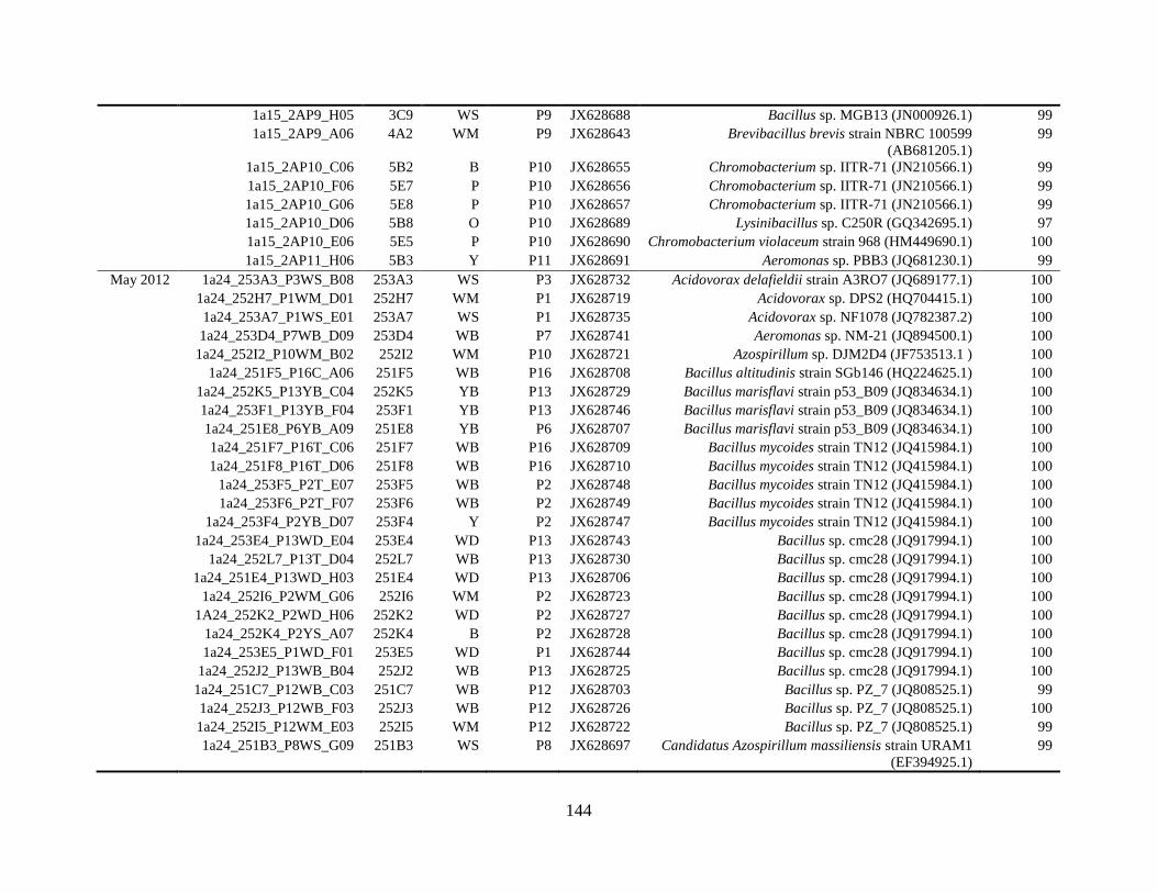
144
1a15_2AP9_H05 3C9 WS P9 JX628688 Bacillus sp. MGB13 (JN000926.1) 99
1a15_2AP9_A06 4A2 WM P9 JX628643 Brevibacillus brevis strain NBRC 100599
(AB681205.1)
99
1a15_2AP10_C06 5B2 B P10 JX628655 Chromobacterium sp. IITR-71 (JN210566.1) 99
1a15_2AP10_F06 5E7 P P10 JX628656 Chromobacterium sp. IITR-71 (JN210566.1) 99
1a15_2AP10_G06 5E8 P P10 JX628657 Chromobacterium sp. IITR-71 (JN210566.1) 99
1a15_2AP10_D06 5B8 O P10 JX628689 Lysinibacillus sp. C250R (GQ342695.1) 97
1a15_2AP10_E06 5E5 P P10 JX628690 Chromobacterium violaceum strain 968 (HM449690.1) 100
1a15_2AP11_H06 5B3 Y P11 JX628691 Aeromonas sp. PBB3 (JQ681230.1) 99
May 2012 1a24_253A3_P3WS_B08 253A3 WS P3 JX628732 Acidovorax delafieldii strain A3RO7 (JQ689177.1) 100
1a24_252H7_P1WM_D01 252H7 WM P1 JX628719 Acidovorax sp. DPS2 (HQ704415.1) 100
1a24_253A7_P1WS_E01 253A7 WS P1 JX628735 Acidovorax sp. NF1078 (JQ782387.2) 100
1a24_253D4_P7WB_D09 253D4 WB P7 JX628741 Aeromonas sp. NM-21 (JQ894500.1) 100
1a24_252I2_P10WM_B02 252I2 WM P10 JX628721 Azospirillum sp. DJM2D4 (JF753513.1 ) 100
1a24_251F5_P16C_A06 251F5 WB P16 JX628708 Bacillus altitudinis strain SGb146 (HQ224625.1) 100
1a24_252K5_P13YB_C04 252K5 YB P13 JX628729 Bacillus marisflavi strain p53_B09 (JQ834634.1) 100
1a24_253F1_P13YB_F04 253F1 YB P13 JX628746 Bacillus marisflavi strain p53_B09 (JQ834634.1) 100
1a24_251E8_P6YB_A09 251E8 YB P6 JX628707 Bacillus marisflavi strain p53_B09 (JQ834634.1) 100
1a24_251F7_P16T_C06 251F7 WB P16 JX628709 Bacillus mycoides strain TN12 (JQ415984.1) 100
1a24_251F8_P16T_D06 251F8 WB P16 JX628710 Bacillus mycoides strain TN12 (JQ415984.1) 100
1a24_253F5_P2T_E07 253F5 WB P2 JX628748 Bacillus mycoides strain TN12 (JQ415984.1) 100
1a24_253F6_P2T_F07 253F6 WB P2 JX628749 Bacillus mycoides strain TN12 (JQ415984.1) 100
1a24_253F4_P2YB_D07 253F4 Y P2 JX628747 Bacillus mycoides strain TN12 (JQ415984.1) 100
1a24_253E4_P13WD_E04 253E4 WD P13 JX628743 Bacillus sp. cmc28 (JQ917994.1) 100
1a24_252L7_P13T_D04 252L7 WB P13 JX628730 Bacillus sp. cmc28 (JQ917994.1) 100
1a24_251E4_P13WD_H03 251E4 WD P13 JX628706 Bacillus sp. cmc28 (JQ917994.1) 100
1a24_252I6_P2WM_G06 252I6 WM P2 JX628723 Bacillus sp. cmc28 (JQ917994.1) 100
1A24_252K2_P2WD_H06 252K2 WD P2 JX628727 Bacillus sp. cmc28 (JQ917994.1) 100
1a24_252K4_P2YS_A07 252K4 B P2 JX628728 Bacillus sp. cmc28 (JQ917994.1) 100
1a24_253E5_P1WD_F01 253E5 WD P1 JX628744 Bacillus sp. cmc28 (JQ917994.1) 100
1a24_252J2_P13WB_B04 252J2 WB P13 JX628725 Bacillus sp. cmc28 (JQ917994.1) 100
1a24_251C7_P12WB_C03 251C7 WB P12 JX628703 Bacillus sp. PZ_7 (JQ808525.1) 99
1a24_252J3_P12WB_F03 252J3 WB P12 JX628726 Bacillus sp. PZ_7 (JQ808525.1) 100
1a24_252I5_P12WM_E03 252I5 WM P12 JX628722 Bacillus sp. PZ_7 (JQ808525.1) 99
1a24_251B3_P8WS_G09 251B3 WS P8 JX628697 Candidatus Azospirillum massiliensis strain URAM1
(EF394925.1)
99
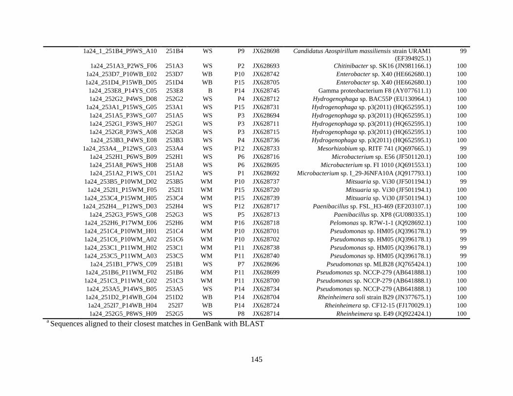
145
1a24_1_251B4_P9WS_A10 251B4 WS P9 JX628698 Candidatus Azospirillum massiliensis strain URAM1
(EF394925.1)
99
1a24_251A3_P2WS_F06 251A3 WS P2 JX628693 Chitinibacter sp. SK16 (JN981166.1) 100
1a24_253D7_P10WB_E02 253D7 WB P10 JX628742 Enterobacter sp. X40 (HE662680.1) 100
1a24_251D4_P15WB_D05 251D4 WB P15 JX628705 Enterobacter sp. X40 (HE662680.1) 100
1a24_253E8_P14YS_C05 253E8 B P14 JX628745 Gamma proteobacterium F8 (AY077611.1) 100
1a24_252G2_P4WS_D08 252G2 WS P4 JX628712 Hydrogenophaga sp. BAC55P (EU130964.1) 100
1a24_253A1_P15WS_G05 253A1 WS P15 JX628731 Hydrogenophaga sp. p3(2011) (HQ652595.1) 100
1a24_251A5_P3WS_G07 251A5 WS P3 JX628694 Hydrogenophaga sp. p3(2011) (HQ652595.1) 100
1a24_252G1_P3WS_H07 252G1 WS P3 JX628711 Hydrogenophaga sp. p3(2011) (HQ652595.1) 100
1a24_252G8_P3WS_A08 252G8 WS P3 JX628715 Hydrogenophaga sp. p3(2011) (HQ652595.1) 100
1a24_253B3_P4WS_E08 253B3 WS P4 JX628736 Hydrogenophaga sp. p3(2011) (HQ652595.1) 100
1a24_253A4__P12WS_G03 253A4 WS P12 JX628733 Mesorhizobium sp. RITF 741 (JQ697665.1) 99
1a24_252H1_P6WS_B09 252H1 WS P6 JX628716 Microbacterium sp. E56 (JF501120.1) 100
1a24_251A8_P6WS_H08 251A8 WS P6 JX628695 Microbacterium sp. FI 1010 (JQ691553.1) 100
1a24_251A2_P1WS_C01 251A2 WS P1 JX628692 Microbacterium sp. I_29-J6NFA10A (JQ917793.1) 100
1a24_253B5_P10WM_D02 253B5 WM P10 JX628737 Mitsuaria sp. Vi30 (JF501194.1) 99
1a24_252I1_P15WM_F05 252I1 WM P15 JX628720 Mitsuaria sp. Vi30 (JF501194.1) 100
1a24_253C4_P15WM_H05 253C4 WM P15 JX628739 Mitsuaria sp. Vi30 (JF501194.1) 100
1a24_252H4__P12WS_D03 252H4 WS P12 JX628717 Paenibacillus sp. FSL_H3-469 (EF203107.1) 100
1a24_252G3_P5WS_G08 252G3 WS P5 JX628713 Paenibacillus sp. XP8 (GU080335.1) 100
1a24_252H6_P17WM_E06 252H6 WM P16 JX628718 Pelomonas sp. R7W-1-1 (JQ928692.1) 100
1a24_251C4_P10WM_H01 251C4 WM P10 JX628701 Pseudomonas sp. HM05 (JQ396178.1) 99
1a24_251C6_P10WM_A02 251C6 WM P10 JX628702 Pseudomonas sp. HM05 (JQ396178.1) 99
1a24_253C1_P11WM_H02 253C1 WM P11 JX628738 Pseudomonas sp. HM05 (JQ396178.1) 99
1a24_253C5_P11WM_A03 253C5 WM P11 JX628740 Pseudomonas sp. HM05 (JQ396178.1) 99
1a24_251B1_P7WS_C09 251B1 WS P7 JX628696 Pseudomonas sp. MLB28 (JQ765424.1) 100
1a24_251B6_P11WM_F02 251B6 WM P11 JX628699 Pseudomonas sp. NCCP-279 (AB641888.1) 100
1a24_251C3_P11WM_G02 251C3 WM P11 JX628700 Pseudomonas sp. NCCP-279 (AB641888.1) 100
1a24_253A5_P14WS_B05 253A5 WS P14 JX628734 Pseudomonas sp. NCCP-279 (AB641888.1) 100
1a24_251D2_P14WB_G04 251D2 WB P14 JX628704 Rheinheimera soli strain B29 (JN377675.1) 100
1a24_252I7_P14WB_H04 252I7 WB P14 JX628724 Rheinheimera sp. CF12-15 (FJ170029.1) 100
1a24_252G5_P8WS_H09 252G5 WS P8 JX628714 Rheinheimera sp. E49 (JQ922424.1) 100 a Sequences aligned to their closest matches in GenBank with BLAST
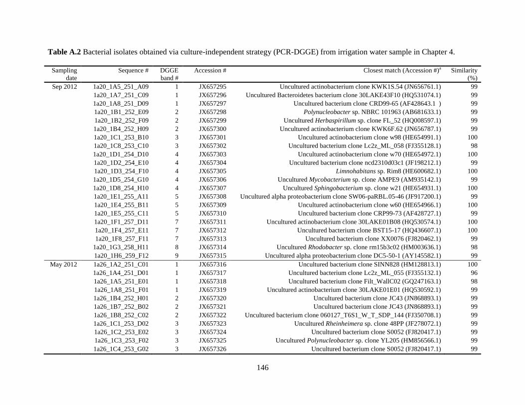
146
Table A.2 Bacterial isolates obtained via culture-independent strategy (PCR-DGGE) from irrigation water sample in Chapter 4.
Sampling
date
Sequence # DGGE
band #
Accession # Closest match (Accession #)a Similarity
(%)
Sep 2012 1a20_1A5_251_A09 1 JX657295 Uncultured actinobacterium clone KWK1S.54 (JN656761.1) 99
1a20_1A7_251_C09 1 JX657296 Uncultured Bacteroidetes bacterium clone 30LAKE43F10 (HQ531074.1) 99
1a20_1A8_251_D09 1 JX657297 Uncultured bacterium clone CRD99-65 (AF428643.1 ) 99
1a20_1B1_252_E09 2 JX657298 Polynucleobacter sp. NBRC 101963 (AB681633.1) 99
1a20_1B2_252_F09 2 JX657299 Uncultured Herbaspirillum sp. clone FL_52 (HQ008597.1) 99
1a20_1B4_252_H09 2 JX657300 Uncultured actinobacterium clone KWK6F.62 (JN656787.1) 99
1a20_1C1_253_B10 3 JX657301 Uncultured actinobacterium clone w98 (HE654991.1) 100
1a20_1C8_253_C10 3 JX657302 Uncultured bacterium clone Lc2z_ML_058 (FJ355128.1) 98
1a20_1D1_254_D10 4 JX657303 Uncultured actinobacterium clone w70 (HE654972.1) 100
1a20_1D2_254_E10 4 JX657304 Uncultured bacterium clone ncd2310d03c1 (JF198212.1) 99
1a20_1D3_254_F10 4 JX657305 Limnohabitans sp. Rim8 (HE600682.1) 100
1a20_1D5_254_G10 4 JX657306 Uncultured Mycobacterium sp. clone AMPE9 (AM935142.1) 99
1a20_1D8_254_H10 4 JX657307 Uncultured Sphingobacterium sp. clone w21 (HE654931.1) 100
1a20_1E1_255_A11 5 JX657308 Uncultured alpha proteobacterium clone SW06-paRBL.05-46 (JF917200.1) 99
1a20_1E4_255_B11 5 JX657309 Uncultured actinobacterium clone w60 (HE654966.1) 100
1a20_1E5_255_C11 5 JX657310 Uncultured bacterium clone CRP99-73 (AF428727.1) 99
1a20_1F1_257_D11 7 JX657311 Uncultured actinobacterium clone 30LAKE01B08 (HQ530574.1) 100
1a20_1F4_257_E11 7 JX657312 Uncultured bacterium clone BST15-17 (HQ436607.1) 100
1a20_1F8_257_F11 7 JX657313 Uncultured bacterium clone XX0076 (FJ820462.1) 99
1a20_1G3_258_H11 8 JX657314 Uncultured Rhodobacter sp. clone rm15b3c02 (HM003636.1) 98
1a20_1H6_259_F12 9 JX657315 Uncultured alpha proteobacterium clone DC5-50-1 (AY145582.1) 99
May 2012 1a26_1A2_251_C01 1 JX657316 Uncultured bacterium clone SINN828 (HM128813.1) 100
1a26_1A4_251_D01 1 JX657317 Uncultured bacterium clone Lc2z_ML_055 (FJ355132.1) 96
1a26_1A5_251_E01 1 JX657318 Uncultured bacterium clone Filt_WallC02 (GQ247163.1) 98
1a26_1A8_251_F01 1 JX657319 Uncultured actinobacterium clone 30LAKE01E01 (HQ530592.1) 99
1a26_1B4_252_H01 2 JX657320 Uncultured bacterium clone JC43 (JN868893.1) 99
1a26_1B7_252_B02 2 JX657321 Uncultured bacterium clone JC43 (JN868893.1) 99
1a26_1B8_252_C02 2 JX657322 Uncultured bacterium clone 060127_T6S1_W_T_SDP_144 (FJ350708.1) 99
1a26_1C1_253_D02 3 JX657323 Uncultured Rheinheimera sp. clone 48PP (JF278072.1) 99
1a26_1C2_253_E02 3 JX657324 Uncultured bacterium clone S0052 (FJ820417.1) 99
1a26_1C3_253_F02 3 JX657325 Uncultured Polynucleobacter sp. clone YL205 (HM856566.1) 99
1a26_1C4_253_G02 3 JX657326 Uncultured bacterium clone S0052 (FJ820417.1) 99
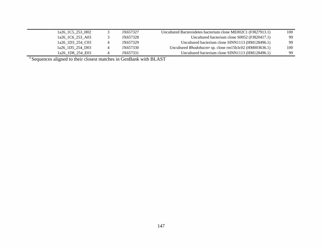
147
1a26_1C5_253_H02 3 JX657327 Uncultured Bacteroidetes bacterium clone ME002C1 (FJ827913.1) 100
1a26_1C6_253_A03 3 JX657328 Uncultured bacterium clone S0052 (FJ820417.1) 99
1a26_1D3_254_C03 4 JX657329 Uncultured bacterium clone SINN1113 (HM128496.1) 99
1a26_1D5_254_D03 4 JX657330 Uncultured Rhodobacter sp. clone rm15b3c02 (HM003636.1) 100
1a26_1D8_254_E03 4 JX657331 Uncultured bacterium clone SINN1113 (HM128496.1) 99 a Sequences aligned to their closest matches in GenBank with BLAST
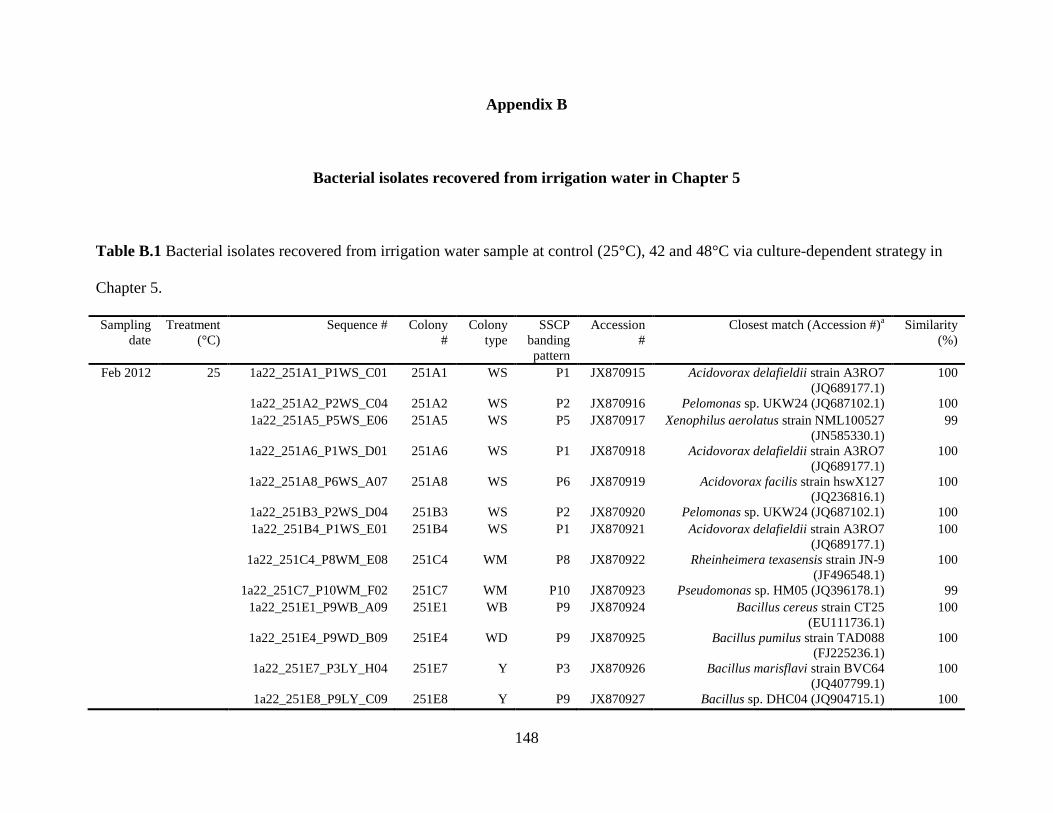
148
Appendix B
Bacterial isolates recovered from irrigation water in Chapter 5
Table B.1 Bacterial isolates recovered from irrigation water sample at control (25°C), 42 and 48°C via culture-dependent strategy in
Chapter 5.
Sampling
date
Treatment
(°C)
Sequence # Colony
#
Colony
type
SSCP
banding
pattern
Accession
#
Closest match (Accession #)a Similarity
(%)
Feb 2012 25 1a22_251A1_P1WS_C01 251A1 WS P1 JX870915 Acidovorax delafieldii strain A3RO7
(JQ689177.1)
100
1a22_251A2_P2WS_C04 251A2 WS P2 JX870916 Pelomonas sp. UKW24 (JQ687102.1) 100
1a22_251A5_P5WS_E06 251A5 WS P5 JX870917 Xenophilus aerolatus strain NML100527
(JN585330.1)
99
1a22_251A6_P1WS_D01 251A6 WS P1 JX870918 Acidovorax delafieldii strain A3RO7
(JQ689177.1)
100
1a22_251A8_P6WS_A07 251A8 WS P6 JX870919 Acidovorax facilis strain hswX127
(JQ236816.1)
100
1a22_251B3_P2WS_D04 251B3 WS P2 JX870920 Pelomonas sp. UKW24 (JQ687102.1) 100
1a22_251B4_P1WS_E01 251B4 WS P1 JX870921 Acidovorax delafieldii strain A3RO7
(JQ689177.1)
100
1a22_251C4_P8WM_E08 251C4 WM P8 JX870922 Rheinheimera texasensis strain JN-9
(JF496548.1)
100
1a22_251C7_P10WM_F02 251C7 WM P10 JX870923 Pseudomonas sp. HM05 (JQ396178.1) 99
1a22_251E1_P9WB_A09 251E1 WB P9 JX870924 Bacillus cereus strain CT25
(EU111736.1)
100
1a22_251E4_P9WD_B09 251E4 WD P9 JX870925 Bacillus pumilus strain TAD088
(FJ225236.1)
100
1a22_251E7_P3LY_H04 251E7 Y P3 JX870926 Bacillus marisflavi strain BVC64
(JQ407799.1)
100
1a22_251E8_P9LY_C09 251E8 Y P9 JX870927 Bacillus sp. DHC04 (JQ904715.1) 100
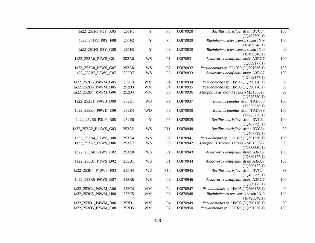
149
1a22_251F1_P3Y_A05 251F1 Y P3 JX870928 Bacillus marisflavi strain BVC64
(JQ407799.1)
100
1a22_251F2_P8Y_F08 251F2 Y P8 JX870929 Rheinheimera texasensis strain JN-9
(JF496548.1)
100
1a22_251F3_P8Y_G08 251F3 Y P8 JX870930 Rheinheimera texasensis strain JN-9
(JF496548.1)
99
1a22_252A6_P1WS_G01 252A6 WS P1 JX870931 Acidovorax delafieldii strain A3RO7
(JQ689177.1)
100
1a22_252A8_P7WS_G07 252A8 WS P7 JX870932 Pseudomonas sp. FI 1029 (JQ691536.1) 100
1a22_252B7_P6WS_C07 252B7 WS P6 JX870933 Acidovorax delafieldii strain A3RO7
(JQ689177.1)
100
1a22_252C3_P4WM_G05 252C3 WM P4 JX870934 Pseudomonas sp. HM05 (JQ396178.1) 99
1a22_252D3_P4WM_H05 252D3 WM P4 JX870935 Pseudomonas sp. HM05 (JQ396178.1) 99
1a22_252D4_P5WM_G06 252D4 WM P5 JX870936 Xenophilus aerolatus strain NML100527
(JN585330.1)
99
1a22_252E1_P9WB_D09 252E1 WB P9 JX870937 Bacillus pumilus strain TAD088
(FJ225236.1)
100
1a22_252E4_P9WD_E09 252E4 WD P9 JX870938 Bacillus pumilus strain TAD088
(FJ225236.1)
100
1a22_252E6_P3LY_B05 252E6 Y P3 JX870939 Bacillus marisflavi strain BVC64
(JQ407799.1)
100
1a22_253A2_P11WS_G03 253A2 WS P11 JX870940 Bacillus marisflavi strain BVC64
(JQ407799.1)
99
1a22_253A4_P7WS_B08 253A4 WS P7 JX870941 Pseudomonas sp. FI 1029 (JQ691536.1) 100
1a22_253A7_P5WS_H06 253A7 WS P5 JX870942 Xenophilus aerolatus strain NML100527
(JN585330.1)
99
1a22_253A8_P1WS_C02 253A8 WS P1 JX870943 Acidovorax delafieldii strain A3RO7
(JQ689177.1)
100
1a22_253B1_P1WS_D02 253B1 WS P1 JX870944 Acidovorax delafieldii strain A3RO7
(JQ689177.1)
100
1a22_253B4_P10WS_F03 253B4 WS P10 JX870945 Bacillus marisflavi strain BVC64
(JQ407799.1)
99
1a22_253B5_P6WS_D07 253B5 WS P6 JX870946 Acidovorax delafieldii strain A3RO7
(JQ689177.1)
100
1a22_253C4_P4WM_A06 253C4 WM P4 JX870947 Pseudomonas sp. HM05 (JQ396178.1) 99
1a22_253C5_P8WM_H08 253C5 WM P8 JX870948 Rheinheimera texasensis strain JN-9
(JF496548.1)
100
1a22_253D1_P4WM_B06 253D1 WM P4 JX870949 Pseudomonas sp. HM05 (JQ396178.1) 99
1a22_253D5_P7WM_C08 253D5 WM P7 JX870950 Pseudomonas sp. FI 1029 (JQ691536.1) 100
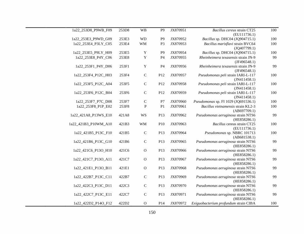
150
1a22_253D8_P9WB_F09 253D8 WB P9 JX870951 Bacillus cereus strain CT25
(EU111736.1)
100
1a22_253E3_P9WD_G09 253E3 WD P9 JX870952 Bacillus sp. DHC04 (JQ904715.1) 100
1a22_253E4_P3LY_C05 253E4 WM P3 JX870953 Bacillus marisflavi strain BVC64
(JQ407799.1)
100
1a22_253E5_P9LY_H09 253E5 Y P9 JX870954 Bacillus sp. DHC04 (JQ904715.1) 100
1a22_253E8_P4Y_C06 253E8 Y P4 JX870955 Rheinheimera texasensis strain JN-9
(JF496548.1)
99
1a22_253F1_P4Y_D06 253F1 Y P4 JX870956 Rheinheimera texasensis strain JN-9
(JF496548.1)
99
1a22_253F4_P12C_H03 253F4 C P12 JX870957 Pseudomonas peli strain IARI-L-117
(JN411458.1)
100
1a22_253F5_P12C_A04 253F5 C P12 JX870958 Pseudomonas peli strain IARI-L-117
(JN411458.1)
100
1a22_253F6_P12C_B04 253F6 C P12 JX870959 Pseudomonas peli strain IARI-L-117
(JN411458.1)
100
1a22_253F7_P7C_D08 253F7 C P7 JX870960 Pseudomonas sp. FI 1029 (JQ691536.1) 100
1a22_253F8_P1P_E02 253F8 P P1 JX870961 Bacillus vietnamensis strain KL2-3
(AB697709.1)
100
1a22_421A8_P13WS_E10 421A8 WS P13 JX870962 Pseudomonas aeruginosa strain NTS6
(HE858286.1)
99
1a22_421B3_P10WM_A10 421B3 WM P10 JX870963 Bacillus cereus strain CT25
(EU111736.1)
100
1a22_421B5_P13C_F10 421B5 C P13 JX870964 Pseudomonas sp. NBRC 101713
(AB681538.1)
100
1a22_421B6_P13C_G10 421B6 C P13 JX870965 Pseudomonas aeruginosa strain NTS6
(HE858286.1)
99
1a22_421C6_P13O_H10 421C6 O P13 JX870966 Pseudomonas aeruginosa strain NTS6
(HE858286.1)
99
1a22_421C7_P13O_A11 421C7 O P13 JX870967 Pseudomonas aeruginosa strain NTS6
(HE858286.1)
99
1a22_421E1_P13O_B11 421E1 O P13 JX870968 Pseudomonas aeruginosa strain NTS6
(HE858286.1)
99
1a22_422B7_P13C_C11 422B7 C P13 JX870969 Pseudomonas aeruginosa strain NTS6
(HE858286.1)
99
1a22_422C3_P13C_D11 422C3 C P13 JX870970 Pseudomonas aeruginosa strain NTS6
(HE858286.1)
99
1a22_422C7_P13C_E11 422C7 C P13 JX870971 Pseudomonas aeruginosa strain NTS6
(HE858286.1)
99
1a22_422D2_P14O_F12 422D2 O P14 JX870972 Exiguobacterium profundum strain CIBA 100
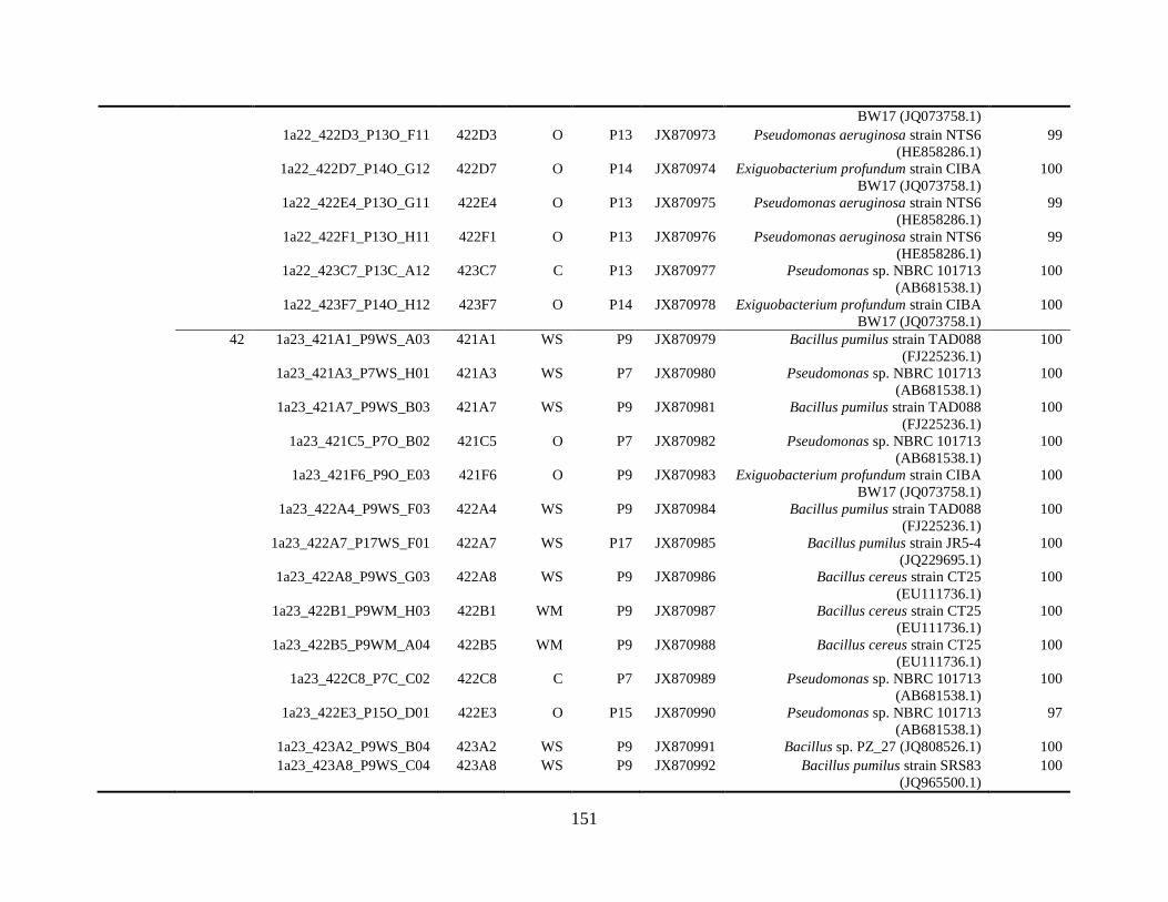
151
BW17 (JQ073758.1)
1a22_422D3_P13O_F11 422D3 O P13 JX870973 Pseudomonas aeruginosa strain NTS6
(HE858286.1)
99
1a22_422D7_P14O_G12 422D7 O P14 JX870974 Exiguobacterium profundum strain CIBA
BW17 (JQ073758.1)
100
1a22_422E4_P13O_G11 422E4 O P13 JX870975 Pseudomonas aeruginosa strain NTS6
(HE858286.1)
99
1a22_422F1_P13O_H11 422F1 O P13 JX870976 Pseudomonas aeruginosa strain NTS6
(HE858286.1)
99
1a22_423C7_P13C_A12 423C7 C P13 JX870977 Pseudomonas sp. NBRC 101713
(AB681538.1)
100
1a22_423F7_P14O_H12 423F7 O P14 JX870978 Exiguobacterium profundum strain CIBA
BW17 (JQ073758.1)
100
42 1a23_421A1_P9WS_A03 421A1 WS P9 JX870979 Bacillus pumilus strain TAD088
(FJ225236.1)
100
1a23_421A3_P7WS_H01 421A3 WS P7 JX870980 Pseudomonas sp. NBRC 101713
(AB681538.1)
100
1a23_421A7_P9WS_B03 421A7 WS P9 JX870981 Bacillus pumilus strain TAD088
(FJ225236.1)
100
1a23_421C5_P7O_B02 421C5 O P7 JX870982 Pseudomonas sp. NBRC 101713
(AB681538.1)
100
1a23_421F6_P9O_E03 421F6 O P9 JX870983 Exiguobacterium profundum strain CIBA
BW17 (JQ073758.1)
100
1a23_422A4_P9WS_F03 422A4 WS P9 JX870984 Bacillus pumilus strain TAD088
(FJ225236.1)
100
1a23_422A7_P17WS_F01 422A7 WS P17 JX870985 Bacillus pumilus strain JR5-4
(JQ229695.1)
100
1a23_422A8_P9WS_G03 422A8 WS P9 JX870986 Bacillus cereus strain CT25
(EU111736.1)
100
1a23_422B1_P9WM_H03 422B1 WM P9 JX870987 Bacillus cereus strain CT25
(EU111736.1)
100
1a23_422B5_P9WM_A04 422B5 WM P9 JX870988 Bacillus cereus strain CT25
(EU111736.1)
100
1a23_422C8_P7C_C02 422C8 C P7 JX870989 Pseudomonas sp. NBRC 101713
(AB681538.1)
100
1a23_422E3_P15O_D01 422E3 O P15 JX870990 Pseudomonas sp. NBRC 101713
(AB681538.1)
97
1a23_423A2_P9WS_B04 423A2 WS P9 JX870991 Bacillus sp. PZ_27 (JQ808526.1) 100
1a23_423A8_P9WS_C04 423A8 WS P9 JX870992 Bacillus pumilus strain SRS83
(JQ965500.1)
100
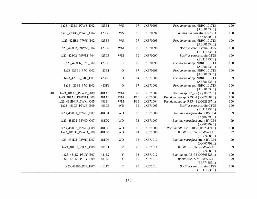
152
1a23_423B1_P7WS_D02 423B1 WS P7 JX870993 Pseudomonas sp. NBRC 101713
(AB681538.1)
100
1a23_423B6_P9WS_D04 423B6 WS P9 JX870994 Bacillus pumilus strain SRS83
(JQ965500.1)
100
1a23_423B8_P7WS_E02 423B8 WS P7 JX870995 Pseudomonas sp. NBRC 101713
(AB681538.1)
100
1a23_423C2_P9WM_E04 423C2 WM P9 JX870996 Bacillus cereus strain CT25
(EU111736.1)
100
1a23_423C3_P9WM_F04 423C3 WM P9 JX870997 Bacillus cereus strain CT25
(EU111736.1)
100
1a23_423C6_P7C_F02 423C6 C P7 JX870998 Pseudomonas sp. NBRC 101713
(AB681538.1)
100
1a23_423E1_P7O_G02 423E1 O P7 JX870999 Pseudomonas sp. NBRC 101713
(AB681538.1)
100
1a23_423E5_P4O_G01 423E5 O P4 JX871000 Pseudomonas sp. NBRC 101713
(AB681538.1)
99
1a23_423F8_P7O_H02 423F8 O P7 JX871001 Pseudomonas sp. NBRC 101713
(AB681538.1)
100
48 1a23_481A5_P9WM_A09 481A5 WM P9 JX871002 Bacillus sp. PZ_27 (JQ808526.1) 100
1a23_481A8_P16WM_F05 481A8 WM P16 JX871003 Pseudomonas sp. R3S4-1 (JQ928687.1) 100
1a23_481B4_P16WM_G05 481B4 WM P16 JX871004 Pseudomonas sp. R3S4-1 (JQ928687.1) 100
1a23_481C6_P9WB_B09 481C6 WB P9 JX871005 Bacillus cereus strain CT25
(EU111736.1)
100
1a23_481D1_P3WD_B07 481D1 WD P3 JX871006 Bacillus marisflavi strain BVC64
(JQ407799.1)
99
1a23_481D2_P3WD_C07 481D2 WD P3 JX871007 Bacillus marisflavi strain BVC64
(JQ407799.1)
99
1a23_481D3_P9WD_C09 481D3 WD P9 JX871008 Paenibacillus sp. 140SI (JF825471.1) 100
1a23_481D5_P4WD_F08 481D5 WD P4 JX871009 Bacillus sp. S-bf-PMW-5.1.1
(FR774585.1)
97
1a23_481D8_P3WD_D07 481D8 WD P3 JX871010 Bacillus marisflavi strain BVC64
(JQ407799.1)
99
1a23_481E1_P9LY_D09 481E1 Y P9 JX871011 Bacillus sp. S-bf-PMW-5.1.1
(FR774585.1)
99
1a23_481E2_P3LY_E07 481E2 Y P3 JX871012 Bacillus sp. PZ_35 (JQ808528.1) 100
1a23_481E3_P9LY_E09 481E3 Y P9 JX871013 Bacillus sp. S-bf-PMW-5.1.1
(FR774585.1)
99
1a23_481F5_P3S_H07 481F5 T P3 JX871014 Bacillus cereus strain CT25
(EU111736.1)
100
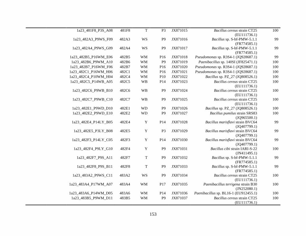
153
1a23_481F8_P3S_A08 481F8 T P3 JX871015 Bacillus cereus strain CT25
(EU111736.1)
100
1a23_482A3_P9WS_F09 482A3 WS P9 JX871016 Bacillus sp. S-bf-PMW-5.1.1
(FR774585.1)
99
1a23_482A4_P9WS_G09 482A4 WS P9 JX871017 Bacillus sp. S-bf-PMW-5.1.1
(FR774585.1)
99
1a23_482B5_P16WM_E06 482B5 WM P16 JX871018 Pseudomonas sp. R3S4-1 (JQ928687.1) 99
1a23_482B6_P9WM_A10 482B6 WM P9 JX871019 Paenibacillus sp. 140SI (JF825471.1) 100
1a23_482B7_P16WM_F06 482B7 WM P16 JX871020 Pseudomonas sp. R3S4-1 (JQ928687.1) 100
1a23_482C1_P16WM_H06 482C1 WM P16 JX871021 Pseudomonas sp. R3S4-1 (JQ928687.1) 100
1a23_482C4_P10WM_H04 482C4 WM P10 JX871022 Bacillus sp. PZ_27 (JQ808526.1) 100
1a23_482C5_P14WB_A05 482C5 WB P14 JX871023 Bacillus cereus strain CT25
(EU111736.1)
100
1a23_482C6_P9WB_B10 482C6 WB P9 JX871024 Bacillus cereus strain CT25
(EU111736.1)
100
1a23_482C7_P9WB_C10 482C7 WB P9 JX871025 Bacillus cereus strain CT25
(EU111736.1)
100
1a23_482E1_P9WD_D10 482E1 WD P9 JX871026 Bacillus sp. PZ_27 (JQ808526.1) 100
1a23_482E2_P9WD_E10 482E2 WD P9 JX871027 Bacillus pumilus strain SRS83
(JQ965500.1)
100
1a23_482E4_P14LY_B05 482E4 Y P14 JX871028 Bacillus marisflavi strain BVC64
(JQ407799.1)
99
1a23_482E5_P3LY_B08 482E5 Y P3 JX871029 Bacillus marisflavi strain BVC64
(JQ407799.1)
99
1a23_482F3_P14LY_C05 482F3 Y P14 JX871030 Bacillus marisflavi strain BVC64
(JQ407799.1)
99
1a23_482F4_P9LY_G10 482F4 Y P9 JX871031 Bacillus cibi strain IARI-S-22
(JN411495.1)
100
1a23_482F7_P9S_A11 482F7 T P9 JX871032 Bacillus sp. S-bf-PMW-5.1.1
(FR774585.1)
99
1a23_482F8_P9S_B11 482F8 T P9 JX871033 Bacillus sp. S-bf-PMW-5.1.1
(FR774585.1)
99
1a23_483A2_P9WS_C11 483A2 WS P9 JX871034 Bacillus cereus strain CT25
(EU111736.1)
100
1a23_483A4_P17WM_A07 483A4 WM P17 JX871035 Paenibacillus terrigena strain B30
(JN252080.1)
100
1a23_483A6_P14WM_D05 483A6 WM P14 JX871036 Paenibacillus sp. BL16-1 (EU912455.1) 100
1a23_483B5_P9WM_D11 483B5 WM P9 JX871037 Bacillus cereus strain CT25
(EU111736.1)
100
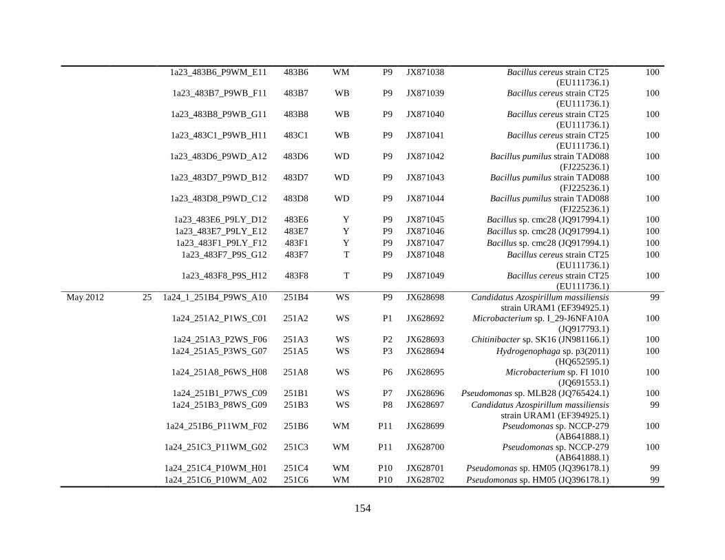
154
1a23_483B6_P9WM_E11 483B6 WM P9 JX871038 Bacillus cereus strain CT25
(EU111736.1)
100
1a23_483B7_P9WB_F11 483B7 WB P9 JX871039 Bacillus cereus strain CT25
(EU111736.1)
100
1a23_483B8_P9WB_G11 483B8 WB P9 JX871040 Bacillus cereus strain CT25
(EU111736.1)
100
1a23_483C1_P9WB_H11 483C1 WB P9 JX871041 Bacillus cereus strain CT25
(EU111736.1)
100
1a23_483D6_P9WD_A12 483D6 WD P9 JX871042 Bacillus pumilus strain TAD088
(FJ225236.1)
100
1a23_483D7_P9WD_B12 483D7 WD P9 JX871043 Bacillus pumilus strain TAD088
(FJ225236.1)
100
1a23_483D8_P9WD_C12 483D8 WD P9 JX871044 Bacillus pumilus strain TAD088
(FJ225236.1)
100
1a23_483E6_P9LY_D12 483E6 Y P9 JX871045 Bacillus sp. cmc28 (JQ917994.1) 100
1a23_483E7_P9LY_E12 483E7 Y P9 JX871046 Bacillus sp. cmc28 (JQ917994.1) 100
1a23_483F1_P9LY_F12 483F1 Y P9 JX871047 Bacillus sp. cmc28 (JQ917994.1) 100
1a23_483F7_P9S_G12 483F7 T P9 JX871048 Bacillus cereus strain CT25
(EU111736.1)
100
1a23_483F8_P9S_H12 483F8 T P9 JX871049 Bacillus cereus strain CT25
(EU111736.1)
100
May 2012 25 1a24_1_251B4_P9WS_A10 251B4 WS P9 JX628698 Candidatus Azospirillum massiliensis
strain URAM1 (EF394925.1)
99
1a24_251A2_P1WS_C01 251A2 WS P1 JX628692 Microbacterium sp. I_29-J6NFA10A
(JQ917793.1)
100
1a24_251A3_P2WS_F06 251A3 WS P2 JX628693 Chitinibacter sp. SK16 (JN981166.1) 100
1a24_251A5_P3WS_G07 251A5 WS P3 JX628694 Hydrogenophaga sp. p3(2011)
(HQ652595.1)
100
1a24_251A8_P6WS_H08 251A8 WS P6 JX628695 Microbacterium sp. FI 1010
(JQ691553.1)
100
1a24_251B1_P7WS_C09 251B1 WS P7 JX628696 Pseudomonas sp. MLB28 (JQ765424.1) 100
1a24_251B3_P8WS_G09 251B3 WS P8 JX628697 Candidatus Azospirillum massiliensis
strain URAM1 (EF394925.1)
99
1a24_251B6_P11WM_F02 251B6 WM P11 JX628699 Pseudomonas sp. NCCP-279
(AB641888.1)
100
1a24_251C3_P11WM_G02 251C3 WM P11 JX628700 Pseudomonas sp. NCCP-279
(AB641888.1)
100
1a24_251C4_P10WM_H01 251C4 WM P10 JX628701 Pseudomonas sp. HM05 (JQ396178.1) 99
1a24_251C6_P10WM_A02 251C6 WM P10 JX628702 Pseudomonas sp. HM05 (JQ396178.1) 99
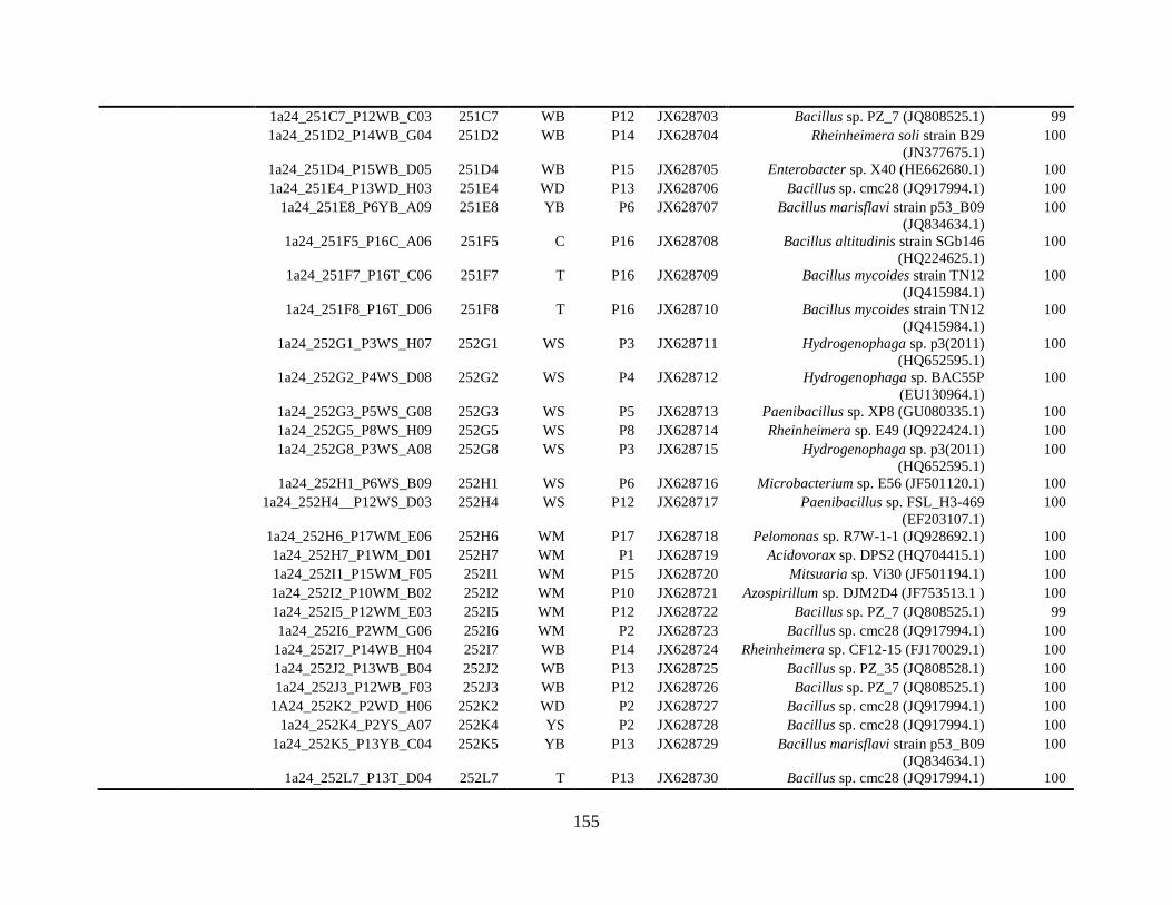
155
1a24_251C7_P12WB_C03 251C7 WB P12 JX628703 Bacillus sp. PZ_7 (JQ808525.1) 99
1a24_251D2_P14WB_G04 251D2 WB P14 JX628704 Rheinheimera soli strain B29
(JN377675.1)
100
1a24_251D4_P15WB_D05 251D4 WB P15 JX628705 Enterobacter sp. X40 (HE662680.1) 100
1a24_251E4_P13WD_H03 251E4 WD P13 JX628706 Bacillus sp. cmc28 (JQ917994.1) 100
1a24_251E8_P6YB_A09 251E8 YB P6 JX628707 Bacillus marisflavi strain p53_B09
(JQ834634.1)
100
1a24_251F5_P16C_A06 251F5 C P16 JX628708 Bacillus altitudinis strain SGb146
(HQ224625.1)
100
1a24_251F7_P16T_C06 251F7 T P16 JX628709 Bacillus mycoides strain TN12
(JQ415984.1)
100
1a24_251F8_P16T_D06 251F8 T P16 JX628710 Bacillus mycoides strain TN12
(JQ415984.1)
100
1a24_252G1_P3WS_H07 252G1 WS P3 JX628711 Hydrogenophaga sp. p3(2011)
(HQ652595.1)
100
1a24_252G2_P4WS_D08 252G2 WS P4 JX628712 Hydrogenophaga sp. BAC55P
(EU130964.1)
100
1a24_252G3_P5WS_G08 252G3 WS P5 JX628713 Paenibacillus sp. XP8 (GU080335.1) 100
1a24_252G5_P8WS_H09 252G5 WS P8 JX628714 Rheinheimera sp. E49 (JQ922424.1) 100
1a24_252G8_P3WS_A08 252G8 WS P3 JX628715 Hydrogenophaga sp. p3(2011)
(HQ652595.1)
100
1a24_252H1_P6WS_B09 252H1 WS P6 JX628716 Microbacterium sp. E56 (JF501120.1) 100
1a24_252H4__P12WS_D03 252H4 WS P12 JX628717 Paenibacillus sp. FSL_H3-469
(EF203107.1)
100
1a24_252H6_P17WM_E06 252H6 WM P17 JX628718 Pelomonas sp. R7W-1-1 (JQ928692.1) 100
1a24_252H7_P1WM_D01 252H7 WM P1 JX628719 Acidovorax sp. DPS2 (HQ704415.1) 100
1a24_252I1_P15WM_F05 252I1 WM P15 JX628720 Mitsuaria sp. Vi30 (JF501194.1) 100
1a24_252I2_P10WM_B02 252I2 WM P10 JX628721 Azospirillum sp. DJM2D4 (JF753513.1 ) 100
1a24_252I5_P12WM_E03 252I5 WM P12 JX628722 Bacillus sp. PZ_7 (JQ808525.1) 99
1a24_252I6_P2WM_G06 252I6 WM P2 JX628723 Bacillus sp. cmc28 (JQ917994.1) 100
1a24_252I7_P14WB_H04 252I7 WB P14 JX628724 Rheinheimera sp. CF12-15 (FJ170029.1) 100
1a24_252J2_P13WB_B04 252J2 WB P13 JX628725 Bacillus sp. PZ_35 (JQ808528.1) 100
1a24_252J3_P12WB_F03 252J3 WB P12 JX628726 Bacillus sp. PZ_7 (JQ808525.1) 100
1A24_252K2_P2WD_H06 252K2 WD P2 JX628727 Bacillus sp. cmc28 (JQ917994.1) 100
1a24_252K4_P2YS_A07 252K4 YS P2 JX628728 Bacillus sp. cmc28 (JQ917994.1) 100
1a24_252K5_P13YB_C04 252K5 YB P13 JX628729 Bacillus marisflavi strain p53_B09
(JQ834634.1)
100
1a24_252L7_P13T_D04 252L7 T P13 JX628730 Bacillus sp. cmc28 (JQ917994.1) 100
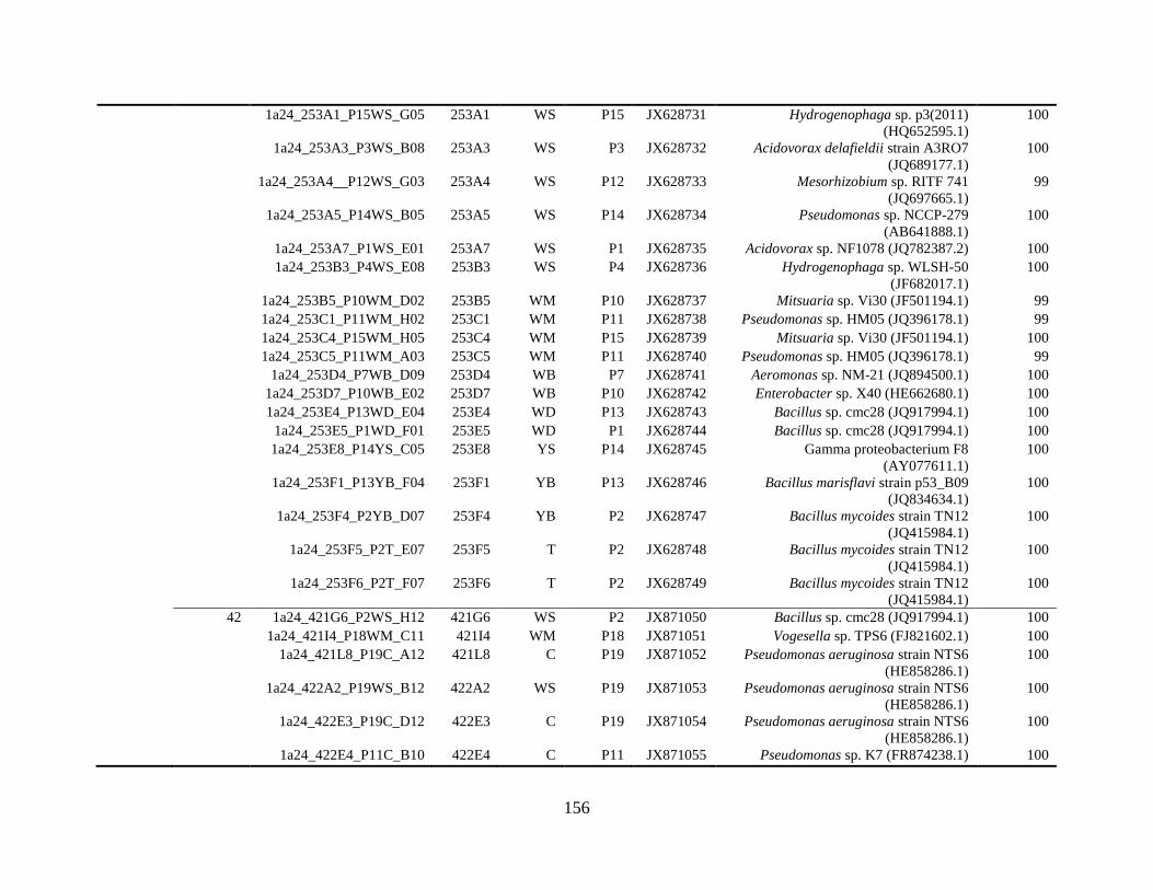
156
1a24_253A1_P15WS_G05 253A1 WS P15 JX628731 Hydrogenophaga sp. p3(2011)
(HQ652595.1)
100
1a24_253A3_P3WS_B08 253A3 WS P3 JX628732 Acidovorax delafieldii strain A3RO7
(JQ689177.1)
100
1a24_253A4__P12WS_G03 253A4 WS P12 JX628733 Mesorhizobium sp. RITF 741
(JQ697665.1)
99
1a24_253A5_P14WS_B05 253A5 WS P14 JX628734 Pseudomonas sp. NCCP-279
(AB641888.1)
100
1a24_253A7_P1WS_E01 253A7 WS P1 JX628735 Acidovorax sp. NF1078 (JQ782387.2) 100
1a24_253B3_P4WS_E08 253B3 WS P4 JX628736 Hydrogenophaga sp. WLSH-50
(JF682017.1)
100
1a24_253B5_P10WM_D02 253B5 WM P10 JX628737 Mitsuaria sp. Vi30 (JF501194.1) 99
1a24_253C1_P11WM_H02 253C1 WM P11 JX628738 Pseudomonas sp. HM05 (JQ396178.1) 99
1a24_253C4_P15WM_H05 253C4 WM P15 JX628739 Mitsuaria sp. Vi30 (JF501194.1) 100
1a24_253C5_P11WM_A03 253C5 WM P11 JX628740 Pseudomonas sp. HM05 (JQ396178.1) 99
1a24_253D4_P7WB_D09 253D4 WB P7 JX628741 Aeromonas sp. NM-21 (JQ894500.1) 100
1a24_253D7_P10WB_E02 253D7 WB P10 JX628742 Enterobacter sp. X40 (HE662680.1) 100
1a24_253E4_P13WD_E04 253E4 WD P13 JX628743 Bacillus sp. cmc28 (JQ917994.1) 100
1a24_253E5_P1WD_F01 253E5 WD P1 JX628744 Bacillus sp. cmc28 (JQ917994.1) 100
1a24_253E8_P14YS_C05 253E8 YS P14 JX628745 Gamma proteobacterium F8
(AY077611.1)
100
1a24_253F1_P13YB_F04 253F1 YB P13 JX628746 Bacillus marisflavi strain p53_B09
(JQ834634.1)
100
1a24_253F4_P2YB_D07 253F4 YB P2 JX628747 Bacillus mycoides strain TN12
(JQ415984.1)
100
1a24_253F5_P2T_E07 253F5 T P2 JX628748 Bacillus mycoides strain TN12
(JQ415984.1)
100
1a24_253F6_P2T_F07 253F6 T P2 JX628749 Bacillus mycoides strain TN12
(JQ415984.1)
100
42 1a24_421G6_P2WS_H12 421G6 WS P2 JX871050 Bacillus sp. cmc28 (JQ917994.1) 100
1a24_421I4_P18WM_C11 421I4 WM P18 JX871051 Vogesella sp. TPS6 (FJ821602.1) 100
1a24_421L8_P19C_A12 421L8 C P19 JX871052 Pseudomonas aeruginosa strain NTS6
(HE858286.1)
100
1a24_422A2_P19WS_B12 422A2 WS P19 JX871053 Pseudomonas aeruginosa strain NTS6
(HE858286.1)
100
1a24_422E3_P19C_D12 422E3 C P19 JX871054 Pseudomonas aeruginosa strain NTS6
(HE858286.1)
100
1a24_422E4_P11C_B10 422E4 C P11 JX871055 Pseudomonas sp. K7 (FR874238.1) 100
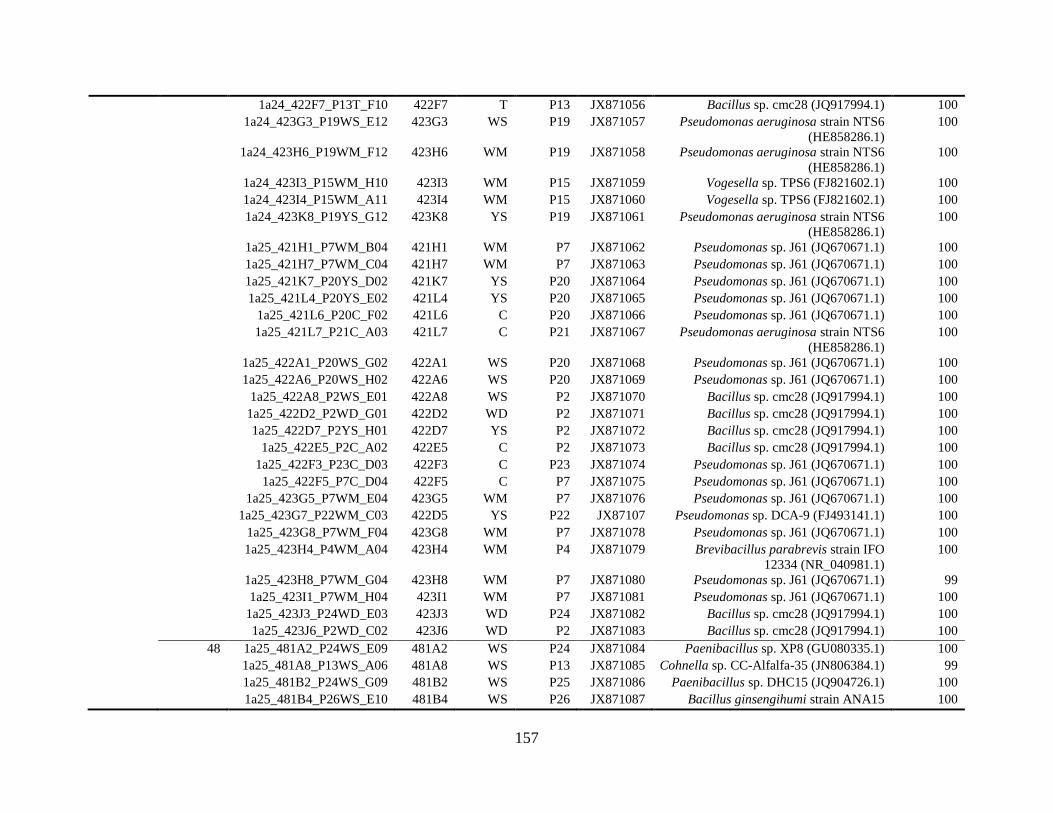
157
1a24_422F7_P13T_F10 422F7 T P13 JX871056 Bacillus sp. cmc28 (JQ917994.1) 100
1a24_423G3_P19WS_E12 423G3 WS P19 JX871057 Pseudomonas aeruginosa strain NTS6
(HE858286.1)
100
1a24_423H6_P19WM_F12 423H6 WM P19 JX871058 Pseudomonas aeruginosa strain NTS6
(HE858286.1)
100
1a24_423I3_P15WM_H10 423I3 WM P15 JX871059 Vogesella sp. TPS6 (FJ821602.1) 100
1a24_423I4_P15WM_A11 423I4 WM P15 JX871060 Vogesella sp. TPS6 (FJ821602.1) 100
1a24_423K8_P19YS_G12 423K8 YS P19 JX871061 Pseudomonas aeruginosa strain NTS6
(HE858286.1)
100
1a25_421H1_P7WM_B04 421H1 WM P7 JX871062 Pseudomonas sp. J61 (JQ670671.1) 100
1a25_421H7_P7WM_C04 421H7 WM P7 JX871063 Pseudomonas sp. J61 (JQ670671.1) 100
1a25_421K7_P20YS_D02 421K7 YS P20 JX871064 Pseudomonas sp. J61 (JQ670671.1) 100
1a25_421L4_P20YS_E02 421L4 YS P20 JX871065 Pseudomonas sp. J61 (JQ670671.1) 100
1a25_421L6_P20C_F02 421L6 C P20 JX871066 Pseudomonas sp. J61 (JQ670671.1) 100
1a25_421L7_P21C_A03 421L7 C P21 JX871067 Pseudomonas aeruginosa strain NTS6
(HE858286.1)
100
1a25_422A1_P20WS_G02 422A1 WS P20 JX871068 Pseudomonas sp. J61 (JQ670671.1) 100
1a25_422A6_P20WS_H02 422A6 WS P20 JX871069 Pseudomonas sp. J61 (JQ670671.1) 100
1a25_422A8_P2WS_E01 422A8 WS P2 JX871070 Bacillus sp. cmc28 (JQ917994.1) 100
1a25_422D2_P2WD_G01 422D2 WD P2 JX871071 Bacillus sp. cmc28 (JQ917994.1) 100
1a25_422D7_P2YS_H01 422D7 YS P2 JX871072 Bacillus sp. cmc28 (JQ917994.1) 100
1a25_422E5_P2C_A02 422E5 C P2 JX871073 Bacillus sp. cmc28 (JQ917994.1) 100
1a25_422F3_P23C_D03 422F3 C P23 JX871074 Pseudomonas sp. J61 (JQ670671.1) 100
1a25_422F5_P7C_D04 422F5 C P7 JX871075 Pseudomonas sp. J61 (JQ670671.1) 100
1a25_423G5_P7WM_E04 423G5 WM P7 JX871076 Pseudomonas sp. J61 (JQ670671.1) 100
1a25_423G7_P22WM_C03 422D5 YS P22 JX87107 Pseudomonas sp. DCA-9 (FJ493141.1) 100
1a25_423G8_P7WM_F04 423G8 WM P7 JX871078 Pseudomonas sp. J61 (JQ670671.1) 100
1a25_423H4_P4WM_A04 423H4 WM P4 JX871079 Brevibacillus parabrevis strain IFO
12334 (NR_040981.1)
100
1a25_423H8_P7WM_G04 423H8 WM P7 JX871080 Pseudomonas sp. J61 (JQ670671.1) 99
1a25_423I1_P7WM_H04 423I1 WM P7 JX871081 Pseudomonas sp. J61 (JQ670671.1) 100
1a25_423J3_P24WD_E03 423J3 WD P24 JX871082 Bacillus sp. cmc28 (JQ917994.1) 100
1a25_423J6_P2WD_C02 423J6 WD P2 JX871083 Bacillus sp. cmc28 (JQ917994.1) 100
48 1a25_481A2_P24WS_E09 481A2 WS P24 JX871084 Paenibacillus sp. XP8 (GU080335.1) 100
1a25_481A8_P13WS_A06 481A8 WS P13 JX871085 Cohnella sp. CC-Alfalfa-35 (JN806384.1) 99
1a25_481B2_P24WS_G09 481B2 WS P25 JX871086 Paenibacillus sp. DHC15 (JQ904726.1) 100
1a25_481B4_P26WS_E10 481B4 WS P26 JX871087 Bacillus ginsengihumi strain ANA15 100
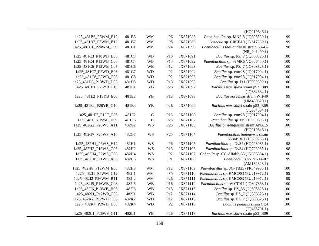
158
(HQ219846.1)
1a25_481B6_P6WM_E12 481B6 WM P6 JX871088 Paenibacillus sp. MN2-8 (JQ396530.1) 99
1a25_481B7_P5WM_B12 481B7 WM P5 JX871089 Cohnella sp. CRCB10 (JN617230.1) 99
1a25_481C1_P24WM_F09 481C1 WM P24 JX871090 Paenibacillus thailandensis strain S3-4A
(NR_041490.1)
98
1a25_481C3_P10WB_B05 481C3 WB P10 JX871091 Bacillus sp. PZ_7 (JQ808525.1) 100
1a25_481C4_P13WB_C06 481C4 WB P13 JX871092 Paenibacillus sp. SaMR6 (JQ806430.1) 100
1a25_481C6_P12WB_C05 481C6 WB P12 JX871093 Bacillus sp. PZ_7 (JQ808525.1) 100
1a25_481C7_P2WD_E08 481C7 WD P2 JX871094 Bacillus sp. cmc28 (JQ917994.1) 100
1a25_481C8_P2WD_F08 481C8 WD P2 JX871095 Bacillus sp. cmc28 (JQ917994.1) 100
1a25_481D8_P13WD_D06 481D8 WD P13 JX871096 Bacillus sp. Pt1 (JF900600.1) 100
1a25_481E1_P26YB_F10 481E1 YB P26 JX871097 Bacillus marisflavi strain p53_B09
(JQ834634.1)
100
1a25_481E2_P13YB_E06 481E2 YB P13 JX871098 Bacillus koreensis strain WIF49
(HM480320.1)
99
1a25_481E4_P26YB_G10 481E4 YB P26 JX871099 Bacillus marisflavi strain p53_B09
(JQ834634.1)
100
1a25_481F2_P13C_F06 481F2 C P13 JX871100 Bacillus sp. cmc28 (JQ917994.1) 100
1a25_481F6_P25C_H09 481F6 C P25 JX871102 Paenibacillus sp. Pt9 (JF900608.1) 99
1a25_482G2_P26WS_A11 482G2 WS P26 JX871103 Bacillus ginsengihumi strain ANA15
(HQ219846.1)
100
1a25_482G7_P25WS_A10 482G7 WS P25 JX871104 Paenibacillus timonensis strain
3584BRRJ (JF309265.1)
100
1a25_482H1_P6WS_H12 482H1 WS P6 JX871105 Paenibacillus sp. Dv34 (HQ728085.1) 98
1a25_482H2_P13WS_G06 482H2 WS P13 JX871106 Paenibacillus sp. Dv34 (HQ728085.1) 98
1a25_482H4_P2WS_G08 482H4 WS P2 JX871107 Cohnella sp. CC-Alfalfa-35 (JN806384.1) 100
1a25_482H6_P1WS_A05 482H6 WS P1 JX871108 Paenibacillus sp. YN14-07
(AM162323.1)
99
1a25_482H8_P12WM_E05 482H8 WM P12 JX871109 Paenibacillus sp. JG-TB25 (FR849935.1) 100
1a25_482I1_P5WM_C12 482I1 WM P5 JX871110 Paenibacillus sp. KMC003 (EU219972.1) 99
1a25_482I2_P26WM_B11 482I2 WM P26 JX871111 Paenibacillus sp. KMC003 (EU219972.1) 99
1a25_482I5_P16WB_C08 482I5 WB P16 JX871112 Paenibacillus sp. WYT011 (JQ807858.1) 100
1a25_482I6_P13WB_H06 482I6 WB P13 JX871113 Bacillus sp. PZ_35 (JQ808528.1) 100
1a25_482J1_P12WB_F05 482J1 WB P12 JX871114 Bacillus sp. PZ_7 (JQ808525.1) 100
1a25_482K2_P12WD_G05 482K2 WD P12 JX871115 Bacillus sp. PZ_7 (JQ808525.1) 100
1a25_482K4_P2WD_H08 482K4 WD P2 JX871116 Bacillus pumilus strain CE4
(JQ435701.1)
100
1a25_482L1_P26WS_C11 482L1 YB P26 JX871117 Bacillus marisflavi strain p53_B09 100
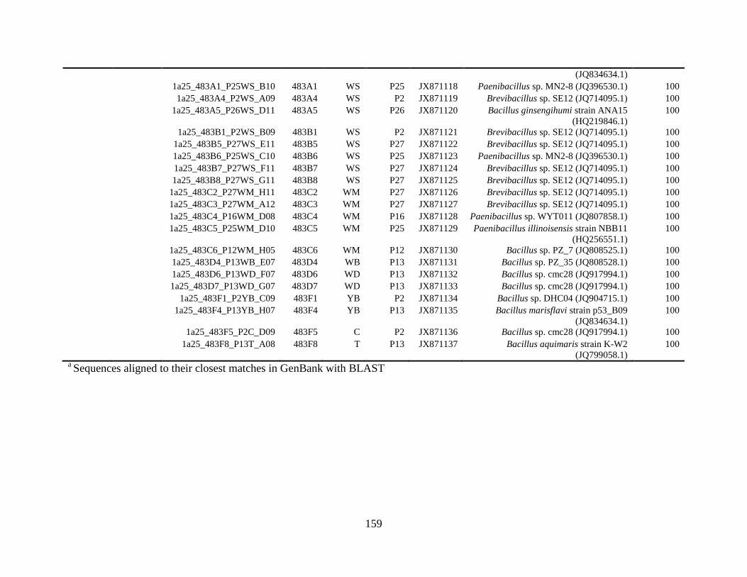
159
(JQ834634.1)
1a25_483A1_P25WS_B10 483A1 WS P25 JX871118 Paenibacillus sp. MN2-8 (JQ396530.1) 100
1a25_483A4_P2WS_A09 483A4 WS P2 JX871119 Brevibacillus sp. SE12 (JQ714095.1) 100
1a25_483A5_P26WS_D11 483A5 WS P26 JX871120 Bacillus ginsengihumi strain ANA15
(HQ219846.1)
100
1a25_483B1_P2WS_B09 483B1 WS P2 JX871121 Brevibacillus sp. SE12 (JQ714095.1) 100
1a25_483B5_P27WS_E11 483B5 WS P27 JX871122 Brevibacillus sp. SE12 (JQ714095.1) 100
1a25_483B6_P25WS_C10 483B6 WS P25 JX871123 Paenibacillus sp. MN2-8 (JQ396530.1) 100
1a25_483B7_P27WS_F11 483B7 WS P27 JX871124 Brevibacillus sp. SE12 (JQ714095.1) 100
1a25_483B8_P27WS_G11 483B8 WS P27 JX871125 Brevibacillus sp. SE12 (JQ714095.1) 100
1a25_483C2_P27WM_H11 483C2 WM P27 JX871126 Brevibacillus sp. SE12 (JQ714095.1) 100
1a25_483C3_P27WM_A12 483C3 WM P27 JX871127 Brevibacillus sp. SE12 (JQ714095.1) 100
1a25_483C4_P16WM_D08 483C4 WM P16 JX871128 Paenibacillus sp. WYT011 (JQ807858.1) 100
1a25_483C5_P25WM_D10 483C5 WM P25 JX871129 Paenibacillus illinoisensis strain NBB11
(HQ256551.1)
100
1a25_483C6_P12WM_H05 483C6 WM P12 JX871130 Bacillus sp. PZ_7 (JQ808525.1) 100
1a25_483D4_P13WB_E07 483D4 WB P13 JX871131 Bacillus sp. PZ_35 (JQ808528.1) 100
1a25_483D6_P13WD_F07 483D6 WD P13 JX871132 Bacillus sp. cmc28 (JQ917994.1) 100
1a25_483D7_P13WD_G07 483D7 WD P13 JX871133 Bacillus sp. cmc28 (JQ917994.1) 100
1a25_483F1_P2YB_C09 483F1 YB P2 JX871134 Bacillus sp. DHC04 (JQ904715.1) 100
1a25_483F4_P13YB_H07 483F4 YB P13 JX871135 Bacillus marisflavi strain p53_B09
(JQ834634.1)
100
1a25_483F5_P2C_D09 483F5 C P2 JX871136 Bacillus sp. cmc28 (JQ917994.1) 100
1a25_483F8_P13T_A08 483F8 T P13 JX871137 Bacillus aquimaris strain K-W2
(JQ799058.1)
100
a Sequences aligned to their closest matches in GenBank with BLAST
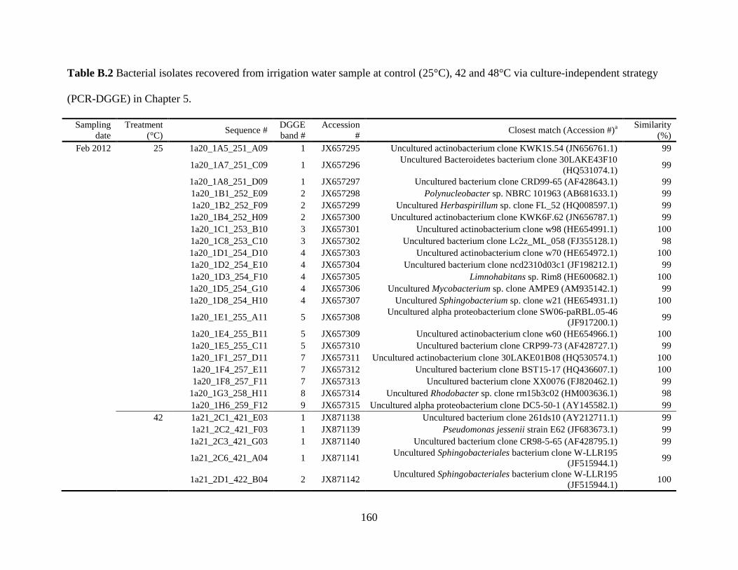
160
Table B.2 Bacterial isolates recovered from irrigation water sample at control (25°C), 42 and 48°C via culture-independent strategy
(PCR-DGGE) in Chapter 5.
Sampling
date
Treatment
(°C) Sequence #
DGGE
band #
Accession
# Closest match (Accession #)
a
Similarity
(%)
Feb 2012 25 1a20_1A5_251_A09 1 JX657295 Uncultured actinobacterium clone KWK1S.54 (JN656761.1) 99
1a20_1A7_251_C09 1 JX657296
Uncultured Bacteroidetes bacterium clone 30LAKE43F10
(HQ531074.1) 99
1a20_1A8_251_D09 1 JX657297 Uncultured bacterium clone CRD99-65 (AF428643.1) 99
1a20_1B1_252_E09 2 JX657298 Polynucleobacter sp. NBRC 101963 (AB681633.1) 99
1a20_1B2_252_F09 2 JX657299 Uncultured Herbaspirillum sp. clone FL_52 (HQ008597.1) 99
1a20_1B4_252_H09 2 JX657300 Uncultured actinobacterium clone KWK6F.62 (JN656787.1) 99
1a20_1C1_253_B10 3 JX657301 Uncultured actinobacterium clone w98 (HE654991.1) 100
1a20_1C8_253_C10 3 JX657302 Uncultured bacterium clone Lc2z_ML_058 (FJ355128.1) 98
1a20_1D1_254_D10 4 JX657303 Uncultured actinobacterium clone w70 (HE654972.1) 100
1a20_1D2_254_E10 4 JX657304 Uncultured bacterium clone ncd2310d03c1 (JF198212.1) 99
1a20_1D3_254_F10 4 JX657305 Limnohabitans sp. Rim8 (HE600682.1) 100
1a20_1D5_254_G10 4 JX657306 Uncultured Mycobacterium sp. clone AMPE9 (AM935142.1) 99
1a20_1D8_254_H10 4 JX657307 Uncultured Sphingobacterium sp. clone w21 (HE654931.1) 100
1a20_1E1_255_A11 5 JX657308
Uncultured alpha proteobacterium clone SW06-paRBL.05-46
(JF917200.1) 99
1a20_1E4_255_B11 5 JX657309 Uncultured actinobacterium clone w60 (HE654966.1) 100
1a20_1E5_255_C11 5 JX657310 Uncultured bacterium clone CRP99-73 (AF428727.1) 99
1a20_1F1_257_D11 7 JX657311 Uncultured actinobacterium clone 30LAKE01B08 (HQ530574.1) 100
1a20_1F4_257_E11 7 JX657312 Uncultured bacterium clone BST15-17 (HQ436607.1) 100
1a20_1F8_257_F11 7 JX657313 Uncultured bacterium clone XX0076 (FJ820462.1) 99
1a20_1G3_258_H11 8 JX657314 Uncultured Rhodobacter sp. clone rm15b3c02 (HM003636.1) 98
1a20_1H6_259_F12 9 JX657315 Uncultured alpha proteobacterium clone DC5-50-1 (AY145582.1) 99
42 1a21_2C1_421_E03 1 JX871138 Uncultured bacterium clone 261ds10 (AY212711.1) 99
1a21_2C2_421_F03 1 JX871139 Pseudomonas jessenii strain E62 (JF683673.1) 99
1a21_2C3_421_G03 1 JX871140 Uncultured bacterium clone CR98-5-65 (AF428795.1) 99
1a21_2C6_421_A04 1 JX871141
Uncultured Sphingobacteriales bacterium clone W-LLR195
(JF515944.1) 99
1a21_2D1_422_B04 2 JX871142
Uncultured Sphingobacteriales bacterium clone W-LLR195
(JF515944.1) 100
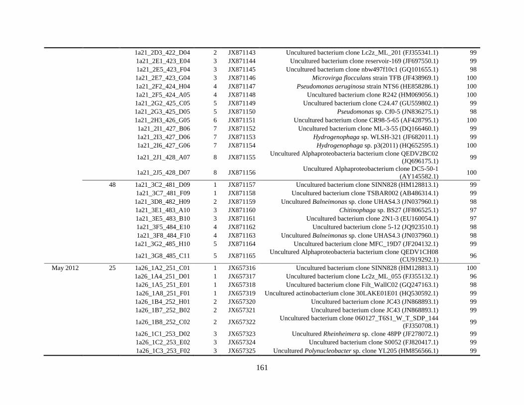
161
1a21_2D3_422_D04 2 JX871143 Uncultured bacterium clone Lc2z_ML_201 (FJ355341.1) 99
1a21_2E1_423_E04 3 JX871144 Uncultured bacterium clone reservoir-169 (JF697550.1) 99
1a21_2E5_423_F04 3 JX871145 Uncultured bacterium clone nbw497f10c1 (GQ101655.1) 98
1a21_2E7_423_G04 3 JX871146 Microvirga flocculans strain TFB (JF438969.1) 100
1a21_2F2_424_H04 4 JX871147 Pseudomonas aeruginosa strain NTS6 (HE858286.1) 100
1a21_2F5_424_A05 4 JX871148 Uncultured bacterium clone R242 (HM069056.1) 100
1a21_2G2_425_C05 5 JX871149 Uncultured bacterium clone C24.47 (GU559802.1) 99
1a21_2G3_425_D05 5 JX871150 Pseudomonas sp. Cf0-5 (JN836275.1) 98
1a21_2H3_426_G05 6 JX871151 Uncultured bacterium clone CR98-5-65 (AF428795.1) 100
1a21_2I1_427_B06 7 JX871152 Uncultured bacterium clone ML-3-55 (DQ166460.1) 99
1a21_2I3_427_D06 7 JX871153 Hydrogenophaga sp. WLSH-321 (JF682011.1) 99
1a21_2I6_427_G06 7 JX871154 Hydrogenophaga sp. p3(2011) (HQ652595.1) 100
1a21_2J1_428_A07 8 JX871155
Uncultured Alphaproteobacteria bacterium clone QEDV2BC02
(JQ696175.1) 99
1a21_2J5_428_D07 8 JX871156
Uncultured Alphaproteobacterium clone DC5-50-1
(AY145582.1) 100
48 1a21_3C2_481_D09 1 JX871157 Uncultured bacterium clone SINN828 (HM128813.1) 99
1a21_3C7_481_F09 1 JX871158 Uncultured bacterium clone TSBAR002 (AB486314.1) 99
1a21_3D8_482_H09 2 JX871159 Uncultured Balneimonas sp. clone UHAS4.3 (JN037960.1) 98
1a21_3E1_483_A10 3 JX871160 Chitinophaga sp. BS27 (JF806525.1) 97
1a21_3E5_483_B10 3 JX871161 Uncultured bacterium clone 2N1-3 (EU160054.1) 97
1a21_3F5_484_E10 4 JX871162 Uncultured bacterium clone 5-12 (JQ923510.1) 98
1a21_3F8_484_F10 4 JX871163 Uncultured Balneimonas sp. clone UHAS4.3 (JN037960.1) 98
1a21_3G2_485_H10 5 JX871164 Uncultured bacterium clone MFC_19D7 (JF204132.1) 99
1a21_3G8_485_C11 5 JX871165
Uncultured Alphaproteobacteria bacterium clone QEDV1CH08
(CU919292.1) 96
May 2012 25 1a26_1A2_251_C01 1 JX657316 Uncultured bacterium clone SINN828 (HM128813.1) 100
1a26_1A4_251_D01 1 JX657317 Uncultured bacterium clone Lc2z_ML_055 (FJ355132.1) 96
1a26_1A5_251_E01 1 JX657318 Uncultured bacterium clone Filt_WallC02 (GQ247163.1) 98
1a26_1A8_251_F01 1 JX657319 Uncultured actinobacterium clone 30LAKE01E01 (HQ530592.1) 99
1a26_1B4_252_H01 2 JX657320 Uncultured bacterium clone JC43 (JN868893.1) 99
1a26_1B7_252_B02 2 JX657321 Uncultured bacterium clone JC43 (JN868893.1) 99
1a26_1B8_252_C02 2 JX657322
Uncultured bacterium clone 060127_T6S1_W_T_SDP_144
(FJ350708.1) 99
1a26_1C1_253_D02 3 JX657323 Uncultured Rheinheimera sp. clone 48PP (JF278072.1) 99
1a26_1C2_253_E02 3 JX657324 Uncultured bacterium clone S0052 (FJ820417.1) 99
1a26_1C3_253_F02 3 JX657325 Uncultured Polynucleobacter sp. clone YL205 (HM856566.1) 99
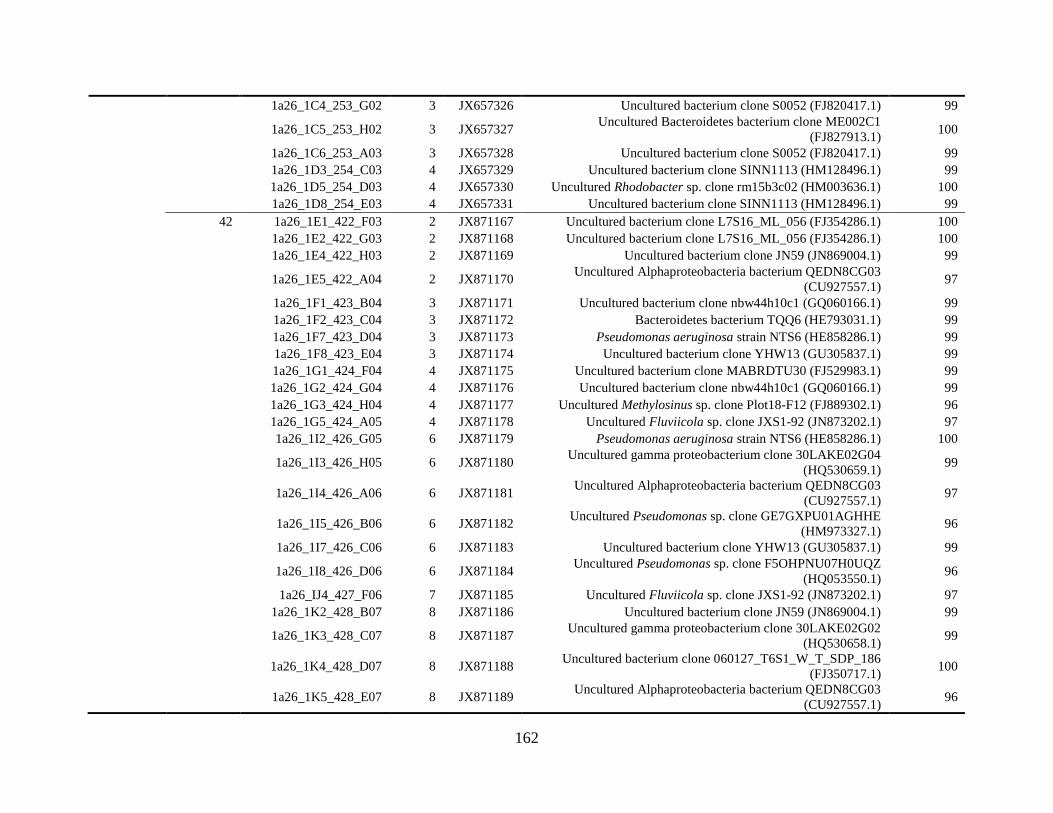
162
1a26_1C4_253_G02 3 JX657326 Uncultured bacterium clone S0052 (FJ820417.1) 99
1a26_1C5_253_H02 3 JX657327
Uncultured Bacteroidetes bacterium clone ME002C1
(FJ827913.1) 100
1a26_1C6_253_A03 3 JX657328 Uncultured bacterium clone S0052 (FJ820417.1) 99
1a26_1D3_254_C03 4 JX657329 Uncultured bacterium clone SINN1113 (HM128496.1) 99
1a26_1D5_254_D03 4 JX657330 Uncultured Rhodobacter sp. clone rm15b3c02 (HM003636.1) 100
1a26_1D8_254_E03 4 JX657331 Uncultured bacterium clone SINN1113 (HM128496.1) 99
42 1a26_1E1_422_F03 2 JX871167 Uncultured bacterium clone L7S16_ML_056 (FJ354286.1) 100
1a26_1E2_422_G03 2 JX871168 Uncultured bacterium clone L7S16_ML_056 (FJ354286.1) 100
1a26_1E4_422_H03 2 JX871169 Uncultured bacterium clone JN59 (JN869004.1) 99
1a26_1E5_422_A04 2 JX871170
Uncultured Alphaproteobacteria bacterium QEDN8CG03
(CU927557.1) 97
1a26_1F1_423_B04 3 JX871171 Uncultured bacterium clone nbw44h10c1 (GQ060166.1) 99
1a26_1F2_423_C04 3 JX871172 Bacteroidetes bacterium TQQ6 (HE793031.1) 99
1a26_1F7_423_D04 3 JX871173 Pseudomonas aeruginosa strain NTS6 (HE858286.1) 99
1a26_1F8_423_E04 3 JX871174 Uncultured bacterium clone YHW13 (GU305837.1) 99
1a26_1G1_424_F04 4 JX871175 Uncultured bacterium clone MABRDTU30 (FJ529983.1) 99
1a26_1G2_424_G04 4 JX871176 Uncultured bacterium clone nbw44h10c1 (GQ060166.1) 99
1a26_1G3_424_H04 4 JX871177 Uncultured Methylosinus sp. clone Plot18-F12 (FJ889302.1) 96
1a26_1G5_424_A05 4 JX871178 Uncultured Fluviicola sp. clone JXS1-92 (JN873202.1) 97
1a26_1I2_426_G05 6 JX871179 Pseudomonas aeruginosa strain NTS6 (HE858286.1) 100
1a26_1I3_426_H05 6 JX871180
Uncultured gamma proteobacterium clone 30LAKE02G04
(HQ530659.1) 99
1a26_1I4_426_A06 6 JX871181
Uncultured Alphaproteobacteria bacterium QEDN8CG03
(CU927557.1) 97
1a26_1I5_426_B06 6 JX871182
Uncultured Pseudomonas sp. clone GE7GXPU01AGHHE
(HM973327.1) 96
1a26_1I7_426_C06 6 JX871183 Uncultured bacterium clone YHW13 (GU305837.1) 99
1a26_1I8_426_D06 6 JX871184
Uncultured Pseudomonas sp. clone F5OHPNU07H0UQZ
(HQ053550.1) 96
1a26_IJ4_427_F06 7 JX871185 Uncultured Fluviicola sp. clone JXS1-92 (JN873202.1) 97
1a26_1K2_428_B07 8 JX871186 Uncultured bacterium clone JN59 (JN869004.1) 99
1a26_1K3_428_C07 8 JX871187
Uncultured gamma proteobacterium clone 30LAKE02G02
(HQ530658.1) 99
1a26_1K4_428_D07 8 JX871188
Uncultured bacterium clone 060127_T6S1_W_T_SDP_186
(FJ350717.1) 100
1a26_1K5_428_E07 8 JX871189
Uncultured Alphaproteobacteria bacterium QEDN8CG03
(CU927557.1) 96
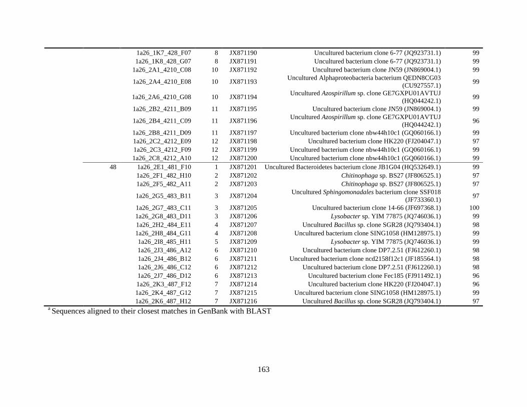
163
1a26_1K7_428_F07 8 JX871190 Uncultured bacterium clone 6-77 (JQ923731.1) 99
1a26_1K8_428_G07 8 JX871191 Uncultured bacterium clone 6-77 (JQ923731.1) 99
1a26_2A1_4210_C08 10 JX871192 Uncultured bacterium clone JN59 (JN869004.1) 99
1a26_2A4_4210_E08 10 JX871193
Uncultured Alphaproteobacteria bacterium QEDN8CG03
(CU927557.1) 99
1a26_2A6_4210_G08 10 JX871194
Uncultured Azospirillum sp. clone GE7GXPU01AVTUJ
(HQ044242.1) 99
1a26_2B2_4211_B09 11 JX871195 Uncultured bacterium clone JN59 (JN869004.1) 99
1a26_2B4_4211_C09 11 JX871196
Uncultured Azospirillum sp. clone GE7GXPU01AVTUJ
(HQ044242.1) 96
1a26_2B8_4211_D09 11 JX871197 Uncultured bacterium clone nbw44h10c1 (GQ060166.1) 99
1a26_2C2_4212_E09 12 JX871198 Uncultured bacterium clone HK220 (FJ204047.1) 97
1a26_2C3_4212_F09 12 JX871199 Uncultured bacterium clone nbw44h10c1 (GQ060166.1) 99
1a26_2C8_4212_A10 12 JX871200 Uncultured bacterium clone nbw44h10c1 (GQ060166.1) 99
48 1a26_2E1_481_F10 1 JX871201 Uncultured Bacteroidetes bacterium clone JB1G04 (HQ532649.1) 99
1a26_2F1_482_H10 2 JX871202 Chitinophaga sp. BS27 (JF806525.1) 97
1a26_2F5_482_A11 2 JX871203 Chitinophaga sp. BS27 (JF806525.1) 97
1a26_2G5_483_B11 3 JX871204
Uncultured Sphingomonadales bacterium clone SSF018
(JF733360.1) 97
1a26_2G7_483_C11 3 JX871205 Uncultured bacterium clone 14-66 (JF697368.1) 100
1a26_2G8_483_D11 3 JX871206 Lysobacter sp. YIM 77875 (JQ746036.1) 99
1a26_2H2_484_E11 4 JX871207 Uncultured Bacillus sp. clone SGR28 (JQ793404.1) 98
1a26_2H8_484_G11 4 JX871208 Uncultured bacterium clone SING1058 (HM128975.1) 99
1a26_2I8_485_H11 5 JX871209 Lysobacter sp. YIM 77875 (JQ746036.1) 99
1a26_2J3_486_A12 6 JX871210 Uncultured bacterium clone DP7.2.51 (FJ612260.1) 98
1a26_2J4_486_B12 6 JX871211 Uncultured bacterium clone ncd2158f12c1 (JF185564.1) 98
1a26_2J6_486_C12 6 JX871212 Uncultured bacterium clone DP7.2.51 (FJ612260.1) 98
1a26_2J7_486_D12 6 JX871213 Uncultured bacterium clone Fec185 (FJ911492.1) 96
1a26_2K3_487_F12 7 JX871214 Uncultured bacterium clone HK220 (FJ204047.1) 96
1a26_2K4_487_G12 7 JX871215 Uncultured bacterium clone SING1058 (HM128975.1) 99
1a26_2K6_487_H12 7 JX871216 Uncultured Bacillus sp. clone SGR28 (JQ793404.1) 97
a Sequences aligned to their closest matches in GenBank with BLAST