A Personal Scientific Odyssey - UCLAdna.kdna.ucla.edu/parasite_course-old/personal...
26
Protist, Vol. 162, 188–206, January 2011 http://www.elsevier.de/protis Published online date 20 October 2010 FROM THE ARCHIVES A Personal Scientific Odyssey Larry Simpson 1 Department of Microbiology, Immunology and Molecular Genetics, Geffen School of Medicine, University of California, Los Angeles 90095 The Early Days When I was just a little kid growing up in Philadel- phia, I had heroes just like any other kid. But my real hero was old Albert Einstein himself. I liked him even more than my basketball heroes on the Philadelphia Warriors - Paul Arizin, Tom Gola and of course Wilt Chamberlin. I had taken up basket- ball in Elementary School mainly to avoid having to pay the tough neighborhood guys bribes to take the trolley. After I somehow got accepted to the all-academic Central High and left the tough kids behind, I amazingly became really good at it. My afternoons and weekends were spent in fierce pick- up games at the local playground. In winter, we actually used to shovel the snow off the ground to play ball. I developed a shot known around the neighborhood as the “Pretzel Shot” and somehow made the Central High Varsity team. The high point in my career (and my life!) came when the Philadel- phia Inquirer for some reason took a picture of one of my unsuccessful pretzel shots in a game against Germantown High and put it on the front page of the local section (Supplementary Fig. S1). It was all downhill after that for my basketball career. But I never lost my hero worship of Albert Ein- stein and early on decided to become a scientist. And when I read George Gamow’s fantastic book, “One, Two, Three Infinity”, I was hooked for life. I did not know what field of science; I just knew that it had to be a field where I learned the meaning of every- thing. But alas, reality intruded and, after having a great Biology teacher at Central, Dr. Sam Lepow (Supplementary Fig. S2), I realized that even I could do Biology and that it was really interesting. Central 1 e-mail [email protected] turned out to be a formative life experience and was definitely the toughest school I ever went to, includ- ing College. I graduated older and a little wiser, and with a flat top haircut (Supplementary Fig. S3). Princeton University and Oak Ridge National Laboratory In my last year I somehow had the audacity to apply to some really top Universities, thinking that after I was rejected I probably could go to our local subway college, Temple U. But Princeton actually accepted me, with a fellowship, the chance to clean tables in the dining halls and a slew of student loans (that I was still paying off in my middle age). So off I went to Princeton. Princeton was great, but I never joined an Eating Club (Princeton’s high class fraternities) and had to eat and recreate in the University alter- native to Eating Clubs - Woodrow Wilson Lodge. Princeton was at that time all male and I sometimes felt like a monk in a cave. My greatest failure at Princeton was to not make the basketball team. This was somewhat decided by my breaking and dislocating my right wrist in the summer while making one of my “pretzel shots” at my old Philly Playground, when I should have been selling Good Humor Ice Cream in my truck (Supplementary Fig. S4). But I did make the shot!. Back at Princeton, after a flirtation with Philoso- phy, I went back to my early love, Biology, as my major. This was solidified by spending two sum- mers working in the Biology Division at Oak Ridge National Laboratories, where I did thesis research under Hal Blum at Princeton and his friend at Oak Ridge, John Kirby-Smith. My project was to use Electron Spin Resonance to study Photodynamic © 2010 Elsevier GmbH. All rights reserved. doi:10.1016/j.protis.2010.08.001
Transcript of A Personal Scientific Odyssey - UCLAdna.kdna.ucla.edu/parasite_course-old/personal...

1
Protist, Vol. 162, 188–206, January 2011http://www.elsevier.de/protisPublished online date 20 October 2010
FROM THE ARCHIVES
A Personal Scientific Odyssey
Larry Simpson1
Department of Microbiology, Immunology and Molecular Genetics, Geffen School of Medicine,University of California, Los Angeles 90095
The Early Days
When I was just a little kid growing up in Philadel-phia, I had heroes just like any other kid. But myreal hero was old Albert Einstein himself. I likedhim even more than my basketball heroes on thePhiladelphia Warriors - Paul Arizin, Tom Gola andof course Wilt Chamberlin. I had taken up basket-ball in Elementary School mainly to avoid havingto pay the tough neighborhood guys bribes to takethe trolley. After I somehow got accepted to theall-academic Central High and left the tough kidsbehind, I amazingly became really good at it. Myafternoons and weekends were spent in fierce pick-up games at the local playground. In winter, weactually used to shovel the snow off the groundto play ball. I developed a shot known around theneighborhood as the “Pretzel Shot” and somehowmade the Central High Varsity team. The high pointin my career (and my life!) came when the Philadel-phia Inquirer for some reason took a picture of oneof my unsuccessful pretzel shots in a game againstGermantown High and put it on the front page ofthe local section (Supplementary Fig. S1). It wasall downhill after that for my basketball career.
But I never lost my hero worship of Albert Ein-stein and early on decided to become a scientist.And when I read George Gamow’s fantastic book,“One, Two, Three Infinity”, I was hooked for life. I didnot know what field of science; I just knew that it hadto be a field where I learned the meaning of every-thing. But alas, reality intruded and, after havinga great Biology teacher at Central, Dr. Sam Lepow(Supplementary Fig. S2), I realized that even I coulddo Biology and that it was really interesting. Central
e-mail [email protected]
turned out to be a formative life experience and wasdefinitely the toughest school I ever went to, includ-ing College. I graduated older and a little wiser, andwith a flat top haircut (Supplementary Fig. S3).
Princeton University and Oak RidgeNational Laboratory
In my last year I somehow had the audacity to applyto some really top Universities, thinking that after Iwas rejected I probably could go to our local subwaycollege, Temple U. But Princeton actually acceptedme, with a fellowship, the chance to clean tables inthe dining halls and a slew of student loans (that Iwas still paying off in my middle age). So off I wentto Princeton. Princeton was great, but I never joinedan Eating Club (Princeton’s high class fraternities)and had to eat and recreate in the University alter-native to Eating Clubs - Woodrow Wilson Lodge.Princeton was at that time all male and I sometimesfelt like a monk in a cave.
My greatest failure at Princeton was to not makethe basketball team. This was somewhat decidedby my breaking and dislocating my right wrist inthe summer while making one of my “pretzel shots”at my old Philly Playground, when I should havebeen selling Good Humor Ice Cream in my truck(Supplementary Fig. S4). But I did make the shot!.
Back at Princeton, after a flirtation with Philoso-phy, I went back to my early love, Biology, as mymajor. This was solidified by spending two sum-mers working in the Biology Division at Oak RidgeNational Laboratories, where I did thesis researchunder Hal Blum at Princeton and his friend at OakRidge, John Kirby-Smith. My project was to useElectron Spin Resonance to study Photodynamic
© 2010 Elsevier GmbH. All rights reserved.doi:10.1016/j.protis.2010.08.001

A Personal Scientific Odyssey 189
Dyes. I wound up with blue and purple stainedhands, a Nature paper (Simpson et al. 1963) anda real appreciation for this National Laboratory -but was really disappointed that the Biology Divi-sion was outside the security fence and I neverlearned any neat national atomic secrets. In addi-tion to Kirby-Smith, I gained another science hero- the biophysicist, Dick Setlow. Dick was brilliantand yet down to earth. I went hiking with him onweekends in the Cumberland Mountains wheneverI could. I wanted to go for a PhD but was not yet surewhat field or graduate school would accept me. So Iapplied to several top schools that had many differ-ent fields, including one school recommended bymy Princeton advisor, Hal Blum - Rockefeller Uni-versity. All except Rock Tech required the GraduateRecord Exam, and when I woke up the day of theexam at 10 AM and it had started at 9, I decidedto go to Rockefeller! I had a brief interview with thePresident, Detlev Bronk, and he accepted me onthe spot - mainly on the word of his friend, Hal Blum.So it was off to New York.
Rockefeller University and Brazil
Rockefeller was an interesting and novel gradu-ate school that had few if any formal courses andseemed to allow students to learn by osmosis in theweekly dinners and luncheons with famous scien-tists. In any case, after a year learning by osmosis,I found an announcement on a bulletin board froman Institute in Brazil in the Amazon that was lookingfor American researchers. I had always dreamed ofhanging from trees in the jungle like Tarzan andimmediately applied. I was accepted and actuallywas asked what I would like to do research on! I didnot let my limited scientific knowledge from inter-fering with my adventure in the jungle, so I askedpeople around Rockefeller what I could do. Thesuggestions ranged from collecting fern spores tocollecting Drosophila sibling species, and finally Dr.William Trager, a famous parasitologist at Rocke-feller, suggested that I work with parasites and lookfor the forest reservoir of mucocutaneous leishma-niasis, a disfiguring disease caused by Leishmaniabraziliensis. In fact he had an Assistant Professorin his lab, Dr. George Jackson, a nematologist, whowas going to Rio de Janeiro in September for amalaria meeting and would be happy to accompanyme and save my reputation while teaching me aboutparasites. So I got a textbook on parasitology andflew down to Manaus, 1,000 miles up the Amazon,where the National Institute of Amazon Research orINPA was located. I read the chapters on protozoan
parasites and was up to the chapters on worm par-asites when the plane arrived in Brazil. That is whyto this day I know very little about worm parasites!George could only come down a few weeks later soI had to brazen it out somehow. They called me “Dr.Larry” probably because I wore a tie, and I did notdiscourage them. They gave us a lab in the Institutein Manaus and a person who could trap animals forus in the forest. My hunter’s name was Mozart Melloand he had two assistants and a jeep and driver totake us to some primary rain forest within 70 kmfrom the city (Supplementary Fig. S5). Wow! I musthave been the only guy from Philadelphia who had ahunter and two assistant hunters!! When they askedwhat I wanted to do, I of course said “field work” untilGeorge arrived, so off to the forest we went. Georgearrived in a few weeks and the real work began. We(i.e. Mozart) trapped animals and took them backto the lab and examined them for parasites, espe-cially blood borne parasites, in an ultimately futilesearch for the forest reservoir of L. braziliensis.
This summer, however, started my love affair withBrazil and opened my eyes to the beauty and fasci-nation of parasites. I decided to ask Bill Trager whenwe returned to New York if I could do my PhD thesiswith him on Leishmania. Trager suggested I workon an unusual organelle that contained DNA butabout which very little was known - the kinetoplast.At that time all we knew was that the Leishmaniamitochondrion contained a large compact mass ofDNA known as kinetoplast DNA that stained wellwith certain dyes and appeared in the EM as a fib-rillar DNA structure. Our favorite model system wasand still is the nonpathogenic trypanosome, Leish-mania tarentolae, which was originally a parasite ofa gecko, since Trager had succeeded in develop-ing a completely synthetic axenic culture mediumfor this parasite (Trager 1957). My thesis researchinvolved two separate projects: The effect of acri-flavin on the kinetoplast of L. tarentolae (Simpson1968b) and the differentiation of Leishmania dono-vani from the amastigote stage to the promastigotestage (Simpson 1968c). The latter project had theadded flavor in that the parasite was infectious tohumans, which kept me alert during the 30 hr longexperiments.
After receiving my PhD from Rockefeller Univer-sity, I went to Brussels for a postdoctoral year withMaurice Steinert, a leading researcher on kineto-plast DNA. During this year I learned how to isolatethe kDNA as a single giant network and visualize itby light microscopy. Maurice also showed me howto collect eatable mushrooms from the Floret deSoignes, which his wife, Gilbert, would cook up forSunday lunch.

190 L. Simpson
UCLA and Kinetoplast DNA
I arrived at UCLA as an Assistant Professor in 1969,bright-eyed and bushy-tailed (Supplementary Figs.S6, S7), and my first graduate student, Agda daSilva, and I began to analyze kinetoplast DNA froman insect parasite, Crithidia fasciculata, and fromL. tarentolae. We and others soon showed thatthis DNA consists of thousands (5000-10,000) ofsmall circular DNA molecules known as minicir-cles, all linked together by catenation like rings ina chain, forming a giant network of DNA, and asmaller number (20-50) of large “edge loops” whichwe now attribute to maxicircles (Simpson and daSilva 1971) (Fig. 1A,B). We found that we couldeasily isolate this DNA due to its high sedimenta-tion value and visualize it in the light microscope asI had learned in the Steinert lab. The genetic roleof the minicircles was entirely mysterious at thattime, but nevertheless the kDNA network was sobizarre that we decided to study the replication andsegregation of minicircles.
Replication of Kinetoplast DNA
The study of replication of minicircles occupied myearly years at UCLA. We showed that L. tarentolaecould be synchronized in culture with hydroxyureaand that the kDNA S phase was fairly synchronouswith the nuclear DNA S phase (Fig. 2A) (Simpsonand Braly 1970). We also developed a method to
isolate the single mitochondrion of L. tarentolae(Braly et al. 1974). This method was based onan observation I made during my thesis researchthat the parasites would swell in hypotonic bufferand that their single mitochondrion would alsoswell and would shrink again if isotonic condi-tions were restored (Fig. 2B) (Simpson 1968a). Wealso found that a mild shear force would sepa-rate the mitochondrial vesicle from the cell ghostand flagellum. Finally equilibrium centrifugationin a Renografin density gradient yielded a veryclean mitochondrial fraction. This basic methodis still used today. An alternative method usinga Percoll gradient was used when we wanted toobtain enzymatically active mitochondria for somepurposes.
When cloning and restriction enzymes becameavailable in the 1970’s, we applied these techniquesto the study of the L. tarentolae minicircles. Therestriction patterns were highly complex, althoughall of the circles were the same size, and thesepatterns actually changed somewhat after severalyears’ culture in the lab (Simpson et al. 1980). Wesequenced several minicircles and found that theywere organized into a single conserved region anda single variable region. The conserved regionswere found by the Englund (Ntambi and Englund1985) and Ray (Birkenmeyer and Ray 1986; Ray1989) labs to contain the origins of replication ofthe two strands of DNA.
We had previously discovered that the large sizeof the networks lent itself to studying kDNA repli-
Figure 1. Kinetoplast DNA. A. 3D reconstruction of the single mitochondrion of L. tarentolae (by Frank Kretzer,UCLA) showing the kDNA nucleoid body in the matrix adjacent to the basal body. B. C. fasciculata kDNAnetwork spread on grid and shadowed with platinum-palladium. Maxicircle edge loops are shown extendingfrom the network. Inserts show the catenated minicircles.

A Personal Scientific Odyssey 191
Figure 2. A. Hydroxyurea-synchronized L. tarentolae showing synchronization of kinetoplast DNA and nuclearDNA synthesis. Cells treated with 200 �g/ml HU for 10 hr. and then washed. kDNA and nDNA synthesismeasured by autoradiography. Symbols: � = 2K-1N + 2K-2N; © = 1K-2N + 2K-2N; � = average number ofsilver grains per nucleus in 15 min pulse with 3H thymidine; � = average number of silver grains per kinetoplastin 15 min pulse. Insert: representative synchronized dividing cells. B. Upper panels: L. tarentolae cells treatedwith hypotonic Tris-EDTA, phase contrast. The swollen vesicles (labeled “K”) usually attached to the flagellumprobably represent the entire single mitochondrion (or at least the kinetoplast region) with the kDNA seenas a dense strip (labeled “D”). Lower panel: Lysed cells resuspended in 0.25 M sucrose, resulting in osmoticshrinking of the kinetoplast-mitochondrion (K). From J Protozool 17: 511-517 (1970), J Protozool (1968) 15132-136, with permission.
cation. We showed that a short pulse label withH3 thymidine led to two antipodal nodes (Simpsonand Simpson 1976) (Fig. 3A). A chase with unla-beled thymidine led to an annular ring of grainsthat progressed towards the central region (Fig. 3B)
(Simpson et al. 1974). We speculated that therewas some type of mobility of minicircles within thenetwork, but this was not really understood until theelegant work of Englund beginning in the 1980’s(Morris et al. 2001).
Figure 3. A. Autoradiographs of kDNA networks isolated from C. fasciculata cells pulse-labeled with 3H thymi-dine for 30 sec. (upper) to 10 min. (lower). B. Same as (A) but cells were pulse-labeled for 10 min (a,b) andchased with unlabeled thymidine for increasing periods (c-h). From J Protozool (1976) 23: 583-597, BiochimBiophys Acta (1974) 349: 161-172, with permission.

192 L. Simpson
Maxicircle Kinetoplast DNA
Around that time - 1973 - a graduate student, RonWesley, and I were investigating the thousands ofenigmatic minicircles in the kDNA network to try tolearn their genetic function. Renaturation kinetic or“CoT” curves were run of sonicated kDNA to seeif the minicircles were homogeneous or heteroge-neous. To our surprise there were two componentsto the renaturation curve, the majority of the DNArenatured fairly rapidly but a minor portion (∼ 5%)renatured significantly slower, suggesting the pres-ence of a minor higher complexity component inaddition to minicircles (Wesley and Simpson 1973).
Around the same time, Kleisen and Borst in Ams-terdam discovered the presence of a minor highercomplexity DNA species in the kDNA network bydirect gel analysis after digestion with restrictionenzymes, and they baptized it the “maxicircle” DNA(Kleisen and Borst 1975; Kleisen et al. 1976). Thisclearly was the DNA responsible for our slowlyrenaturing component. They calculated that therewere around 10-20,000 catenated minicircles andaround 20 maxicircles, which appeared in the EMof kDNA networks as “edge loops”.
In 1979, we found that maxicircle DNA cut with asingle site enzyme showed a lower buoyant densityin a dye-CsCl equilibrium gradient than the bulk ofthe minicircles released from the network due to themaxicircle’s higher A + T content. This led to a sim-ple method to isolate the maxicircle DNA (Simpson1979).
Minicircle DNA of Trypanosoma cruzi -Schizodeme Analysis
In 1977, Carlos Morel, a young Brazilian scientistthen at the University of Brasilia, published a paperwhich showed that strains of T. cruzi, the causalagent of Chagas disease, could be distinguishedby restriction enzyme gel profiles of kDNA. I foundthis of interest and invited him to come to my lab.In 1979, after finally having obtained a travel permitfrom the Brazilian military government and a grantfrom CNPq/NSF, Carlos arrived carrying a slew of T.cruzi cell lines from patients in liquid nitrogen. Morelwas a gifted investigator and soon confirmed andextended his results by using additional enzymesand high resolution gels. The gel profiles werehighly complex and were indeed characteristic ofdifferent strains of the parasite (Fig. 4A). This clas-sification method proved to be more sensitive thanthe classical “zymodeme” enzyme analysis. Wecoined the term, “schizodeme”, to indicate groups
Figure 4. A. Acrylamide gel profile of Hinf I - digestedkDNA from the Y and CL lab strains of T. cruzi and from4 strains which represent zymodemes 1-4 isolatedfrom Chagasic patients. B. Acrylamide gel (4-10%)of cytosolic RNA (lane 2) and kRNA (lane 3) from L.tarentolae. E. coli rRNA (lane 1), 4S (lane 4) and 5SRNA (lane 5) are shown as markers. From Proc NatlAcad Sci USA (1980) 77 6810-6811, Cell (1978) 14169-179, with permission.
of T. cruzi that showed similar kDNA restriction pro-files and schizodeme analysis of kDNA became astandard method to classify strains of T. cruzi (Morelet al. 1980). So, in spite of our lack of knowledge ofthe genetic role of minicircles, they could be usedto classify strains of T. cruzi! I would call this “utilitywithout knowledge”.
One day around 1988 I received a phone call froma friend at Cal Tech who asked if I wanted to look forT. cruzi in ancient mummies from Chile. I told himthat we had been doing PCR of T. cruzi minicirclesand that may provide a method of detection, sincethere are thousands of minicircle targets per cell.He sent over some mummy tissue which lookedlike horsehair, and we proceeded to get nowherequickly in isolating DNA from this material, whichturned into a black goo when detergent was added.But it came to me that perhaps the minicircle PCRmethod could also be used to detect T. cruzi par-asites in live Chagas Disease patients not just inmummies. My graduate student, Nancy Sturm andWim Degrave, a visiting member of Morel’s lab inBrazil, and later, Herb Avila, a graduate student,were instrumental in this work, and soon discoveredthat the minicircles in this species contain four con-served regions and four variable regions (Degraveet al. 1988). We decided to target the conserved

A Personal Scientific Odyssey 193
regions to detect all T. cruzi stains and the vari-able regions to do schizodeme classifications. Theydeveloped a method to isolate DNA from a largeamount of blood and soon showed that they coulddiagnose the presence of T. cruzi in patients’ bloodat a high sensitivity and high specificity, using PCRof kDNA minicircles (Avila et al. 1993). This methodis still being used in Chagas endemic countriesfor diagnosis and classification. Attendance at theannual Brazilian Chagas Disease Meetings at Cax-ambu became of course obligatory (a tough duty,but someone had to do it!), and a continual streamof students working on T. cruzi traveled betweenMorel’s lab in Rio and my lab in Los Angeles.
The Maxicircle-Encoded 9S and 12SMitochondrial Ribosomal RNAs
Work on L. tarentolae kDNA was, however, themajor research thrust of my lab. Since we haddeveloped a method to isolate a highly purifiedfraction of mitochondria from L. tarentolae, AgdaSimpson decided to examine the question of tran-scription of the kDNA. There were two major RNAsvisible in electrophoretic gels, which we called the9S and 12S RNAs (Fig. 4B) (Simpson and Simpson1978). Initially we thought that these were minicircletranscripts but soon learned that they were derivedfrom maxicircle DNA (Simpson and Simpson 1978).
These RNAs turned out to be the mitochon-drial ribosomal RNAs, in spite of their small size.Several students including Vidal de la Cruz, MikeMuhich and Nick Neckelmann started to analyzethese RNAs. They found that the 9S RNA couldbe folded into a standard small rRNA mode, albeitwith structural features missing (de la Cruz et al.1985a). The 12S RNA was more difficult to fold,but several highly conserved regions were foundto fit the general large rRNA model (de la Cruzet al. 1985b). Similar results were obtained for C.fasciculata (Sloof et al. 1985; White et al. 1986).
Work on the Leishmania 9S and 12S rRNAs hascontinued in the lab of Dmitri Maslov, a postdoc-torate who is now a Professor at the Universityof California at Riverside. In 2009, in a real tourde force (pardon my French!), in collaborationwith Raj Agrawal, an expert on cryo-EM of ribo-somes, Maslov obtained a 3D reconstruction of themitochondrial ribosome of L. tarentolae with bothribosomal RNAs precisely localized. (Sharma etal. 2009). This mitochondrial ribosome was moreporous than other known ribosome structures andmajor portions of key functionally conserved sites,such as the channel where mRNA enters, the trans-
fer RNA passage, and the region where nascentpolypeptides exit, contain Leishmania-specific pro-teins, rather than familiar ones. These results reallygave me a warm and fuzzy feeling!
Identification of Maxicircle Genes andthe Missing Genes in Trypanosomabrucei
To return to the exciting years of the 1980’s,we and others had shown that the maxicirclemolecules, which range in size from 23,000 bp to36,000 bp in different species, appeared to repre-sent the informational DNA in the mitochondrionand contain several of the same genes also foundin other mitochondrial DNA molecules: the largeand small mitochondrial ribosomal RNAs (9S and12S RNAs), three subunits of cytochrome oxidase,apocytochrome b, four subunits of NADH dehy-drogenase. There were also several proteins withstill unknown functions. All of the identified struc-tural genes are involved with electron transport inthe inner membrane of the organelle, as in humancells. I will never forget the initial discovery of theapocytochrome b gene by my graduate students,Vidal de la Cruz, Nick Neckelmann and myself. Wehad a sequence of the pLt120 maxicircle fragmentand ran hydropathy plots of all six reading frames(de la Cruz et al. 1984). The dot matrix printoutwas stretched out on the hallway of the Life Sci-ence Building in front of my lab, and we walked upand down the hall trying to recognize by eye thecharacteristic hydropathy profiles of known mito-chondrial genes. It was very exciting when we sawa short stretch that fit the Cyb pattern, and all theother genes rapidly fell in place. Clearly this wasvery different from the sophisticated bioinformaticsmethods used today for gene identification, but thiswas 1983!.
There were several early warning signs thatsomething was unusual about this mitochondrialgenome: two of the genes (COII, ND7) apparentlyhad an extra or a missing nucleotide which createda reading frame shift which would terminate trans-lation if not corrected. These -1 or +1 frame shiftsturned out to be present at the same relative loca-tions in the genes from different genera which weknew were separated by at least 100 million yearsof evolution. Another problem was that several ofthe genes lacked AUG methionine codons for ini-tiation of translation. And, finally, in a collaborationwith Ken Stuart in the Seattle Biomedical ResearchInstitute, who had done the T. brucei maxicirclesequence, we showed by dot matrix analysis that,

194 L. Simpson
Figure 5. A dot matrix comparison of informationalportion of maxicircle genomes from L. tarentolae andT. brucei. Window of 31 amino acids and stringencyof 11. From J Biol Chem (1987) 262: 6182-6196, withpermission.
although the relative localizations of genes in themaxicircle genomes of L. tarentolae and T. bruceiwere very similar, three genes present in L. taren-tolae were missing in the maxicircle genome of T.brucei (ND7, COIII and MURF4) (Simpson et al.1987). These genes were substituted in T. bruceiby shorter sequences that were relatively rich in G’s(Fig. 5). Short non-conserved G-rich sequenceswere also present between genes.
U-insertion/deletion RNA Editing
The problem was solved and a Pandora’s box ofadditional problems was opened up in 1986 whenRob Benne’s laboratory at the University of Ams-terdam published a sequence of the COII mRNA
and found 4 extra U’s in the conserved frameshiftregion that were not encoded in the DNA (Benneet al. 1986)!. The presence of these 4 U’s insertedat 3 sites neatly overcame the -1 reading frameshiftand allowed the mRNA to be translated.
My graduate student, Janet Shaw, in collabora-tion with Ken Stuart and his postdoctorate, JeanFeagin, soon confirmed this by sequencing both themaxicircle DNA and the mRNA from two species,and Janet also found several other more dramaticexamples of this phenomenon that Benne hadtermed “RNA editing”. For example, the Cyb mRNAwas edited within the 5′ end by the insertion of 39U’s at 15 sites, thereby creating 20 new amino acidsat the amino end of the protein, including an AuGtranslation initiation codon (Feagin et al. 1988b).
Deletions of U’s were also found to occur in somegenes, such as the COIII gene of L. tarentolae,although at a lower frequency (Fig. 6) (Shaw etal. 1988). We coined the word “cryptogene” (hid-den gene) to describe genes whose transcripts areedited within coding regions, and we called thatregion of the mRNA which is to be edited the “pre-edited region”.
And then one day in 1988, I got a call from JeanFeagin who told me that she had found the miss-ing COIII gene in T. brucei. It was actually there allthe time but was a truly hidden cryptogene sincethe transcript was so extensively edited with hun-dreds of U additions over almost the entire lengththat the mature edited mRNA was nearly twice thesize of the gene (Feagin et al. 1988a). The editedT. brucei COIII sequence made the cover of Celland suddenly trypanosome RNA editing becamethe “talk of the town”. These were heady times. Wecould now go to meetings and not be relegated tothe last talk on the last day, after the buses had left,with the introduction “and now for something reallydifferent”. The Stuart lab soon found that this wasthe case also for the other two missing genes inT. brucei (Bhat et al. 1990; Koslowsky et al. 1990).Janet Shaw and I decided to call this extensive type
Figure 6. Editing of Leishmania tarentolae and Crithidia fasciculata COIII mRNAs. A. Lt maxicircle COIII DNAand edited COIII mRNA sequences aligned. The inserted u’s are indicated by black dots. The deleted u’s (or T’s)indicated by open dots. B. Same as (A) for Cf DNA and RNA. C. Translated amino acids from edited mRNAswith conserved residues indicated by *. From Cell (1988) 53: 401-411, with permission.
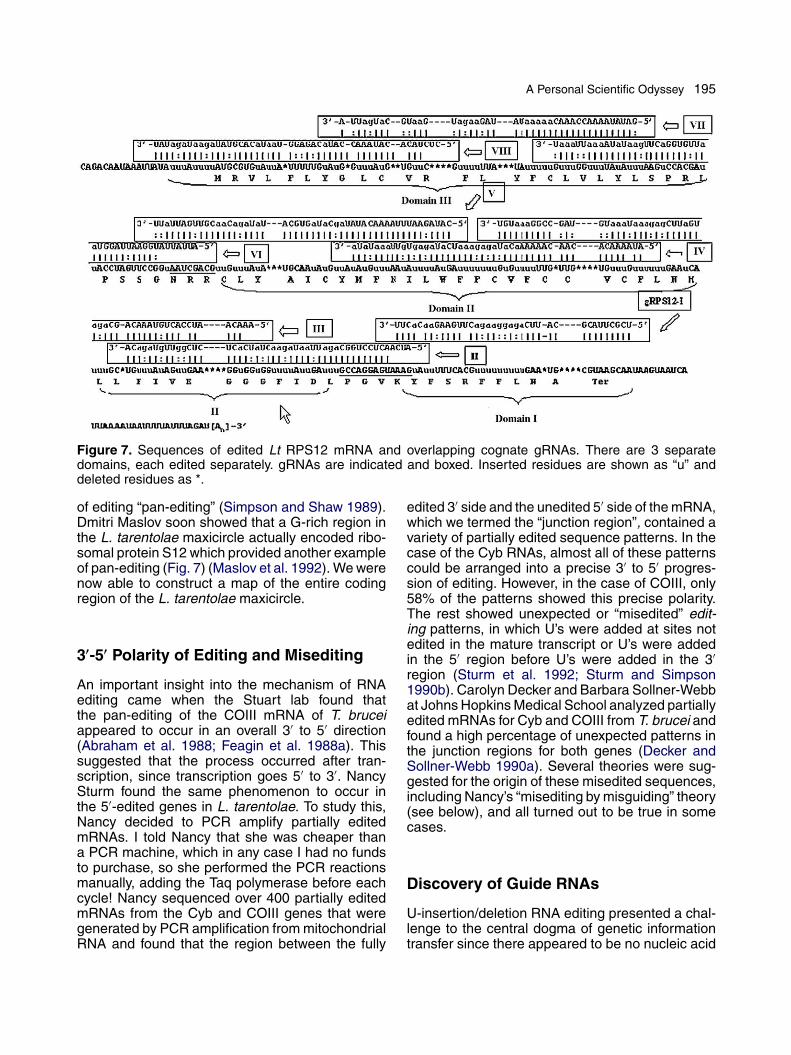
A Personal Scientific Odyssey 195
Figure 7. Sequences of edited Lt RPS12 mRNA and overlapping cognate gRNAs. There are 3 separatedomains, each edited separately. gRNAs are indicated and boxed. Inserted residues are shown as “u” anddeleted residues as *.
of editing “pan-editing” (Simpson and Shaw 1989).Dmitri Maslov soon showed that a G-rich region inthe L. tarentolae maxicircle actually encoded ribo-somal protein S12 which provided another exampleof pan-editing (Fig. 7) (Maslov et al. 1992). We werenow able to construct a map of the entire codingregion of the L. tarentolae maxicircle.
3′-5′ Polarity of Editing and Misediting
An important insight into the mechanism of RNAediting came when the Stuart lab found thatthe pan-editing of the COIII mRNA of T. bruceiappeared to occur in an overall 3′ to 5′ direction(Abraham et al. 1988; Feagin et al. 1988a). Thissuggested that the process occurred after tran-scription, since transcription goes 5′ to 3′. NancySturm found the same phenomenon to occur inthe 5′-edited genes in L. tarentolae. To study this,Nancy decided to PCR amplify partially editedmRNAs. I told Nancy that she was cheaper thana PCR machine, which in any case I had no fundsto purchase, so she performed the PCR reactionsmanually, adding the Taq polymerase before eachcycle! Nancy sequenced over 400 partially editedmRNAs from the Cyb and COIII genes that weregenerated by PCR amplification from mitochondrialRNA and found that the region between the fully
edited 3′ side and the unedited 5′ side of the mRNA,which we termed the “junction region”, contained avariety of partially edited sequence patterns. In thecase of the Cyb RNAs, almost all of these patternscould be arranged into a precise 3′ to 5′ progres-sion of editing. However, in the case of COIII, only58% of the patterns showed this precise polarity.The rest showed unexpected or “misedited” edit-ing patterns, in which U’s were added at sites notedited in the mature transcript or U’s were addedin the 5′ region before U’s were added in the 3′region (Sturm et al. 1992; Sturm and Simpson1990b). Carolyn Decker and Barbara Sollner-Webbat Johns Hopkins Medical School analyzed partiallyedited mRNAs for Cyb and COIII from T. brucei andfound a high percentage of unexpected patterns inthe junction regions for both genes (Decker andSollner-Webb 1990a). Several theories were sug-gested for the origin of these misedited sequences,including Nancy’s “misediting by misguiding” theory(see below), and all turned out to be true in somecases.
Discovery of Guide RNAs
U-insertion/deletion RNA editing presented a chal-lenge to the central dogma of genetic informationtransfer since there appeared to be no nucleic acid

196 L. Simpson
template for this newly added sequence informa-tion. We had, however, not given up on the centraldogma and two postdoctoral fellows in my labo-ratory, Beat Blum and Norbert Bakalara, decidedto give it one last try. They performed a com-puter search of the known L. tarentolae maxicirclesequences for short DNA sequences that couldgive rise to RNAs with complementarity to eitherentire or portions of known edited RNA sequences.In addition to the classical Watson-Crick base pairsC-G and A-U, they decided to allow for G-U basepairs (Fig. 8A) since these are bonafide base pairsin rRNAs and tRNAs. This turned out to be thetrick! Seven such short sequences for four of theknown edited genes were immediately found scat-
Figure 8. Discovery of guide RNAs. A. Upper: Align-ment of edited Lt mRNA sequence versus Ltmaxicircle DNA sequence. One of the best “hits” isshown. Mismatches are boxed. Lower: Alignment ofedited Lt mRNA versus a putative gRNA transcribedfrom the Lt maxicircle DNA sequence in (A). Notethat the mismatches in (A) are base pairs if G-U(*) is allowed. B. Lt kRNA run in acrylamide gel.Left lanes: ethidium bromide stained, showing tRNAs.Right lanes: Blots were probed with riboprobes fromtwo cloned kDNA minicircles, showing gRNAs. C. Themissing gRNA for COIII editing sites 1-8 is encoded ina partially sequenced minicircle within the “variable”region. The minicircle “conserved region” is boxed andthe highly conserved CSB-3 sequence shown. FromNucleic Acids Res (1989) 17 5427-5445, Cell (1990)61: 879-884, with permission.
tered throughout the maxicircle between knowngenes. In a few days we had synthesized oligonu-cleotides for primer extension to verify the existenceof the RNAs and within one very exciting week wehad definitive evidence for the existence of smallRNAs in the mitochondrion which were transcriptsof these sequences. These RNAs also containedsequences at their 5′ end which could form duplexregions with the mRNAs just downstream of thepre-edited regions, which we termed the “anchorregions”, since these provided an ideal way toanchor the short RNAs to the mRNAs by form-ing a double-stranded hybrid just downstream ofthe region that is to be edited. These small RNAshad an unusual mobility in gel electrophoresis; theymigrated in the form of 20-30 bands each differingby a single nucleotide in size. Based on the previ-ous use of the term, “internal guide sequences”, inGroup I self splicing introns (Davies et al. 1987), wecalled these molecules “guide RNAs” or “gRNAs”since they contained the sequence information forediting (Blum et al. 1990). The secret to the editingsequence information was simple base-pairing toshort gRNAs. One gRNA was found to be in cis atthe 3′ end of the COII mRNA.
Base-pairing by guide-like RNAs in trans hassince been found to explain the specificity of siRNAdegradation of mRNA and the snoRNA-mediatedspecificity of methylation and pseudouridylation ofrRNAs. And the in cis–guiding COII gRNA hasprovided a precedent for the specificity of A to Imodification editing of mammalian mRNAs beingdetermined by fold back of a downstream comple-mentary sequence. But we still did not understandhow the gRNAs could mediate the insertion anddeletion of U’s at precise sites.
Let me now digress from the historical develop-ment to return to the mid 1980’s when we had justfinished the sequence of the majority of the L. taren-tolae maxicircle. One striking observation was thatthere were no obvious tRNA genes. The apparentabsence of tRNA genes was really surprising sinceall mitochondrial genomes studied to that time con-tained tRNAs which are involved in mitochondrialprotein synthesis. To examine whether some of theminicircle molecules or regions of the maxicirclewhich we had not yet sequenced encoded tRNAs,My graduate student, Homero Dewes and AgdaSimpson hybridized labeled minicircle and maxi-circle DNA to low molecular weight RNA from L.tarentolae mitochondria which was separated byelectrophoresis in acrylamide. To our surprise, theminicircle probes lit up a cluster of 20-30 bandsone nucleotide apart that migrated well ahead ofthe abundant mitochondrial tRNAs, indicating that

A Personal Scientific Odyssey 197
these RNAs were even smaller than tRNAs; thisRNA was such a minor component that we could noteven see these bands by staining the gel! (Fig. 8B)(Simpson et al. 1989).
We now know that this was our first look at gRNAswhich were present at low abundance in our kRNApreparations, but at the time we had no idea whatthese transcripts were. The heterogeneity of thegRNAs was soon found to be due to the presence ofnon-encoded 3′ oligo-[U] tails 15 - 30 nucleotidesin length. And the fact that these appeared to beminicircle transcripts was also not appreciated atthe time.
It was clear, however, that no mitochondrialtRNAs were encoded in maxicircle or minicircleDNA and therefore it seemed likely that tRNAswere imported into the trypanosome mitochondrionfrom the cytoplasm, as had been suggested severalyears before for Tetrahymena by my collaboratorfrom the University of Pennsylvania, Yosh Suyama(Chiu et al. 1975; Suyama 1967). This was a hereti-cal idea at the time, but an idea which is nowgenerally accepted (Alfonzo and Soll 2009).
Guide RNAs are Encoded inMinicircles
At this time we had identified seven gRNA genesscattered all over the maxicircle with no positionalrelation to the cryptogenes for which they encodedinformation. These gRNAs had information for fourof the five known cryptogenes, but we could notfind a gRNA for the 5′ edited COIII gene in L.tarentolae. We had sequenced several kDNA mini-circles by that time (Kidane et al. 1984), and NancySturm decided to see if the misedited patterns shewas analyzing could be encoded by gRNAs. Toher surprise, she discovered the missing gRNAfor COIII editing sites 1-8 encoded in a minicir-cle which we had partially sequenced! (Fig. 8C)(Sturm and Simpson 1990a). Both Nancy and Ihad completely forgotten our previous results thatminicircles encoded short transcripts migrating inacrylamide as 15-30 bands one nucleotide apartbut now with hindsight these results made sense.
Nancy’s discovery was the first indication of agenetic function of the hitherto enigmatic minicircleDNA and explained neatly the previously observedsequence heterogeneity of minicircle DNA - eachsequence class encoded a different gRNA withinthe variable region! Previous work by my graduatestudents, Getachew Kidane and Michael Muhichand further work by Nancy Sturm and DmitriMaslov, resulted in the identification of a total of
17 different minicircle sequence classes of differ-ing abundances in the L. tarentolae UC strain bycloning and sequencing.
Soon, minicircle-encoded gRNAs were alsofound in T. brucei and the closely related species,T. equiperdum, by the Hajduk (Pollard and Hajduk1991) and Stuart (Shu and Stuart 1993) labs. Thereis one major interesting difference in that each mini-circle in T. brucei encodes three different gRNAsrather than a single gRNA and the genes arelocated in the single variable region between threesets of 18 nt inverted repeats which are not presentin L. tarentolae. Another difference is that previ-ous workers had claimed that there were over threehundred different minicircle sequence classes in T.brucei versus the limited number found in L. taren-tolae, suggesting that the total number of differentgRNAs in T. brucei may be over 900. Some ofthese additional gRNAs are probably required forthe three pan-edited genes that are only 5′-editedin L. tarentolae, and for the five pan-edited G-richgenes discussed below, but the function of the restremains to be investigated. In 2003, we showed thatthe original estimate of minicircle sequence hetero-geneity in T. brucei was an overestimate, but thenumber is still much larger than that found for L.tarentolae (Hong and Simpson 2003).
It was shown by Rob Benne that Crithidia fasic-ulata, a parasite of insects, contains the samemaxicircle DNA-encoded gRNA genes as L. tar-entolae, located at identical relative positions inthe maxicircle genome (Van der Spek et al. 1991).The gRNA anchor sequences in C. fasciculata haveseveral differences compared to the L. tarento-lae sequences, but these are compensated for bymutations in the cryptogene sequences so as topreserve base-pairing. These results nicely confirmthe role of gRNAs in editing by the same type ofevolutionary argument that was previously used byNoller and Woese (Woese et al. 1983) to confirmthe structure of double stranded regions in riboso-mal RNAs.
G-rich Intergenic MaxicircleSequences are Pan-EditedCryptogenes
In our initial comparison of the mitochondrialgenomes of L. tarentolae and T. brucei, wehad noted that there were several stretches ofsequences that were relatively rich in G residues(Simpson et al. 1987). Three of these in T. bruceiwere shown by the Stuart lab to be the three hid-den pan-edited cryptogenes, ND7 (Koslowsky et

198 L. Simpson
al. 1990) COIII (Feagin et al. 1988a) and MURF4(Bhat et al. 1990) (now known as A6), but therewere another six G-rich regions that were locatedbetween known genes in both species. Maslov,Sturm and Marian Peris showed that the transcriptof G-rich region 6 in L. tarentolae is pan-edited bythe addition of 117 U’s at 49 sites and the dele-tion of 32 U’s at 13 sites in three editing domains,producing an mRNA which encodes a protein forthe small subunit of the mitochondrial ribosome(Fig. 7) (Maslov et al. 1992). It appeared thatall six G-rich regions, perhaps in all kinetoplas-tid species, are pan-edited cryptogenes, encodingproteins important for the proper functioning ofthe mitochondrion. These additional cryptogeneswould require approximately 50 additional over-lapping gRNAs for mediating the required editing,which did not agree with the 17 different minicir-cle sequence classes found in the old L. tarentolaeUC lab strain. In T. brucei there are only twogRNA genes in the maxicircle, but each minicir-cle encodes 3-4 gRNAs. It appears that in theAfrican trypanosome there has been a movementof gRNA genes between the maxicircle and mini-circle genomes. In fact the short inverted repeatsequences found adjacent to the gRNA genes in
the minicircles of T. brucei could possibly representthe evolutionary remnants of transposition eventsin which gRNA genes moved from the maxicircle tothe minicircle or between minicircles.
Loss of Editing in Old LaboratoryStrain of Leishmania tarentolae
The 17 identified minicircle-encoded gRNAs in theold lab UC strain of L. tarentolae were clearlyinsufficient to account for the complete editing ofall cryptogenes. This problem was solved by mygraduate student, Otavio Thiemann, who is now aProfessor in San Carlos, Brazil. He showed thatthe UC strain was defective in the editing of theG1-G5 cryptogene transcripts due to the absenceof gRNAs for these editing events, whereas therecently isolated LEM 125 strain contained atleast 32 additional gRNAs which encoded pro-ductive editing of the G1-G5 cryptogenes (Fig. 9)(Thiemann et al. 1994). The loss of gRNAs,accompanied by the loss of editing, was actuallythe first genetic evidence for the involvement ofgRNAs in the editing process. We speculated thatthese minicircle-encoded gRNAs were lost during
Figure 9. Diagrammatic comparison of gRNA-mediated editing of maxicircle cryptogenes in old lab UC strainof L. tarentolae and recently isolated LEM125 strain. The identified overlapping gRNAs that cause the 3′ to 5′polarity of editing are indicated. All of the 80 predicted gRNAs in the LEM strain are shown although only 47have been identified. From Proc Natl Acad Sci USA (2000) 97 6986-6993, with permission.

A Personal Scientific Odyssey 199
the long culture history of the UC strain by lossof the specific minicircle classes. Nick Savill, acomputer science collaborator, showed in fact bycomputer modeling with a few basic assumptions,that minicircle classes would fluctuate dramaticallyin copy number in the course of many generationsin culture and that low frequency minicircle classescould be lost entirely in the absence of selectiondue to the random nature of the network minicir-cle segregation process during the cell cycle (Savilland Higgs 1999; Simpson et al. 2000).
Enzyme-Cascade Model for RNAEditing
Prior to the discovery of gRNAs, Norbert Bakalara,a postdoctorate in my lab, and Agda Simpson hadidentified an enzyme activity from purified mito-chondria of L. tarentolae which could add U’s to the3′ terminus of any RNA molecule - a terminal uridy-lyl transferase or TUTase (Bakalara et al. 1989).This enzyme possibly was responsible for the addi-tion of U’s to the 3′ end of the gRNAs. They alsoshowed the presence of a mitochondrial RNA lig-ase which could covalently link together two RNAmolecules.
Armed with the knowledge of the existence ofgRNAs and these enzymatic activities in the mito-chondrion and of the 3′ to 5′ progression of editingon the mRNA, Beat Blum, Bakalara and I came upwith a model for the role of gRNAs in RNA edit-ing (Fig. 10) (Blum et al. 1990). We called this the‘enzyme cascade’ model since it postulates a seriesof enzymatic reactions occuring in a multienzymecomplex bound to the mRNA. We proposed that theinitial interaction involves the formation of an anchorhybrid by a specific gRNA just 3′ of the pre-editedregion on the mRNA. In addition to RNA/RNA inter-actions involved in the formation of an anchor, webelieve protein factors which Agda Simpson hadfound to be complexed to the gRNAs assist in thisinitial specific interaction, perhaps by recognizingsecondary structures formed by the mRNA itselfor by the gRNA/mRNA hybrid. The next step wasproposed to be a specific cleavage at the first mis-matched base in the mRNA which liberates a free3′ OH group. This cleaved mRNA fragment is agood substrate for the 3′ TUTase enzyme whichcould add one or more U’s to the 3′ end. Theseadded U’s would then base pair with the guide Aor G nucleotides in the gRNA, and then the twoends of the mRNA would be religated by the RNAligase. This would result in a zippering up of thedouble helix in a 3′ to 5′ direction (on the mRNA),
Figure 10. The original “enzyme cascade” model ofRNA editing. U-deletion shown on left and U-insertionon right. Predicted enzymatic activities are indicated.We also suggested that the 3′ U-tail may base pairwith the pre-edited sequence, thereby stabilizing theinitial interaction. From Annal NY Acad Sci (1999) 870:190-205, with permission.
and the whole process would then reinitiate at thenext mismatched base.
This model provided an explanation for the 3′to 5′ polarity of pan-editing, as due to the cre-ation by the downstream gRNA of edited mRNAsequence that was complementary to the anchorsequence of the adjacent upstream gRNA. Thiswas shown clearly by Maslov for the A6 and RPS12(Fig. 7) mRNA editing in L. tarentolae (Maslovand Simpson 1992). The model also explained thepresence of unexpected editing patterns within thejunction regions of partially edited mRNAs (Sturmand Simpson 1990b). Sturm and I had suggestedthat some of these patterns actually represent nor-mal editing by inappropriate gRNAs or appropriategRNAs in the wrong location or wrong readingframe, a process which we termed “misediting bymisguiding”, and which is enhanced by the pres-ence of ‘wobble’ G-U and perhaps A-C base pairs.The formation of an anchor hybrid by the incorrectgRNA or the formation of a secondary anchor inthe wrong location by the correct gRNA could leadto the formation of an unexpected editing pattern,which would terminate the editing process sincea correct anchor for the next gRNA would not beformed. However, misedited sequences within thejunction region could be re-edited with the correctgRNA. Many examples of misediting/misguiding

200 L. Simpson
which are consistent with this hypothesis havebeen found. However, another interpretation ofunexpected patterns was proposed by Decker andSollner-Webb (Decker and Sollner-Webb 1990b).They suggested that editing is completely randomand occurs between every nucleotide within an edit-ing domain, and that when the correct sequence isformed it is “frozen” by the formation of base pairswith the gRNA. This issue will not be resolved untilwe have a complete knowledge of the total gRNAcontent of the mitochondrion and can compare allunexpected patterns to known gRNA sequences,and also a deeper understanding of the mechanismof editing.
The enzyme cascade model is consistent withmost observations, including the known 3′ to 5′polarity of editing, but it does not really explain theexistence of the 3′ oligo-[U] tail on the gRNA (Blumand Simpson 1990). We had proposed a role forthe 3′ oligo-[U] tail in stabilizing the initial hybrid,since the U’s would form base pairs with G’s andA’s in the pre-edited region. However, soon afterwe had proposed the “enzyme cascade” model,Blum, who had this problem of always thinking toomuch, had the idea that perhaps the oligo-[U] tailplayed a more active role and actually was thesource of the U’s added during editing. We thereforeproposed another model in which the 3′ terminalOH of the gRNA attacked a phosphate within themRNA at the site of the first mismatch betweenthe gRNA and mRNA, resulting in exchange of theOH for the phosphate by transesterification (Blumand Simpson 1992; Blum et al. 1991). The transes-terification reaction is similar to that which occursin self-splicing of RNA molecules in other cells. Aprediction of this model is the existence of interme-diate chimeric molecules which consist of gRNAscovalently linked to mRNAs at editing sites by the3′ oligo-[U] tail. Blum, Sturm and Agda Simpsonimmediately searched for and found these chimericmolecules for three genes. This was gratifying, butdid not really prove the transesterification model,since chimeric molecules could possibly be formedin other ways, especially in a system which we knewcontains a cleavage activity and an RNA ligaseactivity.
At about this time I visited Tom Cech in Boulder.Cech, who had discovered that RNA could haveenzymatic activity, told me that he had solved theediting problem and proceeded to draw on a sheetof paper the entire transesterification model! (Cech1991). As happens frequently in science, whenideas are ripe, they germinate simultaneously inseveral gardens. The transesterification model wastheoretically attractive since it employed the same
chemistry and the same type of guide sequencesused in the well understood self splicing of introns,whereas the enzyme cascade model was a novelset of protein-mediated reactions. However, thiswas an example of a beautiful theory which crashedon the hard rocks of facts, since it turned out notto be true (Frech and Simpson 1996). Our originalcleavage-ligation model has proved to be essen-tially correct in almost all details.
Evolution of RNA Editing inTrypanosomes
The question whether RNA editing is a primitiveor derived mechanism is an important one andyet to be resolved. To address this question welooked at Trypanoplasma borreli, the only speciesin the group of bodonids which could be grown inaxenic culture. This paraphyletic group is relatedto trypanosomatids and is thought to have evolvedprior to the emergence of the monophyletic try-panosomatids. T. borreli turned out to have twoclasses of molecules in the mitochondrion: 80 kbcircles which encode homologues of trypanosomemaxicircle genes, and 260 kb megacircles whichencode guide RNA genes (Fig. 11) (Maslov andSimpson 1994). Other labs showed that severalother bodonid species also contain maxicirclehomologues and non-catenated minicircles withgRNA genes. It appears that the separation ofthe mitochondrial genome into maxicircle-encodedcryptogenes and minicircle- or megacircle-encodedgRNA genes occurred in the bodonid lineage, andcatenation of minicircles to form the kDNA networkfirst evolved in the ancestor of the trypanosomatids(Fig. 12). A postdoctoral fellow from Japan, ShinjiYasuhira, and Maslov also examined mitochon-dria of Euglena and Diplonema, the other memberof Euglenozoa (Maslov et al. 1999; Yasuhira andSimpson 1997). No evidence for editing was foundbut it is hard to prove a negative. But since this typeof RNA editing is not seen in other eukaryotes, it islikely that U-insertion/deletion editing arose in themitochondrion of the ancestor of the kinetoplastidprotists.
Maslov and I also did a comparative study ofediting of several genes in different trypanosomatidspecies (Simpson and Maslov 1994). We founda gradual loss of editing from the 3’ end and wespeculated that this was due to the loss of spe-cific gRNA-encoding minicircle classes in evolution,which could be compensated by retroposition ofcDNAs of partially edited mRNAs replacing theoriginal pan-edited cryptogenes in the maxicircle

A Personal Scientific Odyssey 201
Figure 11. Comparison of gRNA gene organization in several trypanosomatid species and in the bodonid,Trypanoplasma borreli. From Mol Biochem Parasitol (1997) 86: 133-141, with permission.
Figure 12. Phylogenetic tree of Kinetoplastida based on SSU rRNA sequences. Maximum likelihood method.Some unpublished data of D. Dolezel, M. Jirku and J. Lukes, with thanks. From Proc Natl Acad Sci USA (2000)97: 6986-6993, with permission.

202 L. Simpson
Figure 13. Retroposition model for evolution of RNA editing. A pan-edited domain mediated by 7 overlappinggRNAs is shown. Loss of minicircle class IV indicated by crossing out. Proposed double crossover of partiallyedited cDNA and pan-edited maxicircle gene indicated by X. From Annals NY Acad Sci (1999) 870: 190-205,with permission.
genome (Fig. 13). This model fits all available data,but raises the unsettling question of why editing hasbeen maintained at all in evolution.
All tRNAs are Imported into theKinetoplast-Mitochondrion ofTrypanosomes
During the decade from 1990 to 2000, we wereoccupied trying to develop in vitro editing reactionsand to learn more details of the reaction mecha-nism. In addition we started a side project whichwas based on our discovery that there were notRNA genes in the maxicircle. A Brazilian postdoc-torate, Beatriz Lima, showed that L. tarentolae cellscould be transfected with a plasmid expressingmitochondrial tRNAs and these would enter themitochondrion in vivo (Lima and Simpson 1996). Agraduate student, Mary Anne Rubio, subsequentlydeveloped an in vitro import system (Rubio et al.2000) using isolated kinetoplast-mitochondria andshowed that different tRNAs had different specifici-ties. Another graduate student, Steve Kapushoc,did a quantitative assay of the localization of tRNAsin the cytosol or mitochondrion (Kapushoc et al.2002).
The mitochondrial genetic code in trypanosomesas in other organisms uses the UGA stop codon astryptophan, and the importation of all tRNAs into themitochondrion presented a theoretical problem for
decoding UGA. Juan Alphonzo, then a postdoctor-ate in my lab and now a Professor at Ohio StateUniversity, had the ingenious realization that a pos-sible solution would be that the imported tRNATryp
undergoes a single C to U substitution or editingevent in the first position of the anticodon (Fig. 14)(Alfonzo et al. 1999). This, surprisingly, turned outto be true and was the first indication that C to Uediting occurs in trypanosomes as in several otherorganisms.
Figure 14. C to U editing of the anticodon of themitochondrial-imported tRNATrp. A. Editing of C34leads to destruction of HinfI site. B. C34 to U34 editingallows decoding of UGA codon as tryptophan. FromEMBO J (1999) 18 7056-7062, with permission.

A Personal Scientific Odyssey 203
Biochemistry of Editing Enzymes
The editing project received a boost when Rus-lan Aphasizhev, then a postdoctorate and now aProfessor at the University of California at Irvine,and Marian Peris, a graduate student, initiateda major lab project to biochemically isolate the3’ TUTase from L. tarentolae mitochondria. Thisproject involved the development of large scaleculture and mitochondrial isolation methods andwas ultimately successful (Aphasizhev et al. 2002).Interestingly, this TUTase, which we have termedRET1, turned out to be responsible for the 3’ addi-tion of U’s to gRNAs (Aphasizhev et al. 2003c).
Then we decided to utilize a new double epi-tope tagging technique, the TAP method, to isolatethe editing complex. Several labs had at that timecloned and sequenced the two mitochondrial RNAligases, REL1 and REL2, and, based on thissequence data, we transfected L. tarentolae cellswith a REL1-TAP fusion protein construct. This ledto the isolation of a high molecular weight complex(Aphasizhev et al. 2003a) containing at least 16 pro-teins which we called the L-complex and now callthe RNA Editing Core Complex or RECC (Simpsonet al. 2009). Utilizing the power of mass spectrom-etry combined with the Leishmania major genomeproject, we were able to identify all of componentproteins. One of the proteins was identified as a 3’TUTase, and this enzyme, which we termed RET2,proved to be responsible for the insertion of U’s atediting sites (Aphasizhev et al. 2003c). At the sametime we isolated the MRP RNA-binding complex,and showed that this and also the RET1 complexinteract via RNA linkers with the RECC (Aphasizhevet al. 2003b). All these results were and are veryexciting and we and others have worked exten-sively on the molecular biochemistry of the editingmachinery.
My current research project is to determine themolecular structure of the L. tarentolae RECC (Li etal. 2009). We are doing this in a close collaborationwith the lab of Hong Zhou at UCLA, a fantastic cryo-electron microscopist with some equally fantasticmicroscopes.
Epilogue
A scientific career is somewhat like a river thatstarts as a small creek, grows as more creeks joinit, occasionally meanders and changes direction,and often splits into separate streams, some ofwhich continue downhill and others end in the fens.My career has been somewhat unusual in that it
has been monomaniacally focused throughout ona single biological phenomenon, the kinetoplast-mitochondrion of the kinetoplastid protists. But myinterests have changed from basic parasitology andcell biology, to DNA structure and replication, totranscription and RNA biology, and finally to pro-tein biochemistry. It is hard to summarize a life inscience that is still actively progressing, but I cansay that it has been a lot of fun and that I was verylucky to have such excellent graduate students andpostdoctoral fellows to share the excitement of theadventure.
Appendix A. Supplementary data
Supplementary data associated with this arti-cle can be found, in the online version, atdoi:10.1016/j.protis.2010.08.001.
References
Abraham J, Feagin J, Stuart K (1988) Characterization ofcytochrome c oxidase III transcripts that are edited only in the3’ region. Cell 55:267–272
Alfonzo JD, Blanc V, Estévez AM, Rubio MAT, Simpson L(1999) C to U editing of the anticodon of imported mitochondrialtRNATrp allows decoding of the UGA stop codon in Leishmaniatarentolae. EMBO J 18:7056–7062
Alfonzo JD, Soll D (2009) Mitochondrial tRNA import–the chal-lenge to understand has just begun. Biol Chem 390:717–722
Aphasizhev R, Aphasizheva I, Nelson RE, Gao G, SimpsonAM, Kang X, Falick AM, Sbicego S, Simpson L (2003a) Isola-tion of a U-insertion/deletion editing complex from Leishmaniatarentolae mitochondria. EMBO J 22:913–924
Aphasizhev R, Aphasizheva I, Nelson RE, Simpson L(2003b) A 100-kD complex of two RNA-binding proteins frommitochondria of Leishmania tarentolae catalyzes RNA anneal-ing and interacts with several RNA editing components. RNA9:62–76
Aphasizhev R, Aphasizheva I, Simpson L (2003c) A tale oftwo TUTases. Proc Natl Acad Sci USA 100:10617–10622
Aphasizhev R, Sbicego S, Peris M, Jang SH, AphasizhevaI, Simpson AM, Rivlin A, Simpson L (2002) Trypanosomemitochondrial 3’ terminal uridylyl transferase (TUTase): The keyenzyme in U-insertion/deletion RNA editing. Cell 108:637–648
Avila HA, Pereira JB, Thiemann O, De Paiva E, DegraveW, Morel CM, Simpson L (1993) Detection of Trypanosomacruzi in blood specimens of chronic chagasic patients by poly-merase chain reaction amplification of kinetoplast minicircleDNA: Comparison with serology and xenodiagnosis. J ClinMicrobiol 31:2421–2426
Bakalara N, Simpson AM, Simpson L (1989) The Leishmaniakinetoplast-mitochondrion contains terminal uridylyltransferaseand RNA ligase activities. J Biol Chem 264:18679–18686

204 L. Simpson
Benne R, Van den Burg J, Brakenhoff J, Sloof P, Van BoomJ, Tromp M (1986) Major transcript of the frameshifted coxIIgene from trypanosome mitochondria contains four nucleotidesthat are not encoded in the DNA. Cell 46:819–826
Bhat GJ, Koslowsky DJ, Feagin JE, Smiley BL, Stuart K(1990) An extensively edited mitochondrial transcript in kineto-plastids encodes a protein homologous to ATPase subunit 6.Cell 61:885–894
Birkenmeyer L, Ray DS (1986) Replication of kinetoplast DNAin isolated kinetoplasts from Crithidia fasciculata. Identifica-tion of minicircle DNA replication intermediates. J Biol Chem261:2362–2368
Blum B, Simpson L (1990) Guide RNAs in kineto-plastid mitochondria have a nonencoded 3’ oligo-(U) tailinvolved in recognition of the pre-edited region. Cell 62:391–397
Blum B, Simpson L (1992) Formation of gRNA/mRNAchimeric molecules in vitro, the initial step of RNA editing, isdependent on an anchor sequence. Proc Natl Acad Sci USA89:11944–11948
Blum B, Bakalara N, Simpson L (1990) A model for RNAediting in kinetoplastid mitochondria: “Guide” RNA moleculestranscribed from maxicircle DNA provide the edited information.Cell 60:189–198
Blum B, Sturm NR, Simpson AM, Simpson L (1991) ChimericgRNA-mRNA molecules with oligo(U) tails covalently linked atsites of RNA editing suggest that U addition occurs by transes-terification. Cell 65:543–550
Braly P, Simpson L, Kretzer F (1974) Isolation of kinetoplast-mitochondrial complexes from Leishmania tarentolae. JProtozool 21:782–790
Cech TR (1991) RNA editing: World’s smallest introns. Cell64:667–669
Chiu N, Chiu A, Suyama Y (1975) Native and imported transferRNA in mitochondria. J Mol Biol 99:37–50
Davies RW, Waring RB, Towner P (1987) Internal guidesequence and reaction specificity of group I self-splicing introns.Cold Spring Harb Symp Quant Biol 52:165–171
de la Cruz V, Neckelmann N, Simpson L (1984) Sequencesof six structural genes and several open reading frames in thekinetoplast maxicircle DNA of Leishmania tarentolae. J BiolChem 259:15136–15147
de la Cruz V, Lake JA, Simpson AM, Simpson L (1985a) Aminimal ribosomal RNA: sequence and secondary structure ofthe 9S kinetoplast ribosomal RNA from Leishmania tarentolae.Proc Natl Acad Sci USA 82:1401–1405
de la Cruz V, Simpson A, Lake J, Simpson L (1985b) Pri-mary sequence and partial secondary structure of the 12Skinetoplast (mitochondrial) ribosomal RNA from Leishmaniatarentolae: Conservation of peptidyl-transferase structural ele-ments. Nucleic Acids Res 13:2337–2356
Decker CJ, Sollner-Webb B (1990a) RNA editing involvesindiscriminate U changes throughout precisely defined editingdomains. Cell 61:1001–1011
Decker CJ, Sollner-Webb B (1990b) RNA editing involvesindiscriminate U changes throughout precisely defined editingdomains. Cell 61:1001–1011
Degrave W, Fragoso S, Britto C, Van Heuverswyn H, KidaneG, Cardoso M, Mueller R, Simpson L, Morel C (1988) Pecu-liar sequence organization of kinetoplast DNA minicircles fromTrypanosoma cruzi. Mol Biochem Parasitol 27:63–70
Feagin JE, Abraham J, Stuart K (1988a) Extensive editing ofthe cytochrome c oxidase III transcript in Trypanosoma brucei.Cell 53:413–422
Feagin JE, Shaw JM, Simpson L, Stuart K (1988b) Cre-ation of AUG initiation codons by addition of uridines withincytochrome b transcripts of kinetoplastids. Proc Natl Acad SciUSA 85:539–543
Frech GC, Simpson L (1996) Uridine insertion into preeditedmRNA by a mitochondrial extract from Leishmania tarentolae:Stereochemical evidence for the enzyme cascade model. MolCell Biol 16:4584–4589
Hong M, Simpson L (2003) Genomic organization ofTrypanosoma brucei kinetoplast DNA minicircles. Protist154:265–279
Kapushoc ST, Alfonzo JD, Simpson L (2002) Differentiallocalization of nuclear-encoded tRNAs between the cytosol andmitochondrion in Leishmania tarentolae. RNA 8:57–68
Kidane G, Hughes D, Simpson L (1984) Sequence hetero-geneity and anomalous electrophoretic mobility of kinetoplastminicircle DNA in Leishmania tarentolae. Gene 27:265–277
Kleisen C, Borst P (1975) Sequence heterogeneity of theminicircle of kinetoplast DNA of Crithidia luciliae and evidencefor presence of a component more complex than minicircleDNA in the kinetoplast DNA network. Biochim Biophys Acta407:473–478
Kleisen C, Weislogel P, Fonck K, Borst P (1976) Character-ization of a novel component of high complexity present in thekinetoplast DNA network of Crithidia luciliae. Eur J Biochem64:153–160
Koslowsky DJ, Bhat GJ, Perrollaz AL, Feagin JE, Stuart K(1990) The MURF3 gene of Trypanosoma brucei contains multi-ple domains of extensive editing and is homologous to a subunitof NADH dehydrogenase. Cell 62:901–911
Li F, Ge P, Hui W, Atanasov A, Rogers K, Guo Q, OsatoD, Falick AM, Zhou H, Simpson L (2009) Structure of thecore editing complex (L-Complex) involved in uridine inser-tion/deletion RNA editing in trypanosomatid mitochondria. ProcNatl Acad Sci USA 106:12306–12310
Lima BD, Simpson L (1996) Sequence-dependent in vivoimportation of tRNAs into the mitochondrion of Leishmania tar-entolae. RNA 2:429–440
Maslov DA, Simpson L (1992) The polarity of editing within amultiple gRNA-mediated domain is due to formation of anchorsfor upstream gRNAs by downstream editing. Cell 70:459–467
Maslov DA, Simpson L (1994) RNA editing and mitochondrialgenomic organization in the cryptobiid kinetoplastid protozoan,Trypanoplasma borreli. Mol Cell Biol 14:8174–8182
Maslov DA, Yasuhira S, Simpson L (1999) Phylogenetic affini-ties of Diplonema within the Euglenozoa as inferred from theSSU rRNA gene and partial COI protein sequences. Protist150:33–42
Maslov DA, Sturm NR, Niner BM, Gruszynski ES, PerisM, Simpson L (1992) An intergenic G-rich region in Leish-
A Personal Scientific Odyssey 205
mania tarentolae kinetoplast maxicircle DNA is a pan-editedcryptogene encoding ribosomal protein S12. Mol Cell Biol 12:56–67
Morel C, Chiari E, Camargo E, Mattei D, Romanha A, Simp-son L (1980) Strains and clones of Trypanosoma cruzi can becharacterized by restriction endonuclease fingerprinting of kine-toplast DNA minicircles. Proc Natl Acad Sci USA 77:6810–6814
Morris JC, Drew ME, Klingbeil MM, Motyka SA, SaxowskyTT, Wang Z, Englund PT (2001) Replication of kinetoplastDNA: an update for the new millennium. Int J Parasitol31:453–458
Ntambi J, Englund P (1985) A gap at a unique locationin newly replicated kinetoplast DNA minicircles from Try-panosoma equiperdum. J Biol Chem 260:5574–5579
Pollard VW, Hajduk SL (1991) Trypanosoma equiperdum mini-circles encode three distinct primary transcripts which exhibitguide RNA characteristics. Mol Cell Biol 11:1668–1675
Ray DS (1989) Conserved sequence blocks in kinetoplast mini-circles from diverse species of trypanosomes. Mol Cell Biol9:1365–1367
Rubio MA, Liu X, Yuzawa H, Alfonzo JD, Simpson L (2000)Selective importation of RNA into isolated mitochondria fromLeishmania tarentolae. RNA 6:988–1003
Savill NJ, Higgs PG (1999) A theoretical study of random seg-regation of minicircles in trypanosomatids. Proc R Soc Lond BBiol Sci 266:611–620
Sharma MR, Booth TM, Simpson L, Maslov DA, AgrawalRK (2009) Structure of a mitochondrial ribosome with minimalRNA. Proc Natl Acad Sci USA 106:9637–9642
Shaw J, Feagin JE, Stuart K, Simpson L (1988) Editing ofmitochondrial mRNAs by uridine addition and deletion gen-erates conserved amino acid sequences and AUG initiationcodons. Cell 53:401–411
Shu HH, Stuart K (1993) A Trypanosoma brucei minicircleencodes the same gRNAs as do minicircles of Trypnosomaequiperdum ATCC 30019 and Trypanosoma evansi type-A mini-circles. Nucleic Acids Res 21:2951
Simpson A, Simpson L (1976) Pulse-labeling of kinetoplastDNA:Localization of two sites of synthesis within the net-works and kinetics of labeling of closed minicircles. J Protozool23:583–587
Simpson AM, Suyama Y, Dewes H, Campbell D, SimpsonL (1989) Kinetoplastid mitochondria contain functional tRNAswhich are encoded in nuclear DNA and also small minicircleand maxicircle transcripts of unknown function. Nucleic AcidsRes 17:5427–5445
Simpson L (1968a) Behavior of the kinetoplast of Leishmaniatarentolae. J Protozool 15:132–136
Simpson L (1968b) The effects of acriflavin on the kinetoplastDNA of Leishmania tarentolae: mechanism of action and phys-iological correlates of the loss of kinetoplast DNA. J Cell Biol37:660–682
Simpson L (1968c) The leishmania-leptomonad transfor-mation of Leishmania donovani: nutritional requirements,respiration changes and antigenic changes. J Protozool15:201–207
Simpson L (1979) Isolation of maxicircle component of kine-toplast DNA from hemoflagellate protozoa. Proc Natl Acad SciUSA 76:1585–1588
Simpson L, Braly P (1970) Synchronization of Leishmaniatarentolae by hydroxyurea. J Protozool 17:511–517
Simpson L, da Silva AM (1971) Isolation and characteriza-tion of kinetoplast DNA from Leishmania tarentolae. J Mol Biol56:443–473
Simpson L, Maslov DA (1994) RNA editing and the evolutionof parasites. Science 264:1870–1871
Simpson L, Shaw J (1989) RNA editing and the mitochondrialcryptogenes of kinetoplastid protozoa. Cell 57:355–366
Simpson L, Simpson A (1978) Kinetoplast RNA from Leish-mania tarentolae. Cell 14:169–178
Simpson L, Simpson A, Wesley R (1974) Replication of thekinetoplast DNA of Leishmania tarentolae and Crithidia fascic-ulata. Biochim Biophys Acta 349:161–172
Simpson L, Aphasizhev R, Lukes J, Cruz Reyes J (2009)Guide to the nomenclature of kinetoplastid RNA editing: A pro-posal. Protist 161:2–6
Simpson L, Simpson AM, Kidane G, Livingston L, SpithillTW (1980) The kinetoplast DNA of the hemoflagellate protozoa.Am J Trop Med Hyg 29:1053–1063
Simpson L, Thiemann OH, Savill NJ, Alfonzo JD, Maslov DA(2000) Evolution of RNA editing in trypanosome mitochondria.Proc Natl Acad Sci USA 97:6986–6993
Simpson L, Neckelmann N, de la Cruz V, Simpson A, Fea-gin J, Jasmer D, Stuart K (1987) Comparison of the maxicircle(mitochondrial) genomes of Leishmania tarentolae and Try-panosoma brucei at the level of nucleotide sequence. J BiolChem 262:6182–6196
Simpson LP, Kirby-Smith JS, Randolph ML (1963) Electronspin resonance in photodynamic dyes. Nature 199:243–245
Sloof P, Van den Burg J, Voogd A, Benne R, AgostinelliM, Borst P, Gutel R, Noller H (1985) Further characterizationof the extremely small mitochondrial ribosomal RNAs from try-panosomes: a detailed comparison of the 9S and 12S RNAsfrom Crithidia fasciculata and Trypanosoma brucei with rRNAsfrom other organisms. Nucleic Acids Res 13:4171–4190
Sturm NR, Simpson L (1990a) Kinetoplast DNA minicirclesencode guide RNAs for editing of cytochrome oxidase subunitIII mRNA. Cell 61:879–884
Sturm NR, Simpson L (1990b) Partially edited mRNAs forcytochrome b and subunit III of cytochrome oxidase from Leish-mania tarentolae mitochondria: RNA editing intermediates. Cell61:871–878
Sturm NR, Maslov DA, Blum B, Simpson L (1992) Gener-ation of unexpected editing patterns in Leishmania tarentolaemitochondrial mRNAs: misediting produced by misguiding. Cell70:469–476
Suyama Y (1967) The origins of mitochondrial ribonucleic acidsin Tetrahymena pyriformis. Biochemistry 6:2829–2839
Thiemann OH, Maslov DA, Simpson L (1994) Disruption ofRNA editing in Leishmania tarentolae by the loss of minicircle-encoded guide RNA genes. EMBO J 13:5689–5700

206 L. Simpson
Trager W (1957) Nutrition of a hemoflagellate (Leishmania tar-entolae) having an interchangeable requirement for choline orpyridoxal. J Protozool 4:269–276
Van der Spek H, Arts GJ, Zwaal RR, Van den Burg J, SloofP, Benne R (1991) Conserved genes encode guide RNAsin mitochondria of Crithidia fasciculata. EMBO J 10:1217–1224
Wesley RD, Simpson L (1973) Studies on kinetoplast DNA. III.Kinetic complexity of kinetoplast and nuclear DNA from Leish-mania tarentolae. Biochim Biophys Acta 319:267–276
White TC, Rudenko G, Borst P (1986) Three small RNAswithin the 10 kb trypanosome rRNA transcription unit are anal-ogous to domain VII of other eukaryotic 28S rRNAs. NucleicAcids Res 14:9471–9489
Woese C, Gutell R, Gupta R, Noller H (1983) Detailed analysisof the higher order structure of 16s- like ribosomal ribonucleicacids. Microbiol Rev 47:621–669
Yasuhira S, Simpson L (1997) Phylogenetic affinity ofmitochondria of Euglena gracilis and kinetoplastids usingcytochrome oxidase I and hsp60. J Mol Evol 44:341–347

Supplemental Figure S1

Supplemental Figure S2

Supplemental Figure S3

Supplemental Figure S4

INPA Forest Preserve
Supplemental Figure S5

Supplementary Figure S6

Supplementary Figure S7