Biotic abiotic transcriptional post transcriptional regulation

Erratum
A Pbx1-dependent genetic and transcriptional network regulates spleenontogenyAndrea Brendolan, Elisabetta Ferretti, Valentina Salsi, Kelvin Moses, Susan Quaggin, Francesco Blasi, Michael L. Clearly andLicia Selleri Development 132, 3113-3126.
Fig. S1 in the supplementary material accompanying this paper was published incorrectly when the paper first appeared on-line.
This has now been rectified and we apologise to the authors and to readers for this mistake.

3113
IntroductionIn vertebrates, the spleen is a lymphoid organ that servesimportant roles in hematopoiesis and the generation of primaryimmune responses, as well as acting as a filter that removesand processes aged and abnormal blood cells (VanRooijen etal., 1989; Zapata and Cooper, 1990). During development, thespleen originates from splanchnic mesoderm (mesenchymethat surrounds the gut endoderm), which arises from splittingof the lateral plate mesoderm into somatic mesoderm (bodywall) and splanchnic mesoderm (Funayama et al., 1999). Thelatter undergoes gut-specific mesodermal differentiation(Apelqvist et al., 1997; Ramalho-Santos et al., 2000) inresponse to signals from the endoderm that gives rise to theepithelium of the gut and associated organs. Histologicalstudies using frog, chick and mammalian embryos indicatethat development of the splenic anlage is first detectable atapproximately gestational day (E) 11, as progenitor cellsderived from splanchnic mesoderm form a single condensationwithin the dorsal mesogastrium (Dm; the mesenchymal sheetthat attaches the stomach to the dorsal body wall), adjacent to
the stomach and dorsal pancreas (Thiel and Downey, 1921;Manning and Horton, 1969; Sty and Conway, 1985; Vellguthet al., 1985; Yassine et al., 1989).
In vertebrates, the mesodermally derived spleen normallydisplays left-handed asymmetry (Boorman and Shimeld, 2002)and has been considered to be a landmark organ for detectinglaterality defects (Aylsworth, 2001). But, interestingly, micewith left-right (LR) asymmetry defects, such as the Inv/Inv(Yokoyama et al., 1993) and ActRIIB (Oh et al., 2002) mutantmice exhibit either normally developed spleens or,infrequently, splenic hypoplasia. Furthermore, asplenic mousemodels, such as the Dh spontaneous mutant (Green, 1967) andthe Nkx3.2 (Bapx1 – Mouse Genome Informatics) mutantmouse (Lettice et al., 1999; Tribioli et al., 1999), appear toexhibit only regional perturbations of LR asymmetry in theprimordial splenopancreatic mesoderm (Hecksher-Sorensen etal., 2004). Other asplenic mouse models, such as those mutantfor Hox11 (Tlx1 – Mouse Genome Informatics) (Roberts et al.,1994; Dear et al., 1995), display asplenia as the sole organabnormality. Likewise, in humans, asplenia may present as thesole organ anomaly, without perturbations of LR asymmetry
The genetic control of cell fate specification, morphogenesisand expansion of the spleen, a crucial lymphoid organ, ispoorly understood. Recent studies of mutant mice implicatevarious transcription factors in spleen development, but thehierarchical relationships between these factors have notbeen explored. In this report, we establish a geneticnetwork that regulates spleen ontogeny, by analyzingasplenic mice mutant for the transcription factors Pbx1,Hox11 (Tlx1), Nkx3.2 (Bapx1) and Pod1 (capsulin, Tcf21).We show that Hox11 and Nkx2.5, among the earliest knownmarkers for splenic progenitor cells, are absent in thesplenic anlage of Pbx1 homozygous mutant (–/–) embryos,implicating the TALE homeoprotein Pbx1 in spleniccell specification. Pbx1 and Hox11 genetically interact inspleen formation and loss of either is associated with asimilar reduction of progenitor cell proliferation andfailed expansion of the splenic anlage. Chromatin
immunoprecipitation assays show that Pbx1 binds to theHox11 promoter in spleen mesenchymal cells, which co-express Pbx1 and Hox11. Furthermore, Hox11 binds itsown promoter in vivo and acts synergistically with TALEproteins to activate transcription, supporting its role in anauto-regulatory circuit. These studies establish a Pbx1-Hox11-dependent genetic and transcriptional pathway inspleen ontogeny. Additionally, we demonstrate that whileNkx3.2 and Pod1 control spleen development via separatepathways, Pbx1 genetically regulates key players in bothpathways, and thus emerges as a central hierarchical co-regulator in spleen genesis.
Key words: Spleen ontogeny, Organogenesis, Pbx1, Hox11 (Tlx1),Nkx2.5, Nkx3.2 (Bapx1), Pod1 (capsulin, Tcf21), Wt1,Transcriptional regulation, Pbx1 targets, Proliferation, Mouse
Summary
A Pbx1-dependent genetic and transcriptional network regulatesspleen ontogenyAndrea Brendolan1, Elisabetta Ferretti1, Valentina Salsi2, Kelvin Moses3, Susan Quaggin4, Francesco Blasi5,Michael L. Cleary6 and Licia Selleri1,*1Department of Cell and Developmental Biology, Cornell University, Weill Medical School, New York, NY, 10021, USA2Dipartimento di Biologia Animale, Universita’ di Modena e Reggio Emilia, Via Università 4, 41100, Modena, Italy3Department of Molecular and Cellular Biology, Baylor College of Medicine, Houston, TX 77030, USA4The Samuel Lunenfeld Research Institute, Mt. Sinai Hospital, University of Toronto, Toronto, Ontario M5G 1X5, Canada5Università Vita-Salute San Raffaele, 20132 Milan, Italy 6Department of Pathology, Stanford University School of Medicine, Stanford, CA 94305, USA*Author for correspondence (e-mail: [email protected])
Accepted 29 April 2005
Development 132, 3113-3126Published by The Company of Biologists 2005doi:10.1242/dev.01884
Research article
Dev
elop
men
t

3114
(Rose et al., 1975; Waldman et al., 1977). Overall, thesefindings underscore the notion that, both in mice and humans,mechanisms other than the regulation of LR asymmetry mustbe responsible for the control of splenic cell fate specificationand morphogenesis.
Recent advances in mouse genetics have led to the discoveryof novel genes required for early spleen ontogeny. Theseinclude Hox11 (Roberts et al., 1994; Dear et al., 1995; Kanzlerand Dear, 2001), Nkx3.2 (Lettice et al., 1999; Tribioli et al.,1999), Pod1 (Quaggin et al., 1999; Lu et al., 2000) and Wt1(Herzer et al., 1999); however, the hierarchical relationshipsamong these genes remain unknown. This limited collection ofgenes also includes Pbx1 (Nourse et al., 1990; Kamps et al.,1990), which encodes a TALE class (Burglin, 1997)homeodomain protein, the absence of which results inembryonic asplenia with 100% penetrance (Selleri et al., 2001).Although the role of Pbx1 in spleen development is undefined,its reported biochemical in vitro interaction with homeodomainprotein Hox11 through the hexapeptide motif (Shen et al., 1996)raises the possibility that these two homeoproteins maycooperate in spleen ontogeny, as Hox11 is also required forspleen formation (Roberts et al., 1994; Dear et al., 1995).
Heterodimers of Pbx and other TALE proteins of the Meinoxfamily, such as Meis (Bischof et al., 1998; Chang et al., 1997)and Pknox/Prep1 (Berthelsen et al., 1998; Knoepfler et al.,1997; Fognani et al., 2002), form stable nuclear complexes, andbiochemical analyses suggest that these complexes regulateseveral genes (Swift et al., 1998). Indeed, we have found thatloss of Pbx1 causes multiple organogenesis defects in themouse and lethality in utero at E15.5 (Selleri et al., 2001).These defects include abnormalities in patterning anddevelopment of the skeleton (Selleri et al., 2001), in pancreasmorphogenesis and function (Kim and Selleri et al., 2002), inadrenal/urogenital development (Schnabel et al., 2003a;Schnabel et al., 2003b), and in caudal pharyngeal pouch-derived organ formation and patterning (Manley and Selleri etal., 2004), as well as impaired hematopoiesis (DiMartino andSelleri et al., 2001). These findings underscore the notion thatPbx1 serves as a key developmental regulator, although thecrucial genetic and transcriptional pathways underlying itsspecific developmental roles have not been established.
In this report, we investigated spleen ontogeny by analyzingasplenic mouse models lacking Pbx1, Hox11, Nkx3.2 or Pod1(capsulin). Our studies define a genetic hierarchy in whichPbx1 serves a central and crucial role as a common co-regulator in spleen ontogeny.
Materials and methodsMiceAsplenic knockout mice for Hox11 (Roberts et al., 1994), Hox11lacZ
(lacZ insertion into the Hox11 locus) (Dear et al., 1995), Pbx1 (Selleriet al., 2001) and Pod1 (Quaggin et al., 1999) have been described andwere genotyped according to previously published protocols. Nkx3.2(Bapx1) asplenic knockout mice were generated and kindly providedby Drs W. Zimmer and R. Schwartz (unpublished). Pbx2 knockoutmice, which develop normally, were genotyped as describedpreviously (Selleri et al., 2004).
Histological analysis, immunohistochemistry and in situhybridizationEmbryos from E10.5 to E15.5 were harvested and fixed overnight
at 4°C in phosphate-buffered saline (PBS) containing 4% (w/v)paraformaldehyde (PFA). For histological analysis andimmunohistochemistry with a mouse anti-Pbx1b primary antibody(Jacobs et al., 1999), protocols were followed as described (Selleri etal., 2001). Single-stranded sense and antisense riboprobes for in situhybridization on frozen sections were specific for Hox11 (Dear et al.,1995), Nkx3.2 (Tribioli et al., 1997), Nkx2.5 (Lyons et al., 1995), Pbx1(3′ UTR), Pod1 (Quaggin et al., 1998) and Wt1 (Herzer et al., 1999).
Assessment of β-galactosidase activityEmbryos heterozygous for Hox11lacZ were collected at E11.5 andE12.5 and stained for β-galactosidase as described previously (Dearet al., 1995).
Staining of germinal centersMice 6-8 weeks old were immunized by intravenous (i.v.) injectionof 5�107 sheep red blood cells (SRBC) in PBS. Mice were sacrificedand the spleens processed for immunohistochemistry withbiotinylated lectin peanut agglutinin (PNA; Vector Laboratories,Burlingame, CA) as described (Inada et al., 1998).
BrdU analysisPregnant Pbx1+/– and Hox11+/– female mice, carrying embryos atE12.5 and E13.5, respectively, were injected intraperitoneally with 5-bromo-2-deoxy-uridine (BrdU) (50 µg/g of body weight) and BrdUincorporation was assayed as previously described (Selleri et al.,2001). The number of BrdU-positive cells (dark brown nuclei) withinthe developing spleen were counted in six to eight sagittal sections(10 µm thickness) for each genotype. Quantitative analysis of BrdUimmunoperoxidase-stained sections was made on a Nikon microscopeequipped with a video camera. TUNEL assays were performed asdescribed by Gavrieli et al. (Gavrieli et al., 1992).
Cell culture and immunostainingEmbryonic spleens at day E16-17 were dissected and trypsinized(0.25% final concentration) for 10-15 minutes at 37°C. The cellsuspension was washed twice and cultured in Dulbecco’s ModifiedEagle Medium (D-MEM), supplemented with 10% fetal calf serum(Celbio), 2 mM L-glutamine (Invitrogen), 100 U/ml penicillin and 100mg/ml streptomycin (Invitrogen) in humidified 5% CO2, and used asa primary cell culture for chromatin immunoprecipitation (ChIP)assays. Primary spleen cultures were immunophenotyped by using anα-smooth muscle actin Ab (ASMA; Santa Cruz Biotech), which stainsmesoderm-derived cells. To generate immortalized spleen embryoniccell lines from Pbx2–/– embryos, the NIH 3T3 protocol (Todaro et al.,1963) was used.
Western blot analysisWestern blot analysis was performed as described previously(Berthelsen et al., 1996; Jacobs et al., 1999). The following antibodieswere used: anti-Hox11 (1:1,000) (Santa Cruz Biotech, CA), anti-Pbx1b (1:1,500) (Jacobs et al., 1999) and anti-Prep 1 (1:1,500;Upstate Biotechnology).
In vitro transcription assaysNIH 3T3 cells were cultured in D-MEM supplemented with 5% fetalcalf serum and 5% delipidated fetal calf serum. Transient transfectionswere performed using Lipofectamine 2000 (Invitrogen) according tothe manufacturer’s instructions. For Hox11 promoter analysis, thefollowing constructs were used: a luciferase reporter vector (pGL2);a pGL2 construct carrying an EcoRI-SalI 0.9 kb fragment,corresponding to the Hox11 promoter region (pGL2-540) (Arai et al.,1997); pcDNA3 constructs containing the cDNA of Pbx1a or Prep1(Berthelsen et al., 1998) and a pBlueScript-Hox11 constructcontaining the full-length cDNA of Hox11 (obtained from Dr N.Hoehler) (Koehler et al., 2000). Cells were lysed 40-45 hours aftertransfection and assayed for luciferase activity (Benasciutti et al.,
Development 132 (13) Research article
Dev
elop
men
t

3115Pbx1 regulation of spleen development
2004). Values were normalized for β-gal activity. Datarepresent means of triplicate values from a representativeexperiment. All transfections were independently performedthree times.
Electrophoretic mobility shift assaysElectrophoretic mobility shift assays (EMSA) were performedas described (Berthelsen et al., 1996; Jacobs et al., 1999) usingnuclear extracts from spleen cells. The followingoligonucleotides, spanning Pbx1-binding sites within theHox11 promoter (AB 000681) (Arai et al., 1997), wereemployed in EMSA reactions: 5′-CCAAAGGCTTGTGA-CTGCTTTTCAGG-3′ PX1 and 5′-CCAAAGGCTTGTCG-ACGCTTTTCAGG-3′ PX1 mutated.
The following antibodies were used: rabbit polyclonalantibodies specific for Pbx1 (P-20) and Hox11 (C-18; SantaCruz Biotech, CA).
Chromatin immunoprecipitation assayFormaldehyde crosslinking and chromatinimmunoprecipitation were performed as described (Frank etal., 2001). Samples were immunoprecipitated overnight at 4°Cwith the following antibodies: mouse monoclonal specific forPbx1b (Jacobs et al., 1999), rabbit polyclonal specific forHox11 (Santa Cruz Biotech, CA), mouse monoclonal anti-Green Fluorescent Protein (anti-GFP; Santa Cruz Biotech, CA)and normal rabbit serum (Covenge Research). Immunecomplexes were recovered by adding 30 µl of blocked proteinG beads and incubated for 2 hours at 4°C. Beads were washedand eluted, and crosslinks were reversed as described (Aparicioet al., 1999). The eluted DNA was resuspended in 30-60 µlwater. A region within the Hox11 promoter containing thePbx1-binding sites (–359 s/–115 as and –258 s/–54 as) and acontrol region within the same promoter (–1170 s/–891 as)were amplified by PCR using specific primer pairs. One primerpair that amplifies a region of the Bmp4 promoter was also usedas additional negative control: 5′-ACGCACTTCCCTGA-TTCTCGTC-3′ (–359 s) and 5′-AGCAGTCACAAGCCTT-TGGATTAC-3′ (–115 as) product size 244 bp; 5′-TCTCACAAACCCAGAGCCATTC-3′ (–258 s) and 5′-TAG-CAGCCACTCCAACTCAGTCTC-3′ (–54 as) product size204 bp; 5′-TGAGAACAACTACCTGCTTCGTGC-3′ (–1170s) and 5′-TGGAGACTTGACTTGCCCAACC-3′ (–891 as)product size 279 bp; and 5′-AATGAACAAACACCACT-CTCCCTC (Bmp4 s) and 5′-AACACCAGACCGAAAAGAT-GACTG (Bmp4 as) product size 350 bp.
ResultsLoss of Pbx1 causes agenesis of the spleenAnalysis of Pbx1–/– embryos at E15.5 demonstrated thatthey are asplenic (Fig. 1B,D), compared with wild-typecontrols, which exhibited a fully developed ribbon-shaped spleen (Fig. 1A,C). The defect in spleen developmentwas evident at E13.5 because of the absence of the splenicanlage (Fig. 1F), which is easily recognizable in closeproximity to the stomach on histological sections of wild-typeembryos (Fig. 1E). Earlier in development, at E11-12.5,histological and in situ analyses (Fig. 2) indicated that thesplenic primordium, which starts to be recognizable as acondensation of mesenchymal cells within the normal Dm,lateral to the stomach (Thiel and Downey, 1921), washypoplastic in Pbx1–/– embryos (Fig. 2). At even earlier stagesof organogenesis (E10-10.5), however, the differentiation ofthe splanchnic mesoderm into the epithelial-like plate of cells
that is well distinguished from the surrounding unorganizedmesenchyme (Green, 1967), was completely preserved inPbx1–/– embryos (Fig. 1G-J). In fact, at this developmentalstage, the general architecture of the splanchnic mesoderm thatsurrounds the stomach enlargement (Fig. 1G-J) and midgutprimordia (not shown) within the coelomic cavity did notappear morphologically abnormal in Pbx1–/– embryos. Thiscontrasts with other cases of murine asplenia, which exhibitmarked abnormalities of the splanchnic mesoderm, such as thatdocumented in the Dh mutant, which lacks the epithelial-likeplate of cells at E10-10.5 (Green, 1967; Hecksher-Sorensen etal., 2004).
Fig. 1. Agenesis of the spleen in Pbx1–/– embryos. (A,B) Comparative whole-mount preparation of upper abdominal organs at E15.5 shows the lack of aspleen, together with liver hypoplasia, in Pbx1–/– embryos (B). The spleenforms lateral to the stomach, and appears as a reddish ribbon-shaped organ inwild-type embryos (black dashes in A). (C,D) Histology of Hematoxylin andEosin-stained transverse sections at E15.5 show agenesis of the spleen inPbx1–/– embryos compared with wild-type littermates. (E,F) Histology ofHematoxylin and Eosin-stained transverse sections of E13.5 wild-type andPbx1–/– embryos. A visible spleen primordium forms as a mesenchymalcondensation within the dorsal mesogastrium (Dm, arrows) in wild-typeembryos (E). In Pbx1–/– littermates (F), no mesenchymal condensation isdetectable within the Dm (arrowheads). (G-J) Histology of Hematoxylin-stained transverse sections of wild-type and Pbx1–/– embryos in the region ofthe stomach enlargement at E10-10.5. The splanchnic mesoderm lateral tothe stomach enlargement (G,I) consists of a thick epithelial-like plate of cells(arrows) that encloses unorganized mesenchyme. The black boxes indicatethe regions magnified (40�) in H,J, which highlight the organized epithelial-like cellular structure of the splanchnic mesoderm. C, coelomic cavity; Dm,dorsal mesogastrium; L, liver; Sp, spleen; St, stomach.
Dev
elop
men
t

3116
Pbx1 is required for onset of Hox11 (Tlx1) andNkx2.5 gene expression in the splenic anlageVisualization of gene transcripts known to be present in thecondensing splenic mesenchyme within the Dm was conductedto trace spleen ontogeny at early embryonic stages (E11-12.5)and to establish hierarchical requirements for specifictranscription factors in splenic cell fate specification andmorphogenesis. Using in situ hybridization, we assessedexpression of Hox11 (Koehler et al., 2000; Kanzler and Dear2001) (Fig. 2A-D) and Nkx2.5 (Patterson et al., 2000;Hecksher-Sorensen et al., 2004), which are first observed inDm mesenchyme between E10.5 and E11 and are regarded asthe earliest known markers for splenic cell fate.
By E11, Nkx2.5 is normally detectable in a domain of themurine Dm (Figs 2 and 3) that overlaps with the expression ofHox11, lateral to the stomach (Hecksher-Sorensen et al., 2004).In Pbx1–/– embryos, however, Hox11 and Nkx2.5 were absent
in the condensing splenic mesenchyme of the Dm from E11 toE12.5 (Hox11, Fig. 2A-D; Nkx2.5, Fig. 3K,L; analysis ofNkx2.5 at E11-11.5 is not shown). Hox11 expression was notdetected up to E13.5 by in situ hybridization in Dm condensingsplenic mesenchyme of Pbx1–/– embryos (not shown),indicating a complete absence of Hox11, rather than a temporaldelay of its expression in the spleen anlage. Thus, Hox11expression is completely absent in splenic condensingmesenchyme and later in the spleen anlage. Conversely, Hox11expression was unperturbed in other organ primordia, such asthe developing pancreas at E12.5 and the branchial arches (notshown). By contrast, no alterations of Pbx1 expression wereobserved in the Dm splenic condensing mesenchyme ofHox11–/– embryos (Fig. 3A,B). Taken together, these resultsindicate that the onset and continued expression of Hox11 inthe splenic anlage, unlike in non-splenic domains, is Pbx1dependent, whereas Pbx1 expression is not dependent on the
Development 132 (13) Research article
Fig. 2. Perturbed expression of Hox11 and Wt1, known regulators of spleen development, in Pbx1–/– embryos. Transcripts for the spleenregulators Hox11, Wt1, Pod1 and Nkx3.2 were detected by in situ hybridization to frozen sagittal sections of wild-type and Pbx1–/– upperabdominal organs at E11-11.5 and E12-12.5, as indicated. The splenic primordium is outlined by black dashes. (A-D) Both at E11-11.5 andE12-12.5 Hox11 expression is absent in the spleen primordia of Pbx1–/– embryos, compared with wild-type littermates. (E-H) Wt1 expression isalso absent in spleen anlage of Pbx1–/– embryos compared with wild-type littermates both at E11-11.5 and E12-12.5. It is noteworthy that Wt1expression is still present in the mesothelial lining that surrounds the mesenchyme of the splenic anlage (arrow). (I-L) Pod1 and (M-P) Nkx3.2are normally expressed within the developing spleen primordia of Pbx1–/– embryos compared with wild-type littermates, both at E11-11.5 andE12-12.5. L, liver; Sp, spleen; St, stomach.
Dev
elop
men
t

3117Pbx1 regulation of spleen development
presence of Hox11 protein. Furthermore, our findings alsosuggest that, in the absence of Pbx1, splenic cell fatespecification is, at least in part, compromised, as two of theearliest known markers of splenic cell fate, Nkx2.5 and Hox11,are absent in Dm condensing mesenchyme. Conversely,expression of other genes that mark splenic condensingmesenchyme, such as Pod1 (Fig. 2I-L) and Nkx3.2 (Fig. 2M-P) is maintained in Pbx1–/– embryos, demonstrating thatcondensing mesenchymal cells are still present in Dm ofPbx1–/– embryos and that splenic gene expression is notglobally impaired by Pbx1 loss.
Pbx1 regulates Wt1 gene expression in spleniccondensing mesenchymal cells, but not in the outermesothelial liningWt1 is expressed during early spleen development in themouse (Rackely et al., 1993) and is required for spleendevelopment (Herzer et al., 1999). It is normally expressed ina domain of the Dm that, by E11, overlaps with that of Hox11within the splenic condensing mesenchyme lateral to thestomach (Fig. 2E,G). Wt1 is regulated by Hox11 in spleendevelopment, as its expression is diminished in Hox11–/–
embryos (Koehler, 2000) (data not shown). In addition, Wt1,like Pbx1, is highly expressed in the outer mesothelial liningof the splenic anlage (Fig. 2E,G) that will give rise to thesplenic capsule (Sadler, 1995). By contrast, Hox11 is not
expressed in the mesothelial lining of the developing spleen(Fig. 4A, arrowhead).
Expression of Wt1 was severely down regulated in thecondensing mesenchyme of Pbx1–/– Dm, when comparedwith wild-type littermates. However, its expression was wellmaintained in the outer mesothelial lining of the Pbx1–/–
splenic primordium, both at E11-11.5 and E12-12.5 (Fig. 2E-H). As Hox11 is absent in Pbx1–/– embryos, and Wt1 isregulated by Hox11 (Koehler, 2000) (data not shown), thesefindings suggest a genetic hierarchy whereby Pbx1 regulatesHox11, which in turn regulates Wt1 in condensing splenicmesenchyme. Nevertheless, while Wt1 expression in Dmsplenic mesenchyme is hierarchically dependent upon Pbx1,probably through Hox11, its expression is independent of Pbx1in the developing splenic capsule.
Pbx1 and Pod1 independently regulate Nkx2.5 in thesplenic anlageExpression of Pod1 (Robb et al., 1998; Lu et al., 2000) (Fig.2I-L) and Nkx3.2 (Fig. 2M-P) overlaps with Hox11 and Nkx2.5in the condensing splenic mesenchyme. In addition, genetargeting studies have shown that Pod1 (Quaggin et al., 1999;Lu et al., 2000) and Nkx3.2 (Lettice et al., 1999; Tribioli et al.,1999) are required for spleen development. Pod1 (Fig. 2I-L)and Nkx3.2 (Fig. 2M-P) expression is maintained in Pbx1–/–
embryos, and Pbx1 expression is unperturbed in the
Fig. 3. Analysis of Pbx1 and Nkx2.5 gene expression during early spleen organogenesis. In situ hybridization was performed on frozen sagittalsections of E12-12.5 embryonic upper abdominal organs. (A-F) Normal expression of Pbx1 within the developing splenic anlage of Hox11–/–,Pod1–/– and Nkx3.2–/– embryos. (G-L) Perturbed expression of Nkx2.5 in Pod1–/– and Pbx1–/– embryos. Nkx2.5 expression is unperturbed in thedeveloping splenic anlage of Hox11–/– embryos (H), while it is absent in Pod1–/– (J) and Pbx1–/– (L) embryos, compared with their wild-typelittermates (G,I,K). L, liver; Sp, spleen; St, stomach.
Dev
elop
men
t

3118
condensing mesenchyme of both Pod1–/– (Fig. 3C,D) andNkx3.2–/– (Fig. 3E,F) embryos at E12-12.5. Thus, Pbx1expression in Dm condensing mesenchyme is not dependenton the presence of either Pod1 or Nkx3.2. We also confirm thatNkx3.2 is expressed in the condensing mesenchyme of Pod1–/–
embryos at E12-12.5 (see Fig. S1A,B in the supplementarymaterial), as already reported by others (Lu et al., 2000).Additionally, Pod1–/– expression is unperturbed in Nkx3.2–/–
embryos at E12-12.5 (see Fig. S1C,D in the supplementarymaterial). Thus, the Pod1 and Nkx3.2 pathways appear to beseparate in early spleen development. Furthermore, our presentstudies (Fig. 2A-D) and work by Lettice et al. (Lettice et al.,1999) reveal that Pbx1 and Nkx3.2 independently regulateHox11 in a hierarchical fashion. Therefore, Hox11 expressionis dependent on both Pbx1 (Fig. 2A-D) and Nkx3.2 (Lettice etal., 1999). Moreover, no expression of Nkx2.5 was observed inthe condensing splenic mesenchyme of Pod1–/– or Pbx1–/–
embryos (Fig. 3I,J; K,L). Conversely, we demonstrated thatNkx2.5 is still expressed in the condensing splenicmesenchyme of Nkx3.2–/– embryos (see Fig. S1E,F in the
supplementary material) at E12-12.5. Thus, Nkx2.5 expressionis dependent on both Pbx1 (Fig. 3K,L) and Pod1 (Fig. 3I,J),although it is not dependent on Nkx3.2 (see Fig. S1E,F in thesupplementary material). Taken together, these results suggestthat Pbx1 impinges on the control of the separate Pod1 andNkx3.2 pathways, both essential for spleen development, byfunctioning upstream of Hox11 and Nkx2.5, respectively.
Genetic interaction of Pbx1 and Hox11 duringspleen organogenesisGiven the dependence of Hox11 expression on Pbx1, thepotential colocalization of these two transcription factors wasassessed in splenic mesenchymal cells. Sections from E11.5and E12.5 Hox11lacZ/+ embryos (in which the lacZ gene wasinserted into the Hox11 locus) (Dear et al., 1995) wereimmunostained with an anti-Pbx1b monoclonal antibody(Jacobs et al., 1999) and simultaneously stained for β-galactosidase activity. In these mice, lacZ expression faithfullyrecapitulates expression of Hox11 in the splenic anlage(Kanzler and Dear, 2001). Pbx1 (visualized by brown staining)and Hox11 (visualized by blue staining) were present andcolocalized (Fig. 4B,D; arrow) in cells of the condensingsplenic mesenchyme. However, not all Pbx1-positive cellsexpressed Hox11 (Fig. 4B,D; arrowhead), which was notablyabsent in the thickened mesothelial lining that surrounds themesenchyme of the splenic anlage (Fig. 4A,C; arrowhead).
Given the requirement for both Hox11 (Roberts et al., 1994;Dear et al., 1995) and Pbx1 in spleen development and theircellular colocalization in the splenic anlage, their potentialgenetic interaction during spleen ontogeny was assessed.Pbx1+/– and Hox11+/– mice were intercrossed and offspringwere examined at 6 to 8 weeks of age (Table 1). A highpercentage (80%) of Pbx1+/–;Hox11+/– double heterozygousmice displayed hypoplastic and malformed spleens, comparedwith wild-type or single heterozygous littermates (Fig. 4E,F;Table 1), of which a very low percentage exhibited splenic
Development 132 (13) Research article
Fig. 4. Genetic interaction of Pbx1 and Hox11 during spleenorganogenesis. (A-D) Immunostaining of sagittal sections of E11.5(A,B) and E 12.5 (C,D) Hox11lacZ/+ embryonic upper abdominalorgans. Arrowheads in A-D indicate cells expressing Pbx1 but notHox11. Pbx1 is visualized in the splenic anlage with DAB (brownstaining), and Hox11 by β-galactosidase staining (blue).(B,D) Enlargements of the black rectangle depicted on the splenicanlage in A,C. Arrows in B,D indicate cells in which Pbx1 andHox11 colocalize. (E,F) The spectrum of malformations of doubleheterozygous spleens (F) is compared with the normal grossmorphology of Pbx1+/– spleens (E). The Pbx1+/–;Hox11+/– doubleheterozygous spleens are hypoplastic (F), and display sickle shapes,indentations, tubercles and nodules, as well as fusions of two spleens(arrowheads). All spleens were isolated from 6- to 8-week-old mice.(G,H) Hematoxylin and Eosin-stained spleen sections reveal noabnormalities in splenic structure of Pbx1+/–;Hox11+/– doubleheterozygous mice. Distribution of white (arrows) and red(arrowheads) pulp is normal within the spleen parenchyma of doubleheterozygous mice (H) compared with Pbx1+/– (not shown), Hox11+/–
(not shown) and wild-type (G) littermates. (I,J) PNA-stained spleensections of mice immunized with sheep red blood cells (SRBC).Formation of germinal centers (GC; arrows), as indicated by PNAstaining (brown), appears normal in Pbx1+/–;Hox11+/– doubleheterozygous mice (J) compared with Pbx1+/– (not shown), Hox11+/–
(not shown) and wild-type (I) littermate controls. L, liver; Sp, spleen;St, stomach.
Dev
elop
men
t

3119Pbx1 regulation of spleen development
morphological abnormalities such as minorindentations (Table 1). The spectrum of malformationsof double heterozygous spleens (Fig. 4F) comprisedsickle shapes, presence of indentations, tubercles andnodules, as well as fusions of two spleens(polysplenia). Thus, Pbx1 and Hox11 geneticallyinteract in spleen development. Despite the observedmorphological abnormalities, Pbx1+/–;Hox11+/– doubleheterozygous mice exhibited normal splenicarchitecture (Fig. 4G,H), germinal center (GC) (Dentet al., 1997) formation (Fig. 4I,J) and primary immunefunction (not shown).
Mesenchymal cell proliferation is similarlyimpaired in the splenic anlage of embryos thatlack Pbx1 or Hox11Early expansion of the splenic anlage, prior tohematopoietic invasion at E14.5 (Sasaki andMatsumura, 1988), is dependent primarily onproliferation of splenic mesenchymal progenitor cells.Evaluation of mesenchymal cell proliferation by BrdUin vivo labeling showed a marked reduction in thepercentage of S-phase cells in Pbx1–/– splenicmesenchyme compared with wild-type controls atE13.5 (Fig. 5A,B). No significant reduction of S-phasecells in Pbx1–/– splenic mesenchyme was found at E12.5(not shown). Conversely, no detectable increase ofapoptosis was found in Pbx1–/– splenic mesenchyme byTUNEL assay (Gavrieli et al., 1992), either at E12.5 orat E13.5 (not shown). Taken together, these resultsindicate that although loss of Pbx1 does not affectmesenchymal cell survival, by E13.5 it severely affectscell proliferation and, therefore, expansion of thesplenic anlage.
Given the in vivo genetic interaction of Pbx1 andHox11 in spleen development and their cellularcolocalization, BrdU in vivo labeling was alsoperformed in E13.5 Hox11–/– embryos. The splenicanlage of Hox11–/– embryos (Fig. 5C,D) wasremarkably similar to that of Pbx1–/– embryos (Fig.5A,B), with a reduction in the percentage of S-phasecells of ~50% in both mutants. Interestingly, the splenicprimordium of E13.5 Hox11–/– embryos, unlike that ofPbx1–/– embryos, also exhibited a modest increase inapoptosis, mostly localized to the mesotheliumsurrounding the mesenchyme of the splenic primordium(not shown). In sum, loss of either Pbx1 or Hox11presents a comparable phenotype, affecting the
Fig. 5. Similar reduction of spleen progenitor cell proliferation in Pbx1–/–
and Hox11–/– embryos. (A,C) BrdU in vivo labeling of sagittal sections ofupper abdominal organs shows a striking difference in the percentage ofBrdU-positive nuclei (brown) within the spleen anlage of Pbx1–/– andHox11–/– embryos, compared with wild-type littermates at E13.5. (B,D)Proliferation of spleen mesenchymal progenitors is reduced by ~50% inPbx1–/– and Hox11–/– embryos, respectively, when compared with wild-typelittermates. The results are expressed as total BrdU-positive cells per mm2 ofspleen anlage. Data are mean±s.e.m. of four E 13.5 embryos analyzed foreach genotype. Black bars, wild type; grey bars, Pbx1–/– and Hox11–/–
embryos; Sp, spleen; St, stomach.
Table 1. Genetic interaction of Pbx1 and Hox11 in vivo during spleen organogenesis
Spleen phenotypePbx1–/–; Hox11
wild typePbx1 wild
type; Hox11–/–Pbx1 wild type;Hox11 wild type
Pbx1+/–; Hox11wild type
Pbx1 wild type;Hox11+/– Pbx1+/–; Hox11+/–
Asplenia 100% 100% 0% 0% 0% 0%Spleen malformations 4%
Indentations9%
Indentations6%
Indentations80%
Sickle form,indentations,
tubercles, nodules,protuberances, fusion
of two spleens
Percentage of mice exhibiting the indicated spleen phenotype. Data are from 30-60 mice (6 to 8 weeks old) of each genotype.
Dev
elop
men
t

3120
proliferation of mesenchymal splenic progenitor cells andpreventing normal expansion of the splenic anlage.
Hox11 is a direct in vivo target of Pbx1 in spleenontogenyTo investigate the possibility that Pbx1 may directly regulateHox11 expression, sequences of the Hox11 promoter that areconserved between the mouse and human genes wereexamined for Pbx-binding sites. A potential Pbx-binding site(PX1) was identified within the 540 bp Hox11 region thatdisplays promoter activity (Arai et al., 1997), as indicated inFig. 6A. To determine if the PX1 element could support theformation of a Pbx1 DNA-binding complex, EMSA assayswere performed using nuclear extracts from primaryembryonic spleen cells. A slow-migrating band containingPbx1, as demonstrated by its specific competition withan anti-Pbx1 antibody, was observed to form on an
oligonucleotide containing the PX1 site (Fig. 6B: left panel,lanes 1 and 2).
Hox11 was also present in this DNA-binding complex, asindicated by the finding that an anti-Hox11 antibody inhibitedcomplex formation (Fig. 6B: right panel, lane 2). The complexwas not observed to form on a PX1 oligo mutated within thePbx1 site (mPX1) (Fig. 6B: middle panel, lane 1), confirmingthe specificity of Pbx1-Hox11 binding. Additionally, complexformation was inhibited by excess unlabeled wild-type PX1oligo, but not by its mutated form mPX1 (not shown), furtherconfirming binding specificity. These results demonstrate thatan endogenous Pbx1-Hox11 heterodimer assembles on thePX1 site of the Hox11 promoter in vitro.
Possible in vivo Hox11 promoter-specific binding by Pbx1and Hox11 was examined by chromatin immunoprecipitation(ChIP) performed on primary cell cultures established frommurine embryonic spleens at E16. These primary murine spleen
Development 132 (13) Research article
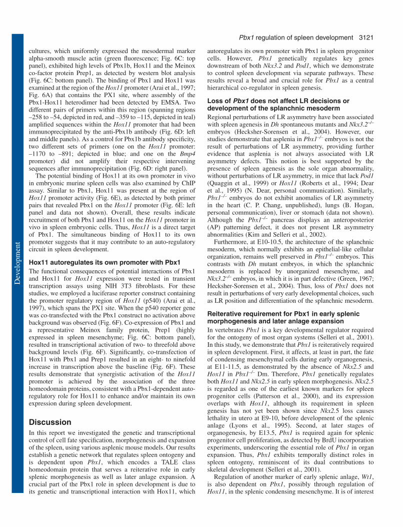
Fig. 6. Recruitment of Pbx1 andHox11 to the mouse Hox11 promoter.(A) Schematic illustration of 1.2 kbof the Hox11 genomic segment withknown promoter activity thatcontains Pbx1-binding sites, and a 5′upstream region. Primers used forPCR analysis are indicated by arrows(each pair in a different color) andthe oligoprobe (PX1) used for EMSAis indicated by a black box.(B) Binding of Pbx1 and Hox11 tothe (PX1) oligo within the Hox11promoter. Nuclear extracts derivedfrom wild-type primary embryonicspleen cells were subjected to EMSAwith a radiolabeled PX1 probecontaining a Pbx1 wild-type (PX1) ormutated (mPX1) core binding site(underlined), as indicated above gellanes. Asterisk indicates non-specificband. (C) Primary cells isolated fromembryonic spleen stained in culturefor the mesodermal marker α smoothmuscle actin (green fluorescence).Western blot analysis (panel below)demonstrates that these cells producePbx1, Prep1 and Hox11 proteins.Two isoforms of Hox11 are presentin embryonic spleen, as indicated(Yamamoto et al., 1995). (D,E) ForChIP analysis, chromatin wassubjected to immunoprecipitation(IP) using antibodies specific forPbx1b (anti-Pbx1b) (D) or Hox11(anti-Hox11) (E). As negativecontrols, IPs were also performedwith an anti-GFP antibody, rabbitserum (RS) or no antibody (No Ab).A primer pair that amplifies a regionwithin the Bmp4 promoter was usedas an additional negative control. (F) Synergistic activation of the Hox11 promoter by Pbx1, Prep1 and Hox11 proteins. Luciferase activity wasassayed from transiently transfected NIH 3T3 cells. Co-transfection assays were performed in the presence (+) of the indicated expressionvectors encoding Pbx1, Prep1 or Hox11, and with a vector containing the promoter regulatory region of Hox11 (p540), or with vector alone (p-GL2). Data are expressed as the fold activation over the p540 basal luciferase activity. Bars represent the mean of three independenttransfections (performed in duplicate)±s.e.m. normalized for β-galactosidase activity (internal control) within each experiment.
Dev
elop
men
t
3121Pbx1 regulation of spleen development
cultures, which uniformly expressed the mesodermal markeralpha-smooth muscle actin (green fluorescence; Fig. 6C: toppanel), exhibited high levels of Pbx1b, Hox11 and the Meinoxco-factor protein Prep1, as detected by western blot analysis(Fig. 6C: bottom panel). The binding of Pbx1 and Hox11 wasexamined at the region of the Hox11 promoter (Arai et al., 1997;Fig. 6A) that contains the PX1 site, where assembly of thePbx1-Hox11 heterodimer had been detected by EMSA. Twodifferent pairs of primers within this region (spanning regions–258 to –54, depicted in red, and –359 to –115, depicted in teal)amplified sequences within the Hox11 promoter that had beenimmunoprecipitated by the anti-Pbx1b antibody (Fig. 6D: leftand middle panels). As a control for Pbx1b antibody specificity,two different sets of primers (one on the Hox11 promoter:–1170 to –891; depicted in blue; and one on the Bmp4promoter) did not amplify their respective interveningsequences after immunoprecipitation (Fig. 6D: right panel).
The potential binding of Hox11 at its own promoter in vivoin embryonic murine spleen cells was also examined by ChIPassay. Similar to Pbx1, Hox11 was present at the region ofHox11 promoter activity (Fig. 6E), as detected by both primerpairs that revealed Pbx1 on the Hox11 promoter (Fig. 6E: leftpanel and data not shown). Overall, these results indicaterecruitment of both Pbx1 and Hox11 on the Hox11 promoter invivo in spleen embryonic cells. Thus, Hox11 is a direct targetof Pbx1. The simultaneous binding of Hox11 to its ownpromoter suggests that it may contribute to an auto-regulatorycircuit in spleen development.
Hox11 autoregulates its own promoter with Pbx1The functional consequences of potential interactions of Pbx1and Hox11 for Hox11 expression were tested in transienttranscription assays using NIH 3T3 fibroblasts. For thesestudies, we employed a luciferase reporter construct containingthe promoter regulatory region of Hox11 (p540) (Arai et al.,1997), which spans the PX1 site. When the p540 reporter genewas co-transfected with the Pbx1 construct no activation abovebackground was observed (Fig. 6F). Co-expression of Pbx1 anda representative Meinox family protein, Prep1 (highlyexpressed in spleen mesenchyme; Fig. 6C: bottom panel),resulted in transcriptional activation of two- to threefold abovebackground levels (Fig. 6F). Significantly, co-transfection ofHox11 with Pbx1 and Prep1 resulted in an eight- to ninefoldincrease in transcription above the baseline (Fig. 6F). Theseresults demonstrate that synergistic activation of the Hox11promoter is achieved by the association of the threehomeodomain proteins, consistent with a Pbx1-dependent auto-regulatory role for Hox11 to enhance and/or maintain its ownexpression during spleen development.
DiscussionIn this report we investigated the genetic and transcriptionalcontrol of cell fate specification, morphogenesis and expansionof the spleen, using various asplenic mouse models. Our resultsestablish a genetic network that regulates spleen ontogeny andis dependent upon Pbx1, which encodes a TALE classhomeodomain protein that serves a reiterative role in earlysplenic morphogenesis as well as later anlage expansion. Acrucial part of the Pbx1 role in spleen development is due toits genetic and transcriptional interaction with Hox11, which
autoregulates its own promoter with Pbx1 in spleen progenitorcells. However, Pbx1 genetically regulates key genesdownstream of both Nkx3.2 and Pod1, which we demonstrateto control spleen development via separate pathways. Theseresults reveal a broad and crucial role for Pbx1 as a centralhierarchical co-regulator in spleen genesis.
Loss of Pbx1 does not affect LR decisions ordevelopment of the splanchnic mesodermRegional perturbations of LR asymmetry have been associatedwith spleen agenesis in Dh spontaneous mutants and Nkx3.2–/–
embryos (Hecksher-Sorensen et al., 2004). However, ourstudies demonstrate that asplenia in Pbx1–/– embryos is not theresult of perturbations of LR asymmetry, providing furtherevidence that asplenia is not always associated with LRasymmetry defects. This notion is best supported by thepresence of spleen agenesis as the sole organ abnormality,without perturbations of LR asymmetry, in mice that lack Pod1(Quaggin et al., 1999) or Hox11 (Roberts et al., 1994; Dearet al., 1995) (N. Dear, personal communication). Similarly,Pbx1–/– embryos do not exhibit anomalies of LR asymmetryin the heart (C. P. Chang, unpublished), lungs (B. Hogan,personal communication), liver or stomach (data not shown).Although the Pbx1–/– pancreas displays an anteroposterior(AP) patterning defect, it does not present LR asymmetryabnormalities (Kim and Selleri et al., 2002).
Furthermore, at E10-10.5, the architecture of the splanchnicmesoderm, which normally exhibits an epithelial-like cellularorganization, remains well preserved in Pbx1–/– embryos. Thiscontrasts with Dh mutant embryos, in which the splanchnicmesoderm is replaced by unorganized mesenchyme, andNkx3.2–/– embryos, in which it is in part defective (Green, 1967;Hecksher-Sorensen et al., 2004). Thus, loss of Pbx1 does notresult in perturbations of very early developmental choices, suchas LR position and differentiation of the splanchnic mesoderm.
Reiterative requirement for Pbx1 in early splenicmorphogenesis and later anlage expansionIn vertebrates Pbx1 is a key developmental regulator requiredfor the ontogeny of most organ systems (Selleri et al., 2001).In this study, we demonstrate that Pbx1 is reiteratively requiredin spleen development. First, it affects, at least in part, the fateof condensing mesenchymal cells during early organogenesis,at E11-11.5, as demonstrated by the absence of Nkx2.5 andHox11 in Pbx1–/– Dm. Therefore, Pbx1 genetically regulatesboth Hox11 and Nkx2.5 in early spleen morphogenesis. Nkx2.5is regarded as one of the earliest known markers for spleenprogenitor cells (Patterson et al., 2000), and its expressionoverlaps with Hox11, although its requirement in spleengenesis has not yet been shown since Nkx2.5 loss causeslethality in utero at E9-10, before development of the splenicanlage (Lyons et al., 1995). Second, at later stages oforganogenesis, by E13.5, Pbx1 is required again for splenicprogenitor cell proliferation, as detected by BrdU incorporationexperiments, underscoring the essential role of Pbx1 in organexpansion. Thus, Pbx1 exhibits temporally distinct roles inspleen ontogeny, reminiscent of its dual contributions toskeletal development (Selleri et al., 2001).
Regulation of another marker of early splenic anlage, Wt1,is also dependent on Pbx1, possibly through regulation ofHox11, in the splenic condensing mesenchyme. It is of interest
Dev
elop
men
t

3122
that regulation of Wt1 expression is independent of Pbx1 in theouter mesothelial lining of the splenic anlage, which normallydoes not express Hox11, and will give rise to the spleniccapsule. Taken together, these results support a scenario wherePbx1 regulates Hox11, which in turn regulates Wt1 in thesplenic mesenchyme, whereas in the mesothelial lining of thedeveloping spleen regulation of Wt1 is uncoupled from Pbx1.Thus, Pbx1 can be considered as the uppermost known geneticregulator within the Hox11-Wt1 pathway in the non-mesothelial splenic mesenchyme (Fig. 7A).
Expression of other genes that mark early condensingsplenic mesenchyme, such as Pod1 and Nkx3.2, is wellmaintained in Pbx1–/– embryos, demonstrating that spleniccondensing mesenchymal cells are still present in Dm ofPbx1–/– embryos and splenic gene expression is not globallyimpaired by Pbx1 loss. Thus, requirement for Pod1 andNkx3.2 in spleen ontogeny is not dependent upon Pbx1.Finally, the findings by Lu et al. (Lu et al., 2000), confirmedby our present studies, that Nkx3.2 is expressed in thecondensing mesenchyme of Pod1–/– embryos at E12-12.5, andour results that Pod1–/– expression is unperturbed in Nkx3.2–/–
embryos, indicate that the Pod1 and Nkx3.2 transcriptionfactors use separate pathways to regulate early spleendevelopment.
Although previous work has demonstrated asplenia inHox11 mutants (Roberts et al., 1994; Dear et al., 1995), untilnow the cellular basis of this defect was mostly unknown.Indeed, apoptosis was not detected in Hox11–/– splenicprimordium in a previous study (Roberts et al., 1995), while itwas documented in another report that used a different Hox11-deficient model (Dear et al., 1995). In the present study, weobserved a modest increase of apoptosis in Hox11–/– splenicprimordium. The apoptotic cells were mostly localized to theouter mesothelial lining of the splenic anlage, which gives riseto the spleen capsule and does not normally express Hox11.Thus, it appears that such a subtle increase of apoptosis cannotbe responsible for the complete lack of spleen development inHox11-deficient embryos. Conversely, our finding that, by
E13.5, Hox11–/– spleen progenitor cells exhibit a markeddefect in cellular proliferation comparable with that inPbx1–/– embryos is consistent with the hypoplasia ofHox11–/– splenic anlage (Roberts et al., 1994; Dear et al.,1995) and the demonstrated involvement of Hox11 incellular proliferation and cell cycle control (Kawabe etal., 1997; Hough et al., 1998; Owens et al., 2003).Finally, the finding of a common cellular defect (i.e.
impaired progenitor cell proliferation) in spleen developmentof Pbx1–/– and Hox11–/– embryos further corroborates theobservation that Pbx1 genetically regulates Hox11.
In sum, the requirement for Pbx1 in spleen ontogeny appearsto be reiterative. This reiterative role can account for thecomplete absence of the spleen, which would not otherwise beexplained either by a partial impairment of splenic cell fatespecification or by a 50% decrease in progenitor cellularproliferation in the splenic anlage, but probably results fromthe summation of these defects.
Genetic interaction of Pbx1 and Hox11 in spleenontogenyThe finding of a high percentage (80%) of Pbx1+/–;Hox11+/–
double heterozygous mice displaying severely hypoplastic andmalformed spleens (Fig. 4E,F; Table 1), compared with singleheterozygotes, demonstrates that Pbx1 and Hox11 geneticallyinteract in vivo in spleen development. The wide spectrum ofmalformations of Pbx1+/–;Hox11+/– double heterozygousspleens, which includes fusions of two spleens, mimicspolysplenia, a human congenital condition. In polysplenia twoor more splenic masses, hypoplastic and irregularly shaped(splenules), are present lateral to the stomach (Lodewyk et al.,1972). Unlike human asplenia, which involves life-threateninginfections in children (Waldman et al., 1977), polyspleniais associated with normal splenic function (Lodewyk etal., 1972). Despite their morphological abnormalities,Pbx1+/–;Hox11+/– double heterozygous mice exhibit normalsplenic architecture, germinal center formation and primaryimmune function (not shown), thus closely modeling thehuman polysplenic condition.
Hox11 is a direct in vivo target of Pbx1 and auto-regulates its own promoter with Pbx1 in spleenontogenyDespite the growing understanding of Hox and TALEhomeoprotein functions in development (Krumlauf, 1994;Mann and Affolter, 1998; Popperl et al., 2000; Selleri et al.,
Development 132 (13) Research article
Fig. 7. Pbx1-dependent genetic network and transcriptionalpathway regulate spleen ontogeny. (A) Transverse section ofabdominal organs schematically illustrates the location of thespleen primordium during vertebrate development. Adapted,with permission, from Sadler (Sadler, 1995). To the right, thePbx1-Hox11 transcriptional hierarchy regulating spleenontogeny is depicted, with the Pbx1-dependent pathway inred. The Nkx2.5 downstream pathway is illustrated with abroken arrow as the requirement for Nkx2.5, an early markerof splenic progenitor cells, has not yet been demonstrated inspleen development. (B) Within the Pbx1-Hox11transcriptional pathway, Pbx1 directly regulates Hox11 inspleen progenitor cells and Hox11, in turn, regulates its ownpromoter together with Pbx1.
Dev
elop
men
t

3123Pbx1 regulation of spleen development
2001; Waskiewicz et al., 2002; Hisa et al., 2004; Selleri et al.,2004) and their functional interactions (Popperl et al., 1995;Maconochie et al., 1997; Jacobs et al., 1999; Ferretti et al.,2000; Manzanares et al., 2001; Samad et al., 2004), to date,only a few direct target genes have been reported (Rauskolb etal., 1993; Graba et al., 1997; Bromleigh and Freedman, 2000;Theokli et al., 2003). In this study, we provide the first in vivoevidence that Pbx1 directly regulates Hox11 in embryonicspleen cells. Interestingly, at E9.5, Pbx1 is already expressedin the mid-gut mesenchyme (Schnabel et al., 2001), fromwhich the spleen is derived, well before the onset of Hox11expression (Kanzler and Dear, 2001). And indeed, Pbx1controls the onset of Hox11 expression at E11 within the Dm,as demonstrated by our in situ hybridization experiments.Although Pbx1 expression starts to decrease in the splenicanlage after E13.5 (data not shown), Hox11 persists in thespleen until birth (Kanzler and Dear, 2001), suggesting thatPbx1 is required for the onset of Hox11 expression and for itscontinued expression in early spleen development, until E13.5,although it is not necessary for Hox11 maintenance in laterphases of organogenesis.
Hox11 is one of the earliest known markers for spleen cellprogenitors (Dear et al., 1995). A useful tool to monitor Hox11transcription in the developing spleen is provided by Hox11lacZ
mice (Dear et al., 1995), in which lacZ expression is dependenton Hox11 regulatory sequences and faithfully recapitulatesHox11 expression. Analysis of Hox11lacZ/lacZ embryospreviously demonstrated that lacZ expression is normallyinitiated in the absence of Hox11 (Dear et al., 1995), suggestingthat the Hox11 protein is not required for initiation of its owntranscription in the splenic mesenchyme. These findingsindicate that other factors might be necessary for the onset ofHox11 transcription. Here, we identify Pbx1 as one such factorthat activates Hox11 transcriptional onset and early expressionin the splenic anlage until E 13.5.
Pbx1 and Hox11 bind to a potential Pbx-binding site (PX1)within the Hox11 promoter, as shown by EMSA assaysconducted on embryonic spleen primary cells. Additionally,Pbx1 and Hox11 bind the Hox11 promoter in vivo inembryonic spleen cells, as revealed by ChIP assays. Regulatoryinteractions of Hox genes, such as the induction of Hoxb1segmental expression by Hoxb1 and Hoxa1 through auto-and cross-regulatory loops, have been documented indevelopmental processes (Popperl et al., 1995; Studer et al.,1996; Studer et al., 1998). Here, we reveal an autoregulatoryloop for an orphan Hox gene, Hox11, which is non-clusteredbut bears a hexapeptide motif (Shen et al., 1996). Takentogether, our findings establish that Hox11 is a direct target ofPbx1 and that, simultaneously, it regulates its own promoter.Significantly, co-transfection of Hox11 with Pbx1 andPrep1 resulted in a striking increase in transcription abovebaseline, demonstrating that synergistic activation of theHox11 promoter is achieved by the association of thethree homeoproteins, consistent with a Pbx1-dependentautoregulatory role for Hox11 to enhance and/or maintain, atleast in part, its own expression during spleen development.Interestingly, additional potential Pbx-Meinox-binding siteswere identified within the Hox11 promoter downstream of thePX1 site (not shown), suggesting that multiple binding siteswithin close proximity might be used, simultaneously or at
different times, for a complex, multi-faceted transcriptionalregulation of Hox11 in spleen development.
Establishment of a Pbx1-dependent genetic andtranscriptional network that regulates spleenontogenyIn addition to establishing that Pbx1 is the most upstreamknown direct regulator of Hox11 in spleen ontogeny (Fig. 7A),our studies demonstrate an even broader role for Pbx1 in spleendevelopment. Pbx1 regulates key genes downstream of Nkx3.2and Pod1, which we show to control spleen developmentthrough separate genetic pathways (Fig. 7A). Both Nkx3.2(Lettice et al., 1999; Tribioli et al., 1999) and Pod1 (Quagginet al., 1999; Lu et al., 2000) are essential for spleendevelopment, and their expression in condensing splenicmesenchyme overlaps with Hox11 and Nkx2.5. The specificmechanisms and cellular behaviors by which the Nkx3.2transcription factor regulates spleen development are as yetunknown, while Pod1 has been proposed to control splenic cellsurvival (Lu et al., 2000). Interestingly, Lu et al. have reportedthat the spleen primordium of Pod1–/– embryos does not furtherexpand after E12.5 and starts to undergo apoptotic cell death.As a result, after E12.5, expression of all splenic markers,including Nkx3.2, disappears from the degenerating splenicprimordium of Pod1–/– embryos (Lu et al., 2000). Our studiesdemonstrate that Pbx1 expression is not dependent on thepresence of either Nkx3.2 or Pod1 in the splenic mesenchyme(Fig. 7A). Likewise, the requirement for both of thesetranscription factors in splenic development is Pbx1independent (Fig. 7A). In addition, our findings that Nkx3.2 isexpressed in the condensing mesenchyme of Pod1–/– embryosat E12-12.5, and that Pod1 expression is also unperturbed inNkx3.2–/– embryos, indicate that the Pod1 and Nkx3.2transcription factors use separate pathways to regulate earlyspleen development.
Furthermore, our studies reveal that Pbx1 and Nkx3.2independently regulate Hox11 in a hierarchical fashion (Fig.2M-P; Fig. 3E,F) (Lettice et al., 1999). And, in a similarscenario, Pbx1 and Pod1, but not Nkx3.2 (see Fig. S1E,F),independently control Nkx2.5 gene expression in a hierarchicalfashion (Fig. 7A). Thus, Pbx1 impinges on the separate Nkx3.2and Pod1 pathways by genetically regulating key players inboth of these pathways, i.e. Hox11 and Nkx2.5 (Fig. 7A). As aresult, Pbx1 emerges as a central hierarchical co-regulator inspleen ontogeny (Fig. 7A). It will be of interest to determinethe roles of additional transcription factors required for spleendevelopment, such as Sox11 (Sock et al., 2004) and Nkx2.3(Pabst et al., 1999; Wang et al., 2000; Tarlinton et al., 2003),within the genetic pathways established by our study.
In conclusion, we demonstrate here the essential role of thePbx1-Hox11 transcriptional pathway in spleen ontogeny. Weprovide evidence that Pbx1 is reiteratively required duringspleen development, as it is implicated, at least in part, insplenic cell fate specification and morphogenesis, and then isessential again, later in organogenesis, for anlage expansionthrough control of progenitor cell proliferation. Finally, wedemonstrate that spleen ontogeny is dependent on theorchestration of a complex network of transcription factors,among which Pbx1 emerges as a central, master co-regulator.Overall, our study takes a significant first step towards
Dev
elop
men
t

3124
understanding the genetic and transcriptional control of spleendevelopment.
We thank W. Zimmer and R. Schwartz for generating and providingNkx3.2 knockout mice (unpublished); S. Korsmeyer and T. H.Rabbitts for providing Hox11 and Hox11lacZ knockout mice,respectively; C. Englert, R. Harvey, H. Popperl, T. H. Rabbitts, J.Rossant and R. Schwartz for in situ hybridization probes; N. Koehlerand M. Hatano for the Hox11 constructs; N. Dear for sharingunpublished observations; K. Manova for immunohistochemistrysupport; C. Nicolas for technical assistance; L. Lacy, A. Koff, L.Niswander, K. Anderson and members of their laboratories forstimulating discussions; J. Giacalone for editing of the manuscript; K.Hadjantonakis, S. Kim, L. Lacy and V. Zappavigna for critical readingof the manuscript. L.S. personally thanks N. Copeland, B. Hogan andT. H. Rabbitts for invaluable input, and V. Zappavigna for sharing hisexpertise for the ChIP experiments. This work was supported bygrants from Telethon and the Italian Ministry of Research (MIUR,PRIN) to F.B.; the National Institutes of Health to M.C. (CA42971and CA90735) and L.S. (HD43997), and a grant from the March ofDimes and Birth Defects Foundation (6-FY03-071) to L.S. L.S. is anIrma T. Hirschl Scholar.
Supplementary materialSupplementary material for this article is available athttp://dev.biologists.org/cgi/content/full/132/13/3113/DC1
ReferencesAparicio, O. M. (1999). Characterization of proteins bound to chromatin by
immunoprecipitation from whole-cell extracts. In Current Protocols inMolecular Biology (ed. F. M. Ausubel, R. Brent, R. E. Kingston, D. M.Moore, J. G. Seidman, J. A. Smith and K. Struhl), pp. 21.23.12–21.23.21.New York: John Wiley.
Apelqvist, A., Ahlgren, U. and Edlund, H. (1997). Sonic hedgehog directsspecialised mesoderm differentiation in the intestine and pancreas. Curr.Biol. 7, 801-804.
Arai, Y., Hatano, M. and Tokuhisa, T. (1997). A negative regulatory regionof the murine Hox11 gene. Gene 193, 73-79.
Aylsworth, A. S. (2001). Clinical aspects of defects in the determination oflaterality. Am. J. Med. Genet. 101, 345-355.
Benasciutti, E., Pages, G., Kenzior, O., Folk, W., Blasi, F. and Crippa, M.P. (2004). MAPK and JNK transduction pathways can phosphorylate Sp1to activate the uPA minimal promoter element and endogenous genetranscription. Blood 104, 256-262.
Berthelsen, J., Vandekerkhove, J. and Blasi, F. (1996). Purification andcharacterization of UEF3, a novel factor involved in the regulation of theurokinase and other AP-1 controlled promoters. J. Biol. Chem. 271, 3822-3830.
Berthelsen, J., Zappavigna, V., Mavilio, F. and Blasi, F. (1998). Prep1, anovel functional partner of Pbx proteins. EMBO J. 17, 1423-1433.
Bischof, L. J., Kagawa, N. and Waterman, M. R. (1998). The bovine CYP17promoter contains a transcriptional regulatory element cooperatively boundby tale homeodomain proteins. Endocrinol. Res. 24, 489-495.
Bishop, M. B. and Lansing, L. S. (1982). The spleen: a correlative overviewof normal and pathologic anatomy. Hum. Pathol. 13, 334-342.
Boorman, C. J. and Shimeld, S. M. (2002). The evolution of left-rightasymmetry in chordates. BioEssays 24, 1004-1011.
Brake, R. L., Kees, U. R. and Watt, P. M. (2002). A complex containingPBX2 contributes to activation of the proto-oncogene HOX11. Biochem.Biophys. Res. Commun. 294, 23-34.
Bromleigh, V. C. and Freedman, L. P. (2000). p21 is a transcriptional targetof HOXA10 in differentiating myelomonocytic cells. Genes Dev. 14, 2581-2586.
Burglin, T. R. (1997). Analysis of TALE superclass homeobox genes (MEIS,PBC, KNOX, Iroquois, TGIF) reveals a novel domain conserved betweenplants and animals. Nucleic Acids Res. 25, 4173-4180.
Chang, C. P., Jacobs, Y., Nakamura, T., Jenkins, N. A., Copeland, N. G.and Cleary, M. L. (1997). Meis proteins are major in vivo DNA bindingpartners for wild-type but not chimeric Pbx proteins. Mol. Cell. Biol. 17,5679-5687.
Dear, T. N., Colledge, W. H., Carlton, M. B., Lavenir, I., Larson, T., Smith,A. J., Warren, A. J., Evans, M. J., Sofroniew, M. V. and Rabbitts, T. H.(1995). The Hox11 gene is essential for cell survival during spleendevelopment. Development 121, 2909-2915.
Dent, A. L., Shaffer, A. L., Yu, X., Allman, D. and Staudt, L. M. (1997).Control of inflammation, cytokine expression, and germinal centerformation by BCL-6. Science 276, 589-592.
DiMartino, J. F., Selleri, L., Traver, D., Firpo, M. T., Rhee, J., Warnke, R.,O’Gorman, S., Weissman, I. L. and Cleary, M. L. (2001). The Hoxcofactor and proto-oncogene Pbx1 is required for maintenance of definitivehematopoiesis in the fetal liver. Blood 98, 618-626.
Ferretti, E., Marshall, H., Popperl, H., Maconochie, M., Krumlauf, R. andBlasi, F. (2000). Segmental expression of Hoxb2 in r4 requires two separatesites that integrate cooperative interactions between Prep1, Pbx and Hoxproteins. Development 127, 155-166.
Fognani, C., Kilstrup-Nielsen, C., Berthelsen, J., Ferretti, E., Zappavigna,V. and Blasi, F. (2002). Characterization of PREP2, a paralog of PREP1,which defines a novel sub-family of the MEINOX TALE homeodomaintranscription factors. Nucleic Acids Res. 30, 2043-2051.
Frank, S. R., Schroeder, M., Fernandez, P., Taubert, S. and Amati, B.(2001). Binding of c-Myc to chromatin mediates mitogen-inducedacetylation of histone H4 and gene activation. Genes Dev. 15, 2069-2082.
Funayama, N., Sato, Y., Matsumoto, K., Ogura, T. and Takahashi, Y.(1999). Coelom formation: binary decision of the lateral plate mesoderm iscontrolled by the ectoderm. Development 126, 4129-4138.
Gavrieli, Y., Sherman, Y. and Ben-Sasson, S. A. (1992). Identification ofprogrammed cell death in situ via specific labeling of nuclear DNAfragmentation. J. Cell Biol. 119, 493-501.
Graba, Y., Aragnol, D. and Pradel, J. (1997). Drosophila Hox complexdownstream targets and the function of homeotic genes. BioEssays 19, 379-388.
Green, M. C. (1967). A defect of the splanchnic mesoderm caused by themutant gene dominant hemimelia in the mouse. Dev. Biol. 15, 62-89.
Hecksher-Sorensen, J., Watson, R. P., Lettice, L. A., Serup, P., Eley, L., DeAngelis, C., Ahlgren, U. and Hill, R. E. (2004). The splanchnicmesodermal plate directs spleen and pancreatic laterality, and is regulatedby Bapx1/Nkx3.2. Development 131, 4665-4675.
Herzer, U., Crocoll, A., Barton, D., Howells, N. and Englert, C. (1999). TheWilms tumor suppressor gene wt1 is required for development of the spleen.Curr. Biol. 9, 837-840.
Hisa, T., Spence, S. E., Rachel, R. A., Fujita, M., Nakamura, T., Ward, J.M., Devor-Henneman, D. E., Saiki, Y., Kutsuna, H., Tessarollo, L. et al.(2004). Hematopoietic, angiogenic and eye defects in Meis1 mutantanimals. EMBO J. 23, 450-459.
Hough, M. R., Reis, M. D., Singaraja, R., Bryce, D. M., Kamel-Reid, S.,Dardick, I., Breitman, M. L. and Dube, I. D. (1998). A model forspontaneous B-lineage lymphomas in IgHmu-HOX11 transgenic mice.Proc. Natl. Acad. Sci. USA 95, 13853-13858.
Inada, K., Okada, S., Phuchareon, J., Hatano, M., Sugimoto, T., Moriya,H. and Tokuhisa, T. (1998). c-Fos induces apoptosis in germinal center Bcells. J. Immunol. 161, 3853-3861.
Ishiko, E., Matsumura, I., Ezoe, S., Gale, K., Ishiko, J., Satoh, Y., Tanaka,H., Shibayama, H., Mizuki, M., Era, T. et al. (2004). Notch signals inhibitthe development of erythroid/megakaryocytic cells by suppressing GATA-1activity through the induction of HES1. J. Biol. Chem. (in press).
Jacobs, Y., Schnabel, C. A. and Cleary, M. L. (1999). Trimeric associationof Hox and TALE homeodomain proteins mediates Hoxb2 hindbrainenhancer activity. Mol. Cell. Biol. 19, 5134-5142.
Kamps, M. P., Murre, C., Sun, X. H. and Baltimore, D. (1990). A newhomeobox gene contributes the DNA binding domain of the t(1;19)translocation protein in pre-B ALL. Cell 60, 547-555.
Kanzler, B. and Dear, T. N. (2001). Hox11 acts cell autonomously in spleendevelopment and its absence results in altered cell fate of mesenchymalspleen precursors. Dev. Biol. 234, 231-243.
Kawabe, T., Muslin, A. J. and Korsmeyer, S. J. (1997). HOX11 interactswith protein phosphatases PP2A and PP1 and disrupts a G2/M cell-cyclecheckpoint. Nature 385, 454-458.
Kim, S. K., Selleri, L., Lee, J. S., Zhang, A. Y., Gu, X., Jacobs, Y. andCleary, M. L. (2002). Pbx1 inactivation disrupts pancreas development andin Ipf1-deficient mice promotes diabetes mellitus. Nat. Genet. 30, 430-435.
Knoepfler, P. S., Calvo, K. R., Chen, H., Antonarakis, S. E. and Kamps,M. P. (1997). Meis1 and pKnox1 bind DNA cooperatively with Pbx1utilizing an interaction surface disrupted in oncoprotein E2a-Pbx1. Proc.Natl. Acad. Sci. USA 94, 14553-14558.
Development 132 (13) Research article
Dev
elop
men
t

3125Pbx1 regulation of spleen development
Koehler, K., Franz, T. and Dear, T. N. (2000). Hox11 is required to maintainnormal Wt1 mRNA levels in the developing spleen. Dev. Dyn. 218, 201-206.
Krumlauf, R. (1994). Hox genes in vertebrate development. Cell 78, 191-201.Lettice, L. A., Purdie, L. A., Carlson, G. J., Kilanowski, F., Dorin, J. and
Hill, R. E. (1999). The mouse bagpipe gene controls development of axialskeleton, skull, and spleen. Proc. Natl. Acad. Sci. USA 96, 9695-9700.
Lodewyk, H. S. V. M., Ira, H. G. and Gerold, L. S. (1972). Asplenia andpolysplenia syndromes. Birth Defects 8, 36-44.
Lu, J., Chang, P., Richardson, J. A., Gan, L., Weiler, H. and Olson, E. N.(2000). The basic helix-loop-helix transcription factor capsulin controlsspleen organogenesis. Proc. Natl. Acad. Sci. USA 97, 9525-9530.
Lyons, I., Parsons, L. M., Hartley, L., Li, R., Andrews, J. E., Robb, L. andHarvey, R. P. (1995). Myogenic and morphogenetic defects in the hearttubes of murine embryos lacking the homeobox gene Nkx2-5. Genes Dev.9, 1654-1666.
Maconochie, M. K., Nonchev, S., Studer, M., Chan, S. K., Popperl, H.,Sham, M. H., Mann, R. S. and Krumlauf, R. (1997). Cross-regulation inthe mouse HoxB complex: the expression of Hoxb2 in rhombomere 4 isregulated by Hoxb1. Genes Dev. 11, 1885-1895.
Manley, N. R., Selleri, L., Brendolan, A., Gordon, J. and Cleary, M. L.(2004). Abnormalities of caudal pharyngeal pouch development in Pbx1knockout mice mimic loss of Hox3 paralogs. Dev. Biol. 276, 301-312.
Mann, R. S. and Affolter, M. (1998). Hox proteins meet more partners. Curr.Opin. Genet. Dev. 8, 423-429.
Manning, M. J. and Horton, J. D. (1969). Histogenesis of lymphoid organsin larvae of the South African clawed toad, Xenopus laevis (Daudin). J.Embryol. Exp. Morphol. 22, 265-277.
Manzanares, M., Bel-Vialar, S., Ariza-McNaughton, L., Ferretti, E.,Marshall, H., Maconochie, M. M., Blasi, F. and Krumlauf, R. (2001).Independent regulation of initiation and maintenance phases of Hoxa3expression in the vertebrate hindbrain involve auto- and cross-regulatorymechanisms. Development 128, 3595-3607.
Nourse, J., Mellentin, J. D., Galili, N., Wilkinson, J., Stanbridge, E., Smith,S. D. and Cleary, M. L. (1990). Chromosomal translocation t(1;19) resultsin synthesis of a homeobox fusion mRNA that codes for a potential chimerictranscription factor. Cell 60, 535-545.
Oh, S. P. and Li, E. (2002). Gene-dosage-sensitive genetic interactionsbetween inversus viscerum (iv), nodal, and activin type IIB receptor(ActRIIB) genes in asymmetrical patterning of the visceral organs along theleft-right axis. Dev. Dyn. 224, 279-290.
Owens, B. M., Zhu, Y. X., Suen, T. C., Wang, P. X., Greenblatt, J. F., Goss,P. E. and Hawley, R. G. (2003). Specific homeodomain-DNA interactionsare required for HOX11-mediated transformation. Blood 101, 4966-4974.
Pabst, O., Zweigerdt, R. and Arnold, H. H. (1999). Targeted disruption ofthe homeobox transcription factor Nkx2-3 in mice results in postnatallethality and abnormal development of small intestine and spleen.Development 126, 2215-2225.
Patterson, K. D., Drysdale, T. A. and Krieg, P. A. (2000). Embryonic originsof spleen asymmetry. Development 127, 167-175.
Popperl, H., Bienz, M., Studer, M., Chan, S. K., Aparicio, S., Brenner, S.,Mann, R. S. and Krumlauf, R. (1995). Segmental expression of Hoxb-1is controlled by a highly conserved autoregulatory loop dependent uponexd/pbx. Cell 81, 1031-1042.
Popperl, H., Rikhof, H., Chang, H., Haffter, P., Kimmel, C. B. and Moens,C. B. (2000). lazarus is a novel pbx gene that globally mediates hox genefunction in zebrafish. Mol. Cell 6, 255-267.
Quaggin, S. E., Vanden Heuvel, G. B. and Igarashi, P. (1998). Pod-1, amesoderm-specific basic-helix-loop-helix protein expressed inmesenchymal and glomerular epithelial cells in the developing kidney.Mech. Dev. 71, 37-48.
Quaggin, S. E., Schwartz, L., Cui, S., Igarashi, P., Deimling, J., Post, M.and Rossant, J. (1999). The basic-helix-loop-helix protein pod1 is criticallyimportant for kidney and lung organogenesis. Development 126, 5771-5783.
Rackley, R. R., Flenniken, A. M., Kuriyan, N. P., Kessler, P. M., Stoler, M.H. and Williams, B. R. (1993). Expression of the Wilms’ tumor suppressorgene WT1 during mouse embryogenesis. Cell Growth Differ. 4, 1023-1031.
Ramalho-Santos, M., Melton, D. A. and McMahon, A. P. (2000). Hedgehogsignals regulate multiple aspects of gastrointestinal development.Development 127, 2763-2772.
Rauskolb, C. and Wieschaus, E. (1994). Coordinate regulation ofdownstream genes by extradenticle and the homeotic selector proteins.EMBO J. 13, 3561-3569.
Robb, L., Mifsud, L., Hartley, L., Biben, C., Copeland, N. G., Gilbert, D.
J., Jenkins, N. A. and Harvey, R. P. (1998). Epicardin: A novel basic helix-loop-helix transcription factor gene expressed in epicardium, branchial archmyoblasts, and mesenchyme of developing lung, gut, kidney, and gonads.Dev. Dyn. 213, 105-113.
Roberts, C. W., Shutter, J. R. and Korsmeyer, S. J. (1994). Hox11 controlsthe genesis of the spleen. Nature 368, 747-749.
Roberts, C. W., Sonder, A. M., Lumsden, A. and Korsmeyer, S. J. (1995).Developmental expression of Hox11 and specification of splenic cell fate.Am. J. Pathol. 146, 1089-1101.
Rose, M. L., Birbeck, M. S., Wallis, V. J., Forrester, J. A. and Davies, A.J. (1980). Peanut lectin binding properties of germinal centres of mouselymphoid tissue. Nature 284, 364-366.
Rose, V., Izukawa, T. and Moes, C. A. (1975). Syndromes of asplenia andpolysplenia. A review of cardiac and non-cardiac malformations in 60 caseswithspecial reference to diagnosis and prognosis. Br. Heart J. 37, 840-852.
Sadler, T. W. (1995). Digestive system. In Langman’s Medical Embryology,7th edn, pp. 242-271. Baltimore, MD: Williams and Wilkins
Samad, O. A., Geisen, M. J., Caronia, G., Varlet, I., Zappavigna, V.,Ericson, J., Goridis. C. and Rijli, F. M. (2004). Integration ofanteroposterior and dorsoventral regulation of Phox2b transcription incranial motoneuron progenitors by homeodomain proteins. Development131, 4071-4083.
Sasaki, K. and Matsumura, G. (1988). Spleen lymphocytes andhaemopoiesis in the mouse embryo. J. Anat. 160, 27-37.
Schnabel, C. A., Selleri, L., Jacobs, Y., Warnke, R. and Cleary, M. L.(2001). Expression of Pbx1b during mammalian organogenesis. Mech. Dev.100, 131-135.
Schnabel, C. A., Godin, R. E. and Cleary, M. L. (2003a). Pbx1 regulatesnephrogenesis and ureteric branching in the developing kidney. Dev. Biol.254, 262-276.
Schnabel, C. A., Selleri, L. and Cleary, M. L. (2003b). Pbx1 is essentialfor adrenal development and urogenital differentiation. Genesis 37, 123-130.
Selleri, L., Depew, M. J., Jacobs, Y., Chanda, S. K., Tsang, K. Y., Cheah,K. S., Rubenstein, J. L., O’Gorman, S. and Cleary, M. L. (2001).Requirement for Pbx1 in skeletal patterning and programming chondrocyteproliferation and differentiation. Development 128, 3543-3557.
Selleri, L., DiMartino, J., van Deursen, J., Brendolan, A., Sanyal, M.,Boon, E., Capellini, T., Smith, K. S., Rhee, J., Popperl, H. et al. (2004).The TALE homeodomain protein Pbx2 is not essential for development andlong-term survival. Mol. Cell. Biol. 24, 5324-5331.
Shen, W. F., Chang, C. P., Rozenfeld, S., Sauvageau, G., Humphries, R.K., Lu, M., Lawrence, H. J., Cleary, M. L. and Largman, C. (1996). Hoxhomeodomain proteins exhibit selective complex stabilities with Pbx andDNA. Nucleic Acids Res. 24, 898-906.
Sock, E., Rettig, S. D., Enderich, J., Bosl, M. R., Tamm, E. R. and Wegner,M. (2004). Gene targeting reveals a widespread role for the high-mobility-group transcription factor Sox11 in tissue remodeling. Mol. Cell. Biol. 24,6635-6644.
Studer, M., Lumsden, A., Ariza-McNaughton, L., Bradley, A. andKrumlauf, R. (1996). Altered segmental identity and abnormal migrationof motor neurons in mice lacking Hoxb-1. Nature 384, 630-634.
Studer, M., Gavalas, A., Marshall, H., Ariza-McNaughton, L., Rijli, F. M.,Chambon, P. and Krumlauf, R. (1998). Genetic interactions betweenHoxa1 and Hoxb1 reveal new roles in regulation of early hindbrainpatterning. Development 125, 1025-1036.
Sty, J. R. and Conway, J. J. (1985). The spleen: development and functionalevaluation. Semin. Nucl. Med. 15, 276-298.
Swift, G. H., Liu, Y., Rose, S. D., Bischof, L. J., Steelman, S., Buchberg,A. M., Wright, C. V. and MacDonald, R. J. (1998). An endocrine-exocrineswitch in the activity of the pancreatic homeodomain protein PDX1 throughformation of a trimeric complex with PBX1b and MRG1 (MEIS2). Mol.Cell. Biol. 18, 5109-5120.
Tarlinton, D., Light, A., Metcalf, D., Harvey, R. P. and Robb, L. (2003).Architectural defects in the spleens of Nkx2-3-deficient mice are intrinsicand associated with defects in both B cell maturation and T cell-dependentimmune responses. J. Immunol. 170, 4002-4010.
Thiel, G. A. and Downey, H. (1921). The development of the mammalianspleen with special references to its hematopoietic activity. Am. J. Anat. 28,279-333.
Theokli, C., Morsi El-Kadi, A. S. and Morgan, R. (2003). TALE classhomeodomain gene Irx5 is an immediate downstream target for Hoxb4transcriptional regulation. Dev. Dyn. 227, 48-55.
Todaro, G. J. and Green, H. (1963). Quantitative studies of the growth of
Dev
elop
men
t

3126
mouse embryo cells in culture and their development into established lines.J. Cell Biol. 17, 299-313.
Tribioli, C. and Lufkin, T. (1999). The murine Bapx1 homeobox gene playsa critical role in embryonic development of the axial skeleton and spleen.Development 126, 5699-5711.
Tribioli, C., Frasch, M. and Lufkin, T. (1997). Bapx1: an evolutionaryconserved homologue of the Drosophila bagpipe homeobox gene isexpressed in splanchnic mesoderm and the embryonic skeleton. Mech. Dev.65, 145-162.
VanRooijen, N., Claassen, E., Kraal, G. and Dijkstra, C. D. (1989).Cytological basis of immune function of the spleen. Progress in Histochem.Cytochem. 19, 1-69.
Vellguth, S., von Gaudecker, B. and Muller-Hermelink, H. K. (1985). Thedevelopment of the human spleen. Ultrastructural studies in fetuses from the14th to 24th week of gestation. Cell Tissue Res. 242, 579-592.
Waldman, J. D., Rosenthal, A., Smith, A. L., Shurin, S. and Nadas, A. S.(1977). Sepsis and congenital asplenia. J. Pediatr. 90, 555-559.
Wang, C. C., Biben, C., Robb, L., Nassir, F., Barnett, L., Davidson, N. O.,Koentgen, F., Tarlinton, D. and Harvey, R. P. (2000). Homeodomainfactor Nkx2-3 controls regional expression of leukocyte homing coreceptorMAdCAM-1 in specialized endothelial cells of the viscera. Dev. Biol. 224,152-167.
Waskiewicz, A. J., Rikhof, H. A. and Moens, C. B. (2002). Eliminatingzebrafish pbx proteins reveals a hindbrain ground state. Dev. Cell 3, 723-733.
Yamamoto, H., Hatano, M., Iitsuka, Y., Mahyar, N. S., Yamamoto, M. andTokuhisa, T. (1995). Two forms of Hox11, a T cell leukemia oncogene, areexpressed in fetal spleen but not in primary lymphocytes. Mol. Immunol. 32,1177-1182.
Yassine, F., Fedecka-Bruner, B. and Dieterlen-Lievre, F. (1989). Ontogenyof the chick embryo spleen: a cytological study. Cell Differ Dev. 27, 29-45.
Yokoyama, T., Copeland, N. G., Jenkins, N. A., Montgomery, C. A., Elder,F. F. and Overbeek, P. A. (1993). Reversal of left-right asymmetry: a situsinversus mutation. Science 260, 679-682.
Zapata, A. G. and Cooper, E. L. (1990). The Immune System: ComparativeHistophysiology. New York: John Wiley and Sons.
Development 132 (13) Research article
Dev
elop
men
t


![Overexpression of HIF-2α-Dependent ... - Cell Physiol Biochem · [15-17]. Hypoxia-inducible factor (HIF) is involved in major mechanism mediating oxygen-dependent transcriptional](https://static.fdocuments.us/doc/165x107/60418717ba206b61c053200c/overexpression-of-hif-2-dependent-cell-physiol-biochem-15-17-hypoxia-inducible.jpg)