A Novel Role for Cardiac Neural Crest in Heart Development
7
214 TCM Vol. 9, No. 7, 1999 induced fatty streak formation than extra- cranial arteries. Circulation 99:2003–2010. Napoli C, Salomone S, Godfraind T, et al.: 1999b. 1,4 Dihydropyridine calcium chan- nel blockers inhibit plasma and LDL oxida- tion and formation of oxidation-specific epitopes in the arterial wall and prolong survival in stroke-prone spontaneously hypertensive rats. Stroke 30:1907–1915. Napoli C, Glass CK, Witztum JL, Deutsch R, D’Armiento FP, Palinski W: 1999c. Influ- ence of maternal hypercholesterolemia during pregnancy on the progression of early atherosclerotic lesions in childhood: Fate of Early Lesions in Children (FELIC) study. Lancet 354:1234–1241. Napoli C, Quehenberger O, de Nigris F, Abete P, Glass CK, Palinski W: 2000. Mildly oxi- dized low-density lipoprotein activates multiple apoptotic signaling pathways in human coronary cells. FASEB J 15:in press. National Cholesterol Education Program (NCEP): 1992. Report of the Expert Panel on blood cholesterol levels in children and adolescents. Pediatrics 89 (Suppl 3):525–570. Palinski W, Hörkkö S, Miller E, et al.: 1996. Cloning of monoclonal autoantibodies to epitopes of oxidized lipoproteins from apo- lipoprotein E-deficient mice. Demonstra- tion of epitopes of oxidized low density lipoprotein in human plasma. J Clin Invest 98:800–814. Palinski W, Ord V, Plump AS, Breslow JL, Steinberg D, Witztum JL: 1994. ApoE defi- cient mice are a model of lipoprotein oxida- tion in atherogenesis. Demonstration of oxi- dation-specific epitopes in lesions and high titers of autoantibodies to malondialdehyde- lysine in serum. Arterioscler Thromb 14: 605–616. Palinski W, Rosenfeld ME, Ylä-Herttuala S, et al.: 1989. Low density lipoprotein under- goes oxidative modification in vivo. Proc Natl Acad Sci USA 86:1372–1376. Palinski W, Witztum JL: 2000. Immune responses to oxidative neoepitopes on LDL and phospholipids modulate the develop- ment of atherosclerosis. J Intern Med 247:371–380. Palinski W, Ylä-Herttuala S, Rosenfeld ME, et al.: 1990. Antisera and monoclonal anti- bodies specific for epitopes generated dur- ing the oxidative modification of low den- sity lipoprotein. Arteriosclerosis 10:325– 335. Pathobiological Determinants of Atheroscle- rosis in Youths (PDAY) Research Group: 1993. Natural history of aortic and coro- nary atherosclerotic lesions in youth. Find- ings from the PDAY study. Arterioscler Thromb 13:1291–1298. Ricote M, Huang J, Fajas L, et al.: 1998. Expression of the peroxisome proliferator- activated receptor g in human atheroscle- rosis and regulation in macrophages by colony stimulating factors and oxidized low density lipoprotein. Proc Natl Acad Sci USA 95:7614–7619. Ross R: 1999. Atherosclerosis—An inflamma- tory disease. N Engl J Med 340:115–126. Rumberger JA, Simons DB, Fitzpatrick LA, Shady PF, Schwartz RS: 1995. Coronary artery calcium area by electron-beam com- puted tomography and coronary atheroscle- rotic plaque area. A histopathologic correla- tive study. Circulation 92:2157–2162. Scandinavian Simvastatin Survival Study Group: 1994. Randomised trial of choles- terol lowering in 4444 patients with coro- nary heart disease: The Scandinavian Sim- vastatin Survival Study (4S). Lancet 344: 1383–1389. Shepherd J, Cobbe SM, Ford I, et al.: 1995. Prevention of coronary heart disease with pravastatin in men with hypercholester- olemia. West of Scotland Coronary Preven- tion Study Group. N Engl J Med 333:1301– 1307. Stephens NG, Parsons A, Schofield PM, Kelly F, Cheeseman K, Mitchinson MJ: 1996. Randomised controlled trial of vitamin E in patients with coronary disease: Cam- bridge Heart Antioxidant Study (CHAOS). Lancet 347:781–786. Tall A, Sharp D, Hayek T, Masucci-Magoulas L, Rubin EM, Breslow JL: 1997. Choles- teryl ester transfer protein and atherogene- sis. Ann NY Acad Sci 811:178–182. Witteman JC, Kannel WB, Wolf PA, et al: 1990. Aortic calcified plaques and cardio- vascular disease (the Framingham Study). Am J Cardiol 66:1060–1064. Witztum JL, Steinberg D: 1991. Role of oxi- dized low density lipoprotein in atherogen- esis. J Clin Invest 88:1785–1792. PII S1050-1738(00)00022-0 TCM Michael Farrell, Karen Waldo, Yin-Xiong Li, and Margaret Kirby are at Developmental Biology Program, Institute of Molecular Medicine and Genetics, Medical College of Georgia, Augusta, GA, USA. *Address correspondence to: Dr. Marga- ret L. Kirby, Developmental Biology Program, Institute of Molecular Medicine and Genet- ics, Medical College of Georgia, Augusta, GA 30912-2640. © 1999, Elsevier Science Inc. All rights reserved. 1050-1738/99/$-see front matter Congenital heart disease affects more than one in 1000 newborn children (Gold- muntz and Emanuel 1997). In a substan- tial portion of these children, such as those that manifest DiGeorge syndrome, the root cause of their cardiovascular phenotype lies in deficient or defective development of the neural crest (Gold- muntz and Emanuel 1997). Among the characteristic anomalies found in these children, conotruncal defects, including A Novel Role for Cardiac Neural Crest in Heart Development Michael Farrell, Karen Waldo, Yin-Xiong Li, and Margaret L. Kirby* It is well known that cardiac neural crest participates in development of the cardiac outflow septation and patterning of the great arteries. Less well known is that ablation of the cardiac neural crest leads to a primary myocardial dysfunction. Recent data suggests that the myo- cardial dysfunction occurs because of the absence of an interaction of neural crest and pharyngeal endoderm to alter signaling from the endo- derm. Continuation of an FGF-like signal from the endoderm past a precise time in development appears to be detrimental to myocardial maturation. (Trends Cardiovasc Med 1999;9:214–220). © 1999, Elsevier Science Inc.
-
Upload
michael-farrell -
Category
Documents
-
view
215 -
download
0
Transcript of A Novel Role for Cardiac Neural Crest in Heart Development
214 TCM Vol. 9, No. 7, 1999
induced fatty streak formation than extra-cranial arteries. Circulation 99:2003–2010.
Napoli C, Salomone S, Godfraind T, et al.:1999b. 1,4 Dihydropyridine calcium chan-nel blockers inhibit plasma and LDL oxida-tion and formation of oxidation-specificepitopes in the arterial wall and prolongsurvival in stroke-prone spontaneouslyhypertensive rats. Stroke 30:1907–1915.
Napoli C, Glass CK, Witztum JL, Deutsch R,D’Armiento FP, Palinski W: 1999c. Influ-ence of maternal hypercholesterolemiaduring pregnancy on the progression ofearly atherosclerotic lesions in childhood:Fate of Early Lesions in Children (FELIC)study. Lancet 354:1234–1241.
Napoli C, Quehenberger O, de Nigris F, AbeteP, Glass CK, Palinski W: 2000. Mildly oxi-dized low-density lipoprotein activatesmultiple apoptotic signaling pathways inhuman coronary cells. FASEB J 15:in press.
National Cholesterol Education Program(NCEP): 1992. Report of the Expert Panelon blood cholesterol levels in children andadolescents. Pediatrics 89 (Suppl 3):525–570.
Palinski W, Hörkkö S, Miller E, et al.: 1996.Cloning of monoclonal autoantibodies toepitopes of oxidized lipoproteins from apo-lipoprotein E-deficient mice. Demonstra-tion of epitopes of oxidized low densitylipoprotein in human plasma. J Clin Invest98:800–814.
Palinski W, Ord V, Plump AS, Breslow JL,Steinberg D, Witztum JL: 1994. ApoE defi-cient mice are a model of lipoprotein oxida-tion in atherogenesis. Demonstration of oxi-dation-specific epitopes in lesions and hightiters of autoantibodies to malondialdehyde-lysine in serum. Arterioscler Thromb 14:605–616.
Palinski W, Rosenfeld ME, Ylä-Herttuala S, etal.: 1989. Low density lipoprotein under-goes oxidative modification in vivo. ProcNatl Acad Sci USA 86:1372–1376.
Palinski W, Witztum JL: 2000. Immuneresponses to oxidative neoepitopes on LDLand phospholipids modulate the develop-ment of atherosclerosis. J Intern Med247:371–380.
Palinski W, Ylä-Herttuala S, Rosenfeld ME, etal.: 1990. Antisera and monoclonal anti-bodies specific for epitopes generated dur-ing the oxidative modification of low den-sity lipoprotein. Arteriosclerosis 10:325–335.
Pathobiological Determinants of Atheroscle-rosis in Youths (PDAY) Research Group:1993. Natural history of aortic and coro-nary atherosclerotic lesions in youth. Find-ings from the PDAY study. ArteriosclerThromb 13:1291–1298.
Ricote M, Huang J, Fajas L, et al.: 1998.Expression of the peroxisome proliferator-
activated receptor g in human atheroscle-rosis and regulation in macrophages bycolony stimulating factors and oxidizedlow density lipoprotein. Proc Natl Acad SciUSA 95:7614–7619.
Ross R: 1999. Atherosclerosis—An inflamma-tory disease. N Engl J Med 340:115–126.
Rumberger JA, Simons DB, Fitzpatrick LA,Shady PF, Schwartz RS: 1995. Coronaryartery calcium area by electron-beam com-puted tomography and coronary atheroscle-rotic plaque area. A histopathologic correla-tive study. Circulation 92:2157–2162.
Scandinavian Simvastatin Survival StudyGroup: 1994. Randomised trial of choles-terol lowering in 4444 patients with coro-nary heart disease: The Scandinavian Sim-vastatin Survival Study (4S). Lancet 344:1383–1389.
Shepherd J, Cobbe SM, Ford I, et al.: 1995.Prevention of coronary heart disease withpravastatin in men with hypercholester-
olemia. West of Scotland Coronary Preven-tion Study Group. N Engl J Med 333:1301–1307.
Stephens NG, Parsons A, Schofield PM, KellyF, Cheeseman K, Mitchinson MJ: 1996.Randomised controlled trial of vitamin Ein patients with coronary disease: Cam-bridge Heart Antioxidant Study (CHAOS).Lancet 347:781–786.
Tall A, Sharp D, Hayek T, Masucci-MagoulasL, Rubin EM, Breslow JL: 1997. Choles-teryl ester transfer protein and atherogene-sis. Ann NY Acad Sci 811:178–182.
Witteman JC, Kannel WB, Wolf PA, et al:1990. Aortic calcified plaques and cardio-vascular disease (the Framingham Study).Am J Cardiol 66:1060–1064.
Witztum JL, Steinberg D: 1991. Role of oxi-dized low density lipoprotein in atherogen-esis. J Clin Invest 88:1785–1792.
PII S1050-1738(00)00022-0 TCM
Michael Farrell, Karen Waldo, Yin-Xiong Li,and Margaret Kirby are at DevelopmentalBiology Program, Institute of MolecularMedicine and Genetics, Medical College ofGeorgia, Augusta, GA, USA.
* Address correspondence to: Dr. Marga-ret L. Kirby, Developmental Biology Program,Institute of Molecular Medicine and Genet-ics, Medical College of Georgia, Augusta, GA30912-2640.
© 1999, Elsevier Science Inc. All rightsreserved. 1050-1738/99/$-see front matter
Congenital heart disease affects morethan one in 1000 newborn children (Gold-muntz and Emanuel 1997). In a substan-tial portion of these children, such asthose that manifest DiGeorge syndrome,the root cause of their cardiovascularphenotype lies in deficient or defectivedevelopment of the neural crest (Gold-muntz and Emanuel 1997). Among thecharacteristic anomalies found in thesechildren, conotruncal defects, including
A Novel Role for Cardiac Neural Crest in Heart DevelopmentMichael Farrell, Karen Waldo, Yin-Xiong Li,and Margaret L. Kirby*
It is well known that cardiac neural crest participates in developmentof the cardiac outflow septation and patterning of the great arteries.Less well known is that ablation of the cardiac neural crest leads to aprimary myocardial dysfunction. Recent data suggests that the myo-cardial dysfunction occurs because of the absence of an interaction ofneural crest and pharyngeal endoderm to alter signaling from the endo-derm. Continuation of an FGF-like signal from the endoderm past aprecise time in development appears to be detrimental to myocardialmaturation. (Trends Cardiovasc Med 1999;9:214–220). © 1999,Elsevier Science Inc.
TCM Vol. 9, No. 7, 1999 215
persistent truncus arteriosus and tetral-ogy of Fallot, are the most common.While the majority of structural cardio-vascular defects can be corrected surgi-cally, ventricular function in many ofthese patients remains significantly im-paired (Vetter 1994). The most convinc-ing evidence that these defects are dueto defective neural crest developmentcomes from experiments in chick em-bryos, where ablation of a portion of thecranial neural crest results in mispat-terning of the great arteries, persistenttruncus arteriosus, and other anomaliesoften associated with DiGeorge syndrome(Kirby and Waldo 1995).
The neural crest is a population ofneuroepithelium-derived cells that liesalong the dorsal aspect of the neuraltube in vertebrate embryos (Horstadius1950, LeDouarin 1982, Weston 1970).Typically, the neural crest has been di-vided into trunk neural crest, which con-tributes to the peripheral nervous systemand melanocytes, and cranial neuralcrest, which contributes to craniofacialdevelopment and the cranial ganglia.However, the caudal portion of cranialneural crest located between the level ofthe midotic placode to the third somitein the chick embryo has been called car-diac neural crest because it plays a criti-cal role in cardiovascular development(Kirby et al. 1983, Kirby and Waldo 1995).Preotic cranial neural crest transplantedto this cardiac region cannot rescue car-diovascular development (Kirby 1989).The cardiac neural crest gives rise to anumber of cell types, including ectomes-enchymal, neural, and smooth musclederivatives, and is critical to the devel-opment of the pharyngeal glands as wellas the cardiovascular system (Bockmanand Kirby 1984). In the chick, cells fromthis region migrate out of the neuraltube into the circumpharyngeal region,where they pause while the caudal pha-ryngeal arches form. Subsequently thesecells migrate into the arch region, spe-cifically between the ventral pharyngealendoderm and the endothelial strandsthat will form the aortic arch arteries (Fig-ure 1). As the neural crest-derived cellsmigrate into this region, these endothe-lial strands open into the bilaterally sym-metrical vascular channels of the aorticarch arteries (Bockman et al. 1989). Si-multaneously, the neural crest cells pro-liferate extensively, ultimately estab-lishing a substantial cellular partition
between the outflow tract and the pha-ryngeal endoderm and endothelium(Waldo et al. 1995 and 1998). Finally, aportion of cardiac neural crest-derivedcells continues to migrate into the out-flow tract where it forms a mesenchymalwedge that gives rise to the outflow tractseptum (Waldo et al. 1998).
• Neural Crest Ablation Causes Abnormal Myocardial Function
In the chick cardiac neural crest ablationmodel, prenatal mortality is often toohigh to be attributed to structural de-fects of the heart alone, suggesting thataltered development of the myocardiumcontributes to mortality. Field-stimulatedintracellular calcium transients in 11- and15-day chick hearts with cardiac neuralcrest-related persistent truncus arteriosusare substantially depressed comparedwith control hearts (Creazzo et al. 1997,Nosek et al. 1997, Creazzo 1990). The re-duced calcium transients are due, atleast partly, to a reduction in L-type cal-cium current, but diminished caffeine-stimulated calcium transients and directstudies in isolated ventricular muscle in-dicate that sarcoplasmic reticulum func-tion is also impaired (Creazzo et al.1997, Nosek et al. 1997).
Earlier myocardial functional abnor-malities have recently been found in car-diac neural crest-ablated embryos (Figure2). Reduced expression of the b-subunitof the L-type calcium channel at stage18 is accompanied by reduced L-typecalcium current after neural crest abla-tion (da Silva and Creazzo, unpub-lished), indicating an early problem withexcitation-contraction coupling. In vivo,subtle but consistent functional changesoccur in the earliest embryos that can beexamined (stage 14). These are morepronounced at stage 16 and are highlysignificant at stage 18 (Leatherbury etal., unpublished). In video images theembryos often exhibit incomplete loop-ing of the cardiac tube, altered conotrun-cal shape, and dilated ventricles at stage18 (Leatherbury et al. 1991, Tomita et al.1991). Peak systolic ventricular pres-sures and end diastolic pressures are notsignificantly different nor is heart rate dif-ferent between neural crest-ablated andcontrol embryos (Leatherbury et al. 1990).Ventricular wall stresses are greater inneural crest-ablated embryos both at endsystole and peak systole. The most striking
functional abnormality observed in neuralcrest-ablated chick embryos at this earlystage is decreased ventricular contractility.
Recently, it has been shown that atstage 14, only 24 h after cardiac neuralcrest ablation, the peak systolic level ofthe myocardial calcium transient mea-sured with the fluorometric indicatorfura 2 is depressed by 50% (Waldo et al.1999). There is no change in the dia-stolic levels. This is accompanied by ab-normal myofibrillogenesis, with disor-ganized bundling and striation of thecontractile apparatus. In contrast, en-docardial development appears to benormal, as evidenced by normal expres-sion of fibrillin 2 and normal formationof cushion mesenchyme and trabeculae.Although trabeculae form from the myo-cardium, their formation is dependent onsignals from the endocardium, and thisserves as another indication that endocar-dial development is normal. While theatrioventricular canal and outflow tractcushions are seeded with mesenchymefrom delaminating endocardial cells at atypical rate, the thickness of the cardiacjelly, another indicator of myocardial de-velopment, is uneven. This renders the ex-perimental hearts readily recognizable bytheir external morphology. These signs ofabnormal myocardial development showthat the presence of cardiac neural crestcells in the pharyngeal region is neces-sary for the normal differentiation andfunction of myocardium during earlyheart development (Waldo et al. 1999).
• Early Myocardial Dysfunction Precedes Outflow Malalignment
Two different experiments have been doneto dissect the relationship of the roles ofneural crest cells in myocardial develop-ment from aortic arch patterning andoutflow septation. When paralogousgroups of hox genes expressed by neuralcrest in the caudal arches are targetedfor down regulation by treatment withcocktails of antisense oligodeoxynucle-otides, the arch arteries undergo predict-able transformations based on the hoxgroup targeted. Even though this leads toaltered aortic arch artery patterning,outflow alignment is normal (Kirby etal. 1997). This finding supports the ideathat outflow alignment is not correlatedwith aortic arch artery patterning.
The importance of cardiac neuralcrest in myocardial development, inde-

216 TCM Vol. 9, No. 7, 1999
Figure 4
Figure 3
Figure 2
Figure 1
TCM Vol. 9, No. 7, 1999 217
pendent from its role in outflow tractseptation, comes from another set of ex-periments. HIRA is a gene located at22q11 and is a DiGeorge syndrome can-didate gene. Functional attenuation ofchick HIRA in the cardiac neural crestcauses an increased incidence of persis-tent truncus arteriosus, a structural de-fect associated with cardiac neural crestablation, but does not cause the othersigns of neural crest ablation, namely,ventricular dysfunction, arch artery dys-morphogenesis, or outflow tract mal-alignment (Farrell et al. 1999b). Theseresults indicate that outflow tract align-ment is not correlated with the presenceor absence of septation, but it is correlatedwith defective myocardial development.The absence of ventricular functional ab-normalities or outflow malalignment inthese hearts suggests that attenuation ofcHIRA expression in the cardiac neuralcrest does not affect the role played byneural crest in early myocardial develop-ment. Furthermore, the fact that out-flow tract alignment tends to be normal,in this case in the absence of outflowtract septation, supports the hypothesisthat early myocardial development playsa major role in outflow tract alignment(Farrell et al. 1999b).
• Myocardial Dysfunction Is Not Due to Altered Hemodynamics
Because the neural crest cells supportdevelopment of the aortic arch arteries,
which are conduits for the entire cardiacoutput early in development, it seemsreasonable to assume that myocardialdysfunction was the result of hemody-namic alterations of the aortic arch ar-teries. However, no differences can befound in dorsal aortic systolic, diastolic,or pulse pressures between experimen-tal and control embryos (Leatherbury etal. 1990). Using pressure measurementsmade simultaneously in the ventricleand dorsal aorta, Hu and Clark (1989)revealed evidence of a systolic pressuregradient across the aortic arch arteries.Thus the systolic pressure in the ventri-cle is higher than that in the dorsal aortain early chick embryos. Leatherbury etal. (1990) found that there was no differ-ence in this gradient in control and neu-ral crest-ablated embryos. Simple volumeoverload, unaccompanied by changesin the myocardium, appears unlikely, asend diastolic pressures were not elevated.Leatherbury et al. (1990) concluded thatwall stresses in the ventricles of neuralcrest-ablated embryos may have beenincreased owing to a “primary myocar-dial defect and inadequate compensa-tory myocardial hypertrophy.”
To determine if increased afterload inthe developing heart could mimic theventricular functional changes seen af-ter neural crest ablation, the right thirdand/or fourth aortic arch arteries wereablated as they formed. These embryoshad a decreased number of right aorticarch arteries. The arteries that were
present had significantly smaller cross-sectional areas. Even so, the ventriculardimensions and wall thicknesses at endsystole and end diastole were not signifi-cantly different between experimentaland control embryos. The ventricularfunction including heart rate, stroke vol-ume, cardiac output, shortening fraction,and ejection fraction were not different,and there was no evidence of decreasedcontractility in the experimental embryos(Hixon et al., in preparation). Thus alter-ation of afterload from increased aorticarch artery resistance does not cause thesame ventricular changes that can beseen after neural crest ablation.
Thus, the preponderance of evidenceleads to the conclusion that alterationsin aortic arch artery hemodynamic pa-rameters are not a direct cause of themyocardial dysfunction that can bemeasured as early as stage 14. This ne-cessitates the alternative hypothesis thatprimary myocardial dysfunction is in-duced by neural crest ablation via amechanism outside of the cardiovascu-lar compartment.
• What Is the Source of Primary Myocardial Dysfunction Associated with Neural Crest Ablation?
Because of the distance involved, itseems difficult to support the idea that afactor released by the migrating cardiacneural crest could affect myocardial de-velopment directly. Alternatively, the neu-ral crest cells could modulate the avail-ability of a factor produced by anothercell population (Figure 3). Because theneural crest cells are a migratory popu-lation, they have the potential to interactwith numerous extracardiac cells. Insearching for a likely group of candidatetissues, it became quickly apparent thatthe ventral pharyngeal endoderm, be-cause of its significance in heart devel-opment and proximity to both cardiacneural crest and heart, is a prime candi-date (Figure 4). The pharyngeal endo-derm is known to produce factors thatinduce myocardial development fromlateral plate mesoderm. The signal forcardiac induction persists long after itis needed, even though stable sarco-mere expression, myofibril organization,and beating are established (Gannon andBader 1995). The endoderm is still capa-ble of myocardial induction from lateralplate mesoderm until the formation of
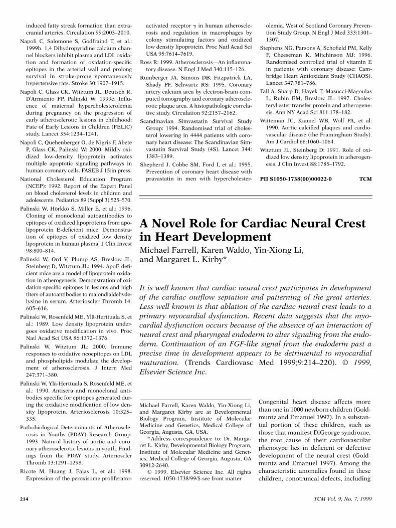
Figure 1. Cardiac neural crest cells (green) migrate from the neural folds at stages 10/11 intothe circumpharyngeal region by about stage 12. Over the next 24 h they populate the caudalpharyngeal arches, where they appear to insulate components of the pharynx from the outflowmyocardium by about stage 18. The cells proliferate and begin their migration into the cardiacoutflow tract by stage 22.
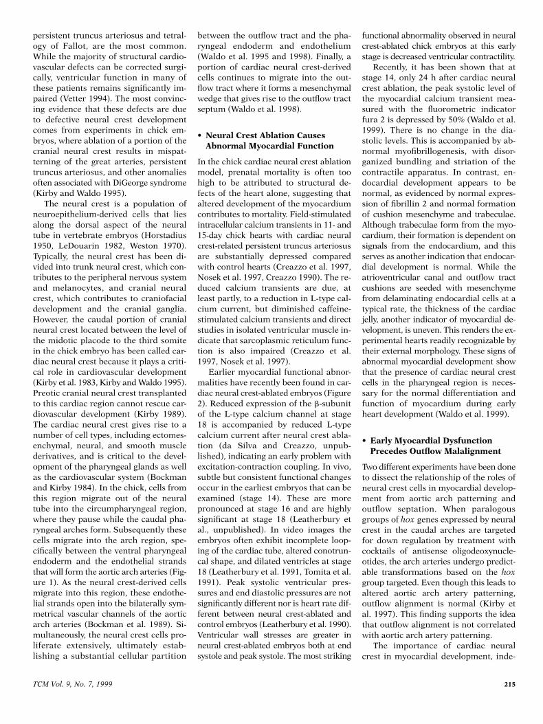
Figure 2. Time line showing the relationship of neural crest ablation to events in chick heartdevelopment with respect to the period when signs of abnormal heart development are firstnoted.
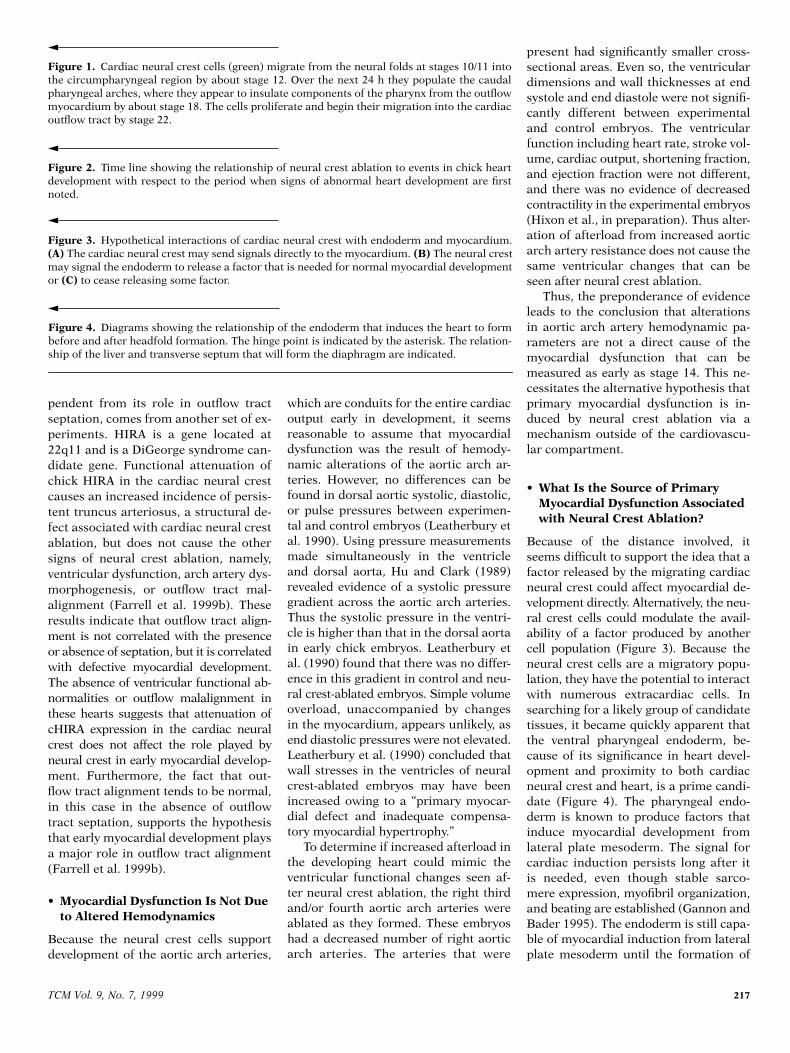
Figure 3. Hypothetical interactions of cardiac neural crest with endoderm and myocardium.(A) The cardiac neural crest may send signals directly to the myocardium. (B) The neural crestmay signal the endoderm to release a factor that is needed for normal myocardial developmentor (C) to cease releasing some factor.
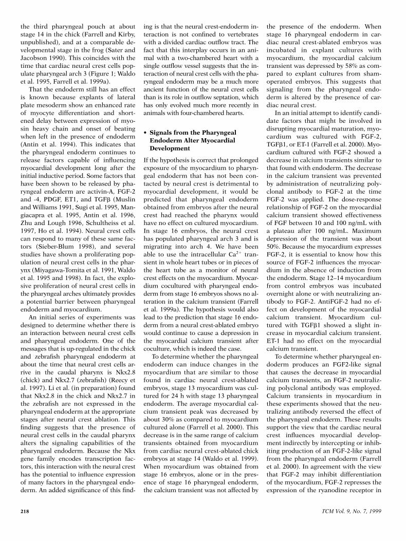
Figure 4. Diagrams showing the relationship of the endoderm that induces the heart to formbefore and after headfold formation. The hinge point is indicated by the asterisk. The relation-ship of the liver and transverse septum that will form the diaphragm are indicated.
218 TCM Vol. 9, No. 7, 1999
the third pharyngeal pouch at aboutstage 14 in the chick (Farrell and Kirby,unpublished), and at a comparable de-velopmental stage in the frog (Sater andJacobson 1990). This coincides with thetime that cardiac neural crest cells pop-ulate pharyngeal arch 3 (Figure 1; Waldoet al. 1995, Farrell et al. 1999a).
That the endoderm still has an effectis known because explants of lateralplate mesoderm show an enhanced rateof myocyte differentiation and short-ened delay between expression of myo-sin heavy chain and onset of beatingwhen left in the presence of endoderm(Antin et al. 1994). This indicates thatthe pharyngeal endoderm continues torelease factors capable of influencingmyocardial development long after theinitial inductive period. Some factors thathave been shown to be released by pha-ryngeal endoderm are activin-A, FGF-2and -4, PDGF, ET1, and TGFb (Muslinand Williams 1991, Sugi et al. 1995, Man-giacapra et al. 1995, Antin et al. 1996,Zhu and Lough 1996, Schultheiss et al.1997, Ho et al. 1994). Neural crest cellscan respond to many of these same fac-tors (Sieber-Blum 1998), and severalstudies have shown a proliferating pop-ulation of neural crest cells in the phar-ynx (Miyagawa-Tomita et al. 1991, Waldoet al. 1995 and 1998). In fact, the explo-sive proliferation of neural crest cells inthe pharyngeal arches ultimately providesa potential barrier between pharyngealendoderm and myocardium.
An initial series of experiments wasdesigned to determine whether there isan interaction between neural crest cellsand pharyngeal endoderm. One of themessages that is up-regulated in the chickand zebrafish pharyngeal endoderm atabout the time that neural crest cells ar-rive in the caudal pharynx is Nkx2.8(chick) and Nkx2.7 (zebrafish) (Reecy etal. 1997). Li et al. (in preparation) foundthat Nkx2.8 in the chick and Nkx2.7 inthe zebrafish are not expressed in thepharyngeal endoderm at the appropriatestages after neural crest ablation. Thisfinding suggests that the presence ofneural crest cells in the caudal pharynxalters the signaling capabilities of thepharyngeal endoderm. Because the Nkxgene family encodes transcription fac-tors, this interaction with the neural cresthas the potential to influence expressionof many factors in the pharyngeal endo-derm. An added significance of this find-
ing is that the neural crest-endoderm in-teraction is not confined to vertebrateswith a divided cardiac outflow tract. Thefact that this interplay occurs in an ani-mal with a two-chambered heart with asingle outflow vessel suggests that the in-teraction of neural crest cells with the pha-ryngeal endoderm may be a much moreancient function of the neural crest cellsthan is its role in outflow septation, whichhas only evolved much more recently inanimals with four-chambered hearts.
• Signals from the Pharyngeal Endoderm Alter Myocardial Development
If the hypothesis is correct that prolongedexposure of the myocardium to pharyn-geal endoderm that has not been con-tacted by neural crest is detrimental tomyocardial development, it would bepredicted that pharyngeal endodermobtained from embryos after the neuralcrest had reached the pharynx wouldhave no effect on cultured myocardium.In stage 16 embryos, the neural cresthas populated pharyngeal arch 3 and ismigrating into arch 4. We have beenable to use the intracellular Ca21 tran-sient in whole heart tubes or in pieces ofthe heart tube as a monitor of neuralcrest effects on the myocardium. Myocar-dium cocultured with pharyngeal endo-derm from stage 16 embryos shows no al-teration in the calcium transient (Farrellet al. 1999a). The hypothesis would alsolead to the prediction that stage 16 endo-derm from a neural crest-ablated embryowould continue to cause a depression inthe myocardial calcium transient aftercoculture, which is indeed the case.
To determine whether the pharyngealendoderm can induce changes in themyocardium that are similar to thosefound in cardiac neural crest-ablatedembryos, stage 13 myocardium was cul-tured for 24 h with stage 13 pharyngealendoderm. The average myocardial cal-cium transient peak was decreased byabout 30% as compared to myocardiumcultured alone (Farrell et al. 2000). Thisdecrease is in the same range of calciumtransients obtained from myocardiumfrom cardiac neural crest-ablated chickembryos at stage 14 (Waldo et al. 1999).When myocardium was obtained fromstage 16 embryos, alone or in the pres-ence of stage 16 pharyngeal endoderm,the calcium transient was not affected by
the presence of the endoderm. Whenstage 16 pharyngeal endoderm in car-diac neural crest-ablated embryos wasincubated in explant cultures withmyocardium, the myocardial calciumtransient was depressed by 58% as com-pared to explant cultures from sham-operated embryos. This suggests thatsignaling from the pharyngeal endo-derm is altered by the presence of car-diac neural crest.
In an initial attempt to identify candi-date factors that might be involved indisrupting myocardial maturation, myo-cardium was cultured with FGF-2,TGFb1, or ET-1 (Farrell et al. 2000). Myo-cardium cultured with FGF-2 showed adecrease in calcium transients similar tothat found with endoderm. The decreasein the calcium transient was preventedby administration of neutralizing poly-clonal antibody to FGF-2 at the timeFGF-2 was applied. The dose-responserelationship of FGF-2 on the myocardialcalcium transient showed effectivenessof FGF between 10 and 100 ng/mL witha plateau after 100 ng/mL. Maximumdepression of the transient was about50%. Because the myocardium expressesFGF-2, it is essential to know how thissource of FGF-2 influences the myocar-dium in the absence of induction fromthe endoderm. Stage 12–14 myocardiumfrom control embryos was incubatedovernight alone or with neutralizing an-tibody to FGF-2. AntiFGF-2 had no ef-fect on development of the myocardialcalcium transient. Myocardium cul-tured with TGFb1 showed a slight in-crease in myocardial calcium transient.ET-1 had no effect on the myocardialcalcium transient.
To determine whether pharyngeal en-doderm produces an FGF2-like signalthat causes the decrease in myocardialcalcium transients, an FGF-2 neutraliz-ing polyclonal antibody was employed.Calcium transients in myocardium inthese experiments showed that the neu-tralizing antibody reversed the effect ofthe pharyngeal endoderm. These resultssupport the view that the cardiac neuralcrest influences myocardial develop-ment indirectly by intercepting or inhib-iting production of an FGF-2-like signalfrom the pharyngeal endoderm (Farrellet al. 2000). In agreement with the viewthat FGF-2 may inhibit differentiationof the myocardium, FGF-2 represses theexpression of the ryanodine receptor in
TCM Vol. 9, No. 7, 1999 219
skeletal myocytes (Marks et al. 1991). Inaddition, FGF Receptor2 (FGFR2) is ex-pressed in the neural crest-derived mes-enchyme of the caudal pharyngeal archesand is expressed in the myocardium un-til stage 15, suggesting that a transitionin the role of FGF-2-like factors takesplace at this stage of development (Par-low et al. 1991, Patstone et al. 1993).
FGF-2 has been shown to increasethe rate of proliferation of cardiac myo-cytes (Pasumarthi et al. 1996). To deter-mine whether pharyngeal endodermhad a similar effect on proliferation, myo-cardium obtained from stage 13 em-bryos was dissected and cultured for 24h. One half was cultured under standardconditions, the other half was culturedin the presence of pharyngeal endoderm(Farrell et al. 2000). At 24 h, the myocar-dium was harvested and prepared forimmunohistochemistry. An antibodyagainst the proliferating cell nuclear an-tigen (PCNA), a cell-cycle marker, wasused to determine the status of prolifer-ation in the myocardium. The percent ofPCNA-positive myocardial cells cocul-tured with pharyngeal endoderm wasover 90%, indicating a very high rate ofproliferation. The percent of PCNA-posi-tive cells in myocardium cultured in theabsence of endoderm was less than 40%.
Although the definitive in vivo experi-ments must still be carried out, the re-sults of Farrell et al. support the viewthat the pharyngeal endoderm producesan FGF-2-like factor that can suppressthe embryonic myocardial calcium tran-sient at a critical stage of development.The results demonstrate that prolongedexposure of the myocardium to the pha-ryngeal endoderm significantly impairsexcitation-contraction coupling. This ef-fect may also be important in vivo, wherethe myocardium may be inappropriatelyexposed to the pharyngeal endoderm inthe cardiac neural crest-ablated embryosduring the stages in development whenneural crest-derived cells would normallybe populating the region between the pha-ryngeal endoderm and the myocardium.
The degree to which the calcium tran-sients are suppressed by coculturingstage 12–13 myocardium with stage 14pharyngeal endoderm is in line with thesuppression found in myocardial cal-cium transients obtained from cardiacneural crest-ablated embryos at stage 14.At this stage, neural crest-derived cells liein intimate contact with the pharyngeal
endoderm and are beginning to inter-pose between the pharyngeal endodermand the splanchnic mesoderm.
• Summary and Conclusions
We have proposed and showed the evi-dence for a novel role of neural crest inmyocardial development. Neural crestablation results in myocardial dysfunc-tion at the time when the circulation isfirst being established. This dysfunctionis not a result of hemodynamic alter-ations in the peripheral circulation.However, it occurs concurrently withthe time when neural crest cells firsthave contact with the pharyngeal endo-derm, a tissue known to release factorsthat influence myocardial differentia-tion. Current data in the neural crest ab-lation model suggest that the pharyngealendoderm, which first induces myocar-dial differentiation, continues to releasesignals that affect myocardial maturationuntil the neural crest migrates into thecaudal pharyngeal arches. The presenceof neural crest in the arches alters thissignaling in such a manner that myocar-dial maturation occurs normally. In theabsence of cardiac neural crest, the sig-naling is prolonged to the detriment ofthe myocardium. It appears that theFGF family is involved in some way inthis signaling, although it is prematureto do more than speculate on the sourcesof this signaling and other factors in-volved in this new role of neural crest incardiovascular development.
• Acknowledgments
We thank Harriett Stadt, Donna Kumiski,Marzena Zdanowicz, and Eileen Mc-Craney for technical assistance in theprojects that led to this review, and TonyCreazzo, Bob Godt, and Linda Leather-bury for stimulating discussions and shar-ing unpublished data. This work wassupported by PHS grants HL36059and HD16057.
References
Antin PB, Taylor RG, Yatskievych T: 1994. Pre-cardiac mesoderm is specified during gas-trulation in quail. Dev Dyn 200:144–154.
Antin PB, Yatskievych T, Dominguez JL, etal.: 1996. Regulation of avian precardiacmesoderm development by insulin and
insulin-like growth factors. J Cell Physiol168:42–50.
Bockman DE, Kirby ML: 1984. Dependenceof thymus development on derivatives ofthe neural crest. Science 223:498–500.
Bockman DE, Redmond ME, Kirby ML:1989. Alteration of early vascular develop-ment after ablation of cranial neural crest.Anat Rec 225:209–217.
Creazzo TL: 1990. Reduced “L” type calciumcurrent in the embryonic chick heart withpersistent truncus arteriosus. Circ Res66:1491–1498.
Creazzo TL, Brotto MA, Burch J: 1997. Exci-tation-contraction coupling in the day 15embryonic chick heart with persistent trun-cus arteriosus. Pediatr Res 42:731–737.
Farrell MJ, Burch JL, Kumiski D, et al.: 1999a.Pharyngeal endoderm produces a factorthat suppresses development of myocardialcalcium transients (in revision).
Farrell MF, Stadt H, Wallis KT, et al.: 1999. ADiGeorge syndrome candidate gene requiredfor cardiac outflow tract septation. CircRes 84:127–135.
Gannon M, Bader D: 1995. Initiation of car-diac differentiation occurs in the absenceof anterior endoderm. Development 121:2439–2450.
Goldmuntz E, Emanuel BS: 1997. Geneticdisorders of cardiac morphogenesis. TheDiGeorge and velocardiofacial syndromes.Circ Res 80:437–443.
Ho L, Symes K, Yordán C, et al.: 1994. Local-ization of PDGF A and PDGFRa mRNA inXenopus embryos suggests signalling fromneural ectoderm and pharyngeal endodermto neural crest cells. Mech Dev 48:165–174.
Horstadius S: 1950. The Neural Crest. ItsProperties and Derivatives in the Light ofExperimental Research. London, OxfordUniversity Press.
Hu N, Clark EB: 1989. Hemodynamics of thestage 12 to stage 29 chick embryo. Circ Res65:1665–1670.
Kirby ML: 1989. Plasticity and predetermi-nation of mesencephalic and trunk neuralcrest transplanted into the region of the car-diac neural crest. Dev Biol 134:402–412.
Kirby ML, Gale TF, Stewart DE: 1983. Neuralcrest cells contribute to aorticopulmonaryseptation. Science 220:1059–1061.
Kirby ML, Hunt P, Wallis KT, Thorogood P:1997. Normal development of the cardiacoutflow tract is not dependent on normalpatterning of the aortic arch arteries. Dev.Dyn. 208:34–47.
Kirby ML, Waldo KL: 1995. Neural crest andcardiovascular patterning. Circ Res 77:211–215.
Leatherbury L, Braden DS, Tomita H, et al.:1990. Wall stresses and pressure gradientsin neural crest-ablated chick embryos. AnnNY Acad Sci 588:305–313.

220 TCM Vol. 9, No. 7, 1999
Leatherbury L, Connuck DM, Gauldin HE, etal.: 1991. Hemodynamic changes and com-pensatory mechanisms during early car-diogenesis after neural crest ablation inchick embryos. Pediatr Res 30:509–512.
LeDouarin NM: 1982. The Neural Crest. Cam-bridge, UK, Cambridge University Press.
Mangiacapra FJ, Fransen ME, Lemanski LF:1995. Activin A and transforming growthfactor-b stimulate heart formation in axo-lotls but do not rescue cardiac lethalmutants. Cell Tissue Res 282:227–236.
Marks AR, Taubman MB, Saito A, et al.: 1991.The ryanodine receptor/junctional channelcomplex is regulated by growth factors in amyogenic cell line. J Cell Biol 114:303–312.
Miyagawa-Tomita S, Waldo K, Tomita H, etal.: 1991. Temporospatial study of themigration and distribution of cardiac neu-ral crest in quail-chick chimeras. Am JAnat 192:79–88.
Muslin AJ, Williams LT: 1991. Well-definedgrowth factors promote cardiac develop-ment in axolotl mesodermal explants.Development 112:1095–1101.
Nosek TM, Fogaca RTH, Hatcher CJ, et al.:1997. Effect of cardiac neural crest abla-tion on contractile force and calciumuptake and release in chick heart. Am JPhysiol 273:H1464–H1471.
Parlow MH, Bolender DL, Kokan-Moore NP,et al.: 1991. Localization of bFGF-like pro-teins as punctate inclusions in the presep-
tation myocardium of the chicken embryo.Dev Biol 146:139–147.
Pasumarthi KB, Kardami E, Cattini PA: 1996.High and low molecular weight fibroblastgrowth factor-2 increase proliferation ofneonatal rat cardiac myocytes but have dif-ferential effects on binucleation and nuclearmorphology. Evidence for both paracrineand intracrine actions of fibroblast growthfactor-2. Circ Res 78:126–136.
Patstone G, Pasquale EB, Maher PA: 1993.Different members of the fibroblast growthfactor receptor family are specific to dis-tinct cell types in the developing chickenembryo. Dev Biol 155:107–123.
Reecy JM, Yamada M, Cummings K, et al.:1997. Chicken Nkx-2.8: A novel homeoboxgene expressed in early heart progenitorcells and pharyngeal pouch-2 and -3 endo-derm. Dev Biol 188:295–311.
Sater AK, Jacobson AG: 1990. The restrictionof the heart morphogenetic field in Xeno-pus laevis. Dev Biol 140:328–336.
Schultheiss TM, Burch JBE, Lassar AB: 1997.A role for bone morphogenetic proteins inthe induction of cardiac myogenesis.Genes Dev 11:451–462.
Sieber-Blum M: 1998. Growth factor synergismand antagonism in early neural crest devel-opment. Biochem Cell Biol 76:1039–1050.
Sugi Y, Sasse J, Barron M, et al.: 1995. Devel-opmental expression of fibroblast growth
factor receptor-1 (cek-1; flg) during heartdevelopment. Dev Dyn 202:115–125.
Tomita H, Connuck DM, Leatherbury L, etal.: 1991. Relation of early hemodynamicchanges to final cardiac phenotype andsurvival after neural crest ablation in chickembryos. Circulation 84:1289–1295.
Vetter VL: 1994. Postoperative arrhythmiasafter surgery for congenital heart defects.Cardiol Review 2:83–97.
Waldo KL, Kumiski K, Kirby ML: 1995. Car-diac neural crest is essential for the persis-tence rather than the formation of an archartery. Dev Dyn 205:281–292.
Waldo KL, Miyagawa-Tomita S, Kumiski D,et al.: 1998. Cardiac neural crest cells pro-vide new insight into septation of the out-flow tract: Aortic sac to ventricular septalclosure. Dev Biol 196:129–144.
Waldo K, Zdanowicz M, Burch J, et al.: 1999.A novel role for cardiac neural crest in heartdevelopment. J Clin Invest 103:1499–1507.
Weston JA: 1970 The migration and differen-tiation of neural crest cells. Adv Morpho-genesis 8:41–117.
Zhu X, Lough J: 1996. Expression of alterna-tively spliced and canonical basic fibroblastgrowth factor mRNAs in the early embryoand developing heart. Dev Dyn 206:139–145.
PII S1050-1738(00)00023-2 TCM
Why wait for it to circulate? Read your own copy of
If you share TCM with others at work, you know that what goes around doesn’t alwayscome around. And if it does, articles are often dated or sometimes clipped.
Why miss a single issue, or wait weeks to read one? By starting your personal subscrip-tion, you’ll be guaranteed to read each and every issue.
TRENDS IN CARDIOVASCULAR MEDICINE’s short reviews on “hot” topics can makea difference in your research and practice. Commissioned researchers and cliniciansreport on advances in endocrinology and its related areas.
Use the business reply cards in this issue or order your personal subscription today—or callElsevier Customer Service at:
(212) 633-3950 or fax (212) 633-3990.