A Novel Method of Medical Image Denoising Using Bilateral ...
6
A novel method of Medical image denoising using bilateral and NLm filtering.omnamasivay aper subtitle) Mahesh Mohan M R M.Tech student Govt Engineering College Trichur Kerala,India [email protected] Sheeba V S Professor : Electronics and Communication Dept Govt Engineering College Trichur Kerala,India [email protected] Abstract— One of the bottleneck in medical imaging is due to low Signal-to-Noise Ratio(SNR) which requires long and repeated acquisition of the same subject to reduce noise and blur. To obtain a high SNR image without lengthy repeated scans, post- processing of data such as denoising plays a critical role. Bilateral and Non-Local means (NLm) filtering are commonly used procedures for medical image denoising. In this paper we propose a new thresholding scheme to wavelet and contourlet based denoising by introducing a scaling factor to universal threshold. Also we propose the aforementioned novel contourlet thresholding scheme as a pre-processing step on bilateral filtering and NLm denoising. Simulation results show that the proposed single entity comprising the novel preprocessing step and bilateral or NLm denoising is superior in terms of PSNR and perceptual quality compared to Bilateral filtering or NLm denoising used alone. Keywords—Denoising,Contourlet transform,Bilateral filtering,Non Local means denoisng,wavelet transform,visushrink. I. INTRODUCTION (HEADING 1) The rapid development of advanced medical imaging technologies such as Magnetic Resonance Imaging(MRI), Positron Emitted Tomography (PET) and Computer Tomography (CT) have provided us an unprecedented way to diagnose illness in-vivo and non-invasively. Some of the most advanced techniques based on this imaging modality remain in the research stage but never reach routine clinical usage. One of the bottlenecks is due to low Signal-to-Noise Ratio (SNR) which requires long and repeated acquisition of the same subject to reduce noise and blur. For example, a high SNR Diffusion Tensor Imaging (DTI) dataset requires an hour of data Acquisition[1]. A high SNR of High-Angular-Resolution Diffusion Imaging (HARDI) data can take 13 hours [2]. To recover high SNR image from noisy and blurry image without lengthy repeated scans, post-processing of data plays a critical role in two aspects: (1) automatic denoising and deblurring algorithms can restore the data computationally to achieve a quality that might take much longer otherwise. (2) computational object segmentation techniques can perform intelligent extraction of data directly and automatically from noisy observations. In medical imaging we often face a relatively low SNR with good contrast, or a low contrast with good SNR. Fortunately the human visual system is highly effective in recognizing structures even in the presence of a considerable amount of noise. But if the SNR is too small or the contrast is too low it becomes very difficult to detect anatomical structures. A definition of overall image quality includes physical and perceptual criteria. Furthermore, it largely depends on specific diagnostic tasks. In some cases a high spatial resolution and a high contrast are required, whereas in other cases more perceptual criteria may be favoured. For a visual analysis of medical images, the clarity of details which mainly comprises edge information and the object visibility are important. Noise mostly occurs in medical images are Additive White Gaussian noise (AWGN), salt and pepper noise[3], speckle noise[4][5][6] etc. There are two basic approaches to image denoising, spatial filtering methods and transform domain filtering methods. The filtering does not respect region boundaries or small structures, and the resulting images appear blurry and diffused. Many researchers have used spatial as well as transform domain filtering in medical imaging[7-10] When developing a filtering method for medical image data, image degradation by blurring or artifacts resulting from a filtering scheme is not acceptable. The following two criteria should ideally be fulfilled: • Minimize information loss by preserving object boundaries and detailed structures. • Efficiently remove noise in regions of homogeneous physical properties. Recent developments based on bilateral filtering [11] overcome the major drawbacks of conventional spatial filtering, and significantly improve image quality while satisfying the main criteria 1 stated above. Bilateral filtering is one of the examples of non-linear non-iterative filtering. It combines domain and range filters simultaneously. It preserves edge information while denoising. Many medical image denoisng algorithm have been proposed using bilateral filtering[6,7]. However it does not give satisfactory results since real gray levels are polluted seriously and the range filter cannot work properly. This would lead to bring side effect to the denoising results like a polishing look to denoised image. For example, image’s tissue regions or brain’s grooves may be 2013 Third International Conference on Advances in Computing and Communications 978-0-7695-5033-6/13 $26.00 © 2013 IEEE DOI 10.1109/ICACC.2013.101 186 2013 Third International Conference on Advances in Computing and Communications 978-0-7695-5033-6/13 $26.00 © 2013 IEEE DOI 10.1109/ICACC.2013.101 186
Transcript of A Novel Method of Medical Image Denoising Using Bilateral ...

A novel method of Medical image denoising using bilateral and NLm filtering.omnamasivay
aper subtitle)
Mahesh Mohan M R M.Tech student
Govt Engineering College Trichur Kerala,India
Sheeba V S Professor : Electronics and Communication Dept
Govt Engineering College Trichur Kerala,India
Abstract— One of the bottleneck in medical imaging is due to low Signal-to-Noise Ratio(SNR) which requires long and repeated acquisition of the same subject to reduce noise and blur. To obtain a high SNR image without lengthy repeated scans, post-processing of data such as denoising plays a critical role. Bilateral and Non-Local means (NLm) filtering are commonly used procedures for medical image denoising. In this paper we propose a new thresholding scheme to wavelet and contourlet based denoising by introducing a scaling factor to universal threshold. Also we propose the aforementioned novel contourlet thresholding scheme as a pre-processing step on bilateral filtering and NLm denoising. Simulation results show that the proposed single entity comprising the novel preprocessing step and bilateral or NLm denoising is superior in terms of PSNR and perceptual quality compared to Bilateral filtering or NLm denoising used alone.
Keywords—Denoising,Contourlet transform,Bilateral filtering,Non Local means denoisng,wavelet transform,visushrink.
I. INTRODUCTION (HEADING 1) The rapid development of advanced medical imaging
technologies such as Magnetic Resonance Imaging(MRI), Positron Emitted Tomography (PET) and Computer Tomography (CT) have provided us an unprecedented way to diagnose illness in-vivo and non-invasively. Some of the most advanced techniques based on this imaging modality remain in the research stage but never reach routine clinical usage. One of the bottlenecks is due to low Signal-to-Noise Ratio (SNR) which requires long and repeated acquisition of the same subject to reduce noise and blur. For example, a high SNR Diffusion Tensor Imaging (DTI) dataset requires an hour of data Acquisition[1]. A high SNR of High-Angular-Resolution Diffusion Imaging (HARDI) data can take 13 hours [2]. To recover high SNR image from noisy and blurry image without lengthy repeated scans, post-processing of data plays a critical role in two aspects: (1) automatic denoising and deblurring algorithms can restore the data computationally to achieve a quality that might take much longer otherwise. (2) computational object segmentation techniques can perform intelligent extraction of data directly and automatically from noisy observations.
In medical imaging we often face a relatively low SNR with good contrast, or a low contrast with good SNR.
Fortunately the human visual system is highly effective in recognizing structures even in the presence of a considerable amount of noise. But if the SNR is too small or the contrast is too low it becomes very difficult to detect anatomical structures. A definition of overall image quality includes physical and perceptual criteria. Furthermore, it largely depends on specific diagnostic tasks. In some cases a high spatial resolution and a high contrast are required, whereas in other cases more perceptual criteria may be favoured. For a visual analysis of medical images, the clarity of details which mainly comprises edge information and the object visibility are important.
Noise mostly occurs in medical images are Additive White Gaussian noise (AWGN), salt and pepper noise[3], speckle noise[4][5][6] etc. There are two basic approaches to image denoising, spatial filtering methods and transform domain filtering methods. The filtering does not respect region boundaries or small structures, and the resulting images appear blurry and diffused. Many researchers have used spatial as well as transform domain filtering in medical imaging[7-10]
When developing a filtering method for medical image data, image degradation by blurring or artifacts resulting from a filtering scheme is not acceptable. The following two criteria should ideally be fulfilled:
• Minimize information loss by preserving object boundaries and detailed structures.
• Efficiently remove noise in regions of homogeneous physical properties.
Recent developments based on bilateral filtering [11] overcome the major drawbacks of conventional spatial filtering, and significantly improve image quality while satisfying the main criteria 1 stated above. Bilateral filtering is one of the examples of non-linear non-iterative filtering. It combines domain and range filters simultaneously. It preserves edge information while denoising. Many medical image denoisng algorithm have been proposed using bilateral filtering[6,7]. However it does not give satisfactory results since real gray levels are polluted seriously and the range filter cannot work properly. This would lead to bring side effect to the denoising results like a polishing look to denoised image. For example, image’s tissue regions or brain’s grooves may be
2013 Third International Conference on Advances in Computing and Communications
978-0-7695-5033-6/13 $26.00 © 2013 IEEE
DOI 10.1109/ICACC.2013.101
186
2013 Third International Conference on Advances in Computing and Communications
978-0-7695-5033-6/13 $26.00 © 2013 IEEE
DOI 10.1109/ICACC.2013.101
186

weakened. This phenomenon is referred as propagation of noise (PoN) [11].
As an extension of bilateral filtering, Buades et al.[12] had put forward Non-Local means image denoising which utilizes structural similarity. Generally speaking, the information in a natural image is redundant to some extent. NL means image denoising algorithm takes full advantage of image redundancy. The basic idea is that images contain repeated structures, and averaging them will reduce the (random) noise. It is an efficient denoising method with the ability to result in proper restoration of the slowly varying signals in homogeneous tissue regions while strongly preserving the tissue boundaries. NLm has been used in medical image denoising in many works [13]. However, the NLM filter may suffer from potential limitations since the calculation for similarity weights is performed in a full-space of neighborhood. Specifically, the accuracy of the similarity weights will be affected by noise.
Donoho and Johnstone proposed an innovative nonlinear denoising scheme in transform domain VisuShrink [14] which thresholds the wavelet detail coefficients for one dimension (1-D) signals. This scheme is simple and efficient. More recently, wavelet based filters have been applied successfully to MR image denoising [15,10,4]. Donoho and Johnstone have shown that VisuShrink results in an estimate asymptotically optimal in the minimax sense. When images are denoised by VisuShrink, in many cases it can outperform the classic linear Wiener filtering, especially for the images with a low peak signal-to-noise ratio (PSNR). However, it is also well known that universal thresholding yields overly smoothed images. This is because its threshold choice can be unwarrantedly large due to its dependence on the number of samples. Many researchers have made claim that the universal threshold is not the perfect threshold and performance can vary around this threshold [4,6].
Minh N. Do and Martin Vetterli put forward a new dimension in image transform named contourlet transform[16,17],which proved to be much better than the wavelet transform in conserving edges and line details. The improvement in approximation by contourlets based on keeping the most significant coefficients will directly lead to improvements in denoising. A simple thresholding scheme applied on the contourlet transform is more effective in removing the noise than it is for the wavelet transform[17]. A number of works have been proposed in image denoising using contourlet transform recently. Atif et al.[18] used the same thresholding scheme which is used in wavelets to contourlet denoising, to substantiate the claim made in paper[17]. The problem is that directly using the universal threshold which suites well in wavelet domain is not suitable in contourlet domain, since the number of elements in transform coefficients for contourlet is much higher than wavelet coefficients.
In this paper we propose a novel technique to medical images corrupted by AWGN. This paper puts forward three contributions
• As the universal thresholding is an estimate which is asymptotically optimal in the minmax sense, we introduce a scaling parameter to the universal threshold by extensive simulation and regression of data with wide range of standard medical images as well as
natural images of different size and corrupted by different noise variances.
• We have also extended this idea to contourlet transformation with a new scaling factor and the same universal thresholding.
• Proposed contourlet thresholding is performed as a pre-processing step prior to bilateral and NLm filtering, which can give significant improvement in PSNR as well as visual quality from bilateral and NLm filter used alone.
The paper is organized as follows. Section II introduces Bilateral filtering, Non Local mean filtering, wavelet shrinkage and contourlet transform. In Section III, Proposed Method is discussed. The Section IV presents the experimental results and discussion. The concluding remarks are given in Section V.
II. BACKGROUND
A. Bilateral Filtering The bilateral �lter is a nonlinear �lter proposed by Tomasi
and Manduchi to smooth images[11].The key idea of the bilateral �lter is that for a pixel to in�uence another pixel, it should not only occupy a nearby location but also have a similar value. The idea underlying bilateral �ltering is to do in the range of an image what traditional �lters do in its domain. Two pixels can be close to one another, that is, occupy nearby spatial location, or they can be similar to one another, that is, have nearby values, possibly in a perceptually meaningful fashion. Closeness refers to vicinity in the domain, similarity to vicinity in the range. Traditional �ltering is domain �ltering, and enforces closeness by weighting pixel values with coef�cients that fall off with distance. Similarly, range �ltering is defined, which averages image values with weights that decay with dissimilarity. Range �lters are nonlinear because their weights depend on image intensity or color. The bilateral �lter is also non-iterative, i.e. it achieves satisfying results with only a single pass. This makes the �lter’s parameters relatively intuitive since their action does not depend on the cumulated e�ects of several iterations. Computationally, they are no more complex than standard non-separable �lters. Most importantly, they preserve edges.
The weight assigned to each neighbour decreases with both the distance in the image plane (the spatial domain S) and the distance on the intensity axis (the range domain R). Using a Gaussian G� as a decreasing function, and considering a gray level image I, the BF[I] of the bilateral �lter is de�ned by:
( ) ( )�∈
−−=S
IIIGGW
IBFq
qqpp
p qp ||||||1][rs σσ (1)
• Ip = value of image I at position: p = ( px , py )
• F [ I ] = output of filter F applied to image I
The parameter �s defines the size of the spatial neighborhood used to filter a pixel, and �r controls how much an adjacent pixel is down weighted because of the intensity difference. Wp normalizes the sum of the weights.
187187
On the other hand, the bilateral filter is nonlinear and its evaluation is computationally expensive since traditional acceleration, such as performing convolution after an FFT, are not applicable. Many fast algorithm have been formulated for bilateral filtering [19,20], one of the recent algorithm is formulated by Choudhary et al. [21] .
B. Non-Local Mean filtering Buades et al. [12] developed a Non-Local means (NLm)
image denoising algorithm which takes full advantage of image redundancy. The basic idea is that images contain repeated structures, and averaging them will reduce the (random) noise. The NL-means filter is an evolution of the Yaroslavsky filter [22] which averages similar image pixels defined according to their local intensity similarity. The main difference between the NLmeans and this filter is that the similarity between pixels has been made more robust to noise by using a region comparison, rather than pixel comparison and also that matching patterns are not restricted to be local. That is, pixels far from the pixel being filtered are not penalized. Given an image Y the filtered value at a point i using the NL-means method is computed as a weighted average of neighbouring pixels Ni in the image following this formula:
�∈
=Nij
jYjiwiYNL )(),()]([ p (2)
1),(0 ≤≤ jiw and 1),( =�∈Nij
jiw
where i is point being filtering and j represents any other image pixel. The weights w(i, j) are based on the similarity between the neighborhoods Ni and Nj of pixels i and j. Ni is defined as a square neighbourhood window indices centered around pixel i. Theoretically, noise filtering performed must be considered as an estimation task. Since the estimation task of weights w(i,j) are computationally expensive, a large number of fast methods have been developed[23,24].
C. Wavelet shrinkage Wavelet transform shows localization in both time and
frequency and hence it has proved itself to be an efficient tool for a number of image processing applications including noise removal[14,15].The methods based on wavelet representations yield very simple algorithms that are often more powerful and easy to work with than traditional methods of function estimation. Visushrink consists of decomposing the observed signal into wavelets and use thresholds to select the coefficients, from which a signal is synthesized. The idea behind wavelet shrinkage is that Wavelet is such a basis because exceptional event generates identifiable exceptional coefficients due to its good localization property in both spatial and frequency domain [5]. But considering noise, as long as it does not generate exceptions, additive white Gaussian noise after applying WT is still AWGN. A large fraction of any L number of random data array with zero mean and variance � 2 , will be smaller than the universal threshold T with high probability; the probability approaching 1 as L increases to infinity[14] where
LT elog2σ= (3)
However, VisuShrink yields overly smoothed images. This is because its threshold choice can be unwarrantedly large due to its dependence on the number of samples and universal thresholding is an estimate which is asymptotically optimal in the minmax sense.
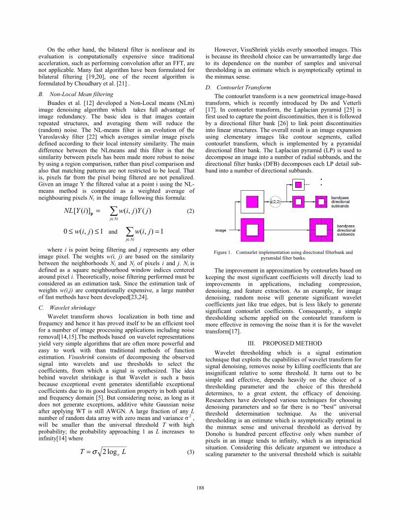
D. Contourlet Transform The contourlet transform is a new geometrical image-based
transform, which is recently introduced by Do and Vetterli [17]. In contourlet transform, the Laplacian pyramid [25] is first used to capture the point discontinuities, then it is followed by a directional filter bank [26] to link point discontinuities into linear structures. The overall result is an image expansion using elementary images like contour segments, called contourlet transform, which is implemented by a pyramidal directional filter bank. The Laplacian pyramid (LP) is used to decompose an image into a number of radial subbands, and the directional filter banks (DFB) decomposes each LP detail sub-band into a number of directional subbands.
Figure 1. Contourlet implementation using directional filterbank and
pyramidal filter banks.
The improvement in approximation by contourlets based on keeping the most significant coefficients will directly lead to improvements in applications, including compression, denoising, and feature extraction. As an example, for image denoising, random noise will generate significant wavelet coefficients just like true edges, but is less likely to generate significant contourlet coefficients. Consequently, a simple thresholding scheme applied on the contourlet transform is more effective in removing the noise than it is for the wavelet transform[17].
III. PROPOSED METHOD Wavelet thresholding which is a signal estimation
technique that exploits the capabilities of wavelet transform for signal denoising, removes noise by killing coefficients that are insignificant relative to some threshold. It turns out to be simple and effective, depends heavily on the choice of a thresholding parameter and the choice of this threshold determines, to a great extent, the efficacy of denoising. Researchers have developed various techniques for choosing denoising parameters and so far there is no “best” universal threshold determination technique. As the universal thresholding is an estimate which is asymptotically optimal in the minmax sense and universal threshold as derived by Donoho is hundred percent effective only when number of pixels in an image tends to infinity, which is an impractical situation. Considering this delicate argument we introduce a scaling parameter to the universal threshold which is suitable
188188

for medical images whose number of pixels are finite, by extensive simulation and tabulation with different standard medical images of different size and corrupted by different noise variances. The experimental procedure we adopted is taking an arbitrary image of known size and corrupted by a known noise variance. Then we introduced scaling factors for the universal threshold and selected the scaling factor corresponding to the highest PSNR. This procedure is repeated for large set of images of different size and corrupted by different noise variance. The compact equation of scaling parameter is obtained by linear regression of the best scaling parameter as a function of noise variance and image size.
For wavelet thresholding, tabulation and regression with different sets of medical images as well as natural images give the threshold �w to be
�w =3.944�10-11 S2 – 5.5285�10-6 S+0.6022 (4) where S is a function of noise variance and number of image pixels, which is given as
NS ×= σ N is the number of pixels of the image under consideration
and � is the standard deviation of noise. So the new threshold is Tw = �w �T.
Same set of experiments done on wavelet thresholding is performed with contourlet transformation. For contourlet thresholding experimental measurements gives the threshold �c to be
�c =3.944�10-11 S2 – 5.5285�10-6 S+0.5522 (5)
where S is a function of noise variance and number of image pixels, which is given as
NS ×= σ N is the number of pixels of the image under
consideration and � is the standard deviation of noise. So the new threshold is Tc= �c�T. The reason for the reduction in scaling factor is that, in contourlet transform, number of transform coefficients are large compared to the wavelet(in which number of transform coefficients equal to image pixels), a similar reduction in weight of coefficients take place in contourlet domain, so as to distribute values over all contourlet coefficients. Simulation results show that proposed contourlet thresholding and wavelet thresholding outperforms the standard Visushrink. Bilateral filtering in which real homogeneous gray levels corrupted by noise is polluted significantly, and fails to efficiently remove noise in regions of homogeneous physical properties. In homogeneous regions in which noise strike, bilateral filtering most likely perform range filtering much greater than spatial filtering. So the effect of noise is retained as if it were an edge information of the image. If contourlet denoising which is computationally efficient and having negligible execution time is done before bilateral filtering, noise in homogeneous regions which is present as un-exceptions can be removed efficiently retaining the edge information as well as texture. Simulation results show that
preprocessing with proposed contourlet denoising is superior to proposed wavelet thresholding. The reason is that the only aim of the denoising prior to bilateral filtering is to remove discrepancies in homogeneous regions, but retaining edge information and texture details as such. But wavelet transform using Visushrink smoothens the image[15] thereby degrading performance of bilateral filtering. But contourlet thresholding is good in retaining directions and edge features. The NLM filter may suffer from potential limitations since the calculation for similarity weights is performed in a full-space of neighborhood. Specifically, the accuracy of the similarity weights will be affected by noise. Performing contourlet denoising prior to NLM filtering can compensate noise, which can increase the accuracy of similarity weights in the full space of neighbourhood and hence increase the performance of NLM filtering. Preprocessing with proposed contourlet thresholding give better results compared to proposed wavelet thresholding because contourlet denoising is much efficient in retaining edges and texture information and hence similarity weights after thresholding.
IV. SIMULATION RESULTS A set of two gray scale images, MRI image of human brain
and CT image of abdomen, shown in Fig. 2, were selected for our simulations. Each image has a size of 512 x 512 and 1024 x 1024 pixels respectively with 256 shades of gray, i.e. 8 bits per pixel. The evaluation criteria are based upon quantitative measure of Peak signal to noise ratio. These measures, as such, do not represent human perception of the images, i.e.visual quality of the images. It is very difficult to guage denoised images mathematically on visual quality as human vision system is not only highly subjective but also exhibit different tolerance levels to various types of noises. Thus authentication of denoising method requires both quantitative measure and qualitative inspection of image. In our work, we apply both criteria; first the PSNR and the image is also viewed for visual acceptance.
a b
Fig. 2 (a) CT image of abdomen(CT) (b) MRI image of human brain(MRI).
Table I gives the comparison of calculated PSNR of images reconstructed utilizing various thresholding. Fig. 3 gives the corresponding images.
Table II gives PSNR comparison of image denoising using standard Bilateral filtering, standard non local mean filtering, and the proposed methods. Fig. 4 gives the corresponding images.
189189

(a) (b)
(c) (d)
(e)
Fig. 3 Comparing threshold performance (a) Noisy image (� = 40) (b)universal thresholding with wavelet (c) universal thresholding with
contourlet (d)Proposed thresholding with wavelet (e)Proposed thresholding with contourlet.
TABLE I. COMPARING THRESHOLD PERFORMANCE (PSNR).
TABLE II. COMPARING PROPOSED METHOD AND STANDARD MEDICAL IMAGE FILTERING (PSNR).
(a) (b)
(c) (d)
(e) Fig. 4 Comparing performance (a) Noisy image (� = 40 ) (b)bilateral filtering
(c) NLm filtering (d)Proposed preprocessing with bilateral filtering (e)Proposed preprocessing with NLm filtering.
Test
image
Wavelet
Universal Threshold
Contourlet Universal Threshold
Wavelet proposed Threshold
Contourlet proposed threshold
� =30 CT 19.2590 18.0033 22.7820 23.2653
MRI 20.8224 19.4452 23.7486 23.8390 � =40
CT 16.1492 14.9014 20.2541 20.7107 MRI 18.2044 16.7866 21.4390 21.5036
� =50 CT 13.8143 12.6572 18.4467 18.6677
MRI 16.2471 14.8722 19.7023 19.8100
Test
image
Bilateral filtering
NLm
filtering
Proposed
preprocessing prior to bilateral filtering
Proposed
preprocessing prior to
NLm filtering
� = 30 CT 23.8733 23.9525 30.0319 30.3344
MRI 22.5029 23.5457 25.8689 26.2227 � = 40
CT 20.0548 19.8352 26.1260 25.1537 MRI 19.7281 20.1621 23.5916 23.5454
� = 50 CT 17.3835 17.1381 22.9191 22.0310
MRI 17.4913 17.6718 21.6140 21.4496
190190

It can be seen that, there is a significant improvement in PSNR values in the proposed threshold compared to the universal thresholding. Also by incorporating proposed contourlet thresholding prior to bilateral filtering and non-local mean filtering gives improved PSNR. Visually it’s evident that the Proposed preprocessing with NLm filtering gives much more visual information, than with bilateral filtering even though pre-processing with bilateral filtering give good PSNR. The reason is that bilateral filtering incorporates a spatial filter apart from range filter which gives a smoothing effect.
V. CONCLUSION AND FURTHER WORKS In this work we propose a novel preprocessing scheme for
bilateral filtering and NLm filtering for medical image denoisning. We used contourlet transform, as a directional non-separable transform, to retain image textures and edges even after thresholding. A new threshold for denoising is devised using simulation results both in contourlet and wavelet space. The method is tested on CT abdomen and brain MR images. It is also compared with universal threshold. It is shown that for many noisy images, universal threshold based on wavelet is not a proper choice. Instead we can use contourlet based methods which can retain edges in the image much better than wavelet thresholding. We have shown that by using contourlet thresholding as a preprocessing step prior to bilateral filtering and Non local means filtering we can effectively denoise the image and also significantly increase the PSNR and perceptual quality.
In general as a future forethought any denoising scheme whose performance very much depend on spatial or structural features, preprocessing with proposed contourlet thresholding happens to be a better candidate in retaining the spatial or structural features and hence enhance the performance of aforementioned denosing scheme. Also it is also possible to device a similar thresholding scheme related with a particular application by simulation and regression with large sets of data.
REFERENCES
[1] O. Christiansen, T. Lee, J. Lie, U. Sinha, and T. Chan, “Total Variation Regularization of Matrix-Valued Images." International Journal of Biomedical Imaging, 2007(27432):11, December 2007.
[2] D. Tuch, “Q-ball imaging." Magnetic Resonance in Medicine, 52(6):1358{1372, 2004.
[3] S.Kalaivani Narayanan, and R.S.D.Wahidabanu, A View on Despeckling in Ultrasound Imaging. International Journal of Signal Processing, Image Processing and Pattern Recognition, 2009, 2(3):85-98.
[4] H. Guo, J. E. Odegard, M.Lang, A. Gopinath, J. W. Selesnick, Wavelet based Speckle Reduction with Application to SAR based ATD/R. First International Conference on Image Processing 1994:75-79.
[5] Rafael C. Gonzalez, Richard E. Woods, Digital Image processing using MATLAB. Second Edition, Mc Graw hill.
[6] Lu Zhang, Jiaming Chen, Yuemin Zhu, Jianhua Luo, Comparisons of Several New Denoising Methods for Medical Images. IEEE, 2009:1-4.
[7] Zhiwu Liao,Shaoxiang Hu,Zhiqiang Yu,Dan Sun, Medical Image Blind Denoising Using Context Bilteteral Filter. International Conference of Medical Image Analysis and Clinical Application, 2010:12-17.
[8] J. B. Weaver, Y. Xu, D. M. Healy, and L. D. Cromwell, “Communications. Filtering noise from images with wavelet transforms,” Magnetic Resonance in Medicine, vol. 21, no. 2, pp. 288–295, 1991.
[9] Zhiling Longa, b and Nicolas H. Younana, “Denoising of images with Multiplicative Noise Corruption”, 13th Europian Signal Processing Conference, 2005,,a1755..
[10] C. S. Anand and J. S. Sahambi, “Wavelet domain non-linear filtering for MRI denoising,”Magnetic Resonance Imaging, vol. 28, no. 6, pp. 842–861, 2010.
[11] C. Tomasi and R. Manduchi, "Bilateral filtering for gray and color images. " Proc Int Conf Computer Vision, pp. 839–846, 1998.
[12] A. Buades, B. Coll, and J. M. Morel, “A review of image denoising algorithms, with a new one,” Multiscale Modeling and Simulation (SIAM Interdisciplinary Journal), Vol. 4, No. 2, 2005, pp 490-530.
[13] Buadès, Antoni, Coll, Bartomeu; Morel, Jean Michel, Image Denoising By Non-Local Averaging Acoustics, Speech, and Signal Processing, 2005. Proceedings. (ICASSP '05) . IEEE International Conference on March 18-23, 2005, 25 - 28
[14] D. L. Donoho and I. M. Johnstone, “Ideal spatial adaptation via wavelet shrinkage,” Biometrika, 81, pp. 425–455, 1994.
[15] M. Alexander, R. Baumgartner, A. R. Summers et al., “A wavelet-basedmethod for improving signal-to-noise ratio and contrast in MR images,” Magnetic Resonance Imaging, vol. 18, no. 2, pp. 169–180, 2000.
[16] M. N. Do and M. Vetterli, "Contourlets", in Beyond Wavelets, Academic Press, New York, 2003.
[17] Minh N. Do, and Martin Vetterli, The Contourlet Transform: An Efficient Directional Multiresolution Image Representation, IEEE Transactions On Image Processing. 2005 Page(s): 2091- 2106
[18] Atif Bin Mansoor, Shoab A Khann, Contoulet Denoising of Natural Images 2008 IEEE 978-1-4244-1724-7/08
[19] Elad, M, On the bilateral filter and ways to improve it, IEEE Trans. On Image Processing 11 (2002) 1141–1151.
[20] Durand, F., Dorsey, J, Fast bilateral filtering for the display of high-dynamic-range images. ACM Trans. on Graphics 21 (2002) Proc. of SIGGRAPH conference.
[21] K.N. Chaudhury, D. Sage, and M. Unser, "Fast O(1) bilateral filtering using trigonometric range kernels," IEEE Trans. Image Processing, vol. 20, no. 11, 2011.
[22] L. P. Yaroslavsky, Digital Picture Processing - An Introduction, Springer Verlag, 1985.
[23] Jin Wang, Yanwen Guo, Yiting Ying, Qunsheng Peng, Fast Non-Local Algorithm For Image Denoising, 2006 IEEE Image Processing Page(s): 1429 - 1432
[24] Jer´ ome Darbon , Alexandre Cunha, Tony F. Chan, Stanley Osher, Grant J. Jensen Rao R M and A S Bopardikar, Fast Nonlocal Filtering Applied To Electron Cryomicroscopy 5th IEEE International Symposium 2008 , Page(s): 1331- 1334.
[25] P. J. Burt and E. H. Adelson, "The Laplacian pyramid as a compact image code", IEEE Trans. Commun., 31(4), pp. 532-540, April 1983.
[26] R. H. Bamberger and M. J. T. Smith, "A filter bank for the directional decomposition of images: Theory and design", IEEE Trans. Signal Proc., 40(4), pp. 882-893, April 1992.
191191


![Study of Curvelet and Wavelet Image Denoising by Using … · 2018-12-15 · novel image denoising method which is based on DCT basis and sparse representation [6]. To achieve a good](https://static.fdocuments.us/doc/165x107/5f03a8f47e708231d40a24d6/study-of-curvelet-and-wavelet-image-denoising-by-using-2018-12-15-novel-image.jpg)