A novel Drosophila model of TDP-43 proteinopathies: N...
11
RESEARCH ARTICLE A novel Drosophila model of TDP-43 proteinopathies: N-terminal sequences combined with the Q/N domain induce protein functional loss and locomotion defects Simona Langellotti 1 , Valentina Romano 1 , Giulia Romano 1 , Raffaella Klima 1 , Fabian Feiguin 1 , Lucia Cragnaz 1 , Maurizio Romano 2, * and Francisco E. Baralle 1, * ABSTRACT Transactive response DNA-binding protein 43 kDa (TDP-43, also known as TBPH in Drosophila melanogaster and TARDBP in mammals) is the main protein component of the pathological inclusions observed in neurons of patients affected by different neurodegenerative disorders, including amyotrophic lateral sclerosis (ALS) and fronto-temporal lobar degeneration (FTLD). The number of studies investigating the molecular mechanisms underlying neurodegeneration is constantly growing; however, the role played by TDP-43 in disease onset and progression is still unclear. A fundamental shortcoming that hampers progress is the lack of animal models showing aggregation of TDP-43 without overexpression. In this manuscript, we have extended our cellular model of aggregation to a transgenic Drosophila line. Our fly model is not based on the overexpression of a wild-type TDP-43 transgene. By contrast, we engineered a construct that includes only the specific TDP-43 amino acid sequences necessary to trigger aggregate formation and capable of trapping endogenous Drosophila TDP-43 into a non-functional insoluble form. Importantly, the resulting recombinant product lacks functional RNA recognition motifs (RRMs) and, thus, does not have specific TDP-43-physiological functions (i.e. splicing regulation ability) that might affect the animal phenotype per se. This novel Drosophila model exhibits an evident degenerative phenotype with reduced lifespan and early locomotion defects. Additionally, we show that important proteins involved in neuromuscular junction function, such as syntaxin (SYX), decrease their levels as a consequence of TDP-43 loss of function implying that the degenerative phenotype is a consequence of TDP-43 sequestration into the aggregates. Our data lend further support to the role of TDP-43 loss-of-function in the pathogenesis of neurodegenerative disorders. The novel transgenic Drosophila model presented in this study will help to gain further insight into the molecular mechanisms underlying neurodegeneration and will provide a valuable system to test potential therapeutic agents to counteract disease. KEY WORDS: TDP-43, TBPH, Drosophila, ALS, FTLD, N-terminus, Aggregation INTRODUCTION Fronto-temporal lobar degeneration (FTLD) and amyotrophic lateral sclerosis (ALS) are two neurodegenerative diseases that can exist both as distinct clinical entities and as a clinical continuum with overlapping pathogenic pathways (Van Langenhove et al., 2012). A common feature of these diseases are cytoplasmic aggregates of TDP-43 (also known as TBPH in Drosophila melanogaster and TARDBP in mammals), resulting in nuclear clearance of the protein (Belzil et al., 2013). TDP-43 is a heterogeneous nuclear ribonucleoprotein (hnRNP) with nuclear and cytoplasmic functions (Buratti and Baralle, 2012) that are evolutionarily conserved from invertebrates to rodents and humans (Ayala et al., 2005). By exploiting the functional overlap between the human (h-) and fruit fly (d-) TDP-43 orthologs (Ayala et al., 2005), different Drosophila models carrying the targeted disruption of the TDP-43 gene have being generated and virtually all exhibit ALS-like neuromuscular deficits (Romano et al., 2012), indicating a loss of function of TDP-43 being central to the pathogenesis (Buratti and Baralle, 2009; Chen-Plotkin et al., 2010; Da Cruz and Cleveland, 2011; Lee et al., 2012). In accordance with this hypothesis, mutations identified within the TARDBP gene in ALS familial cases are in the C-terminal region and some seem to associate with enhanced TDP-43 aggregation (Arai et al., 2006; Neumann et al., 2006; Sreedharan et al., 2008). The C-terminal region of TDP-43 contains a Q- and N-rich region (denoted the Q/N region) involved in protein-protein interaction (D’Ambrogio et al., 2009), and it has been suggested that this sequence resembles a prion-like domain (Gitler and Shorter, 2011; Polymenidou and Cleveland, 2011). The Q/N region is crucial for the aggregation process, as demonstrated by different in vitro models (Igaz et al., 2009; Fuentealba et al., 2010; Budini et al., 2012b, 2015). In particular, we have shown that expression of 12 repetitions of the Q/ N-rich amino acid sequence 331-369 of hTDP-43 (12×Q/N) fused to an EGFP tag (EGFP-12×Q/N) triggers the formation of aggregates that recapitulate the most relevant properties of the inclusions found in patients (Budini et al., 2012a,b). Cells expressing EGFP-12×Q/N show colocalization of endogenous TDP-43 with the cytoplasmic aggregates induced by the transgene. However, no significant loss of TDP-43 function is observed. In a transgenic Drosophila melanogaster line expressing the construct EGFP-12×Q/N in the eye under the control of the GMR-Gal4 driver, it has been shown that EGFP-12×Q/N is able to trigger aggregation similarly to that observed in cells and that there is no intrinsic toxicity of the aggregates (Cragnaz et al., 2014). In a follow up of that study, the EGFP-12×Q/N construct was expressed Received 24 September 2015; Accepted 20 April 2016 1 International Centre for Genetic Engineering and Biotechnology, Padriciano 99, Trieste I-34149, Italy. 2 Department of Life Sciences, University of Trieste, Via A. Valerio 28, Trieste 34127, Italy. *Authors for correspondence ([email protected]; [email protected]) S.L., 0000-0001-6535-1133; V.R., 0000-0002-4820-2897; F.E.B., 0000-0002- 4645-2809 This is an Open Access article distributed under the terms of the Creative Commons Attribution License (http://creativecommons.org/licenses/by/3.0), which permits unrestricted use, distribution and reproduction in any medium provided that the original work is properly attributed. 659 © 2016. Published by The Company of Biologists Ltd | Disease Models & Mechanisms (2016) 9, 659-669 doi:10.1242/dmm.023382 Disease Models & Mechanisms
Transcript of A novel Drosophila model of TDP-43 proteinopathies: N...
RESEARCH ARTICLE
A novel Drosophila model of TDP-43 proteinopathies: N-terminalsequences combined with the Q/N domain induce proteinfunctional loss and locomotion defectsSimona Langellotti1, Valentina Romano1, Giulia Romano1, Raffaella Klima1, Fabian Feiguin1, Lucia Cragnaz1,Maurizio Romano2,* and Francisco E. Baralle1,*
ABSTRACTTransactive response DNA-binding protein 43 kDa (TDP-43, alsoknown as TBPH in Drosophila melanogaster and TARDBP inmammals) is the main protein component of the pathologicalinclusions observed in neurons of patients affected by differentneurodegenerative disorders, including amyotrophic lateralsclerosis (ALS) and fronto-temporal lobar degeneration (FTLD).The number of studies investigating the molecular mechanismsunderlying neurodegeneration is constantly growing; however, therole played by TDP-43 in disease onset and progression is stillunclear. A fundamental shortcoming that hampers progress isthe lack of animal models showing aggregation of TDP-43 withoutoverexpression. In this manuscript, we have extended our cellularmodel of aggregation to a transgenic Drosophila line. Our fly modelis not based on the overexpression of a wild-type TDP-43transgene. By contrast, we engineered a construct that includesonly the specific TDP-43 amino acid sequences necessary totrigger aggregate formation and capable of trapping endogenousDrosophila TDP-43 into a non-functional insoluble form.Importantly, the resulting recombinant product lacks functionalRNA recognition motifs (RRMs) and, thus, does not have specificTDP-43-physiological functions (i.e. splicing regulation ability) thatmight affect the animal phenotype per se. This novel Drosophilamodel exhibits an evident degenerative phenotype with reducedlifespan and early locomotion defects. Additionally, we show thatimportant proteins involved in neuromuscular junction function,such as syntaxin (SYX), decrease their levels as a consequence ofTDP-43 loss of function implying that the degenerative phenotypeis a consequence of TDP-43 sequestration into the aggregates.Our data lend further support to the role of TDP-43 loss-of-functionin the pathogenesis of neurodegenerative disorders. The noveltransgenic Drosophila model presented in this study will help togain further insight into the molecular mechanisms underlyingneurodegeneration and will provide a valuable system to testpotential therapeutic agents to counteract disease.
KEY WORDS: TDP-43, TBPH, Drosophila, ALS, FTLD, N-terminus,Aggregation
INTRODUCTIONFronto-temporal lobar degeneration (FTLD) and amyotrophiclateral sclerosis (ALS) are two neurodegenerative diseases thatcan exist both as distinct clinical entities and as a clinical continuumwith overlapping pathogenic pathways (Van Langenhove et al.,2012). A common feature of these diseases are cytoplasmicaggregates of TDP-43 (also known as TBPH in Drosophilamelanogaster and TARDBP in mammals), resulting in nuclearclearance of the protein (Belzil et al., 2013). TDP-43 is aheterogeneous nuclear ribonucleoprotein (hnRNP) with nuclearand cytoplasmic functions (Buratti and Baralle, 2012) that areevolutionarily conserved from invertebrates to rodents and humans(Ayala et al., 2005). By exploiting the functional overlap betweenthe human (h-) and fruit fly (d-) TDP-43 orthologs (Ayala et al.,2005), different Drosophilamodels carrying the targeted disruptionof the TDP-43 gene have being generated and virtually all exhibitALS-like neuromuscular deficits (Romano et al., 2012), indicating aloss of function of TDP-43 being central to the pathogenesis (Burattiand Baralle, 2009; Chen-Plotkin et al., 2010; Da Cruz andCleveland, 2011; Lee et al., 2012). In accordance with thishypothesis, mutations identified within the TARDBP gene in ALSfamilial cases are in the C-terminal region and some seem toassociate with enhanced TDP-43 aggregation (Arai et al., 2006;Neumann et al., 2006; Sreedharan et al., 2008). The C-terminalregion of TDP-43 contains a Q- and N-rich region (denoted the Q/Nregion) involved in protein-protein interaction (D’Ambrogio et al.,2009), and it has been suggested that this sequence resembles aprion-like domain (Gitler and Shorter, 2011; Polymenidou andCleveland, 2011). The Q/N region is crucial for the aggregationprocess, as demonstrated by different in vitro models (Igaz et al.,2009; Fuentealba et al., 2010; Budini et al., 2012b, 2015). Inparticular, we have shown that expression of 12 repetitions of the Q/N-rich amino acid sequence 331-369 of hTDP-43 (12×Q/N) fusedto an EGFP tag (EGFP-12×Q/N) triggers the formation ofaggregates that recapitulate the most relevant properties of theinclusions found in patients (Budini et al., 2012a,b).
Cells expressing EGFP-12×Q/N show colocalization ofendogenous TDP-43 with the cytoplasmic aggregates induced bythe transgene. However, no significant loss of TDP-43 function isobserved. In a transgenic Drosophila melanogaster line expressingthe construct EGFP-12×Q/N in the eye under the control of theGMR-Gal4 driver, it has been shown that EGFP-12×Q/N is able totrigger aggregation similarly to that observed in cells and that thereis no intrinsic toxicity of the aggregates (Cragnaz et al., 2014). In afollow up of that study, the EGFP-12×Q/N construct was expressedReceived 24 September 2015; Accepted 20 April 2016
1International Centre for Genetic Engineering and Biotechnology, Padriciano 99,Trieste I-34149, Italy. 2Department of Life Sciences, University of Trieste, ViaA. Valerio 28, Trieste 34127, Italy.
*Authors for correspondence ([email protected]; [email protected])
S.L., 0000-0001-6535-1133; V.R., 0000-0002-4820-2897; F.E.B., 0000-0002-4645-2809
This is an Open Access article distributed under the terms of the Creative Commons AttributionLicense (http://creativecommons.org/licenses/by/3.0), which permits unrestricted use,distribution and reproduction in any medium provided that the original work is properly attributed.
659
© 2016. Published by The Company of Biologists Ltd | Disease Models & Mechanisms (2016) 9, 659-669 doi:10.1242/dmm.023382
Disea
seModels&Mechan
isms
as a transgene using the pan-neuronal elav-Gal4 driver. Thetransgenic fly presents a locomotion defect phenotype in mid-adultlife, coinciding with a physiological and age-related fourfoldreduction of dTDP-43 levels (Cragnaz et al., 2015), whereas nosignificant changes in the expression of dTDP-43-regulated genesare detected in these animals (L. Cragnaz, personal communication).This observation suggests that, although trapping of endogenousTDP-43 into EGFP-12×Q/N aggregates occurs, it is not highlyefficient using this transgene. Consequently the phenotypic onset isdetected only when endogenous dTDP-43 levels drop.Further studies in tissue culture cells have shown that in addition
to the TDP-43 C-terminal region, the 1-75 N-terminal portion ofTDP-43 is crucial to triggering the formation of aggregates able toefficiently trap endogenous TDP-43 in a non-functional insolubleform (Budini et al., 2015; Romano et al., 2015). These resultsprompted us to produce a novel transgenicDrosophila line based ona construct carrying the N-terminal domain of TDP-43 in addition tothe 12×Q/N repetitions. The transgene induced an efficient loss offunction of endogenous TDP-43 in Drosophila in a similar way tothat observed in human tissue culture cells. The transgenic fly alsoexhibited an evident degenerative phenotype, with reduced lifespanand early locomotion defects.
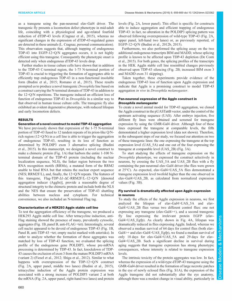
RESULTSGenerationof anovel construct tomodel TDP-43aggregationWe have previously shown that expression of the 1-75 N-terminalportion of TDP-43 fused to 12 tandem repeats of its prion-like Q/N-rich region (12×Q/N) in a cell line is able to trigger the aggregationof endogenous TDP-43, resulting in its loss of function asdetermined by POLDIP3 exon 3 alternative splicing (Budiniet al., 2015). In this manuscript, we designed a novel construct tomake a chimeric protein (Fig. 1A) that harbors the entire 1-100 N-terminal domain of the TDP-43 protein (including the nuclearlocalization sequence, NLS), the linker region between the twoRNA recognition motifs (RRMs), a mutated form of the RRM2unable to bind RNA but that retains the nuclear export sequence(NES; RRM2F/L), and, finally, the 12×Q/N repeats. The features ofthis transgene, Flag-TDP-Δ1-ΔC-RRM2F/L-12×Q/N [denotedaggregation inducer (AggIn)], provide a reasonable degree ofstructural integrity to the chimeric protein and include both the NLSand the NES that ensure the preservation of TDP-43 shuttlingabilities between nucleus and cytoplasm. For technicalconvenience, we also included an N-terminal Flag tag.
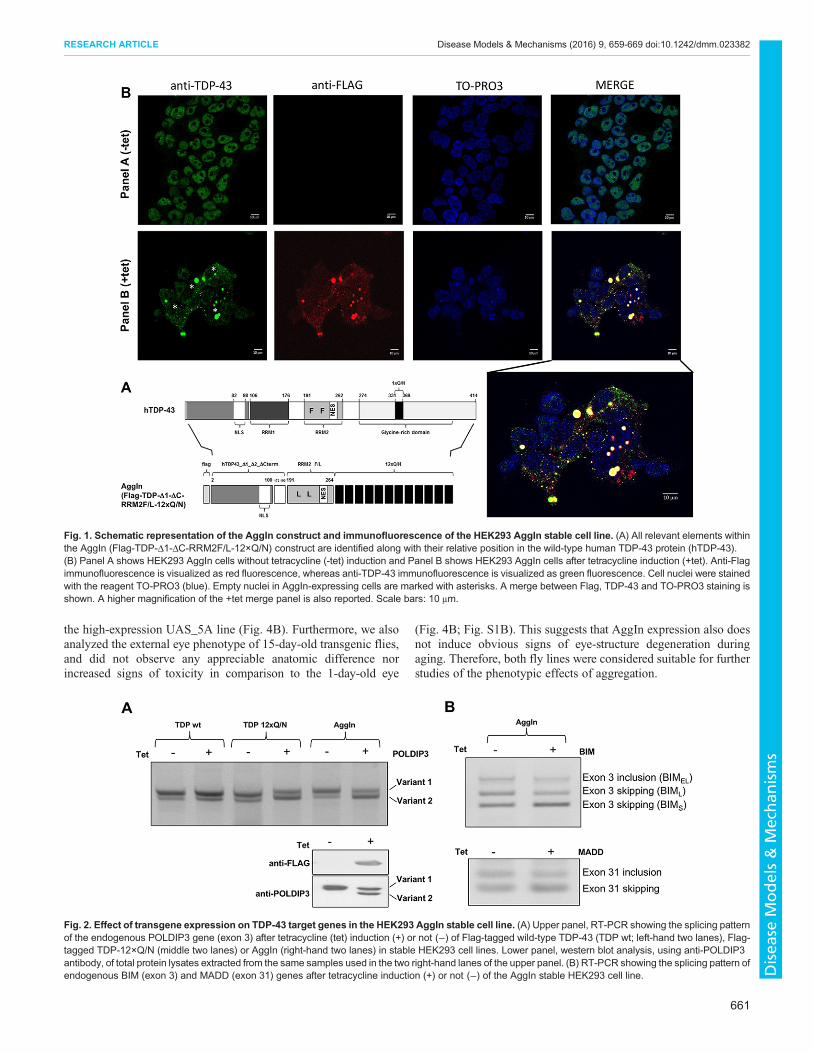
Characterization of a HEK293 AggIn stable cell lineTo test the aggregation efficiency of the transgene, we produced aHEK293 AggIn stable cell line. After tetracycline induction, anti-Flag staining showed the presence of many, prevalently cytosolic,aggregates (Fig. 1B, panel B, anti-FLAG +tet). Interestingly, severalcell nuclei appeared to be devoid of endogenous TDP-43 (Fig. 1B,Panel B, anti-TDP-43 +tet; empty nuclei marked with asterisks). Inorder to analyze whether the formation of these aggregates wasmatched by loss of TDP-43 function, we evaluated the splicingprofile of the endogenous gene POLDIP3, whose pre-mRNAprocessing is determined by TDP-43. In fact, knockdown of TDP-43 causes the exclusion of exon 3 from the mature POLDIP3mRNA(variant 2) (Fiesel et al., 2012; Shiga et al., 2012). Similar to whathappens with overexpression of the TDP-12×Q/N construct(Fig. 2A, upper panel, middle two lanes) (Budini et al., 2015),tetracycline induction of the AggIn protein expression wasassociated with a strong increase of POLDIP3 variant 2 at boththe mRNA (Fig. 2A, upper panel, right-hand two lanes) and protein
levels (Fig. 2A, lower panel). This effect is specific for constructsable to induce aggregation and efficient trapping of endogenousTDP-43: in fact, no alteration in the POLDIP3 splicing pattern wasobserved following overexpression of wild-type TDP-43 (Fig. 2A,upper panel, left-hand two lanes) or, as previously reported, ofEGFP-12×Q/N (Budini et al., 2012b, 2015).
Furthermore, we also performed the splicing assay on the twoadditional endogenous transcripts BIM andMADD, whose splicingprofile is known to be affected upon TDP-43 depletion (De Contiet al., 2015). For both genes, the splicing profiles of the transcriptsin the HEK AggIn stable cell line resembled changes previouslyobserved upon TDP-43 silencing (Fig. 2B; increased BIM exon 3and MADD exon 31 skipping).
Taken together, these experiments provide evidence of theendogenous TDP-43 loss of function upon AggIn expression andindicate that AggIn is a promising construct to model TDP-43aggregation in vivo in Drosophila melanogaster.
Pan-neuronal expression of the AggIn construct inDrosophila melanogasterTo create a novel animal model for TDP-43 aggregation, we clonedthe AggIn construct in the pUASTattB vector, under the control of theupstream activating sequence (UAS). After embryo injection, fivedifferent fly lines were obtained and screened for transgeneexpression by using the GMR-Gal4 driver. Although four of theselines expressed the transgene at comparable levels, the fifthdemonstrated a higher expression level (data not shown). Therefore,in the subsequent steps of our study, we focused our attention on twoof these transgenic lines: the one expressing the transgene at the topexpression level (UAS_5A) and one out of the four expressing thetransgene at comparable level (UAS_2B) (Fig. 3A).
To start studying the effects of transgene expression on theDrosophila phenotype, we expressed the construct selectively inneurons, by crossing the UAS_5A and UAS_2B flies with a flycontaining the pan-neuronal elav-Gal4 driver (the flies were grownat 25°C). As expected, elav-Gal4>UAS_5A flies demonstrated atransgene expression level twofold higher than the one observed inelav-Gal4>UAS_2B, as calculated from normalized expressionvalues (Fig. 3B).
Fly survival is dramatically affected upon AggIn expressionin neuronsTo study the effects of the AggIn expression in neurons, we firstanalyzed the lifespan of elav-Gal4>UAS_5A and elav-Gal4>UAS_2B flies versus two different control flies: one notexpressing any transgene (elav-Gal4>+), and a second transgenicfly line expressing the irrelevant protein EGFP (elav-Gal4>UAS_Egfp). As clearly shown in Fig. 4A, lifespan wasdramatically reduced in flies expressing AggIn. Indeed, whereas weobserved a median survival of 64 days for control flies (both elav-Gal4>+ and elav-Gal4>UAS_Egfp), we found a median survival ofonly 18 days for elav-Gal4>UAS_5A and 29 days for elav-Gal4<UAS_2B. Such a significant decline in survival duringaging suggests that transgene expression has strong phenotypicconsequences, whose intensity is related to transgene expressionlevels.
The intrinsic toxicity of the protein aggregates was low. In fact,whereas the expression of a wild-type dTDP-43 transgene using theGMR-Gal4 driver resulted in the formation of large necrotic patchesin the eye of newly eclosed flies (Fig. S1A), the expression of theAggIn transgene did not substantially alter the eye anatomy,although there was a modest change in visual ability, particularly in
660
RESEARCH ARTICLE Disease Models & Mechanisms (2016) 9, 659-669 doi:10.1242/dmm.023382
Disea
seModels&Mechan
isms
the high-expression UAS_5A line (Fig. 4B). Furthermore, we alsoanalyzed the external eye phenotype of 15-day-old transgenic flies,and did not observe any appreciable anatomic difference norincreased signs of toxicity in comparison to the 1-day-old eye
(Fig. 4B; Fig. S1B). This suggests that AggIn expression also doesnot induce obvious signs of eye-structure degeneration duringaging. Therefore, both fly lines were considered suitable for furtherstudies of the phenotypic effects of aggregation.
Fig. 1. Schematic representation of the AggIn construct and immunofluorescence of the HEK293 AggIn stable cell line. (A) All relevant elements withinthe AggIn (Flag-TDP-Δ1-ΔC-RRM2F/L-12×Q/N) construct are identified along with their relative position in the wild-type human TDP-43 protein (hTDP-43).(B) Panel A shows HEK293 AggIn cells without tetracycline (-tet) induction and Panel B shows HEK293 AggIn cells after tetracycline induction (+tet). Anti-Flagimmunofluorescence is visualized as red fluorescence, whereas anti-TDP-43 immunofluorescence is visualized as green fluorescence. Cell nuclei were stainedwith the reagent TO-PRO3 (blue). Empty nuclei in AggIn-expressing cells are marked with asterisks. A merge between Flag, TDP-43 and TO-PRO3 staining isshown. A higher magnification of the +tet merge panel is also reported. Scale bars: 10 μm.
Fig. 2. Effect of transgene expression on TDP-43 target genes in the HEK293 AggIn stable cell line. (A) Upper panel, RT-PCR showing the splicing patternof the endogenous POLDIP3 gene (exon 3) after tetracycline (tet) induction (+) or not (−) of Flag-tagged wild-type TDP-43 (TDP wt; left-hand two lanes), Flag-tagged TDP-12×Q/N (middle two lanes) or AggIn (right-hand two lanes) in stable HEK293 cell lines. Lower panel, western blot analysis, using anti-POLDIP3antibody, of total protein lysates extracted from the same samples used in the two right-hand lanes of the upper panel. (B) RT-PCR showing the splicing pattern ofendogenous BIM (exon 3) and MADD (exon 31) genes after tetracycline induction (+) or not (−) of the AggIn stable HEK293 cell line.
661
RESEARCH ARTICLE Disease Models & Mechanisms (2016) 9, 659-669 doi:10.1242/dmm.023382
Disea
seModels&Mechan
isms

Transgene expression affects climbing ability of fliesWe then investigated whether a locomotion defect appears at somepoint of the reduced lifespan of these flies using the climbing abilitytest.We assayed the flies at five different time-points after eclosion
(days 3, 7, 11, 15 and 20). Both elav-Gal4>UAS_5A and elav-Gal4>UAS_2B flies already demonstrated a statistically significantimpairment of the climbing ability at the first time-point (day 3). Asexpected, at each time point analyzed, the elav-Gal4>UAS_5Ashowed a more severe impairment than the elav-Gal4>UAS_2B(Fig. 5). Indeed, whereas ∼40% of elav-Gal4>UAS_5A flies wereno longer able to reach the top of the cylinder at day 3, only 15% ofelav-Gal4>UAS_2B flies demonstrated a similar impairment ofclimbing at this time point. Similarly, more than 70% of elav-Gal4>UAS_5A flies were no longer able to reach the top of the tube7 days after eclosion, whereas a similar percentage of elav-Gal4>UAS_2B flies with impaired climbing ability was observedonly after another 8 days (day 15 after eclosion).Taken together, these results suggest that the AggIn product
affects the motility of flies in an expression-dependent manner.
Pan-neuronal expression of the transgene in elav-Gal4>UAS_5A results in early locomotion impairment,which is detectable in the larval stageThe possibility of an early lethality of the AggIn flies was checked:we selected third-instar larvae and transferred them to fresh foodtubes. After 6 days, we calculated the percentages of eclosed flies,pupal lethality and larval lethality (Fig. S2). Whereas the elav-
Gal4>UAS_2B line did not show differences in larval lethalitycompared to controls (elav-Gal4>+, elav-Gal4>UAS_Egfp) andrevealed only a slight increase in pupal lethality, elav-Gal4>UAS_5A animals demonstrated a higher larval and pupallethality compared to both the controls and to the elav-Gal4>UAS_2B line. Nonetheless, we were able to analyze thephenotype of the animals during the larval stage. To this aim, weassayed third-instar larvae movement by counting the number oftheir peristaltic waves in 2 min on a suitable solid substrate (seeMaterials andMethods for details). In addition, as a negative controlof the experiment, we used a transgenic line expressing theirrelevant protein EGFP in neurons (elav-Gal4>UAS_Egfp) and thewild-type control line w1118. We also, as a positive control, analyzedthe movement of TBPHΔ23 larvae, the first-discovered dTDP-43-null allele fly line, which shows a severe neurodegenerativephenotype with a locomotion defect in larval stages and dramaticlocomotive defects after eclosion (Feiguin et al., 2009). We did notobserve any significant difference in larval motility of the elav-Gal4>UAS_2B larvae with respect to the negative controls(Fig. 6A). The elav-Gal4>UAS_5A larvae, however, showed asignificant motility impairment, as compared to the negativecontrols, with a reduced number of peristaltic waves,quantitatively comparable to those counted with dTDP-43-nulllarvae (TBPHΔ23) (Fig. 6A). Therefore, these results show that thelocomotion impairment of the elav-Gal4>UAS_5A fly line iscomparable with that of the dTDP-43-null model.
Biochemical and functional assays support the notion ofloss-of-function in endogenous Drosophila TDP-43 intransgenic AggIn fly modelsThe creation of the HEK293 AggIn stable cell line has shown thattransgene expression efficiently triggers the formation of aggregatesable to recruit and trap the endogenous TDP-43 protein and give riseto a TDP-43 loss-of-function effect (as demonstrated by thealteration of the splicing profile of the endogenous genes POLDIP3,BIM and MADD).
To show that AggIn expression also induces the formation ofinsoluble aggregates able to trap endogenous dTDP-43 in flies, weperformed solubility experiments on transgenic fly heads co-expressing AggIn and a Flag-tagged form of dTDP-43 under thecontrol of the GMR-Gal4 driver. As a control experiment, Flag-dTDP-43 was co-expressed with the unrelated protein EGFP. Thisexperiment clearly shows that AggIn expression results in theformation of insoluble aggregates (Flag-AggIn, *; Fig. 6B) andinduces a very strong shift of dTDP-43 from the soluble to theinsoluble fraction (Flag-TBPH, #; Fig. 6B). As expected, theexpression of control EGFP did not result in the formation ofinsoluble aggregates nor did it alter the solubility pattern ofdTDP-43, which remained mainly soluble.
Western blot analysis of SYX and CSP proteins expressionIn order to investigate whether biochemical evidence of dTDP-43loss of function could also be found in the transgenic flies, weanalyzed the expression levels of genes known to be altered indTDP-43-null fly models and previously characterized as moleculartargets of dTDP-43 potentially related to neurodegenerationpathogenesis.
In particular, we focused our attention on the elav-Gal4>UAS_5A line, which showed a striking degenerativephenotype during both adulthood and larval stage.
SYX (also known as Syx1a) and Cysteine-string protein (CSP)are two presynaptic vesicular proteins. It has been recently reported
Fig. 3. Expression levels of transgene in twoDrosophila lines. (A) Westernblot analysis of total protein extracts from fly heads of GMR-Gal4>UAS_5A andGMR-Gal4>UAS_2B and (B) elav-Gal4>UAS_5A and elav-Gal4>UAS_2Blines. Eye-specific and pan-neuronal expression of the AggIn construct wasachieved using the GMR- and elav-Gal4 drivers, respectively. Western blotdensitometry was performed using the ImageJ software and thenormalized expression of the transgenic protein is reported in the graphs(mean±s.e.m., n=3).
662
RESEARCH ARTICLE Disease Models & Mechanisms (2016) 9, 659-669 doi:10.1242/dmm.023382
Disea
seModels&Mechan
isms

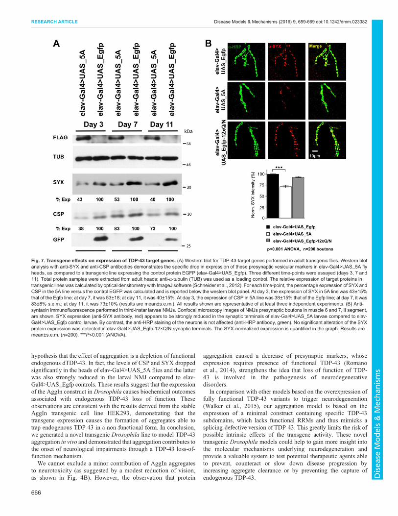
that their downregulation is an early event of dTDP-43 dysfunctionin vivo; in fact, the expression of these proteins was found to besignificantly altered in the heads of our dTDP-43-null fly modelTBPHΔ23 and in neuromuscular junction (NMJ) presynapticboutons in muscle 6 and 7 of third-instar larvae (Romano et al.,2014). Starting from these observations, we verified the endogenousdTDP-43 function in Drosophila expressing the AggIn transgene.In particular, we compared, by western blotting, the expression ofSYX and CSP proteins in the heads of elav-Gal4>UAS_5A versuselav-Gal4>UAS_Egfp control flies. These proteins appeared to besignificantly downregulated in our transgenic model (Fig. 7A).Interestingly, the drop in expression was found at all three timepoints assayed (day 3, 7 and 11), in agreement with the observationthat this fly line already has a severe phenotype by 3 days aftereclosion.
Confocal analysis of SYX protein expression in larval NMJspresynaptic boutonsIn order to support the hypothesis that endogenous dTDP-43 loss offunction was also responsible for the phenotype observed during
the larval stage in elav-Gal4>UAS_5A line, we analyzed SYXprotein expression in the NMJ presynaptic boutons in muscle 6 and7 of these transgenic larvae by immunohistochemistry. Asexpected, SYX levels were strongly reduced in the synapticterminals of elav-Gal4>UAS_5A third-instar larvae compared toelav-Gal4>UAS_Egfp controls (Fig. 7B, both anti-SYX andmerge). By contrast, the anti-HRP staining of the neurons did nothighlight any alteration (Fig. 7B, anti-HRP antibody), confirmingthe specificity of the SYX downregulation in elav-Gal4>UAS_5Aflies. Furthermore, the shape of motoneuron terminals at the NMJdid not appear altered in the transgenic larvae versus control.
To provide evidence that the N-terminal portion of TDP-43 iscrucial to enhance the trapping of endogenous dTDP-43 in aninsoluble non-functional form, we also included elav-Gal4>UAS_Egfp-12×Q/N larvae in the analysis, which do notexhibit any locomotion impairment during the larval stage (Cragnazet al., 2015). This allowed us to perform a side-by-side phenotypiccomparison of EGFP-12×Q/N and AggIn at the NMJ: as expected,and in contrast to what is observed in elav-Gal4>UAS_5A larvae,the EGFP-12×Q/N line did not show any reduction of the SYX
Fig. 4. Effect of transgene expression on Drosophila lifespan and on external eye structure and/or function. (A) Lifespan is dramatically reduced in fliesexpressing the AggIn transgene in neurons (elav-Gal4>UAS_5A and elav-Gal4>UAS_2B) versus a control fly not expressing any transgene (elav-Gal4>+) or atransgenic fly line expressing the control protein EGFP (elav-Gal4>UAS_Egfp). Median survival is 18 days for elav-Gal4>UAS_5A; 29 days for elav-Gal4<UAS_2B; 64 days for controls (both elav-Gal4>+ and elav-Gal4>UAS_Egfp). n>120 animals for each genotype; P<0.001 (log-rank test) for all the followinggenotype pairs: elav-Gal4>+ versus elav-Gal4>UAS_2B; elav-Gal4>+ versus elav-Gal4>UAS_5A; elav-Gal4>UAS_Egfp versus elav-Gal4>UAS_2B; elav-Gal4>UAS_Egfp versus elav-Gal4>UAS_5A; elav-Gal4>UAS_2B versus elav-Gal4>UAS_5A. Time points (days) corresponding to 25%, 50% and 75% survivalare also shown in the graph for each genotype and reported in the flanking summary table. (B) External eye phenotype and phototaxis assay of 1-day-old flies.(a,a′) Oregon (wild-type); (b,b′) GMR-Gal4>UAS_Egfp; (c,c′) GMR-Gal4>UAS_2B; (d,d′) GMR-Gal4>UAS_5A. The AggIn expression in the eye, using theGMR-Gal4 driver, did not result in any substantial alteration of the external eye phenotype (upper panel, compare pictures in c and d versus controls in a and b).However, the vision assay (lower panel) revealed that a minor fraction of the AggIn-expressing population of flies (8.5% of GMR-Gal4>UAS_2B, and 20.4% ofGMR-Gal4>UAS_5A) exhibited vision defects because they did not reach the light within 1 min of time in a phototaxis assay (compare white bars in c′ and d′fractions with the negative controls in a′ and b′). Error bars indicate s.e.m. (n=3).
663
RESEARCH ARTICLE Disease Models & Mechanisms (2016) 9, 659-669 doi:10.1242/dmm.023382
Disea
seModels&Mechan
isms

levels in the synaptic terminals (Fig. 7B), thus supporting the roleascribed to the N-terminal region of TDP-43.Consistently, the observation that dTDP-43 functional loss is
detected in elav-Gal4>UAS_5A but not in elav-Gal4>UAS_Egfp-12×Q/N larvae correlates with the ability of aggregates to sequesterendogenous dTDP-43. Indeed, we recently demonstrated that theclimbing impairment in elav-Gal4>UAS_Egfp-12×Q/N fliesoverlaps with an age-related physiological drop of dTDP-43(Cragnaz et al., 2015). In order to demonstrate that a moreefficient dTDP-43-trapping ability of the AggIn construct mediatesthe earlier onset of degenerative effects observed in this novelanimal model, we analyzed the correlation between phenotype onsetand endogenous dTDP-43 levels in elav-Gal4>UAS_5A flies. Inthis transgenic line, western blot experiments confirmed the sametrend of endogenous dTDP-43 physiological drop observed duringaging in wild-type flies (Fig. S3). Interestingly, whereas thephenotype onset in elav-Gal4>UAS_Egfp-12×Q/N flies matcheswith a strong decrease of endogenous dTDP-43 at day 10 (Cragnazet al., 2015), the elav-Gal4>UAS_5A flies already show an evidentclimbing impairment at day 3 after eclosion (Fig. 5), a time-point bywhich dTDP-43 expression has already started to drop, as thedecrease in its levels appears to occur in at least two main steps (seeday 3 and day 10 in Fig. S3; compare with dTDP-43 expression atday 1), although it is significantly higher than at day 10.
DISCUSSIONIn the past decade several lines of evidence have shown that TDP-43plays a key role in the pathogenesis of several neurodegenerativedisorders, including ALS and FTLD (Neumann et al., 2006; VanLangenhove et al., 2012; Ling et al., 2013). Notwithstanding the
large number of studies devoted to characterization of the molecularmechanisms linking TDP-43 aggregation to neurodegeneration, it isstill unclear what the role of TDP-43 and TDP-43-positiveaggregates is in disease onset and progression.
However, previous studies have identified the structuraldeterminants of the TDP-43 protein that mediate its self-aggregation and trapping into a non-functional insoluble form:they demonstrated that the C-terminal Q/N prion-like domain isimportant in the protein aggregation process and that the N-terminalregion 1-75 is essential to enhance the trapping of endogenous TDP-43 in the aggregates in a non-functional insoluble form (Budiniet al., 2012b, 2015). The sequestered TDP-43 loses its functionalcapacity, but the aggregates do not show intrinsic significanttoxicity in the HEK293 cells (Budini et al., 2012b) or in theDrosophila eye, a tissue that does not need TDP-43 for itsdevelopment (Cragnaz et al., 2014).
Furthermore, whereas the role of the TDP-43 C-terminal part inthe aggregation process is well established (D’Ambrogio et al.,2009; Igaz et al., 2009; Yang et al., 2010; Jiang et al., 2013; Wanget al., 2013), the implication of the N-terminal region in a growingnumber of physiological and pathological functions of the proteinhas been highlighted only by more recent studies showing that theTDP-43 N-terminus, in particular the first ten residues, appear toplay a role not only in RNA recognition (Buratti and Baralle, 2001)and cellular localization (Winton et al., 2008), but also in regulatingTDP-43 folding, homotypic interaction, splicing functionality andcytoplasmic sequestration (Zhang et al., 2013). Other studies havealso suggested that the TDP-43 N-terminus encodes a new type ofubiquitin-like fold that is involved in binding of nucleic acids andthat is normally in equilibrium with an unfolded form: the formation
Fig. 5. Effect of transgene expression on Drosophila climbing ability. A climbing assay performed in flies expressing the AggIn transgene (elav-Gal4>UAS_5A and elav-Gal4>UAS_2B) versus a control fly not expressing any transgene (elav-Gal4>+) and a transgenic fly line expressing the control proteinEGFP (elav-Gal4>UAS_Egfp). A statistically significant impairment of climbing is already observed at day 3 in both the AggIn-expressing fly lines. The climbingdefect gets worse with age, from day 3 to 20. *P<0.05, ***P<0.001 (one-way ANOVA). Error bars indicate s.e.m. (n>120 animals for each genotype).
664
RESEARCH ARTICLE Disease Models & Mechanisms (2016) 9, 659-669 doi:10.1242/dmm.023382
Disea
seModels&Mechan
isms

of irreversible inclusions relevant for both physiological andpathological processes might occur when this equilibrium isaltered (Qin et al., 2014). Finally, we have recently reported thatthe elimination of the first 75 residues of TDP-43 N-terminusreduces the efficiency of intracellular aggregates to interact with andsequester endogenous TDP-43 (Budini et al., 2015). In fact, theinsertion of the N-terminal sequence upstream of an artificialrepetition of the Q/N-rich region of TDP-43 (12×Q/N) results in achimeric protein that induces aggregation and alters the splicingpattern of POLDIP3, a commonly used reporter of TDP-43dysfunction. This experiment demonstrated that the TDP-43 N-terminal is crucial for efficient sequestration of the endogenousTDP-43 within the inclusions (Budini et al., 2015).Taking all the above points into consideration, we have now
optimized an aggregation inducer with minimal TDP-43 sequencesand tested it both in HEK293 cells and in Drosophila. This novelconstruct Flag-TDP-Δ1-ΔC-RRM2F/L-12×Q/N was named, forsimplicity, AggIn. We show here that AggIn is able to triggeraggregation, to deplete nuclei of endogenous TDP-43 and to induceloss of the splicing function of endogenous TDP-43 in HEK293cells (Figs 1B and 2). Importantly, it is also able to induce evidentsurvival and behavioral impairments when expressed as a transgenein Drosophila neurons. We have studied two transgenic fly lines
(UAS_2B and UAS_5A) with different levels of the AggIntransgene expression. This characteristic was useful to modeldifferential levels of the TDP-43 aggregation process. In fact, onlythe elav-Gal4>UAS_5A line (with double the AggIn expression ofelav-Gal4>UAS_2B) showed motility impairment at the larvalstage, whereas both the elav-Gal4>UAS_5A and elav-Gal4>UAS_2B lines resulted in a reduced lifespan and impairedclimbing ability during adulthood. These latter effects were moreseverewhen the aggregation was more efficient. In fact, lifespan wasreduced to about a quarter (elav-Gal4>UAS_5A line) and a half(elav-Gal4>UAS_2B line) with respect to the median survival ofcontrol elav-Gal4>UAS_Egfp flies. Importantly, these effects wereobserved at a physiological growth temperature (25°C).
These results suggest that the stronger effects observed in theUAS_5A line are due to the higher levels of AggIn expression andsupport the hypothesis of a direct correlation between efficiency ofendogenous dTDP-43-trapping and onset, as well as severity of thephenotype. Interestingly, the higher transgene expression in theelav-Gal4>UAS_5A line was able to trigger a very early locomotionimpairment, quantitatively comparable to that observed in thedTDP-43-null TBPHΔ23 larvae (Feiguin et al., 2009). In addition,the genotype–phenotype studies were complemented by theobservation that dTDP-43 function is lost. This supports the
Fig. 6. Effect of transgene expression on Drosophila larval motility and solubility assay on adult fly heads. (A) A larval motility assay was performed onthird-instar larvae. A strong reduction in larval motility of elav-Gal4>UAS_5A (5A) larvae is observed, as compared to a transgenic line expressing the controlprotein EGFP (elav-Gal4>UAS_Egfp) and to the wild-type line (w1118). No impairment in larval motility is observed in elav-Gal4>UAS_2B larvae (2B). ATDP-43-null allele line (TBPHΔ23) was used as a positive reference control. x-axis, genotype; y-axis, peristaltic waves counted in two minutes. Error bars indicate s.e.m.(n=20 animals for each genotype). ***P<0.001 (one-way ANOVA). (B) Solubility assay. Western blot of fractionated proteins obtained from adult fly heads of thefollowing genotypes: GMR-Gal4>UAS_TBPH; UAS_5A, GMR-Gal4>UAS_TBPH; UAS_2B, GMR-Gal4>UAS_TBPH; UAS_Egfp (TBPH is the Drosophila TDP-43). Upper panel, input, soluble and insoluble fractions of each genotype were loaded in a 1:1:1 ratio and probed by immunoblotting. AggIn and TBPH weredetected with anti-FLAG antibody. EGFP was detected using anti-GFP antibody. Anti-tubulin served as protein loading control. Lower panel, to improve theseparation of Flag-AggIn (see *) and Flag-TBPH (see #) protein bands, which have a close molecular mass, the three sample fractions from each genotype werealso loaded on additional gels and were run for a longer time, before anti-Flag immunoblotting. TBPH is mostly insoluble when it is co-expressed with AggIn; bycontrast, it remains mainly soluble when it is co-expressed with the unrelated protein EGFP.
665
RESEARCH ARTICLE Disease Models & Mechanisms (2016) 9, 659-669 doi:10.1242/dmm.023382
Disea
seModels&Mechan
isms
hypothesis that the effect of aggregation is a depletion of functionalendogenous dTDP-43. In fact, the levels of CSP and SYX droppedsignificantly in the heads of elav-Gal4>UAS_5A flies and the latterwas also strongly reduced in the larval NMJ compared to elav-Gal4>UAS_Egfp controls. These results suggest that the expressionof the AggIn construct in Drosophila causes biochemical outcomesassociated with endogenous TDP-43 loss of function. Theseobservations are consistent with the results derived from the stableAggIn transgenic cell line HEK293, demonstrating that thetransgene expression causes the formation of aggregates able totrap endogenous TDP-43 in a non-functional form. In conclusion,we generated a novel transgenic Drosophila line to model TDP-43aggregation in vivo and demonstrated that aggregation contributes tothe onset of neurological impairments through a TDP-43 loss-of-function mechanism.We cannot exclude a minor contribution of AggIn aggregates
to neurotoxicity (as suggested by a modest reduction of vision,as shown in Fig. 4B). However, the observation that protein
aggregation caused a decrease of presynaptic markers, whoseexpression requires presence of functional TDP-43 (Romanoet al., 2014), strengthens the idea that loss of function of TDP-43 is involved in the pathogenesis of neurodegenerativedisorders.
In comparison with other models based on the overexpression offully functional TDP-43 variants to trigger neurodegeneration(Walker et al., 2015), our aggregation model is based on theexpression of a minimal construct containing specific TDP-43subdomains, which lacks functional RRMs and thus mimicks asplicing-defective version of TDP-43. This greatly limits the risk ofpossible intrinsic effects of the transgene activity. These noveltransgenic Drosophila models could help to gain more insight intothe molecular mechanisms underlying neurodegeneration andprovide a valuable system to test potential therapeutic agents ableto prevent, counteract or slow down disease progression byincreasing aggregate clearance or by preventing the capture ofendogenous TDP-43.
Fig. 7. Transgene effects on expression of TDP-43 target genes. (A) Western blot for TDP-43-target genes performed in adult transgenic flies. Western blotanalysis with anti-SYX and anti-CSP antibodies demonstrates the specific drop in expression of these presynaptic vesicular markers in elav-Gal4>UAS_5A flyheads, as compared to a transgenic line expressing the control protein EGFP (elav-Gal4>UAS_Egfp). Three different time-points were assayed (days 3, 7 and11). Total protein samples were extracted from adult heads; anti-α-tubulin (TUB) was used as a loading control. The relative expression of target proteins intransgenic lines was calculated by optical densitometry with ImageJ software (Schneider et al., 2012). For each time-point, the percentage expression of SYX andCSP in the 5A line versus the control EGFP was calculated and is reported below the western blot panel. At day 3, the expression of SYX in 5A line was 43±15%that of the Egfp line; at day 7, it was 53±18; at day 11, it was 40±15%. At day 3, the expression of CSP in 5A line was 38±15% that of the Egfp line; at day 7, it was83±8% s.e.m.; at day 11, it was 73±10% (results are mean±s.e.m.). All results shown are representative of at least three independent experiments. (B) Anti-syntaxin immunofluorescence performed in third-instar larvae NMJs. Confocal microscopy images of NMJs presynaptic boutons in muscle 6 and 7, II segment,are shown. SYX expression (anti-SYX antibody, red) appears to be strongly reduced in the synaptic terminals of elav-Gal4>UAS_5A larvae compared to elav-Gal4>UAS_Egfp control larvae. By contrast, the anti-HRP staining of the neurons is not affected (anti-HRP antibody, green). No significant alteration of the SYXprotein expression was detected in elav-Gal4>UAS_Egfp-12×Q/N synaptic terminals. The SYX-normalized expression is quantified in the graph. Results aremean±s.e.m. (n=200). ***P<0.001 (ANOVA).
666
RESEARCH ARTICLE Disease Models & Mechanisms (2016) 9, 659-669 doi:10.1242/dmm.023382
Disea
seModels&Mechan
isms
MATERIALS AND METHODSExpression plasmid and stable cell line generationThe AggIn (Flag-TDP-Δ1-ΔC-RRM2F/L-12×Q/N) plasmid was generatedby site-directed mutagenesis using the pcDNA5/FRT/TO-Flag-TDP-12×-Q/N plasmid (Budini et al., 2015) as a template. The final constructincluded: an N-terminal Flag tag, the amino acid stretches 2-100, 173-190,191-264 (with phenylalanine in positions 229 and 231 mutated to leucine)of the human TDP-43 protein, and 12 repetitions of the human TDP-43amino acids stretch 331-369 (referred to as 12×QN). Fig. 1A recapitulatesthe main features of the construct.
For generation of stable cell lines, HEK293 flip-in cells were grown inDMEM-Glutamax-I (Gibco-BRL, Life Technologies Inc., Frederick, MD)supplemented with 10% fetal bovine serum (Gibco-BRL) and antibiotic andantimycotic stabilized suspension (Sigma-Aldrich, St Louis, MO). Cellswere transfected using Effectene Transfection reagent (QIAGEN, Inc.,Gaithersburg, MD) following the manufacturer’s instructions. Co-transfection of 0.5 μg of plasmid together with 0.5 μg of pOG44 (ThermoFisher, Scientific, Waltham, MA) vector allowed recombination in thegenome of the cells. After co-transfection, cells were grown in DMEM-Glutamax-I supplemented with 10% fetal bovine serum with antibiotic andantimycotic until they reached 80% of confluence. The stable integration ofthe plasmid was then gradually selected using 100 μg/ml Hygromicin B(Gibco-BRL) and 10 μg/ml of Blasticidin (Gibco-BRL). Once cells wereselected, expression of the protein was achieved by adding 1 μg/ml oftetracycline (Sigma-Aldrich) to the culture medium.
Splicing assayFor the splicing assay, 5×10⁵ Flag-TDP-43-WT, Flag-TDP-43-12×Q/N andAggIn cells were seeded in 6-well plates and induced with tetracycline for72 h. Uninduced cells were used as a control. After induction, cells werecollected and RNA was extracted using Trifast reagent (Euroclone, Milan,Italy) according to the manufacturer’s instruction. Reverse transcriptionwas performed using M-MLV Reverse Transcriptase (Gibco-BRL)following the manufacturer’s protocol. A PCR with TAQ DNAPolymerase (Roche Diagnostics, Mannheim, Germany) was performed for35 amplification cycles (95°C for 30 s, 55°C for 30 s, 72°C for 30 s) toamplify POLDIP3, BIM and MADD cDNAs. The primers used to test thesplicing pattern of POLDIP3, BIM and MADD endogenous genes were:POLDIP3 forward (5′-GCTTAATGCCAGACCGGGAGTTGGA-3′);POLDIP3 reverse (5′-TCATCTTCATCCAGGTCATATAAATT-3′); BIMforward (5′-TCTGAGTGTGACCGAGAAGG-3′); BIM reverse (5′-TCT-TGGGCGATCCATATCTC-3′); MADD forward (5′-GACCTGAATT-GGGTGGCGAGTTCCCT-3′); MADD reverse (5′-CATTGGTGTCT-TGTACTTGTGGCTC-3′).
Protein expression and immunoblotting of HEK293 stable celllinesProtein expression of the AggIn construct was analyzed in 5×10⁵ cells seededin 6-well plates and induced for 72 h. Uninduced cells were also seeded ascontrol. After induction, cells were collected and lysed with 100 μl of RIPAlysis buffer (50 mMTris-HCl pH 7.4, 150 mMNaCl, 1% NP-40, 0.1% SDS,1 mM EDTA pH 8, 1 mM PMSF and 0.5% sodium deoxycholate)supplemented with Complete protease inhibitor cocktail (RocheDiagnostics, Mannheim, Germany). Cell lysates were incubated at +4°C for30 min, then lysed by sonication and centrifuged at 500 g at +4°C for 5 min.Total protein amount in cell lysates was then quantified by the Bradfordmethod, and 20 μg was loaded in a 10% SDS-PAGE. An anti-Flag (1:1000,catalog number F1804; Sigma-Aldrich) primary antibody and a HRP-labeledanti-mouse-IgG (1:2000, catalog number P0260;DAKO,Glostrup, Denmark)secondary antibody were used for protein detection. Western blotting using aprimary anti-POLDIP3 (1:1000, catalog number 5439; Cell SignalingTechnology, Beverly, MA) antibody and a secondary HRP-labeled anti-rabbit-IgG (1:2000, catalog number P0448; DAKO, Glostrup, Denmark)antibody was also performed to detect POLDIP3 isoforms.
Immunofluorescence microscopyFor indirect immunofluorescence, 5×10⁵ HEK-AggIn cells were seeded onslides and induced with tetracycline for 72 h. Non-induced cells were also
seeded as a control. Immunofluorescence was performed as previouslydescribed (Ayala et al., 2008). As primary antibodies, an anti-Flag (1:200,catalog number F1804; Sigma-Aldrich) and an anti-TDP-43 (1:1000,catalog number 10782-2-AP; ProteinTech, Chicago, IL) were used. Thesecondary antibodies were anti-mouse-IgG conjugated to Alexa Fluor 594and anti-rabbit-IgG conjugated to Alexa Fluor 488, and TO-PRO3 dye wasused for nuclei staining, all purchased from Life Technologies. Cells werethen analyzed on a Zeiss LSM 510 Meta confocal microscope.
Fly stocksAggIn and EGFP constructs were cloned in the pUASTattB vector andsubsequently sequenced. The constructs were used to create transgenic fliesby standard embryo injections (BestGene Inc., Chino Hills, CA, USA).Transgenes were subsequently balanced on the required chromosome toobtain fly stocks. w1118 and elav-Gal4 flies were supplied by theBloomington Drosophila Stock Center at Indiana University. Flies werefed on standard fly food (agar 6 g/l; sugar 41.6 g/l; yeast 62.5 g/l; cornmeal29 g/l; propionic acid 4.1 ml/l), and were maintained and crossed in ahumidified incubator at 25°C with a 12-h-light–12-h-dark cycle.
LifespanAdult flies were collected for 2 days from eclosion and transferred to freshfood tubes in a 1:1 male:female ratio (20 total flies/tube). At the third day,death events were scored and viable flies were transferred to fresh tubes. Thesame was done every 3 days. Survival proportions were plotted aspercentage of living flies against days. More than 120 flies were tested foreach genotype.
Phototaxis assayThe assay was performed as previously described (Cragnaz et al., 2014).Briefly, individual flies from each genotypewere introduced into the stem ofa Y-maze with one arm exposed to violet light (400 nm) and the second armcompletely in the dark. The number of flies that moved into the illuminatedchamber within 1 min was determined. At least 50 flies per experiment foreach genotype were tested.
Climbing assayAge-synchronized cohorts of flies were transferred without anesthesia to a50-ml glass cylinder, and tapped to the bottom with cotton. After a period ofadaptation of 30 s, the climbing ability of flies was quantified as number ofanimals that reached the top of the cylinder (10 cm) in 15 s. Flies wereassayed in batches of 20 (1:1 male:female ratio) and the test was repeatedthree times for each batch of animals. More than 120 flies were tested foreach genotype. The number of flies that reached the top was converted into apercentage value, and the mean±s.e.m. percentage was calculated for at leastsix experiments.
Larval movementWandering third-instar larvae were selected, gently washed and transferredto a Petri dish (0.7% agarose in distilled water). After a period of adaptation(30 s), the peristaltic waves were counted within 2 min. At least 20 larvaewere assayed for each genotype. The median number of peristaltic wavesperformed in 2 min by each genotype was plotted on a graph (mean±s.e.m.).
Immunoblotting of fly head samplesDrosophila heads were homogenized in lysis buffer [10 mM Tris-HCl, pH7.4, 150 mMNaCl, 5 mMEDTA, 5 mMEGTA, 10% glycerol, 50 mMNaF,5 mM DTT, 4 M urea and Complete protease inhibitor cocktail (RocheDiagnostics)]. Proteins were separated by 8% SDS-PAGE, transferred tonitrocellulose membranes (Whatman, Clifton, NJ, USA), blocked overnightin a 5% non-fat dried milk solution and probed with the following primaryantibodies: rabbit anti-Drosophila TDP-43 (1:1500, made in-house), mouseanti-SYX 8C3s (1:2500, Developmental Studies Hybridoma Bank, DSHB,Iowa City, IA, USA), anti-CSP2c (1:9000, DSHB), mouse anti-tubulinCP06 (1:4000, Calbiochem, San Diego, CA, USA) and mouse anti-FLAGM2 (1:1000, Sigma-Aldrich) antibodies. The membranes were incubatedwith the secondary antibodies HRP-labeled anti-mouse-IgG (1:1000,
667
RESEARCH ARTICLE Disease Models & Mechanisms (2016) 9, 659-669 doi:10.1242/dmm.023382
Disea
seModels&Mechan
isms
Thermo Scientific, Rockford, IL, USA) or HRP-labeled anti-rabbit-IgG(1:1000, Thermo Scientific). Finally, protein detection was performed withFemto Super Signal substrate (Thermo Scientific) for anti- TDP-43 and anti-CSP2c immunoblotting and with ECL western blotting substrate (ThermoScientific) for anti-syntaxin and anti-tubulin antibodies.
Protein expression was quantified using the NIH ImageJ software(Schneider et al., 2012) and normalized against tubulin. Histograms arerepresentative of three independent experiments.
Solubility testThe solubility assay was performed as previously described (Cragnaz et al.,2014). Briefly, 24 adult fly heads per genotype were homogenized in RIPAbuffer [50 mM Tris-HCl pH 8, 150 mM NaCl, 2 mM EDTA, 1%NonidetP40 (v/v), 0.1% SDS, 1% Na-deoxycholate and a cocktail ofComplete protease inhibitor cocktail (Roche Diagnostics)]. Followingincubation on a rotating wheel for 1 h at 4°C, samples were centrifuged at1000 g for 10 min at 4°C. An aliquot was taken at this point as the input, andafter a further centrifugation step at 100,000 g for 30 min at 4°C, thesupernatant was collected as the soluble fraction. The resulting pellet was re-suspended in urea buffer (9 M urea, 50 mMTris-HCl pH 8, 1% CHAPS andComplete protease inhibitor cocktail) and collected as the insoluble fraction.Proteins were separated by 10% SDS-PAGE. The different samples wereloaded in a 1:1:1 ratio for the input, soluble and insoluble fractions. Proteinswere immunoblotted as already described for fly head samples and probedwith the following reagents. Primary antibodies: mouse anti-FLAG M2(1:1000, Sigma-Aldrich), rabbit anti-GFP (1:2000, sc-8334, Santa CruzBiotechnology Inc., Dallas, TX) and mouse anti-tubulin CP06 (1:4000,Calbiochem, San Diego, CA) antibodies. Secondary antibodies: HRP-labeled anti-mouse-IgG (1:1000, Thermo Scientific) or HRP-labeled anti-rabbit-IgG (1:1000, Thermo Scientific).
NMJ immunohistochemistry and images quantificationThird-instar larvae were selected, briefly washed in water and dissected insaline solution (0.1 mM CaCl2, MgCl2 4 mM, KCl 2 mM, NaCl 128 mM,sucrose 35.5 mM and Hepes 5 mM pH 7.2), fixed for 20 min in 4%paraformaldehyde, washed in PBS with 0.1% Tween 20 and blockedwith 5% normal goat serum (Vector Laboratories, Burlingame, CA) in PBSwith 0.1% Tween. Primary antibodies [anti-HRP (1:150, JacksonImmunoResearch Lab, West Grove, PA, USA) and anti-SYX 8C3s (1:15,DSHB) antibodies] were incubated overnight at 4°C and then the secondaryantibodies, Alexa-Fluor-488-conjugated anti-rabbit-IgG and Alexa-Fluor-555-conjugated anti-mouse-IgG (1:500, Thermo Fisher Scientific), wereincubated for 2 h at room temperature. SlowFade Gold (Gibco-BRL) wasused for the mounting. Images were acquired on a Zeiss LSM 510 MetaConfocal Microscope with a 63× oil lens and 40× lens, then analyzed usingNIH ImageJ software (Schneider et al., 2012).
The larvae analyzed for these experiments were processedsimultaneously and the same microscope settings were employed toacquire all images. The presynaptic terminals of second abdominal segmenton muscle 6 and 7 were analyzed. The samples were double labeled withanti-HRP and anti-SYX antibodies: the mean intensity of both wasquantified and a ratio calculated (adapted from Thomas et al., 1997). Thestatistical analyses were performed using Prism6 (GraphPad, San Diego,CA, USA).
AcknowledgementsWe would like to thank Laura De Conti and Chiara Appocher for their valuablesuggestions during experimental work. We thank Marco Baralle for reviewing thetext.
Competing interestsThe authors declare no competing or financial interests.
Author contributionsS.L. carried out molecular, genetic and behavioral studies with flies, and drafted themanuscript. V.R. carried out molecular studies with cell lines and tested thecharacteristics of the construct. G.R. performed NMJ immunohistochemistryexperiments. R.K. and F.F. participated in the phenotypic characterization of flies.L.C. carried out molecular studies on dTDP-43 age-related variations. M.R.
participated in the design of the study, interpretation of the results, performed thestatistical analysis and helped to draft the manuscript. F.E.B. conceived the study,and participated in its design, in the interpretation of the results, coordination of theproject and helped to draft the manuscript. All authors read and approved the finalmanuscript.
FundingThis work was supported by the Fondation Thierry Latran (project REHNPALS).
Supplementary informationSupplementary information available online athttp://dmm.biologists.org/lookup/suppl/doi:10.1242/dmm.023382/-/DC1
ReferencesArai, T., Hasegawa, M., Akiyama, H., Ikeda, K., Nonaka, T., Mori, H., Mann, D.,
Tsuchiya, K., Yoshida, M., Hashizume, Y. et al. (2006). TDP-43 is a componentof ubiquitin-positive tau-negative inclusions in frontotemporal lobar degenerationand amyotrophic lateral sclerosis. Biochem. Biophys. Res. Commun. 351,602-611.
Ayala, Y. M., Pantano, S., D’Ambrogio, A., Buratti, E., Brindisi, A., Marchetti, C.,Romano, M. and Baralle, F. E. (2005). Human, Drosophila, and C. elegansTDP43: nucleic acid binding properties and splicing regulatory function. J. Mol.Biol. 348, 575-588.
Ayala, Y. M., Zago, P., D’Ambrogio, A., Xu, Y.-F., Petrucelli, L., Buratti, E. andBaralle, F. E. (2008). Structural determinants of the cellular localization andshuttling of TDP-43. J. Cell Sci. 121, 3778-3785.
Belzil, V. V., Gendron, T. F. and Petrucelli, L. (2013). RNA-mediated toxicity inneurodegenerative disease. Mol. Cell. Neurosci. 56, 406-419.
Budini, M., Romano, V., Avendan o-Vazquez, S. E., Bembich, S., Buratti, E. andBaralle, F. E. (2012a). Role of selected mutations in theQ/N rich region of TDP-43in EGFP-12xQ/N-induced aggregate formation. Brain Res. 1462, 139-150.
Budini, M., Buratti, E., Stuani, C., Guarnaccia, C., Romano, V., De Conti, L. andBaralle, F. E. (2012b). Cellular model of TAR DNA-binding protein 43 (TDP-43)aggregation based on its C-terminal Gln/Asn-rich region. J. Biol. Chem. 287,7512-7525.
Budini, M., Romano, V., Quadri, Z., Buratti, E. and Baralle, F. E. (2015). TDP-43loss of cellular function through aggregation requires additional structuraldeterminants beyond its C-terminal Q/N prion-like domain. Hum. Mol. Genet.24, 9-20.
Buratti, E. and Baralle, F. E. (2001). Characterization and functional implications ofthe RNA binding properties of nuclear factor TDP-43, a novel splicing regulator ofCFTR exon 9. J. Biol. Chem. 276, 36337-36343.
Buratti, E. and Baralle, F. E. (2009). The molecular links between TDP-43dysfunction and neurodegeneration. Adv. Genet. 66, 1-34.
Buratti, E. andBaralle, F. E. (2012). TDP-43: gumming up neurons through protein-protein and protein-RNA interactions. Trends Biochem. Sci. 37, 237-247.
Chen-Plotkin, A. S., Lee, V. M.-Y. and Trojanowski, J. Q. (2010). TAR DNA-binding protein 43 in neurodegenerative disease. Nat. Rev. Neurol. 6, 211-220.
Cragnaz, L., Klima, R., Skoko, N., Budini, M., Feiguin, F. and Baralle, F. E.(2014). Aggregate formation prevents dTDP-43 neurotoxicity in the Drosophilamelanogaster eye. Neurobiol. Dis. 71, 74-80.
Cragnaz, L., Klima, R., De Conti, L., Romano, G., Feiguin, F., Buratti, E., Baralle,M. and Baralle, F. E. (2015). An age-related reduction of brain TBPH/TDP-43levels precedes the onset of locomotion defects in a Drosophila ALS model.Neuroscience 311, 415-421.
Da Cruz, S. and Cleveland, D. W. (2011). Understanding the role of TDP-43 andFUS/TLS in ALS and beyond. Curr. Opin. Neurobiol. 21, 904-919.
D’Ambrogio, A., Buratti, E., Stuani, C., Guarnaccia, C., Romano, M., Ayala, Y. M.and Baralle, F. E. (2009). Functional mapping of the interaction between TDP-43and hnRNP A2 in vivo. Nucleic Acids Res. 37, 4116-4126.
De Conti, L., Akinyi, M. V., Mendoza-Maldonado, R., Romano, M., Baralle, M.and Buratti, E. (2015). TDP-43 affects splicing profiles and isoform production ofgenes involved in the apoptotic and mitotic cellular pathways. Nucleic Acids Res.43, 8990-9005.
Feiguin, F., Godena, V. K., Romano, G., D’Ambrogio, A., Klima, R. and Baralle,F. E. (2009). Depletion of TDP-43 affects Drosophila motoneurons terminalsynapsis and locomotive behavior. FEBS Lett. 583, 1586-1592.
Fiesel, F. C., Weber, S. S., Supper, J., Zell, A. and Kahle, P. J. (2012). TDP-43regulates global translational yield by splicing of exon junction complexcomponent SKAR. Nucleic Acids Res. 40, 2668-2682.
Fuentealba, R. A., Udan, M., Bell, S., Wegorzewska, I., Shao, J., Diamond, M. I.,Weihl, C. C. and Baloh, R. H. (2010). Interaction with polyglutamine aggregatesreveals a Q/N-rich domain in TDP-43. J. Biol. Chem. 285, 26304-26314.
Gitler, A. D. andShorter, J. (2011). RNA-binding proteins with prion-like domains inALS and FTLD-U. Prion 5, 179-187.
Igaz, L. M., Kwong, L. K., Chen-Plotkin, A., Winton, M. J., Unger, T. L., Xu, Y.,Neumann, M., Trojanowski, J. Q. and Lee, V. M.-Y. (2009). Expression of TDP-
668
RESEARCH ARTICLE Disease Models & Mechanisms (2016) 9, 659-669 doi:10.1242/dmm.023382
Disea
seModels&Mechan
isms

43 C-terminal fragments in vitro recapitulates pathological features of TDP-43Proteinopathies. J. Biol. Chem. 284, 8516-8524.
Jiang, L.-L., Che, M.-X., Zhao, J., Zhou, C.-J., Xie, M.-Y., Li, H.-Y., He, J.-H. andHu, H.-Y. (2013). Structural transformation of the amyloidogenic core region ofTDP-43 protein initiates its aggregation and cytoplasmic inclusion. J. Biol. Chem.288, 19614-19624.
Lee, E. B., Lee, V. M. and Trojanowski, J. Q. (2012). Gains or losses: molecularmechanisms of TDP43-mediated neurodegeneration. Nat. Rev. Neurosci. 13,38-50.
Ling, S.-C., Polymenidou, M. and Cleveland, D. W. (2013). Convergingmechanisms in ALS and FTD: disrupted RNA and protein homeostasis. Neuron79, 416-438.
Neumann, M., Sampathu, D. M., Kwong, L. K., Truax, A. C., Micsenyi, M. C.,Chou, T. T., Bruce, J., Schuck, T., Grossman, M., Clark, C. M. et al. (2006).Ubiquitinated TDP-43 in frontotemporal lobar degeneration and amyotrophiclateral sclerosis. Science 314, 130-133.
Polymenidou, M. and Cleveland, D. W. (2011). The seeds of neurodegeneration:prion-like spreading in ALS. Cell 147, 498-508.
Qin, H., Lim, L.-Z., Wei, Y. and Song, J. (2014). TDP-43 N terminus encodes anovel ubiquitin-like fold and its unfolded form in equilibrium that can be shifted bybinding to ssDNA. Proc. Natl. Acad. Sci. USA 111, 18619-18624.
Romano, M., Feiguin, F. and Buratti, E. (2012). Drosophila Answers to TDP-43Proteinopathies. J. Amino Acids 2012, 356081.
Romano, G., Klima, R., Buratti, E., Verstreken, P., Baralle, F. E. and Feiguin, F.(2014). Chronological requirements of TDP-43 function in synaptic organizationand locomotive control. Neurobiol. Dis. 71, 95-109.
Romano, V., Quadri, Z., Baralle, F. E. and Buratti, E. (2015). The structuralintegrity of TDP-43 N-terminus is required for efficient aggregate entrapment andconsequent loss of protein function. Prion 9, 1-9.
Schneider, C. A., Rasband,W. S. and Eliceiri, K. W. (2012). NIH Image to ImageJ:25 years of image analysis. Nat. Methods 9, 671-675.
Shiga, A., Ishihara, T., Miyashita, A., Kuwabara, M., Kato, T., Watanabe, N.,Yamahira, A., Kondo, C., Yokoseki, A., Takahashi, M. et al. (2012). Alteration of
POLDIP3 splicing associated with loss of function of TDP-43 in tissues affectedwith ALS. PLoS ONE 7, e43120.
Sreedharan, J., Blair, I. P., Tripathi, V. B., Hu, X., Vance, C., Rogelj, B., Ackerley,S., Durnall, J. C., Williams, K. L., Buratti, E. et al. (2008). TDP-43 mutations infamilial and sporadic amyotrophic lateral sclerosis. Science 319, 1668-1672.
Thomas, U., Kim, E., Kuhlendahl, S., Koh, Y. H., Gundelfinger, E. D., Sheng, M.,Garner, C. C. and Budnik, V. (1997). Synaptic clustering of the cell adhesionmolecule fasciclin II by discs-large and its role in the regulation of presynapticstructure. Neuron 19, 787-799.
Van Langenhove, T., van der Zee, J. and Van Broeckhoven, C. (2012). Themolecular basis of the frontotemporal lobar degeneration-amyotrophic lateralsclerosis spectrum. Ann. Med. 44, 817-828.
Walker, A. K., Spiller, K. J., Ge, G., Zheng, A., Xu, Y., Zhou, M., Tripathy, K.,Kwong, L. K., Trojanowski, J. Q. and Lee, V. M.-Y. (2015). Functional recoveryin new mouse models of ALS/FTLD after clearance of pathological cytoplasmicTDP-43. Acta Neuropathol. 130, 643-660.
Wang, Y.-T., Kuo, P.-H., Chiang, C.-H., Liang, J.-R., Chen, Y.-R., Wang, S., Shen,J. C. K. and Yuan, H. S. (2013). The truncated C-terminal RNA recognition motifof TDP-43 protein plays a key role in forming proteinaceous aggregates. J. Biol.Chem. 288, 9049-9057.
Winton, M. J., Igaz, L. M.,Wong, M.M., Kwong, L. K., Trojanowski, J. Q. and Lee,V. M.-Y. (2008). Disturbance of nuclear and cytoplasmic TARDNA-binding protein(TDP-43) induces disease-like redistribution, sequestration, and aggregateformation. J. Biol. Chem. 283, 13302-13309.
Yang, C., Tan,W.,Whittle, C., Qiu, L., Cao, L., Akbarian, S. andXu, Z. (2010). TheC-terminal TDP-43 fragments have a high aggregation propensity and harmneurons by a dominant-negative mechanism. PLoS ONE 5, e15878.
Zhang, Y.-J., Caulfield, T., Xu, Y.-F., Gendron, T. F., Hubbard, J., Stetler, C.,Sasaguri, H.,Whitelaw, E. C., Cai, S., Lee,W. C. et al. (2013). The dual functionsof the extreme N-terminus of TDP-43 in regulating its biological activity andinclusion formation. Hum. Mol. Genet. 22, 3112-3122.
669
RESEARCH ARTICLE Disease Models & Mechanisms (2016) 9, 659-669 doi:10.1242/dmm.023382
Disea
seModels&Mechan
isms