A novel approach for developing resistance in rice against phloem limited viruses by antagonizing...
18
Abstract Rice production is known to be severely affected by virus transmitting rice pests, brown plant- hopper (BPH) and green leafhopper (GLH) of the order hemiptera, feeding by phloem abstraction. ASAL, a novel lectin from leaves of garlic (Allium sativum) was previously demonstrated to be toxic to- wards hemipteran pests when administered in artificial diet as well as in ASAL expressing transgenic plants. In this report ASAL was targeted under the control of phloem-specific Agrobacterium rolC and rice sucrose synthase-1 (RSs1) promoters at the insect feeding site into popular rice cultivar, susceptible to hemipteran pests. PCR, Southern blot and C-PRINS analyses of transgenic plants have confirmed stable T-DNA inte- gration and the transgenes were co-segregated among self-fertilized progenies. The T 0 and T 1 plants, har- bouring single copy of intact T-DNA expression cas- sette, exhibit stable expression of ASAL in northern and western blot analyses. ELISA showed that the le- vel of expressed ASAL was as high as 1.01% of total soluble protein. Immunohistofluorescence localization of ASAL depicted the expected expression patterns regulated by each promoter type. In-planta bioassay studies revealed that transgenic ASAL adversely affect survival, growth and population of BPH and GLH. GLH resistant T 1 plants were further evaluated for the incidence of tungro disease, caused by co-infection of GLH vectored Rice tungro bacilliform virus (RTBV) and Rice tungro spherical virus (RTSV), which ap- peared to be dramatically reduced. The result pre- sented here is the first report of such GLH mediated resistance to infection by RTBV/RTSV in ASAL expressing transgenic rice plant. Keywords ASAL transgenic rice GLH Immunohistofluorescence RTBV resistance Abbreviations ASAL Allium sativum agglutinin from leaf BAP benzylamino purine BPH brown planthopper BSA bovine serum albumin cv. cultivar C-PRINS cycling-primed in situ labelling dpi days post inoculation ELISA enzyme linked immunosorbent assay FITC fluorescein isothiocyanate GLH green leafhopper Hyg hygromycin MS Murashige and Skoog NAA a-napthaline acetic acid PBS phosphate buffered saline RSs1 rice sucrose synthase-1 promoter RTBV Rice tungro bacilliform virus RTSV Rice tungro spherical virus SDS-PAGE sodium dodecyl sulphate- polyacrylamide gel electrophoresis Electronic Supplementary Material Supplementary material is available to authorised users in the online version of this article at http://dx.doi.org/10.1007/s11103-006-9054-6. P. Saha S. Das (&) Plant Molecular and Cellular Genetics, Bose Institute, P1/12 CIT Scheme VIIM, Kolkata 700054, India e-mail: [email protected] I. Dasgupta Department of Plant Molecular Biology, University Delhi South Campus, Benito Jaurez Road, New Delhi 110021, India Plant Mol Biol (2006) 62:735–752 DOI 10.1007/s11103-006-9054-6 123 A novel approach for developing resistance in rice against phloem limited viruses by antagonizing the phloem feeding hemipteran vectors Prasenjit Saha Indranil Dasgupta Sampa Das Received: 17 March 2006 / Accepted: 10 July 2006 / Published online: 29 August 2006 ȑ Springer Science+Business Media B.V. 2006
-
Upload
prasenjit-saha -
Category
Documents
-
view
212 -
download
0
Transcript of A novel approach for developing resistance in rice against phloem limited viruses by antagonizing...

Abstract Rice production is known to be severely
affected by virus transmitting rice pests, brown plant-
hopper (BPH) and green leafhopper (GLH) of the
order hemiptera, feeding by phloem abstraction.
ASAL, a novel lectin from leaves of garlic (Allium
sativum) was previously demonstrated to be toxic to-
wards hemipteran pests when administered in artificial
diet as well as in ASAL expressing transgenic plants. In
this report ASAL was targeted under the control of
phloem-specific Agrobacterium rolC and rice sucrose
synthase-1 (RSs1) promoters at the insect feeding site
into popular rice cultivar, susceptible to hemipteran
pests. PCR, Southern blot and C-PRINS analyses of
transgenic plants have confirmed stable T-DNA inte-
gration and the transgenes were co-segregated among
self-fertilized progenies. The T0 and T1 plants, har-
bouring single copy of intact T-DNA expression cas-
sette, exhibit stable expression of ASAL in northern
and western blot analyses. ELISA showed that the le-
vel of expressed ASAL was as high as 1.01% of total
soluble protein. Immunohistofluorescence localization
of ASAL depicted the expected expression patterns
regulated by each promoter type. In-planta bioassay
studies revealed that transgenic ASAL adversely affect
survival, growth and population of BPH and GLH.
GLH resistant T1 plants were further evaluated for the
incidence of tungro disease, caused by co-infection of
GLH vectored Rice tungro bacilliform virus (RTBV)
and Rice tungro spherical virus (RTSV), which ap-
peared to be dramatically reduced. The result pre-
sented here is the first report of such GLH mediated
resistance to infection by RTBV/RTSV in ASAL
expressing transgenic rice plant.
Keywords ASAL transgenic rice Æ GLH ÆImmunohistofluorescence Æ RTBV resistance
AbbreviationsASAL Allium sativum agglutinin from leaf
BAP benzylamino purine
BPH brown planthopper
BSA bovine serum albumin
cv. cultivar
C-PRINS cycling-primed in situ labelling
dpi days post inoculation
ELISA enzyme linked immunosorbent assay
FITC fluorescein isothiocyanate
GLH green leafhopper
Hyg hygromycin
MS Murashige and Skoog
NAA a-napthaline acetic acid
PBS phosphate buffered saline
RSs1 rice sucrose synthase-1 promoter
RTBV Rice tungro bacilliform virus
RTSV Rice tungro spherical virus
SDS-PAGE sodium dodecyl sulphate-
polyacrylamide gel electrophoresis
Electronic Supplementary Material Supplementary materialis available to authorised users in the online version of this articleat http://dx.doi.org/10.1007/s11103-006-9054-6.
P. Saha Æ S. Das (&)Plant Molecular and Cellular Genetics, Bose Institute, P1/12CIT Scheme VIIM, Kolkata 700054, Indiae-mail: [email protected]
I. DasguptaDepartment of Plant Molecular Biology, University DelhiSouth Campus, Benito Jaurez Road, New Delhi 110021,India
Plant Mol Biol (2006) 62:735–752
DOI 10.1007/s11103-006-9054-6
123
A novel approach for developing resistance in rice againstphloem limited viruses by antagonizing the phloem feedinghemipteran vectors
Prasenjit Saha Æ Indranil Dasgupta Æ Sampa Das
Received: 17 March 2006 / Accepted: 10 July 2006 / Published online: 29 August 2006� Springer Science+Business Media B.V. 2006
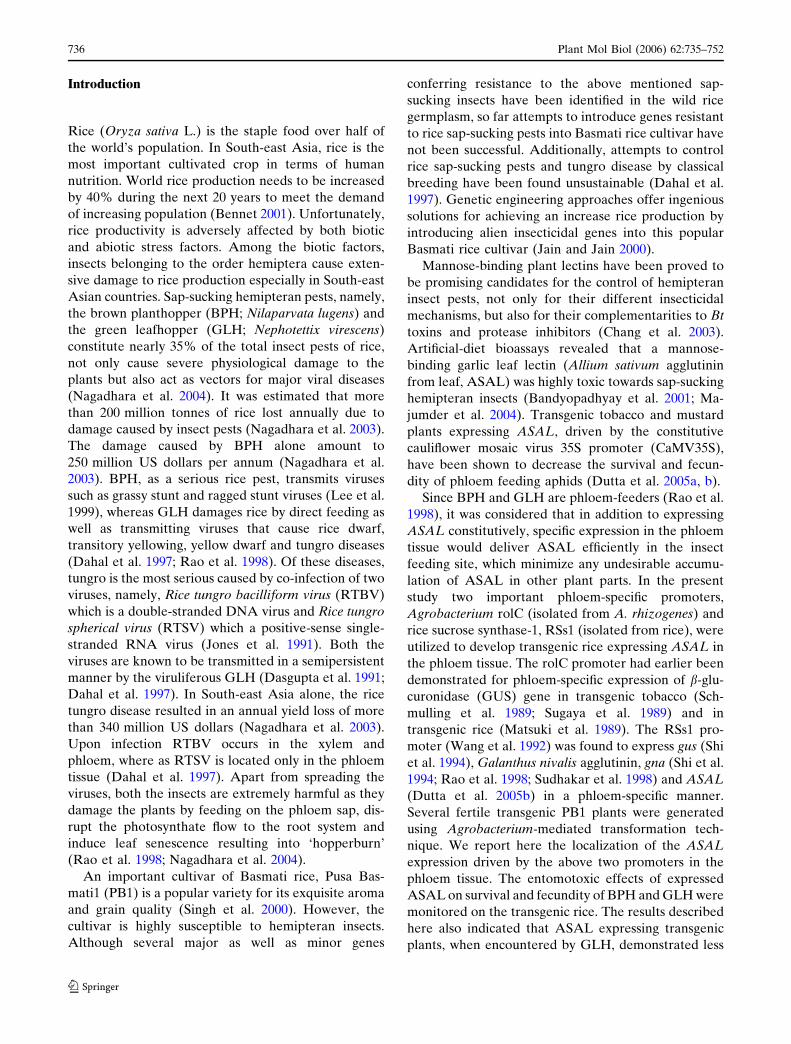
Introduction
Rice (Oryza sativa L.) is the staple food over half of
the world’s population. In South-east Asia, rice is the
most important cultivated crop in terms of human
nutrition. World rice production needs to be increased
by 40% during the next 20 years to meet the demand
of increasing population (Bennet 2001). Unfortunately,
rice productivity is adversely affected by both biotic
and abiotic stress factors. Among the biotic factors,
insects belonging to the order hemiptera cause exten-
sive damage to rice production especially in South-east
Asian countries. Sap-sucking hemipteran pests, namely,
the brown planthopper (BPH; Nilaparvata lugens) and
the green leafhopper (GLH; Nephotettix virescens)
constitute nearly 35% of the total insect pests of rice,
not only cause severe physiological damage to the
plants but also act as vectors for major viral diseases
(Nagadhara et al. 2004). It was estimated that more
than 200 million tonnes of rice lost annually due to
damage caused by insect pests (Nagadhara et al. 2003).
The damage caused by BPH alone amount to
250 million US dollars per annum (Nagadhara et al.
2003). BPH, as a serious rice pest, transmits viruses
such as grassy stunt and ragged stunt viruses (Lee et al.
1999), whereas GLH damages rice by direct feeding as
well as transmitting viruses that cause rice dwarf,
transitory yellowing, yellow dwarf and tungro diseases
(Dahal et al. 1997; Rao et al. 1998). Of these diseases,
tungro is the most serious caused by co-infection of two
viruses, namely, Rice tungro bacilliform virus (RTBV)
which is a double-stranded DNA virus and Rice tungro
spherical virus (RTSV) which a positive-sense single-
stranded RNA virus (Jones et al. 1991). Both the
viruses are known to be transmitted in a semipersistent
manner by the viruliferous GLH (Dasgupta et al. 1991;
Dahal et al. 1997). In South-east Asia alone, the rice
tungro disease resulted in an annual yield loss of more
than 340 million US dollars (Nagadhara et al. 2003).
Upon infection RTBV occurs in the xylem and
phloem, where as RTSV is located only in the phloem
tissue (Dahal et al. 1997). Apart from spreading the
viruses, both the insects are extremely harmful as they
damage the plants by feeding on the phloem sap, dis-
rupt the photosynthate flow to the root system and
induce leaf senescence resulting into ‘hopperburn’
(Rao et al. 1998; Nagadhara et al. 2004).
An important cultivar of Basmati rice, Pusa Bas-
mati1 (PB1) is a popular variety for its exquisite aroma
and grain quality (Singh et al. 2000). However, the
cultivar is highly susceptible to hemipteran insects.
Although several major as well as minor genes
conferring resistance to the above mentioned sap-
sucking insects have been identified in the wild rice
germplasm, so far attempts to introduce genes resistant
to rice sap-sucking pests into Basmati rice cultivar have
not been successful. Additionally, attempts to control
rice sap-sucking pests and tungro disease by classical
breeding have been found unsustainable (Dahal et al.
1997). Genetic engineering approaches offer ingenious
solutions for achieving an increase rice production by
introducing alien insecticidal genes into this popular
Basmati rice cultivar (Jain and Jain 2000).
Mannose-binding plant lectins have been proved to
be promising candidates for the control of hemipteran
insect pests, not only for their different insecticidal
mechanisms, but also for their complementarities to Bt
toxins and protease inhibitors (Chang et al. 2003).
Artificial-diet bioassays revealed that a mannose-
binding garlic leaf lectin (Allium sativum agglutinin
from leaf, ASAL) was highly toxic towards sap-sucking
hemipteran insects (Bandyopadhyay et al. 2001; Ma-
jumder et al. 2004). Transgenic tobacco and mustard
plants expressing ASAL, driven by the constitutive
cauliflower mosaic virus 35S promoter (CaMV35S),
have been shown to decrease the survival and fecun-
dity of phloem feeding aphids (Dutta et al. 2005a, b).
Since BPH and GLH are phloem-feeders (Rao et al.
1998), it was considered that in addition to expressing
ASAL constitutively, specific expression in the phloem
tissue would deliver ASAL efficiently in the insect
feeding site, which minimize any undesirable accumu-
lation of ASAL in other plant parts. In the present
study two important phloem-specific promoters,
Agrobacterium rolC (isolated from A. rhizogenes) and
rice sucrose synthase-1, RSs1 (isolated from rice), were
utilized to develop transgenic rice expressing ASAL in
the phloem tissue. The rolC promoter had earlier been
demonstrated for phloem-specific expression of b-glu-
curonidase (GUS) gene in transgenic tobacco (Sch-
mulling et al. 1989; Sugaya et al. 1989) and in
transgenic rice (Matsuki et al. 1989). The RSs1 pro-
moter (Wang et al. 1992) was found to express gus (Shi
et al. 1994), Galanthus nivalis agglutinin, gna (Shi et al.
1994; Rao et al. 1998; Sudhakar et al. 1998) and ASAL
(Dutta et al. 2005b) in a phloem-specific manner.
Several fertile transgenic PB1 plants were generated
using Agrobacterium-mediated transformation tech-
nique. We report here the localization of the ASAL
expression driven by the above two promoters in the
phloem tissue. The entomotoxic effects of expressed
ASAL on survival and fecundity of BPH and GLH were
monitored on the transgenic rice. The results described
here also indicated that ASAL expressing transgenic
plants, when encountered by GLH, demonstrated less
736 Plant Mol Biol (2006) 62:735–752
123
or no incidence of tungro disease. Therefore, utilization
of phloem-specific promoters for ASAL expression in
phloem tissue for developing genetically superior plants
resistant against phloem feeding insects and phloem
limited viruses is a promising approach in plant biotic
stress improvement programme.
Materials and methods
Plasmid constructs
The cloning of Allium sativum leaf agglutinin
(ASAL) coding sequence (Accession No. AY866499)
under cauliflower mosaic virus 35S (CaMV35S)
promoter in the binary vector, pCAMBIA1301 was
described previously (Dutta et al. 2005a). The p35S-
ASAL construct was used for constitutive expression
of ASAL gene. The rolC promoter sequence was
cloned from TL-DNA of Ri plasmid of Agrobacte-
rium rhizogenes A4 strain by PCR using Pfu DNA
polymerase (MBI Fermentas) and specific forward
and reverse primers (rolC1 and rolC2) including
additional HindIII site and BamHI site respectively
(Table 1). The rolC promoter (Accession No.
DQ160187) fragment was subcloned in pUC18 and
finally inserted into HindIII/BamHI sites of
pCAMBIA-ASAL to develop the prolC-ASAL. The
cloning of rice sucrose synthase-1 (RSs1) promoter
(Accession No. AJ401233) sequence from rice
genomic DNA and subsequent subcloning into
HindIII/BamHI site of binary vector pRSs-ASAL
was reported earlier (Dutta et al. 2005b). Individual
binary vector (p35S-ASAL, prolC-ASAL and pRSs-
ASAL) was mobilized into A. tumefaciens strain
EHA105.
Rice transformation and segregation analysis
Scutellum derived embryogenic calli from mature seed
of indica rice (Oryza sativa L.) cv. Pusa Basmati1 (PB1)
were used for Agrobacterium-mediated transforma-
tion. Approximately 30-days-old calli were infected
with A. tumefaciens strain EHA105 harbouring p35S-
ASAL, prolC-ASAL and pRSs-ASAL gene constructs.
Details of the entire procedure, including selection of
transformed calli and regeneration of plantlets, were
same as described by Mohanty et al. (1999). The only
differences were that full strength MS (Murashige and
Skoog 1962) medium and 30 g l-1maltose (Himedia)
were used instead of 3/2 MS and glucose respectively,
through out the study. The infected calli were co-cul-
tivated on MS medium containing 2 mg l–1 2, 4-D
(Sigma), 100 lM acetosyringone (Aldrich) for 3 days
and subsequently selected and on the same medium
devoid of acetosyringone and supplemented with
50 mg l–1 hygromycin (Hyg) (Roche Diagnostics),
250 mg l–1 cefotaxime for ~45 days including sub-cul-
ture at an interval of ~15 days at 28 ± 2�C under dark.
Plants were regenerated from Hyg resistant calli on a
medium fortified with 2 mg l–1 BAP (Sigma), 1 mg l–1
NAA (Sigma), 50 mg l–1 Hyg and 250 mg l–1 cefotax-
ime at 28 ± 1�C under 16 h photoperiod. Putative
transformants (T0) were transferred to sterile soil and
grown to maturity in the greenhouse. Plants regener-
ated from uninfected scutellum derived calli were em-
ployed as untransformed control (UC).
Segregation analysis was done in antibiotic-amen-
ded medium as well as through gene specific PCR
analysis. In the former method, the surface-sterilized
seeds of transgenic plants as well as untransformed
control plants were inoculated on MS basal medium
for 2 days at 28 ± 1�C under 16 h photoperiod. After
2 days, when seeds had germinated they were transferred
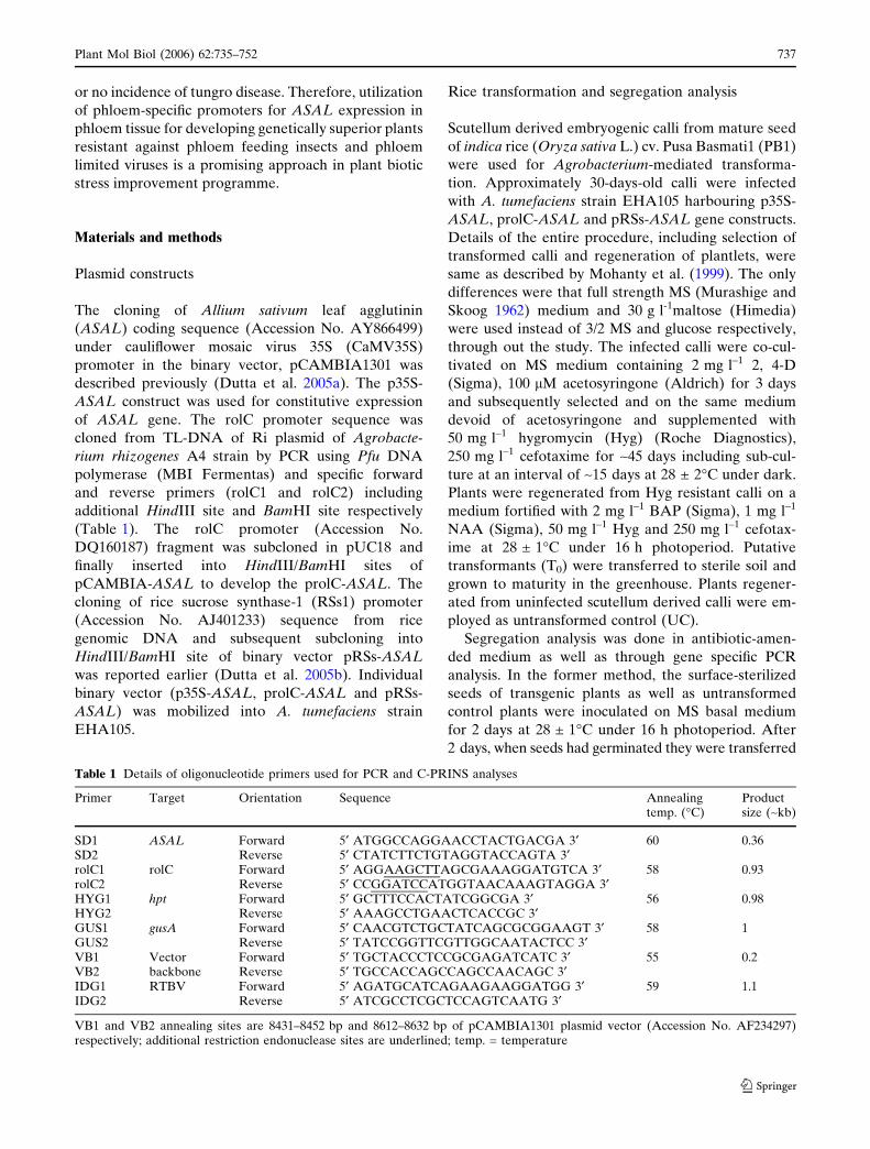
Table 1 Details of oligonucleotide primers used for PCR and C-PRINS analyses
Primer Target Orientation Sequence Annealingtemp. (�C)
Productsize (~kb)
SD1 ASAL Forward 5¢ ATGGCCAGGAACCTACTGACGA 3¢ 60 0.36SD2 Reverse 5¢ CTATCTTCTGTAGGTACCAGTA 3¢rolC1 rolC Forward 5¢ AGGAAGCTTAGCGAAAGGATGTCA 3¢ 58 0.93rolC2 Reverse 5¢ CCGGATCCATGGTAACAAAGTAGGA 3¢HYG1 hpt Forward 5¢ GCTTTCCACTATCGGCGA 3¢ 56 0.98HYG2 Reverse 5¢ AAAGCCTGAACTCACCGC 3¢GUS1 gusA Forward 5¢ CAACGTCTGCTATCAGCGCGGAAGT 3¢ 58 1GUS2 Reverse 5¢ TATCCGGTTCGTTGGCAATACTCC 3¢VB1 Vector Forward 5¢ TGCTACCCTCCGCGAGATCATC 3¢ 55 0.2VB2 backbone Reverse 5¢ TGCCACCAGCCAGCCAACAGC 3¢IDG1 RTBV Forward 5¢ AGATGCATCAGAAGAAGGATGG 3¢ 59 1.1IDG2 Reverse 5¢ ATCGCCTCGCTCCAGTCAATG 3¢
VB1 and VB2 annealing sites are 8431–8452 bp and 8612–8632 bp of pCAMBIA1301 plasmid vector (Accession No. AF234297)respectively; additional restriction endonuclease sites are underlined; temp. = temperature
Plant Mol Biol (2006) 62:735–752 737
123

on to Hyg (50 mg l–1) containing medium. Seeds were
scored for resistance (HygR) and susceptibility (HygS)
after 15 days of incubation. While in the second method,
the seeds were germinated on MS basal medium, DNA
was isolated from the seedlings and PCR was performed
using SD1 and SD2 ASAL specific primers (Table 1).
The segregation patterns in the progeny plants were
calculated from the HygR and PCR detected ASAL
positive and negative (ASALPCR+/–) plants and vali-
dated using v2 test.
Histochemical GUS assay and Nucleic acid analysis
The histochemical assay for gusA gene expression from
putative transgenic rice plants was performed accord-
ing to the method of Jefferson et al. (1987), using 5-
bromo-4-chloro-3-indoxyl-beta-D-glucuronide (X-gluc)
(Biosynth AG) as substrate. Total genomic DNA was
isolated from leaves of transformed and untransformed
rice plants, essentially followed by the method de-
scribed earlier (Dutta et al. 2005a, b) and examined for
the presence of the ASAL, hpt and gusA genes by PCR
using respective oligonucleotide primer pair (Table 1).
In order to determine the transgene copy number
considering left border-junction fragment (LB-JF) and
right border-junction fragment (RB-JF), ~10 lg of
genomic DNA was digested with appropriate restric-
tion enzymes (Roche Diagnostics), fractionated on a
0.8% agarose gel, denatured and transferred onto a
N+-nylon membrane (Amersham Biosciences).
Hybridization of membrane with the [a-32P] dCTP la-
belled BamHI/SacI ~0.36 kb ASAL gene probe, fol-
lowed by washings under stringent conditions, was
carried out according to procedures described earlier
(Dutta et al. 2005a, b).
For monitoring the vector backbone (VB) integra-
tion beyond left border, PCR analyses were performed
using VB1 and VB2 primers designed from left border
sequence and from downstream vector backbone se-
quence of pCAMBIA1301 plasmid (Table 1).
To carry out the northern blot analysis, total RNA
was isolated from young leaves of transformed rice
plants and ~20 lg RNA was fractionated on a 1.2%
formaldehyde–agarose gel, followed by blotting onto a
nylon membrane. Hybridization and washing were
performed as described for the Southern blot proce-
dure (Dutta et al. 2005b).
Chromosome preparation and C-PRINS analysis
To study the stable transgene integration in the
genomic DNA of selected transgenic rice lines,
cycling-primed in situ DNA labelling (C-PRINS)
analyses were carried out using ASAL, hpt and gusA
gene specific oligonucleotide primers (Table 1) on
interphase nuclei and pro-metaphase chromosome
preparations. The preparations of interphase nuclei
and pro-metaphase chromosome plates were per-
formed following the enzyme maceration technique of
Jiang et al. (1995). Actively growing root tips were
excised from transgenic plants and put in ice cold water
for 20–24 h, followed by fixation in ethanol:acetic acid
(3:1) at 14–18 �C for overnight. Root tips were then
washed thoroughly with 0.01 M citrate buffer (sodium
citrate/citric acid, pH 4.8) followed by enzyme macer-
ation with 6% (w/v) cellulase (Sigma), 2% (w/v) pec-
tinase (Sigma) and 0.75% (w/v) macerozyme R200
(Sigma) for 1 h at 37 �C. Finally, the treated root tips
were washed three times in water squashed on Am-
plislides, air dried and stored at –20 �C. Prior to C-
PRINS, the slides were washed three times with 2·SSC
for 5 min at room temperature followed by 50 lg ml–1
RNase (Sigma) treatment in reaction buffer (1 mM
Tris–HCl, 1.5 mM NaCl, pH 7.5) at 37�C for 60 min.
Subsequently, the slides were incubated with 2 lg ml–1
proteinase K (Sigma) at room temperature for 1 min
and rinsed in 1·PCR buffer (10 mM Tris–HCl, pH 8.4,
50 mM KCl, 1.5 mM MgCl2) for 5 min.
The procedure and conditions for C-PRINS were
modified from the method of Abbo et al. (1993) and
Kubalakova et al. (2001). The amplifications were
performed in 50 ll volume containing 1·PCR buffer,
2 Mm MgCl2, 200 lM of primers set for each gene,
100 lM of each dATP, dCTP, dGTP, 34 lM of dTTP
and 2 lM of fluorescein-11-dUTP (Roche Diagnostics)
and 5 U of Amplitaq gold DNA polymerase. Reaction
mixture was sealed under amplicover discs with am-
plicover clips and the C-PRINS reactions were per-
formed following an initial cycle of 18 min at 95�C,
5 min at annealing temperature (Table 1), 10 min at
72�C, followed by 30 cycles each of 1 min at 94�C,
1 min at annealing temperature (Table 1) and 2 min at
72�C with a final extension for 10 min at 72�C. The
reactions were carried out using GeneAmp in situ PCR
system 1000 (Applied Biosystems) and the experiments
were repeated at least three times for each transgenic
line with each set of primer pairs to establish the
reproducibility of the results. The thermal cycle reac-
tions were terminated with stop buffer (0.5 M NaCl,
0.05 M EDTA, pH 8.0) for 5 min at 65�C and washed
with wash buffer (0.1 M maleic acid, 0.15 M NaCl,
0.05% Tween 20, pH 7.5) at room temperature for
5 min. The slides were immediately mounted with
Vectashield antifade solution (Vectolaboratories)
containing 0.2 lg ml–1 4’,6’-diamino-2-phenylindole
738 Plant Mol Biol (2006) 62:735–752
123

(DAPI) and observed with a Zeiss Axioskop 2 fluo-
rescence microscope (Carl Zeiss) using dual excitation
filters (set 27) for FITC and DAPI. Signals were cap-
tured by CCD camera and analyzed in FISH ImagerTM
(v.1.0) software.
Western blot and enzyme linked immunosorbent
assay (ELISA)
The total soluble protein (TSP) was extracted from
leaves of T0 and T1 transgenic rice plants (Dutta et al.
2005a) and the amount of protein in each sample was
determined by Bradford assay (Bradford 1976).
Approximately 10 lg of TSP was separated on 15%
SDS-PAGE and transferred onto a nitrocellulose
membrane by electroblotting. After blocking, the
membrane was probed with anti-ASAL polyclonal
primary antibody, obtained following the method of
Bandyopadhyay et al. (2001), at 1:10,000 dilution and
anti-rabbit IgG-horse radish peroxidase (HRP) conju-
gate (Sigma) as secondary antibody at 1:10,000 dilu-
tions. Bound secondary antibodies were detected by
enhanced chemiluminescence (ECL) reagents (Roche
Diagnostics).
Quantitative level of ASAL expression was esti-
mated by ELISA. The wells of the ELISA plate (Im-
munomaxi) were coated with ~10 lg of TSP from
transgenic leaves and purified native ASAL for over-
night at 4�C. The wells were blocked with 5% (w/v)
non-fat milk (Merck) in PBS and then incubated with
anti-ASAL primary antibody, followed by incubation
with HRP conjugated anti-rabbit secondary antibody.
Washing and incubations were performed according to
standardized protocol (Dutta et al. 2005a). The reading
of the coloured solution developed in microtitre after
reaction with O-phenylenediamine hydrochloride
(OPD) substrate (Sigma) plate was recorded at 415 nm
in a plate reader (BioRad).
Immunohistoflourescent analysis
Hand sections of leaves of plants transformed with
p35S-ASAL, prolC-ASAL and pRSs-ASAL constructs
were incubated in 10% (v/v) trichloroacetic acid (Sig-
ma) for 1 h followed by successive incubation with
absolute alcohol to water through 90, 70, 50 and 30%
(v/v) respectively. Sections were then blocked in
blocking buffer [3% (w/v) BSA (Merck) in 1· PBS],
subsequently incubated with anti-ASAL primary anti-
body at 1:10,000 dilutions in 1 ml blocking buffer
overnight. Immunohistoflourescent localization of
ASAL in transgenic leaf sections was carried out
according to the reported method (Yin et al. 1997) by
incubating with anti-rabbit IgG-biotin conjugate sec-
ondary antibody (Sigma) at 1:1000 dilutions in 1 ml
PBS for 2 h. The sections were then incubated with
avidin-FITC conjugate (Sigma) at 1:200 dilutions in
PBS. Washings were performed following the manu-
facturer’s protocols and sections were mounted on
slides with Vectashield antifade solution. The samples
were observed with a Zeiss Axioskop 2 fluorescence
microscope using excitation filter (set 1) of 450–490 nm
for FITC and photographs were recorded.
Insect bioassay
Insecticidal activity of ASAL toward the major rice
insects BPH and GLH were assayed using in-planta
feeding chamber on transgenic plants under controlled
conditions as described earlier (Rao et al. 1998; Foissac
et al. 2000). For survival, development and fecundity
assays ~35 to 45-days-old, single copy T-DNA bearing
and high ASAL expressive T0 and T1 plants were tested
where as untransformed plants and the T1 plants which
had lost the ASAL gene due to Mendelian segregation
were used as negative controls. A total of 15 first-instar
nymphs were released on each plant with five sets of
replication and the amount of damage to each plant was
counted once every three days. Statistical unpaired t
tests were conducted in order to compare the signifi-
cance of differences between control and treatments for
both the insect bioassay experiments.
Plant inoculation
Challenges of T1 transgenic plants by RTBV/RTSV
viruses using viruliferous GLH were conducted inside
a chamber as described earlier (Sivamani et al. 1999)
with minor modifications. First inoculation was per-
formed with three viruliferous GLH on 15-days-old six
T1 plantlets (first transmission). This was followed by
acquisition of RTBV/RTSV by non viruliferous GLH
(6 GLH/plant) from the above infected transgenic
plants. These viruliferous GLH were then placed
overnight on two 15-day-old untransformed control
PB1 rice plants (3 GLH/plant) in the second trans-
mission assay. In both the first and the second trans-
mission assays, six plants and two untransformed
control PB1 plants (UC) were challenged respectively.
PCR and dot blot analysis for RTBV and RTSV
PCR analyses of the infected plants for detection of
RTBV of first and second transmission assay were
performed following the earlier described simplified
method of Dasgupta et al. (1996) using either undiluted
Plant Mol Biol (2006) 62:735–752 739
123
and/or 100 and 500 fold diluted leaf DNA samples in
10 mM Tris–HCl, 1 mM EDTA, pH 8.0 buffer and
RTBV specific IDG1, IDG2 primers (Table 1). Moni-
toring of the presence of RTSV through dot blot
analysis was carried out with RNA samples isolated
from infected leaf samples of the first transmission
assay using RNeasy Plant Mini Kit (QIAGEN) fol-
lowing the manufacturer’s protocols and recommen-
dations. Approximately 1 lg of total RNA from each
infected and uninfected plants was blotted onto a N+-
nylon membrane using Bio-Dot Micro-filtration appa-
ratus (ATTO). Membranes were then hybridized with
[a-32P] dCTP-labelled BamHI/EcoRI digested ~1 kb
RTSV coat protein 3 (CP3) coding DNA fragment as
probe and autoradiograph was developed.
Results
Gene constructs preparation and rice genotype
used
The DNA fragment containing the ~0.93 kb 5¢ up-
stream sequence of the TL-DNA of Ri plasmid ORF12
gene (rolC) of A. rhizogenes A4 strain was obtained by
PCR using rolC1 and rolC2 primers (Table 1). The
promoter fragment was sequenced and the new se-
quence was submitted to GenBank (Accession No.
DQ160187). The ~1.9 kb RSs1 promoter contains
~0.69 bp 5¢ upstream sequences and ~1.22 kb 3¢downstream sequences to tsp, the intron1. An internal
EcoRI restriction endonuclease site is present at –
0.67 kb position of RSs1. The ASAL gene was initially
cloned under CaMV35S (constitutive) promoter and
nopaline synthase polyA terminator (nosT) in plant
expression vector pCAMBIA1301 to develop p35S-
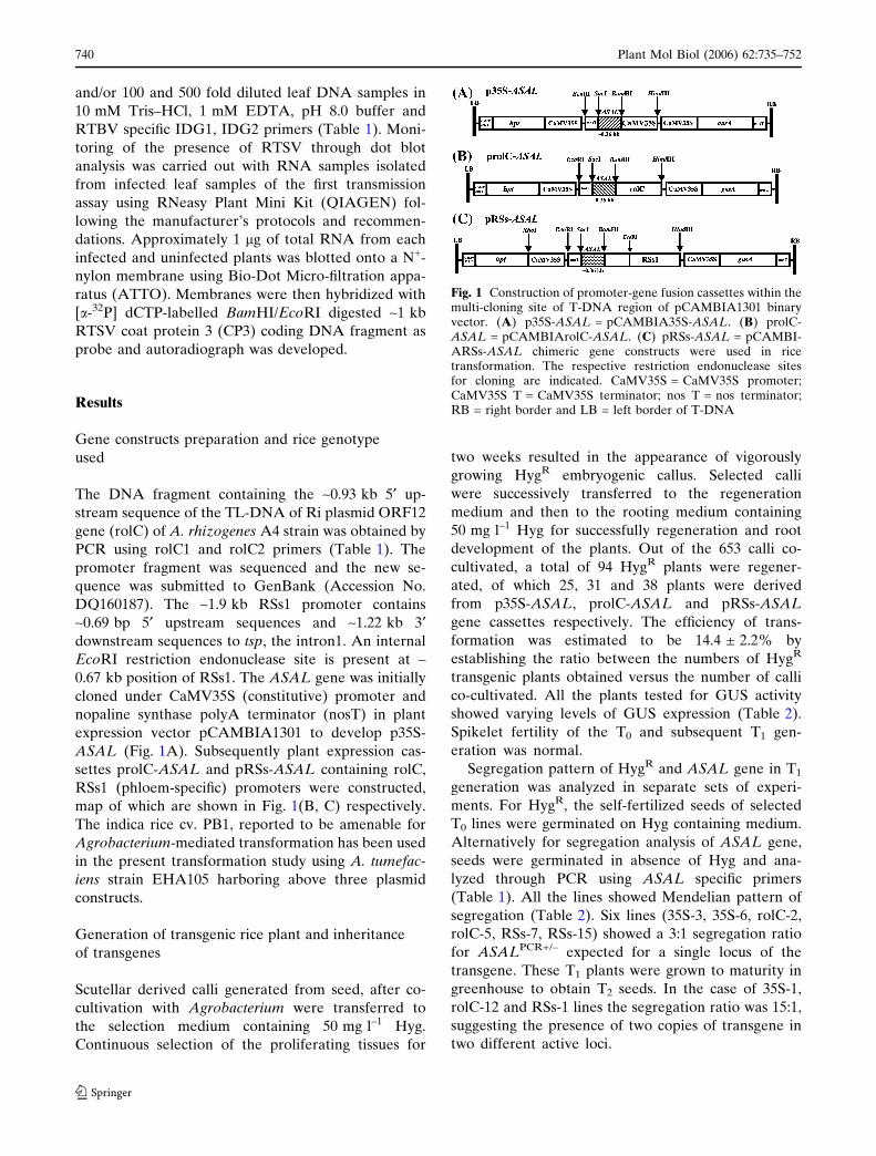
ASAL (Fig. 1A). Subsequently plant expression cas-
settes prolC-ASAL and pRSs-ASAL containing rolC,
RSs1 (phloem-specific) promoters were constructed,
map of which are shown in Fig. 1(B, C) respectively.
The indica rice cv. PB1, reported to be amenable for
Agrobacterium-mediated transformation has been used
in the present transformation study using A. tumefac-
iens strain EHA105 harboring above three plasmid
constructs.
Generation of transgenic rice plant and inheritance
of transgenes
Scutellar derived calli generated from seed, after co-
cultivation with Agrobacterium were transferred to
the selection medium containing 50 mg l–1 Hyg.
Continuous selection of the proliferating tissues for
two weeks resulted in the appearance of vigorously
growing HygR embryogenic callus. Selected calli
were successively transferred to the regeneration
medium and then to the rooting medium containing
50 mg l–1 Hyg for successfully regeneration and root
development of the plants. Out of the 653 calli co-
cultivated, a total of 94 HygR plants were regener-
ated, of which 25, 31 and 38 plants were derived
from p35S-ASAL, prolC-ASAL and pRSs-ASAL
gene cassettes respectively. The efficiency of trans-
formation was estimated to be 14.4 ± 2.2% by
establishing the ratio between the numbers of HygR
transgenic plants obtained versus the number of calli
co-cultivated. All the plants tested for GUS activity
showed varying levels of GUS expression (Table 2).
Spikelet fertility of the T0 and subsequent T1 gen-
eration was normal.
Segregation pattern of HygR and ASAL gene in T1
generation was analyzed in separate sets of experi-
ments. For HygR, the self-fertilized seeds of selected
T0 lines were germinated on Hyg containing medium.
Alternatively for segregation analysis of ASAL gene,
seeds were germinated in absence of Hyg and ana-
lyzed through PCR using ASAL specific primers
(Table 1). All the lines showed Mendelian pattern of
segregation (Table 2). Six lines (35S-3, 35S-6, rolC-2,
rolC-5, RSs-7, RSs-15) showed a 3:1 segregation ratio
for ASALPCR+/– expected for a single locus of the
transgene. These T1 plants were grown to maturity in
greenhouse to obtain T2 seeds. In the case of 35S-1,
rolC-12 and RSs-1 lines the segregation ratio was 15:1,
suggesting the presence of two copies of transgene in
two different active loci.
Fig. 1 Construction of promoter-gene fusion cassettes within themulti-cloning site of T-DNA region of pCAMBIA1301 binaryvector. (A) p35S-ASAL = pCAMBIA35S-ASAL. (B) prolC-ASAL = pCAMBIArolC-ASAL. (C) pRSs-ASAL = pCAMBI-ARSs-ASAL chimeric gene constructs were used in ricetransformation. The respective restriction endonuclease sitesfor cloning are indicated. CaMV35S = CaMV35S promoter;CaMV35S T = CaMV35S terminator; nos T = nos terminator;RB = right border and LB = left border of T-DNA
740 Plant Mol Biol (2006) 62:735–752
123

Molecular analysis of transgenic rice plant
Samples of the genomic DNA, isolated from leaves of
HygR transgenic plants as well as untransformed con-
trol (UC) plant, were analyzed for the presence of
ASAL and gusA genes by PCR using SD1, SD2 and
GUS1, GUS2 oligonucleotide primers, respectively
(Table 1). Presence of ASAL (~0.36 kb) and gusA
(~1 kb) gene fragments in the genome of all primary
transformants confirmed their transgenic status (data
not shown). However, no such bands were found in the
PCR product of UC plant DNA under the identical
conditions.
In an initial screening out of the 25 p35S-ASAL, 31
prolC-ASAL and 38 pRSs-ASAL plants, we examined
18 independent T0 plants (six plants from each plasmid
construct) by Southern blot for ASAL gene. Genomic
DNA (~10 lg) digested with HindIII showed hybrid-
izable bands in all the transformants (Fig. 2 lane 35S-5
to RSs-34) including a ~0.36 kb band in the positive
control (~0.36 kb ASAL) using ASAL gene probe
(Fig. 2, lane +). Most of the transgenic plants carried
one to two copies of the ASAL gene. For detail
molecular analysis, four plants from each of the pro-
moter driven gene construct were selected depending
upon their high HygR and GUS expression (Table 2).
However, to confirm transgene integration and to
determine the copy number of the integrated T-DNA,
the genomic DNA of 35S-1, 35S-3, 35S-4, 35S-6, rolC-2,
rolC-5, rolC-9, rolC-12 and RSs-1, RSs-7, RSs-11,
RSs-15 plants were digested with HindIII/EcoRI
and HindIII/XhoI to generate T-DNA LB-JF/RB-JF
respectively. After hybridization with [a-32P] dCTP
labelled BamHI/SacI 0.36 kb ASAL gene probe, HindIII
digested DNA from above 35S-ASAL, rolC-ASAL and
RSs-ASAL plants exhibited LB-JFs longer than
~3.5 kb, ~3.6 kb and ~4.6 kb, respectively (Fig. 3, pa-
nel H). The T0 lines 35S-3, 35S-6, rolC-2, rolC-5, RSs-7,
RSs-11 and RSs-15 revealed single junction fragment,
suggesting a single copy T-DNA integration event.
However, plants 35S-1, rolC-9, rolC-12 and RSs-1
showed double copy and only the 35S-4 line showed
triple copy T-DNA integration events. Further analysis
using RB-JF revealed hybridization signals longer than
Table 2 Segregation of hygromycin resistance, GUS activity and presence of ASAL gene in T1 progeny plants of transgenic rice lines
Transgenic plants Segregation in T1 generation T2 seed
T0 lines HygR/GUS/ASALPCR/VBPCR HygR/S gusAPCR+/– ASALPCR+/– Ratio (ASAL) v2(P value)
35S-1 +/+/+/+ 30/2 28/2 28/2 15:1 0.53 (> 0.9)35S-3 +/+/+/– 40/11 38/13 38/13 3:1 0.007 (> 0.8) Yes35S-4 +/–/+/+ NT NT NT – –35S-6 +/+/+/– 22/8 23/7 23/7 3:1 0.04 (> 0.8) YesrolC-2 +/+/+/– 32/12 34/10 32/10 3:1 0.94 (> 0.2) YesrolC-5 +/+/+/– 18/7 20/5 20/5 3:1 0.33 (> 0.5) YesrolC-9 +/–/+/+ NT NT NT – –rolC-12 +/–/+/+ 19/2 NT 21/0 15:1 –RSs-1 +/+/+/+ 36/1 34/3 34/3 15:1 0.22 (> 0.5)RSs-7 +/+/+/– 24/9 25/8 25/8 3:1 0.01 (> 0.9) YesRSs-11 +/+/+/+ NT* NT* NT* – –RSs-15 +/+/+/– 38/10 37/11 36/12 3:1 0.00 (–) Yes
ASALPCR+/– = PCR positive/negative for ASAL gene; GUS + = GUS positive by histochemical assay; gusAPCR+/– = PCR positive/negative for gusA gene; HygR/S = hygromycin resistant/susceptible; NT = not tested; * Plants contaminated during tissue culture;VBPCR = PCR positive for vector backbone sequence; v2 value is calculated for inheritance of one gene (3:1) or two genes (15:1)
Fig. 2 Southern blot hybridization of p35S-ASAL, prolC-ASALand pRSs-ASAL bearing primary transgenic plants. ~10 lg DNAfrom the transformed and untransformed rice plant leaf wasdigested with HindIII and processed for Southern blotting usinga-32P labelled BamHI/SacI ASAL gene fragment as probe.Lane + = positive control ~0.36 kb ASAL gene; lane UC = un-transformed control plant DNA; lane 35S-5 to RSs-34 = differ-ent line of 35S-ASAL, rolC-ASAL and RSs-ASAL. Molecularweight markers are indicated on the left
Plant Mol Biol (2006) 62:735–752 741
123

~4.3 kb in 35S-ASAL, ~4.4 kb in rolC-ASAL with
EcoRI digested genomic DNA (Fig. 3 panel E) and
~6.2 kb in RSs-ASAL with XhoI digested genomic
DNA (Fig. 3 panel X). The primary transformant rolC-
9 line showed ~8.8 kb single copy RB-JF with EcoRI
digested genomic DNA but produced double copy
hybridization signal with HindIII digested LB-JF
genomic DNA, suggesting a ‘head-to-head’ double
copy T-DNA (LB-RBMRB-LB) integration event
(Fig. 3I). However, all the RSs-ASAL lines showed a
~1.3 kb hybridization signal when EcoRI digested
genomic DNA was analyzed indicating about the
presence of an internal EcoRI site in the promoter
sequence.
Fig. 3 Detection of T-DNA genes (ASAL, hpt and gusA) copynumber in transgenic rice lines using Southern blot andinterphase nuclei and/or prometaphase C-PRINS analyses. TheHindIII (H) digested and EcoRI (E) or XhoI (X) digestedgenomic DNA blot are shown in panels which documented thecopy number through left border and right border junctionalfragment analysis. The ~0.36 kb ASAL gene was used as probe inSouthern hybridization. Molecular weight markers are indicated
on the right side of each panel. The respective transgenic line andT-DNA gene used in C-PRINS study are indicated in bracket.Arrows on the figures indicate the strong hybridization signals(fluorescent green colour spot) obtained after incorporation offluorescein-11-dUTP. Nuclei or chromosomes are counterstained with DAPI (blue colour). (A, B, C, D, E, F, H) showedsingle copy and (G, I, J, K, L) showed multi-copy T-DNAintegration
742 Plant Mol Biol (2006) 62:735–752
123

In addition to Southern blot analysis, we adopted
the C-PRINS technique to confirm the transgene copy
number and to characterize the transgene status in
detail. C-PRINS analysis using ASAL, hpt and gusA
gene specific oligonucleotide primers (Table 1) re-
vealed single copy (green fluorescent spot) localization
of T-DNA genes (ASAL, hpt and gusA) in lines 35S-3,
35S-6, rolC-2, rolC-5, RSs-7, RSs-11 and RSs-15 after
incorporation of fluorescein-11-dUTP (Fig. 3A–F, H).
The transgenic lines 35S-1, rolC-12, RSs-1 showed
double spot (Fig. 3G, J, K), whereas 35S-4 showed
triple copy (Fig. 3L) localization of ASAL, hpt and
gusA genes on interphase nuclei and pro-metaphase
chromosomes. The line rolC-9 showed localization of
two subtelomeric hpt gene (two green fluorescent spot
very close to each other) on single chromosome using
HYG1 and HYG2 primers in C-PRINS study, sug-
gesting integration of two copies of complete T-DNA
(Fig. 3I). Therefore, the RB-JF and LB-JF analyses in
Southern blot and C-PRINS studies were confirmed
single copy in 35S-3, 35S-6, rolC-2, rolC-5, RSs-7, RSs-
15 lines; double copy in 35S-1, rolC-12, RSs-1 lines;
triple copy in 35S-4 line and head-to-head double copy
T-DNA integration in rolC-9 line. However, Southern
blot analysis with HindIII digested genomic DNA from
four T1 progeny plants showed single hybridizable
band in accordance with that of parental lines 35S-6,
rolC-2 and RSs-15 (Fig. 4A–C). The untransformed
control plant failed to show any hybridizable band
when either of the junction fragment was analyzed.
Furthermore, all the 12 characterized best lines were
checked for the integration of ~0.2 kb vector backbone
(VB) sequences beyond the T-DNA left border in the
genome by PCR analysis using VB specific primers
(Table 1). All the double copy (35S-1, rolC-9, rolC-12,
RSs-1) and triple copy (35S-4) T-DNA integrated lines
were found to be PCR positive for VB sequence
(VBPCR+, Table 2). Only one single copy line (RSs-11)
showed amplification beyond T-DNA border in PCR
analysis but rest of the lines showing clean T-DNA
integration (Table 2).
Table 3 Insect bioassay and RTBV transmission of T1 progeny plants of T0 rice lines
T1 progeny Amount of ASAL(% TSP)a
% of insect mortal-ity
% of insect fecundity RTBV/RTSV trans-mission
BPH GLH BPH GLH susc/inoc % R
Control 0 0(20)b 0(14)b 100(102)c 100(115)c 12/12 035S-3T1 0.66 NT NT NT NT 3/6 5035S-6T1 0.87 41 37 NT 42 3/6 50rolC-2T1 0.45 35 59 29 25 1/6 83rolC-5 T1 0.33 NT 53 NT NT 2/6 67RSs-7T1 0.09 39 46 NT 37 3/6 50RSs-15T1 0.21 NT 40 NT 28 2/6 67
a Percentage mean value of six plants in total soluble protein; b Insect mortality on control plant; c Mean number of nymphs oncontrol plant; NT = not tested; susc/inoc = susceptible plants (positive in PCR for RTBV and dot blot for RTSV) per total number ofinoculated with viruliferous GLH; % R = percentage of plants showing resistance against RTBV/RTSV; % of insect mortality andfecundity on transgenic T1 lines were determined after deducting the bvalue and calculating from cvalue respectively (n = 5)
Fig. 4 Southern blot analyses of genomic DNA from progenyplant leaves of 35S-ASAL, rolC-ASAL and RSs-ASAL T0 lines.Genomic DNA (~10 lg) was digested with HindIII andhybridized with [a-32P] dCTP labelled BamHI/SacI ASAL geneprobe. (A) Four T1 plants of 35S-6 line. (B) Four T1 plants ofrolC-2 line. (C) Four T1 plants of RSs-15 line. Lane + = ~0.36 kbASAL gene was used as positive control; lane UC = untrans-formed genomic DNA as negative control. Molecular weightmarkers are indicated on the left
Plant Mol Biol (2006) 62:735–752 743
123

Expression of ASAL gene in transgenic plants
On the basis of preliminary screening we have chosen
the high HygR, most strongly GUS expressive, with
single copy ASAL gene insertion and without vector
backbone bearing transgenic lines for further analysis.
Two primary transformants from each construct,
namely, 35S-3, 35S-6 (for constitutive expression),
rolC-2, rolC-5 and RSs-7, RSs-15 (for phloem-specific
expression) were tested in northern, western blot and
ELISA for ASAL gene product. Northern blot analysis
showed the presence of intact, full length hybridisable
transcript using ASAL probe (Fig. 5A). The level of
ASALmRNA accumulation was higher in constitu-
tively expressing lines (35S-3, 35S-6) than the phloem-
specific lines.
Western blot analysis of leaf extracts from trans-
genic plants revealed the presence of a polypeptide of
~12 kDa that corresponds to purified ASAL after
treating with specific anti-ASAL antibodies (Fig. 5B).
All the six T0 lines were accumulated detectable level
of ASAL in their leaf tissue. However, no band was
observed in the untransformed control plant in both
northern and western blot analyses (Fig. 5A, B, lane
UC).
To quantify the amount of ASAL in these selected
lines, ELISA with leaf protein extracts was performed,
the results of which are shown in Fig. 5C. Constitutive
ASAL expression from 35S promoter was estimated to
be 0.86–1.01% of total soluble protein (TSP) by cal-
culating the amount of ASAL in ~10 lg of TSP of each
transgenic line from ELISA reading, which was much
higher than RSs1 promoter driven ASAL expression
(0.11–0.31% of TSP). The quantitative level of ex-
pressed ASAL from rolC promoter (0.38–0.52% of
TSP) was found to be in between the 35S and RSs1
promoters.
Northern blot analysis using total RNA from leaf of
T1 progeny plants was detected hybridisable amount of
ASALmRNA similar to their mother plants after
probed with ASAL gene (Fig. 6). In the T1 progenies
of the T0 lines, the level of expressed ASAL was cal-
culated to be in the range of 0.87% to 0.09% of TSP
(Table 3).
Immunohistofluorescence localization of ASAL
in transgenic plant
The spatial ASAL expression patterns driven by 35S,
rolC and RSs1 promoters in transgenic lines were
determined by in situ immunohistofluorescence locali-
zation of ASAL in trichloroacetic acid fixed leaf tissue
Fig. 5 Expression and quantitative estimation of ASAL drivenby 35S, rolC and RSs1 promoters in selected transgenic rice lines.(A) Northern blot analysis of p35S-ASAL, prolC-ASAL andpRSs-ASAL plants. ~20 lg total RNA from individual plantswas probed with radiolabelled ASAL gene. Lane UC = RNAfrom untransformed control plant. The ASAL transcript accu-mulations in mature leaf of selected lines are represented bytheir lanes. (B) Western blot analysis of protein extracts fromleaves of same T0 plants. Lane + = ~1.5 lg purified native ASALused as positive control; lane UC = crude protein from untrans-formed plant used as negative control; transgenic plants arerepresented by their respective lanes. (C) The amount of ASALin total soluble protein of corresponding T0 plants anduntransformed control plant (UC) as determined by ELISA.The results of the three separate ELISA experiments using totalcrude protein extracts from 30-days-old field grown plant leavesare shown. The bars represent the average ELISA read-ing + standard error of the mean. ELISA reading representedafter subtracting the background readings from UC
Fig. 6 Northern blot analysis of T1 plants of selected parentallines. (A) Ethidium bromide strained rRNA profile of fourprogenies of 35S-6, rolC-2, RSs-15 T0 transformants anduntransformed control (UC). (B) Hybridization signals ofASALmRNA with ASAL gene probe in corresponding lanes.Total RNA (~20 lg) of T1 plants derived from self-fertilizationof the three T0 plants of lanes 35S-6, rolC-2 and RSs-15 in Fig.5A, was subjected to northern blot analysis. Lane UC = un-transformed control RNA. The ASAL transcript accumulation inmature leaf of T1 plants are represented by their lanes
744 Plant Mol Biol (2006) 62:735–752
123

sections after reaction with anti-ASAL primary anti-
body, IgG-biotin conjugate secondary antibody and
avidin-FITC conjugate (Fig. 7). We found that the three
promoters conferred quite different ASAL expression
patterns of which the 35S-ASAL (35S-3, 35S-6) showed
the expected constitutive ASAL expression pattern with
green fluorescence spreading all over the cell types,
strong fluorescence in vascular region and weaker fluo-
rescence in all other tissues (Fig. 7B). Accumulation of
ASAL in both rolC-ASAL and RSs-ASAL lines was
detected in phloem cells and other non-lignified vascular
tissue including lesser amount in the epidermal cells.
Detailed examination of leaf tissue sections of the rolC-
ASAL lines demonstrated that the presence of ASAL
within the vascular bundles, vascular parenchyma,
adjacent cortical cells, epidermal cells and trichromes
(Fig. 7C) where as in RSs-ASAL lines ASAL expression
was restricted to the phloem tissue, sieve elements and
companion cells (Fig. 7D). However, tissue sections of
different leaf blades gave identical results. No green
fluorescence in any cell types of untransformed control
plant was visible (Fig. 7A).
Monitoring insect bioassay on ASAL expressive
transgenic plants
Bioassays for resistance to sap-sucking insect pests
were carried out in-planta on the 35 to 45-days-old
T0 and T1 transgenic plants expressing ASAL. All
the plants that were infested with BPH/GLH
nymphs during bioassay showed significant resistance
against survival, growth and fecundity of BPH and
GLH (Fig. 8), were survived and grew to maturity
with normal fertility. BPH and GLH survival was
monitored on three high ASAL expressive primary
transformants, 35S-6 (constitutively expressive) and
rolC-2, RSs-15 (phloem-specific expressive), using
first-instar nymphs to adults at 3 days intervals up to
18 days of survival assay period. Gradual reduction
of survival of BPH nymphs was observed on all the
transgenic lines tested (Fig. 8A), whereas a sharp
decline of survival of GLH nymphs was recorded
only on the rolC-2 line (Fig. 8B). Phloem expressive
lines showed comparatively higher insect (BPH and
GLH) mortality than the constitutively expressive
line (significant at P < 0.05). However, within the
phloem expressive lines, rolC-2 line has documented
elevated level of BPH and GLH resistant by ~5.1%
and ~11.1% compared to RSs-15 line respectively.
The effect of ASAL on GLH development was
monitored by releasing 15 first-instar nymphs onto
ASAL expressive T0 lines. GLH development on
transgenic plants was significantly retarded compared
to control (significant at P < 0.05). From the Fig. 8
(C, D) it is clearly evident that rolC-2 line showed
maximum retardation of GLH development than
Fig. 7 Immunohistofluorescence localization of ASAL in trans-genic plants containing p35S-ASAL, prolC-ASAL and pRSs-ASAL constructs. Transverse leaf sections were treated withanti-ASAL anti-serum as primary antibody and anti-rabbit IgG-biotin conjugated secondary antibody. The presence of ASAL isindicated by the green fluorescence. (A) The transverse sectionsfrom untransformed control showed no green fluorescence. (B)
The leaf section of p35S-ASAL plants showing detection ofuniform green fluorescence of constitutive ASAL expression inall the cells types. (C) Cross sections of leaf blade of plantstransformed with prolC-ASAL showing localization of ASAL inthe phloem cells, epidermis and trichomes. (D) The pRSs-ASALshowing the presence of ASAL in the phloem cells and nonlignified vascular region
Plant Mol Biol (2006) 62:735–752 745
123

35S-6 and RSs-15 lines, as out of 34.7% survivors,
only 13.3% nymphs were reached to adulthood after
15 days of incubation (significant at P < 0.01). The
poor development of GLH nymphs on all the
respective transgenic lines was reflected by 33.4%,
29.3%, 21.3% immature nymphs after 15 days of
assay period. The effect of ASAL on BPH and
GLH fecundity was also assessed by measuring total
nymphs production by adult insects on the trans-
genic lines used for survival assays after 20 day as-
say period. The rolC-2 line exhibited minimum
mean numbers of BPH and GLH nymph production
per plant followed by RSs-15 and 35S-6 lines
(Fig. 8E, F). Therefore, the three tested lines are
categorized as rolC-2 > RSs-15 > 35S-6 depending on
their ability to control insect survival, development
Fig. 8 Effect of ASAL on the survival, development andfecundity of rice phloem feeding insects. (A, B) Survival ofBPH and GLH on T0 transgenic rice lines expressing ASAL.Fifteen first-instar BPH and GLH nymphs were releasedseparately on each plant at day 0. Untransformed control plantsand transgenic plants 35S-6 (constitutive ASAL expression) androlC-2, RSs-15 (phloem-specific ASAL expression) were used forbioassay experiments. Points and bars show mean ± SE (n = 5).The significant (P < 0.05) differences between control andtransgenic plants were assessed by unpaired t-test. (C, D) Effectof ASAL on the development of GLH nymphs. Fifteen first-instar GLH nymphs were released separately on untransformed
control and transgenic plant at day 0, and after 15 days the insectsurvival and the number of nymphs which reached adult stage oncontrol and transgenic plants were plotted on the graph.Differences between control and transgenic plants were signif-icant at P < 0.05 (t-test). Data represent as mean ± SE (n = 5).(E, F) Effect of ASAL on the fecundity of the BPH and GLH.The total number of nymphs produced from adult BPH andGLH on control and transgenic plants were counted at the end ofthe insect bioassay were plotted on the graph. The significant(P < 0.01) differences in nymph production on control andtransgenic plants were assessed by unpaired t test. Data representas mean ± SE (n = 5)
746 Plant Mol Biol (2006) 62:735–752
123

and fecundity. The percentage of insect mortality
and fecundity on ASAL expressive T1 progeny
plants were tabulated in Table 3.
Evaluation of GLH vectored tungro viruses
transmission on ASAL expressive transgenic plants
RTBV/RTSV infection and/or transmission were
assessed on T1 progeny plants derived from self-
fertilization of ASAL expressing GLH resistant T0
lines. Transgenic T1 plants from all of the T0 lines
described in Table 3 were inoculated with GLH (3
GLH/plant) previously fed on source plants known
to contain both RTBV and RTSV. Out of six T1
plants from each high expressing lines which were
challenged with viruliferous GLH, three T1 progenies
of 35S-6, one T1 progeny of rolC-2 and two T1
progenies RSs-15 plants were found to be positive
for RTBV and RTSV using PCR and RNA dot blot
analyses respectively, where as all the infected
control plants were found to be positive for both
viruses after 10 dpi (Table 3). In addition, the in-
fected transgenic plants (Fig. 8A, B; IT) showed
slower tungro symptom development (stunting of
plant, yellowing of leaf) with normal plant vigour
compared to the infected control plants (Fig. 9A, B;
IC) at the end of 20 dpi. Transmission assays were
repeated with additional T1 offspring of other three
T0 lines (35S-3, rolC-5, RSs-7). Thus, altogether the
aforementioned six lines exhibited 50–83% resis-
tance, free from RTBV after 10 dpi of first inocu-
lation period (Table 3). After first infection, infected
transgenics (IT) and infected control (IC) plants
were used as sources for further virus acquisition by
GLH and allowed to infect or transmit tungro
viruses to control plants. The RTBV transmission as
well as viral titre in control plants was determined
by PCR analysis using either crude or diluted leaf
extracts after 10 dpi of second transmission assay.
Presence of ~1.1 kb RTBV CP fragment in the
crude and 100-fold diluted leaf extract in the IT as
well as IC plants was observed, whereas no such
band was detected in 500-fold diluted leaf extracts
of infected transgenic lines in the same conditions
(Fig. 9C). In the second transmission assay only one
infected control plant which acquired inoculums
from infected 35S-6T14 line showed positive ampli-
fication of RTBV CP sequence when crude leaf
extracts and 100 times diluted leaf extracts were
used (Fig. 9D, lanes IC-1c < 35S6T14, IC-
1100 < 35S6T14). In 500-fold diluted leaf extract
there was no amplification (Fig. 9D lane IC-
1500 < 35S6T14). However, in the similar situation all
the infected control (IC) and IC acquired infected
control (IC < IC) plants of both the transmission
assays showed PCR amplified ~1.1 kb product in
crude, 100- and 500-fold diluted leaf extracts
(Fig. 9C, D). This suggests significantly lower viral
titre in ASAL expressive transgenic lines of first
transmission assay and in the transgenic-acquired
infected control plants of second transmission assay.
In contrast, control plants inoculated from infected
control plants as the source showed relatively higher
viral titers. No such symptom development and
presence of RTBV CP sequence in PCR analysis
were observed in untransformed uninfected control
(Fig. 9C, D lane UC). Therefore, in general it be-
came clear that when GLH were fed on transgenics
they did not acquire or transmit any virus (both
Fig. 9 Viruliferous GLH mediated Rice tungro bacilliform virus(RTBV) transmission assay on selected T1 lines and controlplants. Infected T1 transgenic (IT) plant (right) showing resistantphenotype [healthy growth (A) and less yellowing of leaf (B)]same as uninfected control (UC) plant (left) showing normalphenotype [normal growth (A) and green leaf (B)] compared toinfected control (IC) plant (centre) showing susceptible pheno-type [stunted growth (A) and yellowing of leaf (B)]. Data wastaken after 20 days post inoculation (dpi) in first transmissionassay. (C, D) PCR detection of RTBV sequence after 10 dpi infirst and second transmission assay. Lanes ICC, IC100, IC500, ITC,IT100, IC-1C > 35S-6T14, IC-1100 < 5S-6T14 and both the ICshowed distinct amplification of ~1.1 kb RTBV sequence; lanesM = DNA as ladder molecular weight marker; lane + =plasmidpRTBV 203 (RTBV isolate from West Bengal, India); laneUC = untransformed uninfected control; C, 100, 500 = crude, 100fold diluted, 500-fold diluted leaf extracts respectively
Plant Mol Biol (2006) 62:735–752 747
123
RTBV and RTSV), where as on control plants vir-
uliferous GLH were able to do so.
Discussion
Attempts made so far to introduce agronomically
useful genes through cross hybridization into Basmati
rice have met with limited success because of deterio-
ration or dilution in quality traits present in the back-
ground. This stimulated us to employ the cultivar for
agronomic improvement by genetic transformation
strategy rather than cross hybridization. Genetic
transformation has been an important technique to
transfer one or more useful genes into the elite indica
rice cultivar without disturbing its original genetic
background. For developing resistance in Basmati rice
against hemipteran insects, ASAL gene has been ex-
pressed primarily under the control of CaMV35S pro-
moter (Fig. 1) since it drives high level of constitutive
expression of transgenes. However, to control phloem
feeding vectors (BPH and GLH) and vector mediated
transmission of plant viruses (RTBV and RTSV),
phloem-specific expression of ASAL is highly necessi-
tated because it directly affects the phloem feeding
target insects, avoiding unexpected expression in non-
target organs and tissues, thus reducing metabolic load
on the transgenic plants. In the present study, rolC and
RSs1 promoters (Fig. 1) were selected for phloem-
specific ASAL expression at the insect feeding site.
Transgenic plants derived from ASAL gene driven by
three different promoters constructs were found to be
normal in terms of growth and fertility.
It has been anticipated that multiple copies of a
transgene might lead to cosuppression and silencing
(Dai et al. 2001; Vaucheret and Frgard 2001). In
addition, multiple transgene copies in direct or/and
inverted orientation may be subjected to homology
dependent de novo DNA methylation, either in the
promoter or in the coding region and generate varie-
gated phenotypes due to epistatic silencing (Muskens
et al. 2000; Dai et al. 2001). Single copy integration of
transgene is essential to achieve predictable patterns of
their inheritance and to eliminate the problems of gene
silencing in transgenic plants (Finnegan and McElroy
1994). Moreover, the inverse correlation between
transgene copy number and expression levels was re-
ported previously by several authors (Fagard and
Vaucheret 2000; Dai et al. 2001; Shou et al. 2004).
Southern blot hybridization with HindIII digested
genomic DNA (LB-JF) and EcoRI or XhoI digested
genomic DNA (RB-JF) in selected T0 plants indicated
stable T-DNA integration events (Figs. 2, 3). However,
in the present study, the majority (> 70%) of the
transgenic events contained single copy of the trans-
gene. In one plant (rolC-9) there were two hybridiza-
tion signals of the HindIII digested LB-JF analysis,
suggesting a possibility of head-to-head (LB-RBMRB-
LB) T-DNA integration event. Furthermore, integra-
tion of T-DNA as multiple elements in different
patterns of inverted or tandem repeats (head-to-head
or tail-to-tail) within the rice genome has been reported
(Jacob and Veluthambi 2003; Eamens et al. 2004). The
T1 progeny plants showed similar banding profile and
copy number corresponding to their mother plants in
Southern blot analysis (Fig. 4).
The additional factors that influence transgene
expression are the chromatin structure of the sur-
rounding area of transgene insertion (Muskens et al.
2000; Vaucheret and Fagard 2001) and the integration
of vector backbone sequence beyond the T-DNA
border sequence (Kononov et al. 1997; Shou et al.
2004). Areas like subtelomeric regions within the plant
genome exert positive position effects due to their high
transcriptional activity (Travella et al. 2004). Con-
versely, integration in areas rich in heterochromatin
adjacent to centromeres may exert strong negative
position effects and transgene integrated at such sites
may be prone to silencing (Dong et al. 2001; Jin et al.
2002). Agrobacterium-mediated rice transformation
results in low copy transgene insertion (Dai et al. 2001)
into gene rich transcriptionally active regions of rice
genome (Eamens et al. 2004). However to elucidate
the copy number effect and position effect we have
performed the C-PRINS study on interphase nuclei
and on prometaphase chromosomes. Interestingly,
detection of incorporated fluorescein-11-dUTP as dis-
tinct double spot on same chromosomal location after
C-PRINS using HYG1 and HYG2 primers established
the occurrence of head-to-head T-DNA in rolC-9 line
(Fig. 3I). Most of the transgene integration events took
place in the subtelomeric regions of the chromosomes
(Fig. 3), which corroborated with the earlier fluores-
cence in situ hybridization (FISH) localization of
transgenes in rice developed by Agrobacterium-medi-
ated transformation technique (Dong et al. 2001; Jin
et al. 2002). Thus, C-PRINS proved to be an efficient
techniques for quick insight into the structure of
transgenic loci and suitable for rapid detection of
transgene copy number in transgenic population.
Again if the flanking sequence of the left border of
binary vector is not strong enough to attenuate the
transfer of vector backbone sequence during transfor-
mation, it is likely that backbone DNA contamination
might have existed more commonly in Agrobacterium-
mediated transgenic plants (Kononov et al. 1997;
748 Plant Mol Biol (2006) 62:735–752
123
Wenck et al. 1997; Kim et al. 2003). Wenck et al. (1997)
have showed that the frequency of vector backbone co-
transfer ranges between 30% and 60%, depending on
the plant species. Therefore, the transfer of vector
DNA (beyond border transfer) into the genome of the
primary transformants was taken into consideration in
the present study. PCR analysis detected the presence
of vector backbone DNA sequence of the binary vec-
tor (pCAMBIA1301) into the genome of the multi-
copy transgenic plants. However, in most of the events
in the present study, the T-DNA was found to be stably
integrated into the genome as single copy without any
rearrangement. The segregation and inheritance pat-
terns of the transgenes confirmed definitive transgene
transmission in the next generation. HygR test, PCR
and Southern analyses pointed to the fact that the hpt,
gusA and ASAL genes were co-transmitted in a Men-
delian fashion (Table 2). Segregation analysis of
transgenes in T1 progeny clearly showed a monogenic
ratio (3:1) and co-segregation of transgenes, unequiv-
ocally confirming that all the three T-DNA genes (hpt,
ASAL and gusA) are integrated in a single locus. To
overcome the copy number effect, only the high Hyg
tolerant, strong GUS expressive and single copy ASAL
transgene integrated lines were selected for further
study.
Northern and western blot analysis of RNA and
proteins from the transgenic plants confirmed the sta-
ble expression of the ASAL gene (Fig. 5). The amount
of expressed ASAL in RSs-ASAL transgenic plants is
slightly higher than the previously reported expression
levels of GNA using RSs1 promoter (Rao et al. 1998;
Nagadhara et al. 2003). Stable and consistent ASAL
expression was observed through northern (Fig. 6) blot
and ELISA (Table 3) over the generation. Immuno-
histofluorescence analysis detected high level of gene
expression in a variety of vascular cell types, namely
companion cells, vascular parenchyma and bundle
sheath cells in rolC-ASAL lines, whereas ASAL
expression regulated by the RSs1 seems to occur
exclusively in phloem cells (Fig. 7). Our data is con-
sistent with an earlier report, which described the rolC
driven expression in a variety of vascular cell types
(Graham et al. 1997). In addition, rolC was shown to
direct gene expression in phloem parenchyma and
adjacent cortical cells including sieve elements and
companion cells (Matsuki et al. 1989; Sugaya et al.
1989), while RSs1 has been reported earlier to strictly
direct gene expression in sieve elements and compan-
ion cells of the phloem tissue (Shi et al. 1994). There-
fore the present immunohistofluorescent patterns of
RSs-ASAL are in agreement with that of previously
reported data on transgenic rice and mustard, where
transgene expression has been shown to be exclusively
localized in sieve elements and companion cells
respectively (Rao et al. 1998; Sudhakar et al. 1998;
Dutta et al. 2005b). Thus, the specificity and resolution
of immunohistofluorescent analysis proves to be a
superior alternative to the previously reported immu-
nohistochemical analysis (Rao et al. 1998; Sudhakar
et al. 1998; Dutta et al. 2005b) and could be used for
efficient in situ detection of gene expression.
The present study demonstrates the successful
expression of ASAL in transgenic rice conferring
substantial resistance against BPH and GLH, not only
in terms of increased insect mortality, but also in terms
of retarded development and decreasing fecundity
(Fig. 8). The T1 progenies of two high expressive T0
lines, from each plasmid constructs, were monitored
for insect survival and fecundity assays were exhibited
elevated levels of resistance against the rice sap-suck-
ing insect pests (Table 3). In corroboration with our
previous experience, using transgenic tobacco and
mustard (Dutta et al. 2005a, b), transgenic rice
expressing ASAL amply indicating that ASAL is the
most potent controlling agent against sap-sucking in-
sects. However, the entomotoxic and/or antinutritional
effect of plant lectins on sap-sucking insects have been
reported in several cases (Rao et al. 1998; Gatehouse
et al. 1999; Chang et al. 2003). The precise mechanism
of mannose-binding ASAL toxicity towards sap-suck-
ing insect involves the binding of ASAL to midgut
epithelial cells causing disruption of cell function
(Bandyopadhyay et al. 2001; Fitches et al. 2001). A
good correlation does exist between the number of
mannose-binding sites and the biological activity of the
lectin (Barre et al. 1996). Furthemore, the bound lectin
might inhibit the absorption of nutrients or disrupt the
midgut cells by stimulating endocytosis of lectin and
other toxic metabolites (Eisemann et al. 1994). Re-
cently, such a mechanism of ASAL binding to the
mannose moiety of midgut brush border membrane
vesicle of hemipteran insects has been confirmed by us
(Bandyopadhyay et al. 2001; Majumder et al. 2004;
Dutta et al. 2005a, b), which is mediated by a complex
network of hydrogen bonds and by hydrophobic
interactions between the side chains of amino acid
residues comprised of the lectin binding site and the
sugar residues (Weis and Drickaner 1996). Similar
phenomenon of GNA binding to the gut receptors of
BPH and GLH has been evident from previous studies
(Powell et al. 1998; Fossaic et al. 2000). The mechanism
that affects the fecundity of the insects appears to be
different from the mechanism that results in their
mortality and it seemed to be positively correlated
with the influence of the lectin to the physiology of the
Plant Mol Biol (2006) 62:735–752 749
123
insects. The accumulations of GNA through out the fat
bodies and ovarioles of intoxicated BPH (Powell et al.
1998) and the detection of ASAL in the ovarioles of
peach potato aphid (Dutta et al. 2005a) have been
demonstrated.
Furthermore, there have been a number of reports
dealing with the production of RTBV/RTSV resistant
plants by introducing and expressing viral gene se-
quences (Huet et al. 1999; Sivamani et al. 1999). The
unique achievement of the present study is the GLH
vectored resistance to tungro disease caused by com-
plex interaction of RTBV/RTSV in ASAL expressive
T1 plants, which is the first report of its kind. In the most
promising line rolC-2 and its T1 progeny plants showed
elevated level of resistance against BPH, GLH and
83% of resistance to tungro viruses after first trans-
mission (Table 3), which is higher than that of earlier
attempt of developing resistance using transgenic plants
expressing viral gene sequences (Huet et al. 1999; Siv-
amani et al. 1999). The reduced magnitude of infection
of RTBV and RTSV upon GLH feeding on the ASAL
transgenic plants (Fig. 9) described here implied that
ASAL adversely affects tungro disease transmission.
However, the underlying molecular mechanism
responsible for GLH vectored RTBV/RTSV transmis-
sion inhibited by ASAL is not yet well understood. The
existence of a ‘helper component’ (HC) other than
RTSV virion has previously been shown for plant-to-
plant noncirculative semipersistant transmission of
RTBV by GLH (Froissart et al. 2002). Furthermore,
involvement of aphid endosymbiotic SymL (GroEL
homolog) as HC for transmission of potato leafroll
virus (PLRV), barley yellow dwarf virus (BYDV) and
luteovirus has been well documented (Filichkin et al.
1997; Hogenhout et al. 1998; Banerjee et al. 2004).
Banerjee et al. (2004), using ligand blot analysis of
aphid brush border membrane vesicle (BBMV), char-
acterized a glycoprotein endosymbiotic SymL (GroEL
homolog) receptor binds with ASAL mediated by
mannose residues, involved in the transmission of
viruses by the host aphid. The above ASAL binding
may elicit changes in surface accessibility of symbionin,
leading to its inactivation to the luteovirus RTD bind-
ing ability thus reducing the incidence of viral trans-
mission from one plant to another (Banerjee et al.
2004). Presence of yeast-like endosymbionts (YLSs)
which are involved in an obligate association with BPH
fat body and are transmitted to the offspring through
ovary have been reported (Powell et al. 1998; Suh et al.
2001). Intriguingly, from the observation that since
some yeast cell walls contain high levels of mannosyl
glycoproteins (Fleet 1991), it may be speculated that
reduced RTBV/RTSV transmission on ASAL expres-
sive transgenic plants mediated by viral protein-man-
nose–ASAL interactions. However, the possibility of
expressed ASAL in preventing proper feeding of GLH,
as GLH consumes less or starves on ASAL transgenic
plants, and thereby reducing the inoculation of the virus
particle cannot be ruled out.
In summary, we have shown that ASAL transgenic
rice plants, exhibiting resistance against BPH and
GLH, conferring resistance to RTBV/RTSV. The
expression efficiency of ASAL monitored from the two
phloem specific promoters, rolC demonstrated to be
stronger in sensu lato (s.l.) and more effective for
engineering resistance to phloem limited viruses, than
phloem-specific RSs1 promoter in sensu sticto (s.s.),
which is an important observation from the agronomic
point of view. The high level of inhibitory effect of
ASAL on GLH vectored tungro virus transmission in
the present study indicates that ASAL is a novel can-
didate for transgenic engineering against phloem lim-
ited viruses vectored by phloem feeding insects. Thus,
a field of ASAL expressive transgenic rice plants would
certainly have reduced yield loss in geographical
locations where planthopper or leafhopper attack and
tungro disease are predominant.
Acknowledgements Authors are grateful to Council of Scien-tific and Industrial Research; Government of India for providingfellowships to PS. We are grateful to Prof. S. C. Roy, Centre ofAdvanced Study (CAS), Cell and Chromosome Research,Department of Botany, University of Calcutta, 35 BallygungeCircular Road, Kolkata, India for his help to carry out C-PRINSstudy in his laboratory. Authors thank the Programme Coordi-nator, CAS, Dept. Bot. CU for providing above technicalopportunities. We are thankful to Regional Rice Research Sta-tion, Chinsurah, West Bengal, India for providing nuclear stockseed of Pusa Basmati 1 rice cultivar. For back up service of Mr.Arup Kumar Dey, BI is sincerely acknowledged. Authors arealso thankful to Gautam Basu for critically reading the manu-script. The expert technical help of Anand Singh Rana in viralassays and Trilok Singh Rawat in inoculations of DU(SC) areacknowledged.
References
Abbo S, Dunford RP, Miller TE, Reader SM, King IP (1993)Primer-mediated in situ detection of the B-hordein genecluster on barley chromosome1H. Proc Natl Acad Sci USA90:11821–11824
Bandyopadhyay S, Roy A, Das S (2001) Binding of garlic (Al-lium sativum) leaf lectin to the gut receptors of homopteranpests is correlated to its insecticidal activity. Plant Sci161:1025–1033
Banerjee S, Hess D, Majumder P, Roy D, Das S (2004) Theinteractions of Allium sativum leaf agglutinin with a chap-eronin group of unique receptor protein isolated from abacterial endosymbiont of the mustard aphid. J Biol Chem279:23782–23789
750 Plant Mol Biol (2006) 62:735–752
123

Barre A, Van Damme EJM, Peumaus WJ, Rouge P (1996)Structure-function relationship of monocot mannose-bind-ing lectins. Plant Physiol 112:1531–1540
Bennet J (2001) Summing-up: cutting-edge science for riceimprovement-breakthroughs and beneficiaries. In: Goode J,Chadwick D (Eds.), Rice biotechnology: improving yield,stress tolerance and grain quality, Novartis Foundation/JohnWiley, UK, pp. 242–251
Bradford MM (1976) A rapid and sensitive method for thequantitation of proteins using the principle of protein-dyebinding. Anal Biochem 72:248–254
Chang T, Chen L, Chen S, Cai H, Liu X, Xiao G, Zhu Z (2003)Transformation of tobacco with genes encoding Helianthustuberosus agglutinin (HTA) confers resistance to peach-potato aphid (Myzus persicae). Transgenic Res 12:607–614
Dahal G, Hibino H, Aguiero VM (1997) Population character-istics and tungro transmission by Nephotettix virescens(Hemiptera: Cicadellidea) on selected resistant rice culti-vars. Bull Entomol Res 87:387–395
Dai S, Zheng P, Marmey P, Zhang S, Tian W, Chen S, BeachyRN, Fauquet C (2001) Comparative analysis of transgenicrice plants obtained by Agrobacterium-mediated transfor-mation and particle bombardment. Mol Breed 7:25–33
Dasgupta I, Das BK, Nath SP, Mukhopadhyay S, Niaiz FR,Varma A (1996) Detection of rice tungro bacilliform virus infield and glasshouse samples from India using the poly-merase chain reaction. J Virol Methods 58:53–58
Dasgupta I, Hull R, Eastop S, Poggi PC, Blakebrough M,Boulton MI, Davies JW (1991) Rice tungro bacilliform virusDNA independently infects rice after Agrobacterium med-iated transfer. J Gen Virol 72:1215–1221
Dong J, Kharb P, Cervera M, Hall CT (2001) The use of FISH inchromosomal localization of transgenes in rice. MethodsCell Sci 23:105–113
Dutta I, Saha P, Majumder P, Sarkar A, Chakraborti D,Banerjee S, Das S (2005a) The efficacy of a novel insecti-cidal protein, Allium sativum leaf lectin (ASAL), againsthomopteran insects monitored in transgenic tobacco. PlantBiotechnol J 3:601–611
Dutta I, Majumder I, Saha P, Ray K, Das S (2005b) Constitutiveand phloem specific expression of Allium sativum leafagglutinin (ASAL) to engineer aphid (Lipaphis erysimi)resistance in transgenic Indian mustard (Brassica juncea).Plant Sci 169:996–1007
Eamens AL, Blanchard CL, Dennis ES, Upadhyaya NM (2004)A bidirectional gene trap construct for tDNA and Ds-mediated insertional mutagenesis in rice (Oryza sativa L.).Plant Biotechnol J 2:367–380
Eisemann CH, Donaldson RA, Pearson RD, Cadagon LC,Vuocolo T, Pellam RL (1994) Larvicidal action of lectins onLucilia cuprina; mechanism of action. Entomol Exp Appl72:1–11
Fagard M, Vaucheret H (2000) (Trans)gene siliencing in plants:How many mechanisms? Annu Rev Plant Physiol Plant MolBiol 51:167–194
Fleet GH (1991) Cell walls. In: Harrison AHR, Harrison JS(Eds.), Yeast organelles, Academic Press, London, pp 199–277
Filichkin SA, Brumfield S, Filichkin TP, Young MJ (1997) Invitro interactions of the aphid endosymbiotic SymL chap-eronin with barley yellow dwarf virus. J Virol 71:569–577
Finnegan J, McElory D (1994) Transgene inactivation: plantfight back! Bio/Technol 12:883–888
Fitches E, Woodhouse SD, Edwards JP, Gatehoush JA (2001) Invitro and in vivo binding of snowdrop lectin (Galanthusnivalis agglutinin; GNA) and jackbean (Canavalia ensifor-
mis; Con A) lectins within tomato moth (Lacanobia olera-ceae) larvae; mechanisms of insecticidal action. J InsectPhysiol 47:777–787
Foissac X, Loc NT, Christou P, Gatehouse AMR, Gatehouse JA(2000) Resistance to green leafhopper (Nephotettix vires-cens) and brown planthopper (Nilaparvata lugens) in trans-genic rice expressing snowdrop lectin (Galanthus nivalisagglutinin; GNA). J Insect Physiol 46:573–583
Froissart R, Michalakis Y, Blanc S (2002) Helper component-transcomplementation in the vector transmission of plantviruses. Phytopathol 92:576–579
Gatehouse AM, Davison GM, Stewart JN, Gatehouse LN, Ku-mar A, Geoghegan IE, Birch ANE, Gatehouse JA (1999)Concanavalin A inhibits development of tomato moth(Lacanobia oleracea) and peach-potato aphid (Myzus per-sicae) when expressed in transgenic potato plants. MolBreed 5:153–165
Graham MW, Craig S, Waterhouse PM (1997) Expression pat-terns of vascular-specific promoter RolC and Sh in trans-genic potatoes and their use in engineering PLRV-resistantplants. Plant Mol Biol 33:729–735
Hogenhout SA, van den Wilk F, Verbeek M, Goldbach RW, vanden Heuvel JFJM (1998) Potato leafroll virus binds to theequatorial domain of the aphid endosymbiotic GroELhomolog. J Virol 72:358–365
Huet H, Mahendra S, Wang J, Sivamani E, Ong CA, Chen L,de Kochko A, Beachy RN, Fauquet C (1999) Near immu-nity to rice tungro spherical virus achieved in rice by areplicase-mediated resistance strategy. Phytopathol89:1022–1027
Jacob SS, Valuthambi K (2003) A cointegrate Ti plasmid vectorfor Agrobacterium tumefaciens-mediated transformation ofindiac rice cv Pusa Basmati1. J Plant Biochem Biotech 12:1–9
Jain RK, Jain S (2000) Transgenic strategies for geneticimprovement of Basmati rice. Indian J Exp Biol 38:6–17
Jefferson RA, Kavanagh TA, Bevan MW (1987) GUS fusion: b-glucuronidase as a sensitive and versatile gene fusion mar-ker in higher plants. EMBO J 6:3901–3907
Jiang J, Gill BS, Wang G-L, Ronald CP, Ward DC (1995)Metaphase and interphase fluorescence in situ hybridizationmapping of the rice genome with bacterial artificial chro-mosomes. Proc Natl Acad Sci USA 92:4487–4491
Jin WW, Li ZY, Fang Q, Altosaar I, Lui LH, Song YC (2002)Fluorescence in situ hybridization analysis of alien genes inAgrobacterium-mediated Cry1A (b)- transformed rice. AnnBot 90:31–36
Jones MC, Gough K, Dasgupta I, Subba-Rao BL, Cliffe J, Qu R,Shen P, Kaniewska M, Blakebrough M, Davies JW, BeachyRN, Hull R (1991) Rice tungro disease is caused by an RNAand a DNA virus. J Gen Virol 72:757–761
Kim SR, Lee J, Jun SH, Park S, Kang HG, Kwon S, An G (2003)Transgene structures in T-DNA-inserted rice plants. PlantMol Biol 52:761–773
Kononov ME, Bassuner B, Gelvin SB (1997) Integration of T-DNA binary vector ‘backbone’ sequences into the tobaccogenome: evidence for multiple complex patterns of inte-gration. Plant J 11:945–957
Kubalakova M, Vrana J, Cıhalıkova J, Lysak MA, DolezelJ (2001) Localization of DNA sequences on plant chromo-somes using PRINS and C-PRINS. Methods Cell Sci 23:71–82
Lee SI, Lee S-H, Koo JC, Chun HJ, Lim CO, Mun JH, Song YH,Cho MJ (1999) Soybean Kunitz trypsin inhibitor (SKTI)confers resistance to the brown planthoppe (Nilaparvatalugens Stal) in transgenic rice. Mol Breed 5:1–9
Plant Mol Biol (2006) 62:735–752 751
123

Majumder P, Banerjee S, Das S (2004) Identification of receptorsresponsible for binding of the mannose specific lectin to thegut epithelial membrane of the target insects. Glycoconju-gate J 20:525–530
Matsuki R, Onodera H, Yamauchi T, Uchimiya H (1989) Tissue-specific expression of the rolC promoter of the Ri plasmid intransgenic rice plants. Mol Gen Genet 220:12–16
Mohanty A, Sarma NP, Tyagi AK (1999) Agrobacterium-medi-ated high frequency transformation of an elite indica ricevariety Pusa Basmati 1 and transformation of the transgeneto R2 progey. Plant Sci 147:127–137
Murashige T, Skoog F (1962) A revised medium for rapid growthand bioassays with tobacco cultures. Plant Physiol 15:473–497
Muskens MWM, Vissers APA, Mol JNM, Kooter JM (2000)Role of inverted DNA repeats in transcriptional andpost-transcriptional gene silencing. Plant Mol Biol 43:243–260
Nagadhara D, Ramesh S, Pasalu IC, Rao YK, Krishnaiah NV,Sarma NP, Brown DP, Gatehouse JA, Reddy VD, Rao KV(2003) Transgenic indica rice resistant to sap-sucking in-sects. Plant Biotechnol J 1:231–240
Nagadhara D, Ramesh S, Pasalu IC, Rao YK, Sarma NP, ReddyVD, Rao KV (2004) Transgenic rice plants expressing thesnowdrop lectin (gna) exhibit high-level resistance to thewhitebacked planthopper (Sogetella furcifera). Theor ApplGenet 109:1399–1405
Powell KS, Spence J, Bharathi M, Gatehouse AJ, GatehouseAMR (1998) Immunohistochemical and developmentalstudies to elucidate the mechanism of action of the snow-drop lectin on the rice brown planthopper, Nilaparvata lu-gens (Stal). J Insect Physiol 44:529–539
Rao KV, Rathore KS, Hodges TK, Fu X, Stoger E, SudhakarS, Williams P, Christou P, Bharathi M, Bown DP, PowellKS, Spence J, Gatehouse A, Gatehouse JA (1998) Expres-sion of snowdrop lectin (GNA) in transgenic plants confersresistance to rice brown planthopper. Plant J 15:469–477
Schmulling T, Schell J, Spena A (1989) Promoters of the rolA, B,and C genes of Agrobacterium rhizogenes are differentiallyregulated in transgenic plants. Plant Cell 1:665–670
Shi Y, Wang MB, Powell KS, Damme EV, Hilder VA,Gatehouse AMR, Boulter D, Gatehouse JA (1994) Use ofthe rice sucrose synthase-1 promoter to direct phloem-spe-cific expression of b-glucuronidase and snowdrop lectingenes in transgenic tobacco plants. J Exp Bot 45:623–
631Shing RK, Shing US, Khush GS, Rohilla R (2000) Genetics and
biotechnology of quality traits in aromatic rices In: ShingRK, Shing US, Khush GS (Eds.), Aromatic rices, SciencePublishers Inc, Enfield, NH, USA, pp 47–69
Shou H, Frame BR, Whitham SAWK (2004) Assessment oftransgenic maize events produced by particle bombardmentor Agrobacterium-mediated transformation. Mol Breed13:201–208
Sivamani E, Huet H, Shen P, Ong CA, de-Kochko A, Fauquet C,Beachy RN (1999) Rice plants (Oryza sativa L.) containingRice tungro spherical virus (RTSV) coat protein transgenesare resistant to virus infection. Mol Breed 5:177–185
Sudhakar D, Fu X, Stoger E, Williams S, Spence J, Brown DP,Bharathi M, Gatehouse JA, Christou P (1998) Expressionand immunolocalization of the snowdrop lectin, GNA intransgenic rice plants. Transgenic Res 7:371–378
Sugaya S, Hayakawa K, Handa K, Uchimiya H (1989) Cell-specific expression of the rolC gene of the TL-DNA of Riplasmid in transgenic tobacco plants. Plant Cell Physiol30:649–653
Suh S-O, Noda H, Blackwell M (2001) Insect symbiosis: deri-vation of yeast-like endosymbionts within an entomopath-ogenic filamentous lineage. Mol Biol Evol 18:995–1000
Travella S, Ross SM, Harden J, Everett C, Snape JW, HarwoodWA (2004) A comparison of transgenic barley lines pro-duced by particle bombardment and Agrobacterium-medi-ated techniques. Plant Cell Rep 23:780–789
Vaucheret H, Fagard M (2001) Transcriptional gene silencing inplant: target, inducers and regulators. Trends Genet 17:29–35
Wang M, Boulter D, Gatehouse JA (1992) A complete sequenceof the rice sucrose synthase-1 (RSs1) gene. Plant Mol Biol19:881–885
Wenck A, Czako M, Kanevski I, Marton L (1997) Frequentcollinear long transfer of DNA inclusive of the whole binaryvector during Agrobacterium-mediated transformation.Plant Mol Biol 34:913–922
Weis WI, Drickaner K (1996) Structural basis of lectin-carbo-hydrate recognition. Annu Rev Biotechnol 65:441–473
Yin Y, Zhu Q, Dai S, Lamb C, Beachy RN (1997) RF2a, a bZIPtranscriptional activator of the phloem-specific rice tungrobacilliform virus promoter, functions in vascular develop-ment. EMBO J 16:5247–5259
752 Plant Mol Biol (2006) 62:735–752
123