A Nodule-Specific Lipid Transfer Protein AsE246 ... in Transport of Plant-Synthesized Lipids ......
14
A Nodule-Speci fic Lipid Transfer Protein AsE246 Participates in Transport of Plant-Synthesized Lipids to Symbiosome Membrane and Is Essential for Nodule Organogenesis in Chinese Milk Vetch 1[C][W][OPEN] Lei Lei, Ling Chen, Xiaofeng Shi, Yixing Li, Jianyun Wang, Dasong Chen, Fuli Xie, and Youguo Li* State Key Laboratory of Agricultural Microbiology, Huazhong Agricultural University, Wuhan 430070, People’s Republic of China Rhizobia in legume root nodules fix nitrogen in symbiosomes, organelle-like structures in which a membrane from the host plant surrounds the symbiotic bacteria. However, the components that transport plant-synthesized lipids to the symbiosome membrane remain unknown. This study identified and functionally characterized the Chinese milk vetch (Astragalus sinicus) lipid transfer protein AsE246, which is specifically expressed in nodules. It was found that AsE246 can bind lipids in vitro. More importantly, AsE246 can bind the plant-synthesized membrane lipid digalactosyldiacylglycerol in vivo. Immunofluorescence and immunoelectron microscopy showed that AsE246 and digalactosyldiacylglycerol localize in the symbiosome membrane and are present in infection threads. Overexpression of AsE246 resulted in increased nodule numbers; knockdown of AsE246 resulted in reduced nodule numbers, decreased lipids contents in nodules, diminished nitrogen fixation activity, and abnormal development of symbiosomes. AsE246 knockdown also resulted in fewer infection threads, nodule primordia, and nodules, while AsE246 overexpression resulted in more infection threads and nodule primordia, suggesting that AsE246 affects nodule organogenesis associated with infection thread formation. Taken together, these results indicate that AsE246 contributes to lipids transport to the symbiosome membrane, and this transport is required for effective legume-rhizobium symbiosis. Legume crops can act as hosts for nitrogen-fixing soil Rhizobium spp. bacteria, which induce and occupy a specialized organ, the root nodule (Limpens et al., 2009). This endosymbiotic relationship is mutualistic for both the host plant and the Rhizobium spp.; the plant receives a crucial supply of reduced nitrogen from the bacteria and the nodule bacteria receive re- duced carbon and other nutrients (Held et al., 2010). Symbiosis requires specialized host-symbiont com- munication and cellular development. Plant roots are exposed to various microorganisms in the soil, but their strong protective barriers, including cell walls, prevent the entry of most harmful species. To bypass these barriers, the invasion of plant roots by rhizobia begins with a reciprocal exchange of signals that allow the bacteria to enter through the plant root hair cells (Jones et al., 2007). The rhizobia enter the root hair and un- derlying cells via an infection thread (IT), from which they are eventually released into cortical cells via en- docytosis. Each bacterial cell is endocytosed by a target cell into an individual, unwalled membrane compart- ment that originates from the IT. The bacteria are sur- rounded by a membrane of plant origin; this membrane is variously termed the endocytic, peribacteroid, or symbiosome membrane. Also, the entire unit is known as the symbiosome (Verma and Hong, 1996; Jones et al., 2007), and the space between the bacteria and the mem- brane is called the peribacteroid space. The symbiosome membrane forms the structural and functional interface between the host plant and the rhi- zobia. First, it prevents direct contact between the host plant cell cytoplasm and the invading prokaryote, which may otherwise interfere with host cell metabolism and may provoke host defense responses. Second, the sym- biosome membrane controls the exchange of substrate and signal molecules between host plant cell and the bacteria (Verma and Hong, 1996; Gaude et al., 2004). The host plant makes the symbiosome membrane, which has similar properties to the plant vacuolar membrane, but contains several nodule-specific proteins. For example, the syntaxin MtSYP132, a Medicago truncatula homolog of Arabidopsis (Arabidopsis thaliana) SYNTAXIN OF PLANTS132, occurs on M. truncatula symbiosomes throughout their development (Verma and Hong, 1996; 1 This work was supported by funds from the National Basic Re- search Program of China (973 program grant no. 2010CB126502), the National Natural Science Foundation of China (grant nos. 31371549, 31071346, and 30970074), and the State Key Laboratory of Agricul- tural Microbiology (grant no. AMLKF200909). * Address correspondence to [email protected]. The author responsible for distribution of materials integral to the findings presented in this article in accordance with the policy de- scribed in the Instructions for Authors (www.plantphysiol.org) is: Youguo Li ([email protected]). [C] Some figures in this article are displayed in color online but in black and white in the print edition. [W] The online version of this article contains Web-only data. [OPEN] Articles can be viewed online without a subscription. www.plantphysiol.org/cgi/doi/10.1104/pp.113.232637 Plant Physiology Ò , February 2014, Vol. 164, pp. 1045–1058, www.plantphysiol.org Ó 2013 American Society of Plant Biologists. All Rights Reserved. 1045 www.plantphysiol.org on June 24, 2018 - Published by Downloaded from Copyright © 2014 American Society of Plant Biologists. All rights reserved.
Transcript of A Nodule-Specific Lipid Transfer Protein AsE246 ... in Transport of Plant-Synthesized Lipids ......

A Nodule-Specific Lipid Transfer Protein AsE246Participates in Transport of Plant-Synthesized Lipidsto Symbiosome Membrane and Is Essential for NoduleOrganogenesis in Chinese Milk Vetch1[C][W][OPEN]
Lei Lei, Ling Chen, Xiaofeng Shi, Yixing Li, Jianyun Wang, Dasong Chen, Fuli Xie, and Youguo Li*
State Key Laboratory of Agricultural Microbiology, Huazhong Agricultural University, Wuhan 430070,People’s Republic of China
Rhizobia in legume root nodules fix nitrogen in symbiosomes, organelle-like structures in which a membrane from the host plantsurrounds the symbiotic bacteria. However, the components that transport plant-synthesized lipids to the symbiosomemembrane remain unknown. This study identified and functionally characterized the Chinese milk vetch (Astragalus sinicus)lipid transfer protein AsE246, which is specifically expressed in nodules. It was found that AsE246 can bind lipids in vitro. Moreimportantly, AsE246 can bind the plant-synthesized membrane lipid digalactosyldiacylglycerol in vivo. Immunofluorescenceand immunoelectron microscopy showed that AsE246 and digalactosyldiacylglycerol localize in the symbiosome membrane andare present in infection threads. Overexpression of AsE246 resulted in increased nodule numbers; knockdown of AsE246 resultedin reduced nodule numbers, decreased lipids contents in nodules, diminished nitrogen fixation activity, and abnormal developmentof symbiosomes. AsE246 knockdown also resulted in fewer infection threads, nodule primordia, and nodules, while AsE246overexpression resulted in more infection threads and nodule primordia, suggesting that AsE246 affects nodule organogenesisassociated with infection thread formation. Taken together, these results indicate that AsE246 contributes to lipids transport to thesymbiosome membrane, and this transport is required for effective legume-rhizobium symbiosis.
Legume crops can act as hosts for nitrogen-fixingsoil Rhizobium spp. bacteria, which induce and occupya specialized organ, the root nodule (Limpens et al.,2009). This endosymbiotic relationship is mutualisticfor both the host plant and the Rhizobium spp.; theplant receives a crucial supply of reduced nitrogenfrom the bacteria and the nodule bacteria receive re-duced carbon and other nutrients (Held et al., 2010).
Symbiosis requires specialized host-symbiont com-munication and cellular development. Plant roots areexposed to various microorganisms in the soil, but theirstrong protective barriers, including cell walls, preventthe entry of most harmful species. To bypass thesebarriers, the invasion of plant roots by rhizobia begins
with a reciprocal exchange of signals that allow thebacteria to enter through the plant root hair cells (Joneset al., 2007). The rhizobia enter the root hair and un-derlying cells via an infection thread (IT), from whichthey are eventually released into cortical cells via en-docytosis. Each bacterial cell is endocytosed by a targetcell into an individual, unwalled membrane compart-ment that originates from the IT. The bacteria are sur-rounded by a membrane of plant origin; this membraneis variously termed the endocytic, peribacteroid, orsymbiosome membrane. Also, the entire unit is knownas the symbiosome (Verma and Hong, 1996; Jones et al.,2007), and the space between the bacteria and the mem-brane is called the peribacteroid space.
The symbiosome membrane forms the structural andfunctional interface between the host plant and the rhi-zobia. First, it prevents direct contact between the hostplant cell cytoplasm and the invading prokaryote, whichmay otherwise interfere with host cell metabolism andmay provoke host defense responses. Second, the sym-biosome membrane controls the exchange of substrateand signal molecules between host plant cell and thebacteria (Verma and Hong, 1996; Gaude et al., 2004). Thehost plant makes the symbiosome membrane, which hassimilar properties to the plant vacuolar membrane, butcontains several nodule-specific proteins. For example,the syntaxin MtSYP132, aMedicago truncatula homolog ofArabidopsis (Arabidopsis thaliana) SYNTAXIN OFPLANTS132, occurs on M. truncatula symbiosomesthroughout their development (Verma and Hong, 1996;
1 This work was supported by funds from the National Basic Re-search Program of China (973 program grant no. 2010CB126502), theNational Natural Science Foundation of China (grant nos. 31371549,31071346, and 30970074), and the State Key Laboratory of Agricul-tural Microbiology (grant no. AMLKF200909).
* Address correspondence to [email protected] author responsible for distribution of materials integral to the
findings presented in this article in accordance with the policy de-scribed in the Instructions for Authors (www.plantphysiol.org) is:Youguo Li ([email protected]).
[C] Some figures in this article are displayed in color online but inblack and white in the print edition.
[W] The online version of this article contains Web-only data.[OPEN] Articles can be viewed online without a subscription.www.plantphysiol.org/cgi/doi/10.1104/pp.113.232637
Plant Physiology�, February 2014, Vol. 164, pp. 1045–1058, www.plantphysiol.org � 2013 American Society of Plant Biologists. All Rights Reserved. 1045 www.plantphysiol.orgon June 24, 2018 - Published by Downloaded from
Copyright © 2014 American Society of Plant Biologists. All rights reserved.

Whitehead and Day, 1997; Catalano et al., 2007). Thesymbiosome membrane also contains a nonphosphorusgalactoglycerolipid, digalactosyldiacylglycerol (DGDG),which is also found in chloroplast, extraplastidic,tonoplast, and plasma membranes (Gaude et al., 2004;Benning, 2009). The symbiosomemembrane also containssaturated (16:0, palmitic acid; 18:0, stearic acid) and un-saturated fatty acids (16:1D3trans, palmitoleic acid; 18:1D9cis,oleic acid 18:2D9,12, linoleic acid; 18:3D9,12,15, a-linolenicacid), all of which are typically found in higher plants(Whitehead and Day, 1997; Gaude et al., 2004).
During nodule development, the number of rhizobiain infected cells increases dramatically. This increaserequires membrane biosynthesis in the bacteria as partof cell division and also requires membrane biosyn-thesis by the plant to produce symbiosome membraneto enclose the bacteria (Verma, 1992; Gaude et al., 2004).Thus, symbiosis requires large amounts of lipid, but howplant-synthesized lipids are transported to the symbio-some membrane remains unclear.
In eukaryotic cells, vesicular and nonvesicular trans-port mechanisms mediate intracellular lipid trafficking(Voelker, 1990; Lev, 2010). Large amounts of lipidswere thought to be transported between organellesonly by vesicular transport. However, lipid transportcan occur even when vesicular transport is blocked byATP depletion, by reduction in temperature, or bytreatment with specific drugs such as brefeldin A andcolchicine (Kaplan and Simoni, 1985; Vance et al., 1991).Lipid transport can also occur between organelles thatare not connected to the vesicular transport machinery,e.g. chloroplasts, mitochondria, and peroxisomes (Levine,2004; Holthuis and Levine, 2005; Benning, 2009). Theseobservations suggest that nonvesicular transport mecha-nisms have an important role in intracellular lipid traf-ficking. However, to date, no symbiosome-targeting plantlipid transport proteins have been characterized.
Nonvesicular lipid transport could occur, in princi-ple, by spontaneous desorption of a lipid monomerfrom a bilayer and free diffusion through the cytosol,
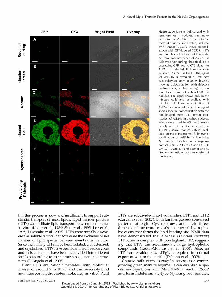
Figure 1. Lipid binding activity assay of AsE246. A, Association of P-96 with DAsE246. Purified DAsE246 (final concentration,0.5 mM) was incubated in 10 mM MOPS, pH 7.2. Increasing amounts of P-96 (from a 30 mM solution in ethanol) were added, andthe fluorescence intensity was measured at 378 nm (excitation at 343 nm). DAsE246, incubated with increasing concentrationsof P-96, exhibited saturation binding defined by Kd = 0.1083 6 0.03 mM (circles). No saturation binding was observed (squares)when P-96 was incubated with GST in the control experiment. B, Binding competition between P-96 and various unlabeledfatty acids. Purified DAsE246 (final concentration 0.5 mM) was incubated in 10 mM MOPS, pH 7.2, with 1 mM P-96 (from a 30 mM
solution in ethanol). Various unlabeled fatty acids were added from concentrated solutions in ethanol (final concentration,10 mM). Fluorescence intensity was measured at 378 nm (excitation at 343 nm) after its stabilization. C, Binding abilitycompetition between P-96 and natural lipid, PC, PE, PI, DGDG, and its precursor MGDG. Purified DAsE246 (final concen-tration, 0.5 mM) was incubated in 10 mM MOPS, pH 7.2, with 1 mM P-96 (from a 30 mM solution in ethanol). Increasing amountsof unlabeled lipids were added from concentrated solutions. D, Co-IP of AsE246 with DGDG. The wild-type Chinese milk vetchnodules inoculated with M. huakuii extracts were incubated first with anti-DGDG antibody and then with immobilized ProteinG beads. Proteins bound to the beads were separated on SDS-PAGE and immunoblotted with anti-AsE246 antibody.
1046 Plant Physiol. Vol. 164, 2014
Lei et al.
www.plantphysiol.orgon June 24, 2018 - Published by Downloaded from Copyright © 2014 American Society of Plant Biologists. All rights reserved.
but this process is slow and insufficient to support sub-stantial transport of most lipids. Lipid transfer proteins(LTPs) can facilitate lipid transport between membranesin vitro (Kader et al., 1984; Shin et al., 1995; Lee et al.,1998; Lascombe et al., 2008). LTPs were initially discov-ered as soluble factors that accelerate the exchange or nettransfer of lipid species between membranes in vitro.Since then, many LTPs have been isolated, characterized,and crystallized. LTPs have been identified in eukaryotesand in bacteria and have been subdivided into differentfamilies according to their protein sequences and struc-tures (D’Angelo et al., 2008).Plant LTPs are cationic peptides, with molecular
masses of around 7 to 10 kD and can reversibly bindand transport hydrophobic molecules in vitro. Plant
LTPs are subdivided into two families, LTP1 and LTP2(Carvalho et al., 2007). Both families possess conservedpatterns of eight Cys residues, and their three-dimensional structure reveals an internal hydropho-bic cavity that forms the lipid binding site. NMR datahave demonstrated that a wheat (Triticum aestivum)LTP forms a complex with prostaglandin B2, suggest-ing that LTPs can accommodate large hydrophobiccompounds (Tassin-Moindrot et al., 2000). Also, anLTP from Arabidopsis, LTPg1, is required for normalexport of wax to the cuticle (Debono et al., 2009).
Chinese milk vetch (Astragalus sinicus) is a winter-growing green manure legume. It can establish a spe-cific endosymbiosis with Mesorhizobium huakuii 7653Rand form indeterminate-type N2-fixing root nodules,
Figure 2. AsE246 is colocalized withsymbiosomes in nodules. Immunolo-calization of AsE246 in the infectedroots of Chinese milk vetch, inducedby M. huakuii 7653R, shows colocali-zation with GFP-labeled 7653R in ITsand nodules but not in root hair curls.A, Immunofluorescence of AsE246 inwild-type hair curling; the rhizobia areexpressing GFP, but no CY3 signal forAsE246 is detected. B, Immunolocali-zation of AsE246 in the IT. The signalfor AsE246 is revealed as red dots(secondary antibody tagged with CY3),showing colocalization with rhizobia(yellow color; in the overlay). C, Im-munolocalization of anti-AsE246 onnodules. The signal shows only in theinfected cells and colocalizes withrhizobia. D, Immunolocalization ofAsE246 in infected cells. The signalshows specific colocalization with thenodule symbiosomes. E, Immunoloca-lization of AsE246 in crushed nodules,which were fixed in 4% (w/v) freshlydepolymerized paraformaldehyde in13 PBS, shows that AsE246 is local-ized on the symbiosome. F, Immuno-localization of AsE246 in free-livingM. huakuii rhizobia as a negativecontrol. Bars = 20 mm (A and B), 200mm (C), 10 mm (D), and 5 mm (E and F).[See online article for color version ofthis figure.]
Plant Physiol. Vol. 164, 2014 1047
A Novel Lipid Transfer Protein in the Nodule Organogenesis
www.plantphysiol.orgon June 24, 2018 - Published by Downloaded from Copyright © 2014 American Society of Plant Biologists. All rights reserved.

which are cylindrical and consist of a gradient of de-velopmental zones with a persistent apical meristem(zone I), an infection zone (zone II), and a fixation zone(zone III). In mature nodules, a senescence zone (zoneIV) is established proximal to zone III. As the noduleages, this zone gradually moves in a proximal-distaldirection until it reaches the apical part and the noduledegenerates (Monahan-Giovanelli et al., 2006; Van deVelde et al., 2006). Chinese milk vetch is mainly dis-tributed in China, Japan, and Korea, where it is widelyplanted in rice fields to increase soil fertility (Li et al.,2008).
To investigate whether LTPs function in symbiosismembrane deposition and nodule organogenesis inChinese milk vetch, we previously identified two can-didate LTP genes, AsE246 and AsIB259, via suppressivesubtractive hybridization. AsE246 is specifically ex-pressed in nodules, but AsIB259 is expressed in bothuninfected roots and nodules, indicating that onlyAsE246 is specific to nodulation (Chou et al., 2006). Here,we report the functional characterization of AsE246, anodule-specific lipid transfer protein that localizes onthe symbiosome membrane and plays an essential rolein symbiosome membrane biogenesis and effectivesymbiosis.
RESULTS
Phylogenetic Characterization of AsE246
AsE246 has a 399-nucleotide open reading framethat encodes a putative protein of 132 amino acids,containing a 23-amino acid-long N-terminal signal
peptide for the secretory pathway. According to a clas-sification scheme based on primary structure (Boutrotet al., 2008), comparison of the deduced sequence ofmature AsE246 to LTPs from Arabidopsis, M. truncatula,Lotus japonicus, and Glycine max showed that AsE246belongs to the Type I LTPs. A further search of the Pfamdatabase (Finn et al., 2010) showed that the Type I LTPsbelong to the LTP1 family, also called the “Tryp a amyl”family (Supplemental Fig. S1; Supplemental Data Set S1).By contrast, MtN5 and AsIB259 belong to Type IV andType V LTPs, respectively, and both are members of theLTP2 family (Supplemental Fig. S1).
AsE246 Has Lipid Binding Activity in Vitro
Protein sequences that contain the conserved eightCys motif may be annotated as LTPs (Chou et al., 2006),but this is not a sufficient predictor of lipid-bindingactivity, as the conserved eight-Cys motif and a-helixstructure occur in LTPs and in several plant proteaseand amylase inhibitors, as well as in proteins of un-known function (José-Estanyol et al., 2004).
To confirm the lipid-binding capacity of AsE246,we used an in vitro lipid binding assay with purifiedAsE246 and a fluorescent lipid substrate. To produce aprocessed, soluble version of AsE246, which we termedDAsE246, the complementary DNA lacking the N-terminalsignal sequence was expressed in Escherichia coli BL21and purified using a glutathione S-transferase (GST)tag. To assess potential lipid-binding activity, DAsE246was incubated with the fluorescent lipophilic probe1-pyrenedodecanoic acid (P-96), which fluoresces weakly
Figure 3. Ultrastructural localization of AsE246and DGDG on the symbiosome membrane. Aand B, Immunogold detection of anti-AsE246(immunogold signal appears as black dots; indi-cated by black arrowheads) in wild-type nodules.The 10-nm gold particles are present over thesymbiosome membranes. C and D, Immunogolddetection of anti-DGDG (immunogold signalappears as black dots; indicated by black arrow-heads) in wild-type nodules. The 10-nm goldparticles are present over the symbiosome mem-branes. Bars = 200 nm (A, B, andD) and 500 nm (C).
1048 Plant Physiol. Vol. 164, 2014
Lei et al.
www.plantphysiol.orgon June 24, 2018 - Published by Downloaded from Copyright © 2014 American Society of Plant Biologists. All rights reserved.

in aqueous solution but fluoresces intensely when boundin a hydrophobic environment. We found that, in bindingassays with increasing amounts of P-96, fluorescenceintensity increased initially and plateaued at greater than0.5 mmol L–1 P-96 (Fig. 1A). A control experiment withan empty GST tag gave P-96 fluorescence at close tobackground levels. This result supports the hypothesisthat AsE246 binds lipids.We next used a competition test to determine
whether common fatty acids bind to DAsE246. Addi-tion of a fatty acid that competes with P-96 forDAsE246 binding will result in lower fluorescence ofP-96 compared with the control with P-96 alone. Sev-eral lipids (each at 10 mM) were used as competitors:saturated fatty acids with C12 to C22 chain length reducedthe signal from 1 mM P-96 (Fig. 1B). These fatty acidsdisplayed different efficiencies in competing with P-96,depending on their chain length (Fig. 1B). Molecules witha 16- to 18-carbon chain length showed higher compe-tition efficiency, but shorter (lauric and myristic) and
longer (arachidic and behenic) fatty acids showed lowercompetition efficiency.
AsE246 Binds to Biological Membrane Lipid in Vitroand in Vivo
We next used the competition assay to test the ca-pacity of AsE246 to bind to biological membrane lipids,phosphatidylcholine (PC), phosphatidylethanolamine(PE), phosphatidylinositol (PI), DGDG, and its precur-sor monogalactosyldiacylglycerol (MGDG; Fig. 1C).Increasing amounts of natural lipid (each to a maxi-mum of 2 mM) were added to a solution of 1 mM P-96plus 1 mM DAsE246. Stearic acid at the same concen-tration was used as a positive control. We found that thefluorescence decreased with the addition of natural lipid(Fig. 1C), showing that they can compete with the fluo-rescent lipophilic probe P-96 and indicating that DAsE246can bind to PC, PE, PI, DGDG, and MGDG in vitro.
Figure 4. DGDG is localized on theplasma membrane and colocalizedwith the symbiosome in infected cells.Immunolocalization of DGDG overthe infected roots of Chinese milk vetchinduced by M. huakuii 7653R showscolocalization with GFP-labeled 7653Rin ITs and nodules but not with free-living M. huakuii. A, Immunofluores-cence of DGDG on the IT; the rhizobiaare expressing GFP, and the signal forDGDG is revealed as red dots (sec-ondary antibody tagged with CY3). CY3signal is detected both on root hairplasma membrane and colocalized withrhizobia (yellow color; in the overlay).B and C, Immunolocalization of DGDGon nodules. The signal shows in bothinfected and uninfected cells andcolocalizes with rhizobia in the infectedcells. D, Immunolocalization of DGDGin crushed nodules, which were fixed in4% (w/v) of freshly depolymerized par-aformaldehyde in 13 PBS. The signalshows that DGDG is localized on thesymbiosome. E, Immunolocalizationof DGDG on free-living M. huakuiirhizobia as a negative control showsthat the free-living rhizobia do not con-tain DGDG. Bars = 20 mm (A and C),50 mm (B), and 5 mm (D and E). [Seeonline article for color version of thisfigure.]
Plant Physiol. Vol. 164, 2014 1049
A Novel Lipid Transfer Protein in the Nodule Organogenesis
www.plantphysiol.orgon June 24, 2018 - Published by Downloaded from Copyright © 2014 American Society of Plant Biologists. All rights reserved.

DGDG was reported to accumulate in the symbio-some membranes of G. max and L. japonicus (Gaudeet al., 2004). More importantly, unlike other biologicalmembrane lipids (PC, PE, or PI), DGDG was not pre-sent in the free-living rhizobia cells (Gaude et al.,2004). To achieve direct evidence to reveal the func-tional association and interaction between LTP andplant-synthesized lipids, we chose DGDG to perform
coimmunoprecipitation (Co-IP) assay and subcellularlocalization and such therefore to further confirm theinteraction between DGDG and AsE246. To conductthe designed experiment, anti-DGDG antibody wasprepared as described in Botté et al. (2008). A noduleextract of wild-type Chinese milk vetch nodules wasprecipitated with immobilized anti-DGDG antibody orwith anti-AsE246 antibody as a positive control. Thecaptured immune complexes were resolved by SDS-PAGEand detected by immunoblotting with anti-AsE246antibody. It was found that AsE246 was detected inthe Co-IP products but not in the negative controlwithout antibody (Fig. 1D). These results show that thenodule-specific lipid transfer protein AsE246 binds thebiological membrane lipid DGDG in vivo.
AsE246 Localizes to the Infected Nodule Cellsand Symbiosome Membrane
To determine the subcellular location of AsE246under symbiotic conditions, an immunofluorescenceanalysis was carried out with wild-type Chinese milkvetch inoculated with GFP-labeled M. huakuii 7653R.The anti-AsE246 antibody is specific for AsE246(Supplemental Fig. S2). AsE246 was expressed in ITsand infected cells in the nodule but was not detected inroot hair curling (Fig. 2, A–D). AsE246 colocalizedwith symbiosomes (Fig. 2E) but was not detectedin free-living rhizobia (Fig. 2F). The secondary anti-body controls did not show any labeling in nodules(Supplemental Fig. S3A). Considering that AsE246could bind to DGDG, a major constituent of the sym-biosome membrane (Gaude et al., 2004), it is possiblethat AsE246 is present on the symbiosome membrane.Immunoelectron microscopy detected AsE246 on thesymbiosome membrane (Fig. 3, A and B). The secondaryantibody controls for immunoelectron microscopy alsodid not show any labeling (Supplemental Fig. S3B). Theseresults are consistent with our findings that AsE246 isexpressed in nodule cells containing symbiosomes.
The Plant-Synthesized Glycolipid DGDG Occurs on theChinese Milk Vetch Nodule Symbiosome Membrane
DGDG accumulates in the symbiosome membranesof G. max and L. japonicus (Gaude et al., 2004), bothof which form determinate-type root nodules, whileChinese milk vetch forms indeterminate-type rootnodules. Therefore, we analyzed the DGDG content ofthe Chinese milk vetch symbiosome by using liquidchromatography-mass spectrometry (LC-MS) and con-firmed its presence occurring on the symbiosome mem-brane through immunolocalization methods.
The symbiosomes were isolated from Chinese milkvetch nodules, and LC-MS confirmed that the totallipids extracted from the symbiosomes and nodulescontained DGDG (Supplemental Figs. S4 and S5). TheLC-MS data also showed the DGDG species in the
Figure 5. Overexpression of AsE246 results in increased numbers ofnodules. A, The empty vector control hair root, inoculated withM. huakuii 7653R and grown in the absence of nitrogen. The photo-graphs were taken 4 weeks after inoculation. B, AsE246-overexpressinghairy root inoculated with M. huakuii 7653R, showing the increase innodule numbers. C, Close-up of boxed area in A. The nodules areindicated by white arrows. D, Close-up of boxed area in B. E, Numbersof nodules per root. The nodule number of AsE246-overexpressingroots (n = 17) is more than that of the empty vector control. F, Thenitrogenase activity of AsE246 overexpressing and control nodulesshowed no remarkable difference. Different letters above bars indicatesignificant differences (P , 0.05, Student’s t test) between pairwisecomparisons. Bars = 1 cm. [See online article for color version of thisfigure.]
1050 Plant Physiol. Vol. 164, 2014
Lei et al.
www.plantphysiol.orgon June 24, 2018 - Published by Downloaded from Copyright © 2014 American Society of Plant Biologists. All rights reserved.

symbiosome were different from those in nodules andleaves. The result showed that in Chinese milk vetchnodules, the symbiosome contains the plant-synthesizedglycolipid DGDG.The immunofluorescence was carried out in the
wild-type Chinese milk vetch inoculated with GFP-labeled M. huakuii 7653R and showed a weak anti-DGDG signal on the plasma membrane of the hostplant cell (Fig. 4A) and in ITs and nodules. The signalwas present in both infected and uninfected cells andcolocalized with rhizobia in the infected cells (Fig. 4,B and C). The anti-DGDG signal was also detected onthe symbiosomes. To examine the symbiosomes, crushednodules were fixed with 1% (w/v) freshly depolymer-ized paraformaldehyde; immunolocalization showedthat the anti-DGDG signal colocalized with the sym-biosomes (Fig. 4D) but was not detected in the free-living M. huakuii control (Fig. 5E). To determinewhether DGDG localized on the symbiosome mem-brane, we examined the ultrastructural localization ofDGDG by electron microscopy (EM). EM immuno-gold labeling with an anti-DGDG antibody was usedto localize DGDG in wild-type nodules (Fig. 3, C andD), and the result showed that plant-synthesized gly-colipid DGDG occurs on the Chinese milk vetch nodulesymbiosome membrane.
Overexpression of AsE246 Results in Increased Numbersof Root Nodules
To investigate the role of AsE246 in symbiosis,we overexpressed AsE246 under the control of the 35Spromoter. Expression levels of AsE246 in transgenicroots and nodules were examined by real-time PCR(Supplemental Fig. S6A). The level of AsE246 transcriptsin transgenic nodules was about 2 to 3 times that incontrol nodules. AsE246 transcripts were also expressedin transgenic roots but were not detected in wild-typeand control roots. The AsE246-overexpressing trans-genic roots formed more root nodules (an average of47.8, n = 17) than the empty vector control (an averageof 21.1, n = 13; Fig. 5, A–E). Nodules in AsE246-over-expressing transgenic roots and empty vector controlsshowed similar nitrogenase activities (Fig. 5F).
Knockdown of AsE246 Impairs Nodulationand Symbiosome Development
To further investigate the role of AsE246 in symbio-some development, we knocked down the expression ofAsE246 using RNA interference (RNAi). Nodule RNAsequencing studies showed that the RNAi fragmentused in this construct was unique (data not shown),
Figure 6. Nodulation phenotype ofAsE246-RNAi hair root. A, The emptyvector control hair root, inoculatedwith M. huakuii 7653R and grown inthe absence of nitrogen. The photo-graphs were taken 4 weeks after inoc-ulation. B, AsE246 RNAi hair rootinoculated with M. huakuii 7653Rformed fewer root nodules comparedwith control, and the nodules are alsowhite colored. C, Close-up of boxedarea in A. The nodules are indicated bywhite arrows. D, Close-up of boxedarea in B. The nodules are indicated bywhite arrows. E, Numbers of nodulesper root. There are fewer nodules inRNAi root (n = 47) compared with theempty control (n = 35). F, The nitroge-nase activity of the AsE246-RNAi nod-ule is lower than that of the controlnodule. Different letters above bars in-dicate significant differences (P , 0.05,Student’s t test) between pairwisecomparisons. G, Lipid PC, PE, PI, andDGDG contents of transgenic nodule.Different letters above bars indicatesignificant differences (P , 0.05, Stu-dent’s t test) between pairwise com-parisons. Bar = 1 cm. [See onlinearticle for color version of this figure.]
Plant Physiol. Vol. 164, 2014 1051
A Novel Lipid Transfer Protein in the Nodule Organogenesis
www.plantphysiol.orgon June 24, 2018 - Published by Downloaded from Copyright © 2014 American Society of Plant Biologists. All rights reserved.

indicating that the RNAi construct would specificallysilence AsE246. Real-time PCR showed that AsE246transcript levels in the transgenic nodules were 16.2%of levels in the control nodules (Supplemental Fig.S6B). The average number of nodules formed on con-trol hairy roots was 22.1 (n = 35) but that on the RNAiplants was 4.2 (n = 47; Fig. 6, A–E). In addition, 14.9%of RNAi roots failed to develop any nodules. Com-pared with the empty vector control, the RNAi nod-ules were smaller and whiter (Fig. 6D), with muchlower nitrogenase activity (Fig. 6F).
We also examined the structure of the nodules formedin AsE246 RNAi plants. Paraffin sections showed thatRNAi nodules at 21 d after infection had fewer maturedinfected cells, as infected cell development in thesenodules remained in zone II (infection zone; Fig. 7, Band D), and the mature fixation zone III was not ob-served. Furthermore, transmission electron microscopy(TEM) showed obvious shrinkage and peribacteroidspace enlargement in the symbiosomes of RNAi nod-ules (Fig. 8, C and D, white arrows). There were many
more unstained granules of poly-b-hydroxybutyrate(PHB) in the symbiosomes of RNAi nodules (Fig. 8,C and D, black arrows). By contrast, the symbiosomesof control nodules developed normal cellular mor-phology with a smoothly rounded boundary and morehomogeneous cytoplasm (Fig. 8, A and B). TEM alsoshowed that, for both the nodules formed in the emptyvector and RNAi plants, there were coated vesicles overthe symbiosomes.
Knockdown of AsE246 Affects Lipids Abundance inthe Nodule
The lipid abundance in transgenic nodules wasmeasured using LC-MS. The results showed that PC,PE, PI, and DGDG contents were significantly lower inAsE246 RNAi nodules than in controls, manifested bytheir corresponding contents in the RNAi nodules,which were 60.62%, 61.00%, 77.28%, and 46.74% of that inthe control nodules, respectively (Fig. 6G; SupplementalData Set S2). These data support the idea that AsE246 isinvolved in lipids transport to the nodule symbiosomemembrane.
AsE246 Is Required for IT Formation
Rhizobial infection of transgenic roots was alsomonitored and quantified (Fig. 9) using GFP-labeledM. huakuii 7653R to visualize formation of ITs at9 d after infection. In AsE246 RNAi roots, ITs initiatedfrom curled root hair tips (Fig. 9F), grew through well-elongated root hairs, and formed ITs (Fig. 9B). Thesefurther penetrated into the root cortex and formednodule primordia (Fig. 9C) and then developed intonodules (Fig. 9D). However, there were significantlyfewer ITs and nodule primordia in RNAi roots com-pared with control roots; also in the overexpressionroots, there were more ITs and nodule primordia (Fig. 9E).These data indicated that AsE246 participates in ITformation but not in root hair curling.
DISCUSSION
AsE246 is specifically expressed in Chinese milkvetch nodules (Chou et al., 2006) and encodes a proteinwith a conserved eight-Cys motif characteristic ofLTPs. Further phylogenetic analysis showed AsE246belongs to the LTP1 family, also called the “Tryp a amyl”family, and is not homologous to another symbiosis-associated LTP, MtN5, a member of the LTP2 family(Chou et al., 2006; Pii et al., 2009). Various in vivo bio-logical roles have been proposed for plant LTPs, includingdefense against pathogens and modulation of plantdevelopment.
In our search for the lipid transport machinery re-quired for symbiosome development in legume plantnodules, we examined the involvement of LTPs. LTPsare abundantly expressed in epidermal cells, secreted
Figure 7. AsE246-RNAi impairs nodule development. A, Longitudinalsection of a control (empty vector) 21-d-old nodule. B, Longitudinalsection of a 21-d-old RNAi nodule, showing the infected cells remainin the meristem and infection zone. C, Magnification of nitrogen fix-ation zone in A, showing developed nitrogen-fixing symbiosomes.D, Magnification in B. M, Meristem; Z2, infection zone; Z3, nitrogenfixation zone. Bars = 500 mm (A), 200 mm (B), and 100 mm in (C andD). [See online article for color version of this figure.]
1052 Plant Physiol. Vol. 164, 2014
Lei et al.
www.plantphysiol.orgon June 24, 2018 - Published by Downloaded from Copyright © 2014 American Society of Plant Biologists. All rights reserved.

to the extracellular matrix (Carvalho et al., 2007), andcapable of binding and carrying lipids in vitro (Kaderet al., 1984). These characteristics provide the rationalefor the proposal that LTPs are potential carriers of lipidconstituents to the nodule symbiosomes. In this study,we provide evidence that one member of the LTPfamily in Chinese milk vetch, AsE246, participates insymbiosome membrane lipid transport and is requiredfor successful legume-rhizobium symbiosis, includinginfection and bacteroid maintenance.The expression of AsE246 without a signal peptide
in an E. coli system allowed us to purify AsE246 andassess its lipid binding capacity. As reported for sev-eral other LTPs (Buhot et al., 2004; Debono et al., 2009;Pii et al., 2009), AsE246 can bind to the fluorescentlipid probe P-96. P-96 binding to DAsE246 could bedisplaced by saturated fatty acids and biological gly-colipids, including PC, PE, PI, DGDG, and its precur-sor MGDG, suggesting that lipids could compete withP-96 for the same hydrophobic binding cavity. The Co-IPexperiment revealed that AsE246 could bind withDGDG to form a complex in vivo.The immunofluorescence and immunoelectron mi-
croscopy images showed that AsE246 was expressedin the IT and nodule-infected cells and localized onthe symbiosome membrane. This is quite similar tothe symbiosome membrane-specific protein SNARESYP132, which is present on the symbiosome mem-brane from the start of symbiosome formation and
throughout its development (Catalano et al., 2007;Limpens et al., 2009). These results indicate thatAsE246 might be involved in the targeting of plant-synthesized lipid during IT and symbiosome devel-opment, which require reorganization and de novoformation of membranes. Therefore, AsE246 can affectlipid homeostasis and the diverse cellular proces-ses associated with it, such as signal transduction,membrane trafficking, and lipid metabolism, by cap-turing and responding to symbiosome membranemodifications.
In contrast with the empty vector control, theAsE246 RNAi roots formed fewer and smaller nodules,and there were fewer infected cells in the RNAi nod-ules. TEM showed obvious symbiosome shrinkageand peribacteroid space enlargement in the symbio-somes of the RNAi nodule. This suggests that thereduction of AsE246 led to changes in membranepermeability and caused symbiosomes to be moresensitive to the osmotic pressure in nodules; this ab-normal development of the symbiosome membraneled to a significant decrease in nitrogen fixation abilityof RNAi nodules. TEM also showed some coatedvesicles over the symbiosome membrane, both in thecontrol and RNAi nodules. This indicates that vesicu-lar transport to the symbiosome was not affected bythe reduction of AsE246.
The accumulation of PHB in AsE246-RNAi nodules isalso of interest for analysis. As an indeterminate-type
Figure 8. AsE246-RNAi impairs symbiosome de-velopment. A and B, TEM images of symbiosomesof the control nodule. CV, Coated vesicle. C andD, TEM images of symbiosomes of the AsE246-RNAi nodule. There is obvious shrinkage andperibacteroid space enlargement in the symbio-somes of the RNAi nodule (white arrowheads)and PHB accumulation (black arrowheads).Bars = 500 nm.
Plant Physiol. Vol. 164, 2014 1053
A Novel Lipid Transfer Protein in the Nodule Organogenesis
www.plantphysiol.orgon June 24, 2018 - Published by Downloaded from Copyright © 2014 American Society of Plant Biologists. All rights reserved.

nodule formation legume plant, Chinese milk vetch(similar to alfalfa [Medicago sativa] interacting with Sino-rhizobium meliloti [Hirsch et al., 1983]) does not usuallyaccumulate PHB during symbiosis with M. huakuii7653R. PHB synthesis does occur but is presumably ac-companied by an equivalent rate of degradation duringnormal bacteroid differentiation, which leads to only avery small accumulation of PHB (Kim and Copeland,1996). This amount is insufficient to allow for the for-mation of granules that are visible by EM. A study ofRhizobium leguminosarum bv viciaemutations in two genesencoding broad-specificity amino acid transporters, mu-tations that block the amino acid cycling pathway be-tween the plant and the symbiosomes, showed that these
mutants possessed PHB granules in mature symbio-somes (Lodwig et al., 2003; Trainer and Charles, 2006).We speculate that the knockdown of AsE246 led tochanges in symbiosome membrane structure and func-tion and also may have blocked the amino acid cyclingpathway between the plant and symbiosomes.
The knockdown of AsE246 resulted in reduced PC, PE,PI, and DGDG contents of transgenic nodules. This resultsupported the idea that AsE246 participates in thetransport of plant-synthesized lipid to the symbiosomemembrane. In this case, the lipid contents in the sym-biosomes of transgenic nodules was not examined be-cause of technical difficulties caused by the low numberof nodules formed in the RNAi plants.
Figure 9. Reduction of AsE246 affects rhizobiainfection. A to D, Infection event observations ofGFP-labeled M. huakuii 7653R strain to track theinfection events at 9 d after infection. Represen-tative root hair curling observed in (empty vector)control roots: root hair curling (A), IT (B), noduleprimordia (C), and nodule (D). E, Frequencies ofinfection events per root. The data are presentedas 16 individual transgenic plants for each con-struct. Different letters above bars indicate sig-nificant differences (P , 0.05, Student’s t test)between pairwise comparisons. Bars = 20 mm.[See online article for color version of this figure.]
1054 Plant Physiol. Vol. 164, 2014
Lei et al.
www.plantphysiol.orgon June 24, 2018 - Published by Downloaded from Copyright © 2014 American Society of Plant Biologists. All rights reserved.

AsE246 also plays an important role in rhizobia in-fection. The knockdown of AsE246 resulted in fewerITs, nodule primordia, and nodules; overexpression ofAsE246 resulted in more ITs and nodule primordia.Moreover, immunofluorescence showed that AsE246colocalized with rhizobia in the IT. Considering thatthe symbiosome membrane is derived from the IT(Whitehead and Day, 1997), we conclude that AsE246is also required for IT formation.M. truncatula MtN5 encodes an LTP and is required
for the symbiotic association between M. truncatulaand the nitrogen-fixing rhizobial bacteria S. meliloti (Piiet al., 2009). However, the LTP mechanisms that un-derlie the symbiotic interaction have not been fullyunraveled. Our results showing that a nodule-onlyLTP transports the plant-synthesized lipid includingDGDG and targets to the symbiosome membranes,even in the ITs, raise deep questions concerning theDGDG biological functions associated with progres-sion of symbiosis. Accumulation of DGDG, instead ofphospholipids, in the symbiosome membrane mighthave two major functions. First, it might be a hostplant protection strategy to facilitate rhizobia entry,differentiation, and functional maintenance inside thetarget host plant cells by avoiding host defense re-sponses, which otherwise may interfere with normalmetabolism of the host cell and provoke host defenseresponses. For example, there is a significant linearrelationship between the amounts of MGDG andDGDG in noninoculated wheat leaves and the level ofresistance to tan spot, which is caused by the fungusPyrenophora triticirepentis (Kim et al., 2013). These find-ings suggest that the amount of DGDG in plant cellsaffects the resistance response to pathogens. The infec-tion of rhizobia and their subsequent existence withinthe host plant cell are quite similar to phytopathogeninfection and even to infection with some intracellularmammalian pathogens (i.e. Mycobacterium tuberculosisand Salmonella spp.; Schumpp and Deakin, 2010). Thus,DGDG in the symbiosome membrane (Figs. 5–7; Gaudeet al., 2004) might be an alternative defense response toinvading rhizobia and facilitate the intracellular sym-biosis between the plant and the rhizobia.Second, the accumulation of the nonphosphorus
galactolipid DGDG, instead of phospholipids, in thesymbiosome membrane might conserve phosphate foressential cellular processes during nodulation. Plantsand algae synthesize DGDG in plastid envelope mem-branes (Maréchal et al., 1997, 2000; Awai et al., 2001;Jouhet et al., 2004, 2007; Benning and Ohta, 2005). Uponphosphate deprivation, DGDG is exported to the plasmamembrane (Andersson et al., 2003) and the mitochondria(Jouhet et al., 2004). During nodule development, thenumber of rhizobia in infected cells increases dramati-cally. This cell division requires membrane biosynthesisby the bacteria and also by the plant for synthesis ofthe symbiosome membrane to enclose the bacteria insymbiosomes (Verma, 1992). As a consequence, nod-ulation requires the uptake of large amounts of phos-phate for the synthesis of phospholipids, nucleic acids,
phosphorylated proteins, and sugar phosphates. It ispossible that cells infected with bacteroids might sufferfrom local phosphate limitation. Previous studies havedemonstrated that phosphate availability is crucial fornormal nodulation (Israel, 1987; Leidi and Rodriguez-Navarro, 2000). Thus, the presence of the nonphosphorusgalactolipid DGDG might conserve phosphate for hostplant metabolism (Gaude et al., 2004).
In conclusion, the novel LTP AsE246 may participatein the transport of the plant-synthesized lipid to thesymbiosome membrane and is involved in legume-rhizobia symbiosis. These results increase our under-standing of the molecular and physiological mechanismsof legume nodule organogenesis and symbiosomedevelopment.
MATERIALS AND METHODS
Phylogenetic Analysis
The Medicago truncatula genome database (assembly release 3.5; Younget al., 2011) was searched for nsLTP gene sequences using the gene annota-tions. The entire Glycine max and Lotus japonicus (release 2.5) proteomewas searched for proteins having the Hidden Markov Model Pfam domainPF00234 (Plant lipid transfer/seed storage/trypsin-a amylase inhibitor).
Protein sequences of all nsLTPs were analyzed for the presence of potentialsignal peptide cleavage sites using the SignalP 4.0 program (Finn et al., 2010).Amino acid sequences were efficiently aligned to the Pfam profile HiddenMarkov model defined from the protease inhibitor/seed storage/LTP family(Finn et al., 2010) using HMMalign from the HMMER package (Eddy, 1998).
Deduced M. truncatula, L. japonicus, G. max, and Arabidopsis (Arabidopsisthaliana) amino acid sequences were aligned to the Pfam glocal model usingHMMalign from the HMMER package (Eddy, 1998). Phylogenetic trees werebuilt from the protein alignment with the maximum-likelihood method usingthe PHYML program (Guindon and Gascuel, 2003). Maximum-likelihood in-ference analyses were conducted under the Le and Gascuel model (Le andGascuel, 2008) with estimation of the proportion of invariant sites and estimationof variation rate among the remaining sites according to a g distribution. Theconfidence level of each node was estimated by bootstrap procedure using 1,000resampling repetitions of the data. The unrooted phylogenetic trees were visu-alized using Mega 5 (Tamura et al., 2011).
Plant Materials
Seeds of Chinese milk vetch (Astralagus sinicus; collected from Xinyang, HenanProvince, China) were surface sterilized in 70% (v/v) alcohol for 3 min, then in 2%(w/v) sodium hypochlorite for 20 min, followed by five washes in distilled water.After immersion in distilled water for several hours, the seeds were placed onmedium with 0.5% Suc and 1.2% (w/v) agar (Cho and Widholm, 2002).
Isolation of Symbiosomes from Chinese Milk Vetch
Approximately 10 g of fresh nodules fromChinesemilk vetch plantswas usedfor the isolation of symbiosomes via three-step Percoll density gradient centrif-ugation (Price et al., 1987; Panter et al., 2000). Intact symbiosomes were har-vested from the 60%/80% Percoll interface. After washing with washing buffer(350 mM mannitol, 25 mM MES-KOH, 3 mM MgSO4, pH 7.0), the suspension wascentrifuged at 4,000g for 5 min at 4°C. The pellet containing symbiosomes wasresuspended in washing buffer and vortexed vigorously for 5 min.
Lipid Extraction and Quantification
Lipids were isolated from frozen plant material, rhizobia (isolated fromliquid culture), or symbiosomes (Bligh and Dyer, 1959). To determine the lipidcomposition changes of transgenic nodule, the 35-d-after-infection transgenicnodules were harvested (100 mg) for lipid extraction and profiling. Sampleswere homogenized using liquid nitrogen and resuspended in 50 mL chloroform
Plant Physiol. Vol. 164, 2014 1055
A Novel Lipid Transfer Protein in the Nodule Organogenesis
www.plantphysiol.orgon June 24, 2018 - Published by Downloaded from Copyright © 2014 American Society of Plant Biologists. All rights reserved.

and 100 mL methanol, which was then sonicated two to three times. Then, tothe mixture is added 50 mL chloroform and 50 mL distilled, deionized water,then transferred to a separating funnel, and after a few minutes for completeseparation and clarification. The lower chloroform phase was dried usingrotary evaporation, and the lipid extract was dissolved in methanol. The lipidextracts were introduced into the Quadrupole Time-of-Flight (Q-TOF) massspectrometer (Agilent 6540 Accurate Mass Q-TOF LC-MS unit) operated in thepositive mode. Lipids were supplied to the mass spectrometer in methanol.Q-TOF LC-MS was performed using the Agilent 6540 UHD accurate-massQ-TOF LC-MS system with a ZORBAX Eclipse Plus C18 column (2.1 3100 mm, 3.5 mm). The 1-mL sample was loaded with a flow rate of 0.3 mL min–1
A (water plus 5%methanol plus 10 mM NH4AcOA), B (methanol); solution B wasat 25% (v/v) at the start, 100% (v/v) at 5 min, 100% (v/v) at 10 min, and 25%(v/v) again at 15 min. The acquisition range (m/z) was 400 to 1,100; nebulizerpressure, 40 pounds per square inch gauge; drying gas, N2 350°C 9 L min–1;electrospray ionization voltage of capillary, 4,000 V; fragment or 214v; skimmer,65 V; and octopole radio frequency programming mode voltage, 750 V. MGDGand DGDGwere analyzed as [M+NH4]
+ and [M+Na]+ ions (Wu and Xue, 2010).
Nitrogenase Activity Measurement
Nitrogenase activitywasmeasured by acetylene reduction activity (Hardy et al.,1973). For each sample at each time point, nine hairy root lines were analyzed.Hypogeal parts of hairy root plants (including nodules and roots) were incubatedin 15-mL glass bottles with rubber seals containing 2 mL of acetylene (C2H2) for 1 hat 28°C. The C2H2 was measured in a Hitachi 163 gas chromatograph.
Protein Expression and Purification
AsE246 coding sequence (accession no. DQ199648) was amplified fromcomplementary DNA obtained by reverse transcribing mRNA extracted fromChinese milk vetch nodules. The upstream primer was 59-GGATCCAAA-CAATTAGTTGCCGT-39 and the downstream primer 59-CTCGAGTTAGA-CCAACAAAGTTTG-39. The PCR product corresponding to the codingsequence of mature AsE246 was double digested with XhoI and BamHI, clonedinto pGEX-6p-1 (GE), and checked by sequencing. The recombinant vectorpGEX-246 was mobilized into the host strain Escherichia coli BL21 DE3. AsE246protein was purified from E. coli lysate using a GST cartridge (Bio-Rad).
Lipid Binding Assay in Vitro
Lipid binding capacity of AsE246 was assayed by monitoring the displace-ment of the fluorescent probe P-96. Fluorescence experiments were performed at25°C in a Shimadzu RF 5301 PC spectrofluorimeter, as previously described(Mikes et al., 1998; Osman et al., 2001), with minor modifications. The excitationand emission wavelengths were set at 343 and 378 nm, respectively.
P-96, with or without fatty acids, was incubated for 1 min in a stirredcuvette containing 2 mL of measurement buffer (10 mM MOPS, pH 7.2) beforethe fluorescence was recorded (F0). Then, AsE246 was added, and after 2 min,fluorescence was recorded at equilibrium (F). Results are expressed either aspercentage of AsE246-P-96 complex fluorescence according to (F–Fc)/F, whereFc is the fluorescence of the AsE246-P-96 complex in absence of fatty acid, oras fluorescence (F, F0, F), in arbitrary units.
Antibody Preparation
AsE246 protein was purified from inclusion bodies using strong denaturingconditions (20 mM Tris HCl, pH 8.0; 0.5 M NaCl; 5 mM imidazole; and 6 M
guanidine hydrochloride) and loaded on an immobilized metal affinity chroma-tography cartridge. AsE246 was refolded, applying a linear gradient from 6 to 0 M
guanidine hydrochloride, and eluted using 250 mM imidazole. The recombi-nant protein was used to produce polyclonal antibodies in rabbit. Pure DGDG(Avanti) used for immunization in New Zealand white rabbits as describedBotté et al. (2008).
Immunofluorescence Experiments
Nodules were hand sectioned using a double-edged razor blade. Nodulesections or roots were fixed in 1% (w/v) freshly depolymerized paraformal-dehyde in 13 phosphate-buffered saline (PBS), pH 7.4, for 30 min at 4°C.Nodule sections were blocked in 3% (w/v) bovine serum albumin and further
incubated in primary antibody overnight at 4°C in 13 PBS containing 0.3%(v/v) Triton X-100. The secondary antibodies CY3 (a red fluorescence dye)-conjugated goat anti-Rabbit were used. Controls were carried out in the absenceof primary antibodies. Nodule sections containing GFP-labeled Mesorhizobiumhuakuii 7653R were examined by confocal microscopy. Primary antibody di-lutions were prepared as follows: anti-AsE246 rabbit, 1:1,000; goat anti-rabbit(CY3), 1:1,000.
Sample Preparation for EM
Nodules were fixed in 4% (w/v) of freshly depolymerized paraformalde-hyde, 0.4% (v/v) glutaraldehyde in 13 PBS, pH 7.4, for 1 h at 4°C. Noduleswere treated with a graded ethanol series for dehydration, embedded usingLR White embedding kit (Fluka), and polymerized at 50°C for 24 h.
EM Immunodetection
Thin sections (60 nm) were cut using a Leica Ultracut microtome. Nickelgrids with sections were blocked in 2% (w/v) bovine serum albumin in PBS.Grids were incubated overnight at 4°C with the primary antibody accordingto dilutions given above. Goat anti-rabbit coupled with 10-nm gold (1:1,000dilution) was used as the secondary antibody. The sections were examinedusing TEM (Hitachi, H-7650).
Construction of the Overexpression Plasmid
The open reading frame of AsE246 was amplified by reverse transcription-PCR using the following primers: 59-GGATCCATGAAATTTGCATATGT-GGTTGTG-39, containing a BamHI site at the 59 end, and 59-CCCGGGCT-AGACCAACAAAGTTTGAAGT-39, containing a SmaI site at the 59 end. Theamplification product was digested with BamHI-SmaI and ligated into thepBI121 vector. This AsE246 construct was placed behind the Cauliflower mosaicvirus 35S promoter. The construct was transferred into Agrobacterium rhizogenesK599 by electroporation.
Construction of the RNAi Plasmid
A 225-bp fragment of the 39 untranslated region with a short part of thecoding region of AsE246was amplified by reverse transcription-PCR using thefollowing primers: 59-GAAGCAATTAAGAGTTTCAAAGTTGT-39, containinga SmaI or PstΙ site at the 59 end, and 59-CCAAATCATTTATTTAATGGCATG-39,containing a BamHI or SalΙ site at the 59 end. The amplification products weredigested with SmaI-BamHI or with PstΙ-SalΙ and ligated into the pCAMBIA1301-35S-int-T7 plasmid vector (gifted by Dr. Da Luo, School of Life Science, Sun Yat-sen University), in which the sense and antisense AsE246 RNA sequences werelocated in tandem with the Arabidopsis actin11 intron between them, and thisRNAi construct was placed behind the Cauliflower mosaic virus 35S promoter.The constructs were transferred into A. rhizogenes K599 by electroporation.
Co-IP
The Chinese milk vetch nodules were extracted using native extractionbuffer. The native extraction buffer contains 50 mM HEPES, 70 mM potassiumacetate, 1 mM sodium fluoride, 20 mM b-glycerophosphate, 5 mM magnesiumacetate, 0.3% (v/v) Triton X-100, 10% (v/v) glycerol, pH 7.4, and proteaseinhibitor cocktail Complete Mini Tablets (Roche). Nodules were ground withliquid nitrogen to a fine powder, and then 6 mL extraction buffer was addedfor each 1-g nodule sample, which was then sonicated one to two times. Thesamples were centrifuged at 10,000g for 10 min at 4°C, and the supernatantwas removed and filtered through a 0.45 mM filter before addition of antibody.Anti-DGDG antibodies were added to the extract (10 mg mL–1 proteins), whichwas incubated with rotation overnight at 4°C. Protein G agarose beads (GE)were added to the mixture at 20 mg mL–1 to immobilize the immune complex.After gentle shaking at 4°C for 3 h, the agarose beads were recovered bycentrifugation at 4,000 rpm for 1 min and washed three times with cold PBS.
Plant Transformation
A. rhizogenes-mediated plant transformation was carried out as describedby Cho and Widholm (2002). Briefly, A. rhizogenes K599 lines containing
1056 Plant Physiol. Vol. 164, 2014
Lei et al.
www.plantphysiol.orgon June 24, 2018 - Published by Downloaded from Copyright © 2014 American Society of Plant Biologists. All rights reserved.

pRNAi, pOVEREXPRESSION, empty pBI121, or pCAMBIA1301-35S-int-T7were cultured in 20 mL of Luria-Bertani medium containing 100 mg mL–1
kanamycin until the culture reached an optical density at 600 nm of approx-imately 1.0. Sterilized 5-d-old seedlings were cut in the middle of the hypo-cotyl and incubated in the bacterial suspension culture for 10 min. Afterblotting with autoclaved filter paper, the seedlings were placed on Murashigeand Skoog basal medium and grown in daily conditions of 16-h/8-h light/darkness at 24°C/20°C, respectively, for cocultivation. Three days later,the explants were transferred to fresh Murashige and Skoog medium with500 mg L–1 carbenicillin (Duchefa) and 30 mg L–1 kanamycin (Duchefa) andgrown for 10 more days until hairy roots developed from hypocotyls. For se-lection of transgenic hairy roots, root tips (2 to 3 mm) were excised for GUSstaining overnight at 37°C in 100 mM sodium phosphate, pH 7.0, 0.1% (v/v)Triton X-100, 0.1% (w/v) N-laurylsarcosine, 10 mM EDTA, 1 mM K3Fe(CN)6,1 mM K4Fe(CN)6, and 0.5 mgmL–1 5-bromo-4-chloro-3-indolyl-b-glucuronic acid.The remaining portion of the hairy root (20 to 30 mm long) attached to theseedling was labeled. If its tip was GUS negative, the whole hairy root wasdiscarded. If the hairy root tip was GUS positive, the remaining portion of thehairy root was saved. One to three transgenic hairy roots were saved for eachseedling. Plants harboring transgenic hairy roots were transferred to pots filledwith vermiculite and sand (1:1) and grown in a chamber in a 16-h/8-h day/night cycle at 22°C. After 5 to 7 d, plants were inoculated withM. huakuii 7653R.Nodule number was scored 4 weeks after inoculation.
Nodulation of Hairy Roots
Three weeks after transformation, plants with well-developed hairy rootswere transferred to aeroponics chambers with sterile sand for nodulation tests.Transgenic plantlets were grown under the same growth conditions as de-scribed earlier. The plantlets were wateredwith Fahraeus nitrogen-free nutrientsolution. After being starved of nitrogen for 7 d, the plantlets were inoculatedwith M. huakuii 7653R. Nodules were collected at various times for paraffin-embedded section slides, EM, and nitrogenase activity detection.
Sequence data from this article can be found in the GenBank/EMBL data li-braries under accession numbers DQ199648.1 (AsE246) and DQ199649.1 (AsIB259).
Supplemental Data
The following materials are available in the online version of this article.
Supplemental Figure S1. AsE246 phylogenetic relationships.
Supplemental Figure S2. Immunoblot analysis of anti-AsE246 antibody.
Supplemental Figure S3. Immunolocalization control in wild-type plantnodule.
Supplemental Figure S4. The symbiosome membrane contains DGDG.
Supplemental Figure S5. Electrospray ionization quadrupole MS precur-sor ion spectra acquired on extracts of A. sinicus.
Supplemental Figure S6. Quantification of AsE246 expression levels intransgenic roots and nodules.
Supplemental Data Set S1. Multiple sequence alignment of LTPs fromgenome-sequenced legume and Arabidopsis.
Supplemental Data Set S2. Different lipids species content in transgenicA. sinicus nodules.
ACKNOWLEDGMENTS
We thank Andy Johnston (University of East Anglia) for critical commentsand reediting of the manuscript.
Received November 15, 2013; accepted December 20, 2013; published Decem-ber 23, 2013.
LITERATURE CITED
Andersson MX, Stridh MH, Larsson KE, Liljenberg C, Sandelius AS(2003) Phosphate-deficient oat replaces a major portion of the plasma
membrane phospholipids with the galactolipid digalactosyldiacylgly-cerol. FEBS Lett 537: 128–132
Awai K, Maréchal E, Block MA, Brun D, Masuda T, Shimada H,Takamiya K, Ohta H, Joyard J (2001) Two types of MGDG synthasegenes, found widely in both 16:3 and 18:3 plants, differentially mediategalactolipid syntheses in photosynthetic and nonphotosynthetic tissuesin Arabidopsis thaliana. Proc Natl Acad Sci USA 98: 10960–10965
Benning C (2009) Mechanisms of lipid transport involved in organellebiogenesis in plant cells. Annu Rev Cell Dev Biol 25: 71–91
Benning C, Ohta H (2005) Three enzyme systems for galactoglycerolipidbiosynthesis are coordinately regulated in plants. J Biol Chem 280:2397–2400
Bligh EG, Dyer WJ (1959) A rapid method of total lipid extraction andpurification. Can J Biochem Physiol 37: 911–917
Botté C, Saïdani N, Mondragon R, Mondragón M, Isaac G, Mui E,McLeod R, Dubremetz JF, Vial H, Welti R, et al (2008) Subcellularlocalization and dynamics of a digalactolipid-like epitope in Toxoplasmagondii. J Lipid Res 49: 746–762
Boutrot F, Chantret N, Gautier MF (2008) Genome-wide analysis of the riceand Arabidopsis non-specific lipid transfer protein (nsLtp) gene familiesand identification of wheat nsLtp genes by EST data mining. BMC Ge-nomics 9: 86
Buhot N, Gomès E, Milat ML, Ponchet M, Marion D, Lequeu J, Delrot S,Coutos-Thévenot P, Blein JP (2004) Modulation of the biologicalactivity of a tobacco LTP1 by lipid complexation. Mol Biol Cell 15:5047–5052
Carvalho AdeO, Gomes VM (2007) Role of plant lipid transfer proteins inplant cell physiology-a concise review. Peptides 28: 1144–1153
Catalano CM, Czymmek KJ, Gann JG, Sherrier DJ (2007) Medicago trun-catula syntaxin SYP132 defines the symbiosome membrane and infectiondroplet membrane in root nodules. Planta 225: 541–550
Cho HJ, Widholm JM (2002) Improved shoot regeneration protocol forhairy roots of the legume Astragalus sinicus. Plant Cell Tissue Organ Cult69: 259–269
Chou MX, Wei XY, Chen DS, Zhou JC (2006) Thirteen nodule-specific ornodule-enhanced genes encoding products homologous to cysteinecluster proteins or plant lipid transfer proteins are identified in Astrag-alus sinicus L. by suppressive subtractive hybridization. J Exp Bot 57:2673–2685
D’Angelo G, Vicinanza M, De Matteis MA (2008) Lipid-transfer proteinsin biosynthetic pathways. Curr Opin Cell Biol 20: 360–370
Debono A, Yeats TH, Rose JK, Bird D, Jetter R, Kunst L, Samuels L (2009)Arabidopsis LTPG is a glycosylphosphatidylinositol-anchored lipidtransfer protein required for export of lipids to the plant surface. PlantCell 21: 1230–1238
Eddy SR (1998) Profile hidden Markov models. Bioinformatics 14: 755–763Finn RD, Mistry J, Tate J, Coggill P, Heger A, Pollington JE, Gavin OL,
Gunasekaran P, Ceric G, Forslund K, et al (2010) The Pfam proteinfamilies database. Nucleic Acids Res 38: D211–D222
Gaude N, Tippmann H, Flemetakis E, Katinakis P, Udvardi M, DörmannP (2004) The galactolipid digalactosyldiacylglycerol accumulates in theperibacteroid membrane of nitrogen-fixing nodules of soybean and Lotus.J Biol Chem 279: 34624–34630
Guindon S, Gascuel O (2003) A simple, fast, and accurate algorithm toestimate large phylogenies by maximum likelihood. Syst Biol 52:696–704
Hardy RW, Burns RC, Holsten RD (1973) Applications of the acetylene-ethylene assay for measurement of nitrogen fixation. Soil Biol Biochem5: 47–81
Held M, Hossain MS, Yokota K, Bonfante P, Stougaard J, Szczyglowski K(2010) Common and not so common symbiotic entry. Trends Plant Sci15: 540–545
Hirsch AM, Bang M, Ausubel FM (1983) Ultrastructural analysis of inef-fective alfalfa nodules formed by nif:Tn5 mutants of Rhizobium meliloti.J Bacteriol 155: 367–380
Holthuis JC, Levine TP (2005) Lipid traffic: floppy drives and a super-highway. Nat Rev Mol Cell Biol 6: 209–220
Israel DW (1987) Investigation of the role of phosphorus in symbiotic di-nitrogen fixation. Plant Physiol 84: 835–840
Jones KM, Kobayashi H, Davies BW, Taga ME, Walker GC (2007) Howrhizobial symbionts invade plants: the Sinorhizobium-Medicago model.Nat Rev Microbiol 5: 619–633
A Novel Lipid Transfer Protein in the Nodule Organogenesis
Plant Physiol. Vol. 164, 2014 1057 www.plantphysiol.orgon June 24, 2018 - Published by Downloaded from
Copyright © 2014 American Society of Plant Biologists. All rights reserved.

José-Estanyol M, Gomis-Rüth FX, Puigdomènech P (2004) The eight-cysteine motif, a versatile structure in plant proteins. Plant PhysiolBiochem 42: 355–365
Jouhet J, Maréchal E, Baldan B, Bligny R, Joyard J, Block MA (2004)Phosphate deprivation induces transfer of DGDG galactolipid fromchloroplast to mitochondria. J Cell Biol 167: 863–874
Jouhet J, Maréchal E, Block MA (2007) Glycerolipid transfer for thebuilding of membranes in plant cells. Prog Lipid Res 46: 37–55
Kader JC, Julienne M, Vergnolle C (1984) Purification and characterizationof a spinach-leaf protein capable of transferring phospholipids fromliposomes to mitochondria or chloroplasts. Eur J Biochem 139: 411–416
Kaplan MR, Simoni RD (1985) Intracellular transport of phosphatidyl-choline to the plasma membrane. J Cell Biol 101: 441–445
Kim D, Jeannotte R, Welti R, Bockus WW (2013) Lipid profiles in wheatcultivars resistant and susceptible to tan spot and the effect of disease onthe profiles. Phytopathology 103: 74–80
Kim SA, Copeland L (1996) Enzymes of poly-b-hydroxybutyrate metabo-lism in soybean and chickpea bacteroids. Appl Environ Microbiol 62:4186–4190
Lascombe MB, Bakan B, Buhot N, Marion D, Blein JP, Larue V, Lamb C,Prangé T (2008) The structure of “defective in induced resistance”protein of Arabidopsis thaliana, DIR1, reveals a new type of lipid transferprotein. Protein Sci 17: 1522–1530
Le SQ, Gascuel O (2008) An improved general amino acid replacementmatrix. Mol Biol Evol 25: 1307–1320
Lee JY, Min K, Cha H, Shin DH, Hwang KY, Suh SW (1998) Ricenon-specific lipid transfer protein: the 1.6 A crystal structure in theunliganded state reveals a small hydrophobic cavity. J Mol Biol 276:437–448
Leidi EO, Rodriguez-Navarro DN (2000) Nitrogen and phosphorus avail-ability limit N2 fixation in bean. New Phytol 147: 337–346
Lev S (2010) Non-vesicular lipid transport by lipid-transfer proteins andbeyond. Nat Rev Mol Cell Biol 11: 739–750
Levine T (2004) Short-range intracellular trafficking of small moleculesacross endoplasmic reticulum junctions. Trends Cell Biol 14: 483–490
Li Y, Zhou L, Li Y, Chen D, Tan X, Lei L, Zhou J (2008) A nodule-specificplant cysteine proteinase, AsNODF32, is involved in nodule senescenceand nitrogen fixation activity of the green manure legume Astragalussinicus. New Phytol 180: 185–192
Limpens E, Ivanov S, van Esse W, Voets G, Fedorova E, Bisseling T (2009)Medicago N2-fixing symbiosomes acquire the endocytic identity markerRab7 but delay the acquisition of vacuolar identity. Plant Cell 21: 2811–2828
Lodwig EM, Hosie AH, Bourdès A, Findlay K, Allaway D, KarunakaranR, Downie JA, Poole PS (2003) Amino-acid cycling drives nitrogenfixation in the legume-Rhizobium symbiosis. Nature 422: 722–726
Maréchal E, Awai K, Block MA, Brun D, Masuda T, Shimada H,Takamiya K, Ohta H, Joyard J (2000) The multigenic family of mo-nogalactosyl diacylglycerol synthases. Biochem Soc Trans 28: 732–738
Maréchal E, Block MA, Dorne AJ, Douce R, Joyard J (1997) Lipid synthesisand metabolism in the plastid envelope. Physiol Plant 1: 65–77
Mikes V, Milat ML, Ponchet M, Panabières F, Ricci P, Blein JP (1998)Elicitins, proteinaceous elicitors of plant defense, are a new class ofsterol carrier proteins. Biochem Biophys Res Commun 245: 133–139
Monahan-Giovanelli H, Pinedo CA, Gage DJ (2006) Architecture of in-fection thread networks in developing root nodules induced by thesymbiotic bacterium Sinorhizobium meliloti on Medicago truncatula. PlantPhysiol 140: 661–670
Osman H, Mikes V, Milat ML, Ponchet M, Marion D, Prangé T, MaumeBF, Vauthrin S, Blein JP (2001) Fatty acids bind to the fungal elicitorcryptogein and compete with sterols. FEBS Lett 489: 55–58
Panter S, Thomson R, de Bruxelles G, Laver D, Trevaskis B, Udvardi M(2000) Identification with proteomics of novel proteins associated withthe peribacteroid membrane of soybean root nodules. Mol Plant MicrobeInteract 13: 325–333
Pii Y, Astegno A, Peroni E, Zaccardelli M, Pandolfini T, Crimi M (2009)The Medicago truncatula N5 gene encoding a root-specific lipid transferprotein is required for the symbiotic interaction with Sinorhizobiummeliloti. Mol Plant Microbe Interact 22: 1577–1587
Price GD, Day DA, Gresshoff PM (1987) Rapid isolation of intact peri-bacteroid envelopes from soybean nodules and demonstration of se-lective permeability to metabolites. J Plant Physiol 130: 157–164
Schumpp O, Deakin WJ (2010) How inefficient rhizobia prolong their ex-istence within nodules. Trends Plant Sci 15: 189–195
Shin DH, Lee JY, Hwang KY, Kim KK, Suh SW (1995) High-resolutioncrystal structure of the non-specific lipid-transfer protein from maizeseedlings. Structure 3: 189–199
Tamura K, Peterson D, Peterson N, Stecher G, Nei M, Kumar S (2011)MEGA5: molecular evolutionary genetics analysis using maximumlikelihood, evolutionary distance, and maximum parsimony methods.Mol Biol Evol 28: 2731–2739
Tassin-Moindrot S, Caille A, Douliez JP, Marion D, Vovelle F (2000) Thewide binding properties of a wheat nonspecific lipid transfer protein.Solution structure of a complex with prostaglandin B2. Eur J Biochem267: 1117–1124
Trainer MA, Charles TC (2006) The role of PHB metabolism in the sym-biosis of rhizobia with legumes. Appl Microbiol Biotechnol 71: 377–386
Van de Velde W, Guerra JC, De Keyser A, De Rycke R, Rombauts S,Maunoury N, Mergaert P, Kondorosi E, Holsters M, Goormachtig S(2006) Aging in legume symbiosis. A molecular view on nodule senes-cence in Medicago truncatula. Plant Physiol 141: 711–720
Vance JE, Aasman EJ, Szarka R (1991) Brefeldin A does not inhibit themovement of phosphatidylethanolamine from its sites for synthesis tothe cell surface. J Biol Chem 266: 8241–8247
Verma D (1992) Signals in root nodule organogenesis and endocytosis ofRhizobium. Plant Cell 4: 373–382
Verma DP, Hong Z (1996) Biogenesis of the peribacteroid membrane inroot nodules. Trends Microbiol 4: 364–368
Voelker DR (1990) Lipid transport pathways in mammalian cells. Experi-entia 46: 569–579
Whitehead LF, Day DA (1997) The peribacteroid membrane. Physiol Plant100: 30–44
Wu GZ, Xue HW (2010) Arabidopsis b-ketoacyl-[acyl carrier protein] syn-thase I is crucial for fatty acid synthesis and plays a role in chloroplastdivision and embryo development. Plant Cell 22: 3726–3744
Young ND, Debellé F, Oldroyd GE, Geurts R, Cannon SB, Udvardi MK,Benedito VA, Mayer KF, Gouzy J, Schoof H, et al (2011) The Medicagogenome provides insight into the evolution of rhizobial symbioses.Nature 480: 520–524
1058 Plant Physiol. Vol. 164, 2014
Lei et al.
www.plantphysiol.orgon June 24, 2018 - Published by Downloaded from Copyright © 2014 American Society of Plant Biologists. All rights reserved.