A new eutriconodont mammal and evolutionary development in early mammals
6
ARTICLES A new eutriconodont mammal and evolutionary development in early mammals Zhe-Xi Luo 1,2 , Peiji Chen 3 , Gang Li 3 & Meng Chen 2 Detachment of the three tiny middle ear bones from the reptilian mandible is an important innovation of modern mammals. Here we describe a Mesozoic eutriconodont nested within crown mammals that clearly illustrates this transition: the middle ear bones are connected to the mandible via an ossified Meckel’s cartilage. The connected ear and jaw structure is similar to the embryonic pattern in modern monotremes (egg-laying mammals) and placental mammals, but is a paedomorphic feature retained in the adult, unlike in monotreme and placental adults. This suggests that reversal to (or retention of) this premammalian ancestral condition is correlated with different developmental timing (heterochrony) in eutriconodonts. This new eutriconodont adds to the evidence of homoplasy of vertebral characters in the thoraco-lumbar transition and unfused lumbar ribs among early mammals. This is similar to the effect of homeobox gene patterning of vertebrae in modern mammals, making it plausible to extrapolate the effects of Hox gene patterning to account for homoplastic evolution of vertebral characters in early mammals. Class Mammalia Order Eutriconodonta (ref. 1) Family Jeholodentidae (nov.) Yanoconodon allini gen. et sp. nov. Etymology. Yan is for the Yan mountains in Northern Hebei Province; conodon is Latin for ‘cuspate tooth’, a common suffix for mammalian taxonomic names; allini refers to Edgar Allin’s studies on mammalian ear evolution. Holotype. Nanjing University - Paleontology Laboratory NJU-P 06001, preserved on a slab (NJU-P06001A, shown in Fig. 1) and counter-slab (NJU-P06001B, shown in the Supplementary Infor- mation) of laminated siltstones. Locality and horizon. Yixian Formation at Daluozigou locality (41u 499 N and 116u 319 E) in Fengning County in Hebei Province, China. Equivalent beds in the western Liaoning Province were dated to 125 to 122 million years ago 2–4 . Diagnosis. Dental formula I 2 .C 1 .P 2 .M 3 /I 2 .C 1 .P 2 .M 3 , with plesio- morphic ‘triconodont’ molars having three laterally compressed main cusps in straight alignment. Among mammaliaforms with ‘triconodont-like’ postcanines, it differs from Sinoconodon and mor- ganucodontans in its absence of a post-dentary trough 1,5,6 . Among eutriconodontans, it differs from all known ‘amphilestids’ and gobi- conodontids in its absence of lower cingular cuspules e and f and the upper lingual cingulum 1,5–12 . Yanoconodon and Jeholodens 13 are ident- ical in molar characteristics and clustered in one clade (Fig. 2) 14–16 . Yanoconodon allini differs from Jeholodens jenkinsi in having a tri- angular outline of scapula and several differences in thoracolumbar vertebrae (full diagnoses of Y. allini and the family Jeholodentidae are given in the Supplementary Information). Description The last molariform in the holotype of Yanoconodon has partially erupted (Fig. 3b), similar to the condition in Jeholodens 13 . How- ever, the exposed interior of the lower and upper jaws shows no replacement at any tooth loci and its functional teeth (including the last tooth) are all permanent. The holotype is either a late-stage subadult or an adult with a delayed eruption of the last molar. It differs from gobiconodontids in the absence of replacements at the anterior molariform loci, although it shares this delayed eruption of the ultimate molariform with older gobiconodontid individuals 8–12 . Yanoconodon is preserved with the middle-ear bones (except the stapes), an ossified Meckel’s cartilages, and the associated (although disarticulated) hyoid elements (Fig. 1; also Supplementary Infor- mation). The malleus has a manubrium that is similar to (although more robust than) that of the adult monotreme Ornithorhynchus 17–19 , and the goniale element (‘prearticular’) is also present (Fig. 3). Similarly to modern mammals, the incus has a crus longum (stape- dial process) and a crus breve (for basicranial articulation). The ectotympanic ring (‘reflected lamina of angular’) forms an arc of about 90u. The dorsal crus of the ectotympanic and the prearticular element of the malleus are fused to each other and both are connected anteriorly with the ossified Meckel’s cartilage (Fig. 3b, g). The Meckel’s cartilage has a mediolaterally compressed anterior (man- dibular) limb, and a dorsoventrally compressed posterior (tympanic) limb. These two limbs are twisted relative to each other and curved at the mid-length of the cartilage (red arrow in Fig. 3). By comparison to Yanoconodon, we recognize that an ossified Meckel’s cartilage is also preserved, although it is detached from the mandible in Jeholodens 13 . The Meckel’s cartilage is similar to those preserved in several gobiconodontids (Fig. 3e, 3f) 9–12 but adds new information on its structural connection with the middle-ear bones, which are as yet unknown in gobiconodontids. Limb and foot structure Yanoconodon has a triangular outline of the scapula, and a gracile and slightly curved clavicle that lacks a rigid articulation to the inter- clavicle, it therefore has a mobile and ‘therian-like’ shoulder girdle (Fig. 1). However, the humerus resembles those of mammaliaforms or cynodonts 20–25 in a spindle-shaped (instead of spherical) head, a broad and shallow intertubercular groove, broad ect- and entepicon- dyles, and a proximo-distal torsion. The femur is similar to those of 1 Carnegie Museum of Natural History, Pittsburgh, Pennysylvania 15213, USA. 2 Department of Earth Sciences, Nanjing University, Nanjing 210093, China. 3 State Key Laboratory of Palaeobiology and Stratigraphy, Nanjing Institute of Geology and Palaeontology, Chinese Academy of Sciences, Nanjing 210008, China. Vol 446 | 15 March 2007 | doi:10.1038/nature05627 288 Nature ©2007 Publishing Group
Transcript of A new eutriconodont mammal and evolutionary development in early mammals

ARTICLES
A new eutriconodont mammal andevolutionary development in early mammalsZhe-Xi Luo1,2, Peiji Chen3, Gang Li3 & Meng Chen2
Detachment of the three tiny middle ear bones from the reptilian mandible is an important innovation of modern mammals.Here we describe a Mesozoic eutriconodont nested within crown mammals that clearly illustrates this transition: the middleear bones are connected to the mandible via an ossified Meckel’s cartilage. The connected ear and jaw structure is similar tothe embryonic pattern in modern monotremes (egg-laying mammals) and placental mammals, but is a paedomorphic featureretained in the adult, unlike in monotreme and placental adults. This suggests that reversal to (or retention of) thispremammalian ancestral condition is correlated with different developmental timing (heterochrony) in eutriconodonts. Thisnew eutriconodont adds to the evidence of homoplasy of vertebral characters in the thoraco-lumbar transition and unfusedlumbar ribs among early mammals. This is similar to the effect of homeobox gene patterning of vertebrae in modernmammals, making it plausible to extrapolate the effects of Hox gene patterning to account for homoplastic evolution ofvertebral characters in early mammals.
Class MammaliaOrder Eutriconodonta (ref. 1)Family Jeholodentidae (nov.)
Yanoconodon allini gen. et sp. nov.
Etymology. Yan is for the Yan mountains in Northern HebeiProvince; conodon is Latin for ‘cuspate tooth’, a common suffix formammalian taxonomic names; allini refers to Edgar Allin’s studieson mammalian ear evolution.Holotype. Nanjing University - Paleontology Laboratory NJU-P06001, preserved on a slab (NJU-P06001A, shown in Fig. 1) andcounter-slab (NJU-P06001B, shown in the Supplementary Infor-mation) of laminated siltstones.Locality and horizon. Yixian Formation at Daluozigou locality(41u 499 N and 116u 319 E) in Fengning County in Hebei Province,China. Equivalent beds in the western Liaoning Province were datedto 125 to 122 million years ago2–4.Diagnosis. Dental formula I2.C1.P2.M3/I2.C1.P2.M3, with plesio-morphic ‘triconodont’ molars having three laterally compressedmain cusps in straight alignment. Among mammaliaforms with‘triconodont-like’ postcanines, it differs from Sinoconodon and mor-ganucodontans in its absence of a post-dentary trough1,5,6. Amongeutriconodontans, it differs from all known ‘amphilestids’ and gobi-conodontids in its absence of lower cingular cuspules e and f and theupper lingual cingulum1,5–12. Yanoconodon and Jeholodens13 are ident-ical in molar characteristics and clustered in one clade (Fig. 2)14–16.Yanoconodon allini differs from Jeholodens jenkinsi in having a tri-angular outline of scapula and several differences in thoracolumbarvertebrae (full diagnoses of Y. allini and the family Jeholodentidae aregiven in the Supplementary Information).
Description
The last molariform in the holotype of Yanoconodon has partiallyerupted (Fig. 3b), similar to the condition in Jeholodens13. How-ever, the exposed interior of the lower and upper jaws shows noreplacement at any tooth loci and its functional teeth (including
the last tooth) are all permanent. The holotype is either a late-stagesubadult or an adult with a delayed eruption of the last molar. Itdiffers from gobiconodontids in the absence of replacements at theanterior molariform loci, although it shares this delayed eruption ofthe ultimate molariform with older gobiconodontid individuals8–12.
Yanoconodon is preserved with the middle-ear bones (except thestapes), an ossified Meckel’s cartilages, and the associated (althoughdisarticulated) hyoid elements (Fig. 1; also Supplementary Infor-mation). The malleus has a manubrium that is similar to (althoughmore robust than) that of the adult monotreme Ornithorhynchus17–19,and the goniale element (‘prearticular’) is also present (Fig. 3).Similarly to modern mammals, the incus has a crus longum (stape-dial process) and a crus breve (for basicranial articulation). Theectotympanic ring (‘reflected lamina of angular’) forms an arc ofabout 90u. The dorsal crus of the ectotympanic and the prearticularelement of the malleus are fused to each other and both are connectedanteriorly with the ossified Meckel’s cartilage (Fig. 3b, g). TheMeckel’s cartilage has a mediolaterally compressed anterior (man-dibular) limb, and a dorsoventrally compressed posterior (tympanic)limb. These two limbs are twisted relative to each other and curved atthe mid-length of the cartilage (red arrow in Fig. 3). By comparisonto Yanoconodon, we recognize that an ossified Meckel’s cartilage isalso preserved, although it is detached from the mandible inJeholodens13. The Meckel’s cartilage is similar to those preserved inseveral gobiconodontids (Fig. 3e, 3f)9–12 but adds new information onits structural connection with the middle-ear bones, which are as yetunknown in gobiconodontids.
Limb and foot structure
Yanoconodon has a triangular outline of the scapula, and a gracile andslightly curved clavicle that lacks a rigid articulation to the inter-clavicle, it therefore has a mobile and ‘therian-like’ shoulder girdle(Fig. 1). However, the humerus resembles those of mammaliaformsor cynodonts20–25 in a spindle-shaped (instead of spherical) head, abroad and shallow intertubercular groove, broad ect- and entepicon-dyles, and a proximo-distal torsion. The femur is similar to those of
1Carnegie Museum of Natural History, Pittsburgh, Pennysylvania 15213, USA. 2Department of Earth Sciences, Nanjing University, Nanjing 210093, China. 3State Key Laboratory ofPalaeobiology and Stratigraphy, Nanjing Institute of Geology and Palaeontology, Chinese Academy of Sciences, Nanjing 210008, China.
Vol 446 | 15 March 2007 | doi:10.1038/nature05627
288Nature ©2007 Publishing Group
cynodonts in that the femoral head is not spherical and has no neck;the greater trochanter has no vertical process. Gobiconodontids andjeholodentids lack the epiphyseal growth plates between the epi-physes (growth caps) and diaphysis (shaft) of long bones, a plesio-morphy of mammaliaforms and cynodonts20–25. But such growthplates are typical in multituberculates, extant monotremes, and ther-ians26–33. Many modern therians have relatively rapid early skeletalgrowth, but terminate this growth upon the fusion of the epiphyses tothe diaphysis. By contrast, eutriconodonts lacked a similar mech-anism for terminating skeletal growth that would presumably slowdown (although not stop completely) in the adult, because of theabsence of the epiphyseal growth plates. The astragalus is oblong inoutline without a distinctive neck or a pulley-like articulation for theupper ankle joint. The calcaneus has a broad peroneal shelf and ashort tuber. Yanoconodon and Jeholodens lack a long list of apomor-phies of limb and foot bones of multituberculates and therian mam-mals26–35. The fore- and hind-limbs both have sprawling posture(Fig. 1b). The metacarpals, metatarsals, and all phalanges are shortand stout, which are primitive features also of cynodonts, mamma-liaforms, gobiconodontids and Jeholodens12,13,20–25, for either terrest-rial or fossorial habits.
The vertebral column of Yanoconodon (NJU-P06001A and B) hasseven cervical vertebrae with unfused ribs. Components of the atlasand axis are not co-ossified (Fig. 1). Owing to a gradational transitionfrom thoracics to lumbars (Figs 1 and 5), designation of the 18thoracic versus eight lumbar vertebrae is somewhat arbitrary, basedon the criterion that lumbars tend to have larger and more robust
centra and wider rib ‘plates’. Yanoconodon is similar to gobico-nodontids12,36 but different from closely related Jeholodens in havingthese primitive features. Similar to many cynodonts and some mam-maliaforms20–25, the anterior seven lumbar vertebrae have mobileribs, several with an expanded proximal portion of the rib as in (butless developed than) the plated lumbar ribs of many cynodonts and thedocodont Castorocauda22–25. Yanoconodon and Repenomamus36 have26 thoracolumbar vertebral segments, an exceptional number if com-pared to the 22 thoracolumbars of Jeholodens, and 19 or 20 thoraco-lumbars in most Tertiary and modern mammals37.
Implications for mammalian ear evolution
The rare preservation of Meckel’s cartilage in association with theectotympanic, malleus and incus in Yanoconodon provides the fol-lowing new observation of the middle ear and its relationship to themandible. The outline and proportion of the ectotympanic, malleusand incus of Yanoconodon are similar to their homologues in adultOrnithorhynchus (except for the gracile tips of the manubrium andectotympanic in the latter) (Fig. 3c, 3h)17–19. Yanoconodon is far morederived than mammaliaforms (Fig. 3a)5,6,22 in that its middle-earbones are mediolaterally separated from the pterygoid part of themandible (Fig. 3b, 3h), despite the plesiomorphic similarity in retain-ing the Meckel’s connection to the mandible.
The homology of post-dentary elements of non-mammalian tetra-pods with the mammalian middle-ear bones has long been estab-lished38–44. Long before the current fossil evidence was discovered,it was hypothesized that the migration of middle-ear bones from the
m1–4 p1–2
i1-2
c?
aghy
ma
in
mx
sq
j?cod
dc
mcmg
fe
fi
cmasmt
ph
fe
ti
fi
cm
as
ils1–3?
eppb
isca3
ca8
lthu
cp
mp
ph
ec
am
ul
t10
clt1
ra
dpc
tr1–2
en
ul
ra cp
mp
phstb
tr6
tr15-18
l1
l8
lr2–7
t18
at
axc5–7cl
amcos
sc
2 cm
a b
2 cmti
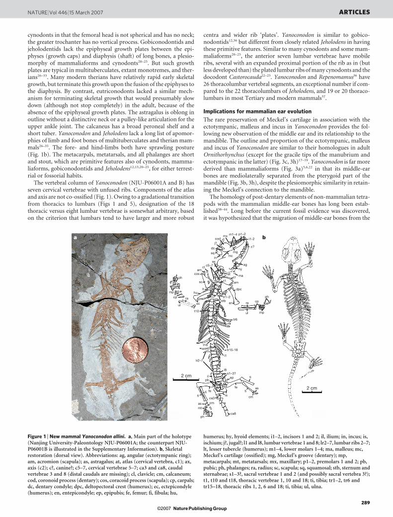
Figure 1 | New mammal Yanoconodon allini. a, Main part of the holotype(Nanjing University-Paleontology NJU-P06001A; the counterpart NJU-P06001B is illustrated in the Supplementary Information). b, Skeletalrestoration (dorsal view). Abbreviations: ag, angular (ectotympanic ring);am, acromion (scapula); as, astragalus; at, atlas (cervical vertebra, c1); ax,axis (c2); c?, canine?; c5–7, cervical vertebrae 5–7; ca3 and ca8, caudalvertebrae 3 and 8 (distal caudals are missing); cl, clavicle; cm, calcaneum;cod, coronoid process (dentary); cos, coracoid process (scapula); cp, carpals;dc, dentary condyle; dpc, deltopectoral crest (humerus); ec, ectepicondyle(humerus); en, entepicondyle; ep, epipubis; fe, femur; fi, fibula; hu,
humerus; hy, hyoid elements; i1–2, incisors 1 and 2; il, ilium; in, incus; is,ischium; j?, jugal?; l1 and l8, lumbar vertebrae 1 and 8; lr2–7, lumbar ribs 2–7;lt, lesser tubercle (humerus); m1–4, lower molars 1–4; ma, malleus; mc,Meckel’s cartilage (ossified); mg, Meckel’s groove (dentary); mp,metacarpals; mt, metatarsals; mx, maxillary; p1–2, premolars 1 and 2; pb,pubic; ph, phalanges; ra, radius; sc, scapula; sq, squamosal; stb, sternum andsternabrae; s1–3?, sacral vertebrae 1 and 2 (and possibly sacral vertebra 3?);t1, t10 and t18, thoracic vertebrae 1, 10 and 18; ti, tibia; tr1–2, tr6 andtr15–18, thoracic ribs 1, 2, 6 and 18; ti, tibia; ul, ulna.
NATURE | Vol 446 | 15 March 2007 ARTICLES
289Nature ©2007 Publishing Group
mandible in cynodont–mammal evolution underwent an intermedi-ate stage in which these bones would be anteriorly connected to thelower jaw, but suspended medial to (and free of) the mandibularpterygoid region (ref. 43 and Fig. 28.11 of ref. 44). Yanoconodoncorroborates Allin’s hypothetical model of an intermediate evolu-tionary stage. Newly discovered fossils of Yanoconodon (Fig. 1) andgobiconodontids9–11, preserved in multiple specimens from two dif-ferent fossil sites, add some crucial and previously unknown struc-tural elements to this ‘transitional’ middle-ear model:
(1) The anterior (mandibular) limb and the posterior (tympanic)limb of Meckel’s cartilage are twisted and curved relative to each otherat the mid-length of the cartilage (red arrow in Fig. 3). (2) The mid-length twist and curvature of Meckel’s cartilage made it feasible forthe anterior limb of the cartilage to be nestled in the Meckel’s grooveon the mandible9–11, while the tympanic limb of the cartilage andits associated ectotympanic and prearticular are separated medio-laterally from the pterygoid region of the mandible (Fig. 3f, g), as
postulated by Allin43. The tympanic membrane suspended by theectotympanic ring and malleus manubrium would be re-orientedmore mediodorsally (see red arrows in b-1 and b-2 of Fig. 3b), similarto the ectotympanic’s migratory path during ontogeny in the mar-supial Monodelphys41, leaving a space between the pterygoid region ofthe mandible and the tympanic membrane for access of the externalauditory meatus and for pterygoid muscle. (3) Owing to the curvatureof Meckel’s cartilage, the basicranial articulation of the incus isnearly co-axial with the fulcrum for movement around the dentary-squamosal jaw hinge, so the jaw movement had little impact on themiddle ear function. (4) The middle ear and the ossified Meckel’scartilage in late-stage subadult or adult of Yanoconodon (Fig. 3g) showa similar structural pattern to the embryos of monotreme (Fig. 3i)and placental mammals. The middle ear is anteriorly connected to acurved Meckel’s cartilage, which is in turn connected to the mandible.In the meantime, the embryonic middle-ear bones are mediolaterallyseparated from the pterygoid region17,18,39,40.
Mammaliforms
Crown mammals
Monotremes
Trechnotherians
Crown Therians
Eutherians (Placentals)
Metatherians (Marsupials)DMME homoplasies in basal mammals
Vomb
atus
Phascolarctos
Macropus
Petauroides
Pseudocheirus
Phalanger
Acrobates
Thylacomyidae
Dromiciops
Perameles
Dasyurus
Caenolestes
Marm
osaDidelphis
Andinodelphys
PucadelphysMayulestesTurgidodonPediomysDidelphodonAlbertatheriumAnchistodelphysAsiatherium
KokopelliaMarsasiaSulestes
Atokatheridium
Deltatheridium
Sinodelphys
Holoclemensia
Euphractus
Chaetophractus
Dasypus
Glyptotherium
Tamandua
Bradyp
us
Felis
Canis
Oryct
olag
usRat
tus
Erin
aceu
s
Prot
ungu
latu
mEo
ungu
latu
mA
span
lest
esLe
ptic
tis
Zal
ambd
ales
tes
Gyp
son
icto
ps
Cim
oles
tes
Asi
oryc
tes
Ken
nal
este
sU
khaa
ther
ium
Eom
aia
Mu
rtoi
lest
es
Pro
kenn
ales
tes
Mon
tana
lest
es
Dau
lest
es
Aegialodon
Kielantherium
Peramus
Nanolestes
Vincelestes
Amphitherium
Dryolestes
Henkelotherium
Zhangheotherium
Maotherium
Spalacotherium
Akidolestes
Priacodon
Trioracodon
Jeholodens
Yanoconodon
Gobiconodon
Repenomamus
Amphilestes
CimolodontansPlagiaulacidansHaramiyaviaTinodon
FruitafossorOrnithorhynchusObdurodonTachyglossus
TeinolophosSteropodon
BishopsAusk
tribosp
henos
Ambondro
Asfalto
mylo
s
Shuother
ium
Hadro
codi
um
Cas
toro
caud
a
Hal
dano
don
Meg
azos
trod
on
Mor
ganu
codo
n
Sin
ocon
odon
Ade
loba
sile
us
Pac
hyg
enel
us
Trit
ylod
onti
ds
Pro
bai
nog
nat
hu
s
Mas
seto
gn
ath
us
Th
rin
axod
on
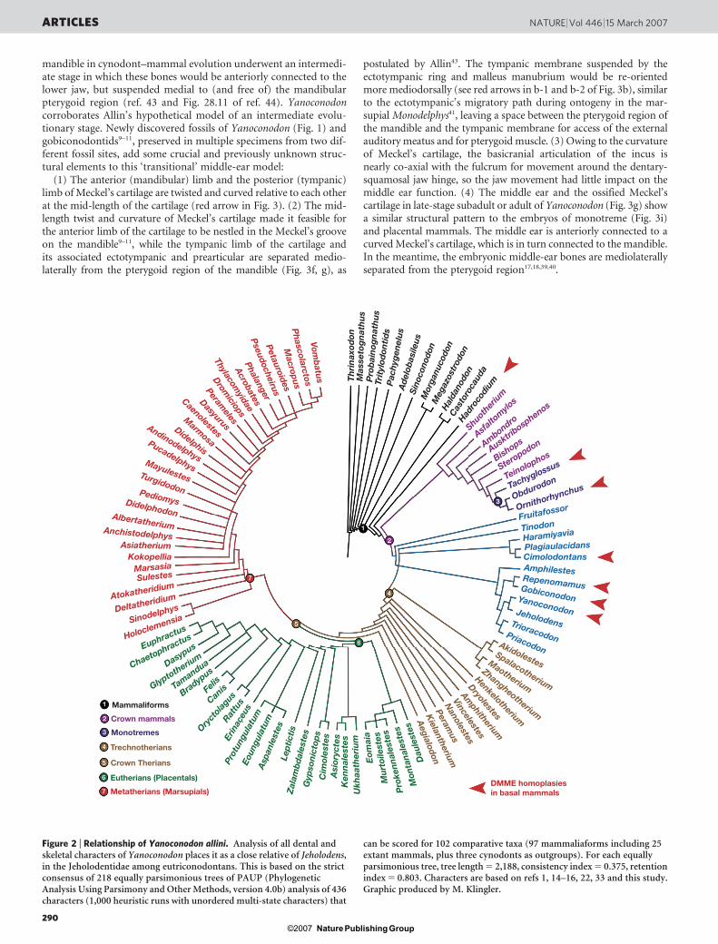
Figure 2 | Relationship of Yanoconodon allini. Analysis of all dental andskeletal characters of Yanoconodon places it as a close relative of Jeholodens,in the Jeholodentidae among eutriconodontans. This is based on the strictconsensus of 218 equally parsimonious trees of PAUP (PhylogeneticAnalysis Using Parsimony and Other Methods, version 4.0b) analysis of 436characters (1,000 heuristic runs with unordered multi-state characters) that
can be scored for 102 comparative taxa (97 mammaliaforms including 25extant mammals, plus three cynodonts as outgroups). For each equallyparsimonious tree, tree length 5 2,188, consistency index 5 0.375, retentionindex 5 0.803. Characters are based on refs 1, 14–16, 22, 33 and this study.Graphic produced by M. Klingler.
ARTICLES NATURE | Vol 446 | 15 March 2007
290Nature ©2007 Publishing Group

Overall, Yanoconodon’s middle ear shows a paedomorphic resem-blance to the embryonic pattern of modern mammals. All that isnecessary for adult Yanoconodon to retain this paedomorphic (albeitpermanent) connection of the middle ear to the mandible is a rela-tively earlier timing in (premature) ossification of the Meckel’s car-tilage and its fusion with the ectotympanic (Fig. 3c–g).
Developmental heterochrony and evolutionary homoplasy
A definite mammalian middle ear (DMME)43,44 is accomplishedby two ontogenetic steps in extant monotremes and placentals:first, a mediolateral separation of the middle ear bones from themandible in early embryonic stages (secondary mediolateral con-tact in marsupials due to inflected mandibular angle; see Fig. 12.10of ref. 40), and second, a loss of anterior connection to the mandi-ble owing to the reabsorption of Meckel’s cartilage in the subadult.In Yanoconodon and gobiconodontids (and by inference alsoJeholodens), the former step of mediolateral separation of the mid-dle ear from the mandible has already occurred. But reabsorptionof Meckel’s cartilage did not happen, resulting in the retentionin Yanoconodon of the middle-ear connection to the mandible,otherwise seen only in the early embryonic or fetal stage of extantmammals17,18,39–42.
Yanoconodon and its eutriconodontan kin are nested within thecrown Mammalia (Fig. 2) by the parsimony of all characters14–16.The absence of DMME in eutriconodonts, an in-group of crownMammalia, is in sharp contrast to modern monotremes and theriansthat have DMME. This phylogeny requires one of the following twoevolutionary scenarios: either (1) DMME was present in the commonancestor of monotremes, eutriconodonts and therians; but eutri-conodonts re-evolved the middle ear attachment to mandible, or(2) DMME was absent in the common ancestor of monotremes,eutriconodonts and therians, and this is retained as a paedomorpho-sis in eutriconodonts; but DMME evolved in extant monotremes44–47,and separately in therians39. Paedomorphosis, or retention of fetal orjuvenile characteristics of ancestors and relatives through develop-mental heterochrony, is a common phenomenon in vertebrate evo-lution. The heterochronic (‘premature’) ossification of Meckel’scartilage in eutriconodonts is the immediate cause for this paedo-morphic connection of middle ear and mandible, and is why there isan overall homoplastic distribution among therians (with DMME),eutriconodonts (without DMME), monotremes (with DMME) andpre-mammalian relatives (without DMME) (triangles in Fig. 2). Thepaedomorphic connection of the middle ear to mandible of eutri-conodonts and mammaliaforms is consistent with their lack of thelong-bone epiphyses for terminating skeletal growth, as seen in mod-ern mammals.
Hox gene patterning of axial skeleton in extinct mammals
Modern mammals have a highly conserved pattern of vertebralidentities: seven cervical, 13 to 14 thoracic, and five or six lumbarvertebrae (without separate lumbar ribs) for a combined 19 or 20thoracolumbar vertebrae37. These regional vertebral identities arepatterned by homeobox genes37,48–50. Homeotic changes in vertebralidentities, such as shift of the thoracolumbar boundary or grada-tional transition, are now correlated with the loss and gain of Hoxgenes function in mice48. The distinctive boundary of thoracic versuslumbar regions and the absence of lumbar ribs are patterned by theHox10 paralogues in modern placental mammals (Fig. 4)48. The tripleknockout of Hox10 paralogues can alter the thoracic versus lumbarboundary, and triple knockout of Hox11 can alter lumbar versussacral vertebral identities. A loss of Hox10 gene function can regen-erate the lumbar ribs and a more gradational thoraco-lumbar trans-ition in laboratory mice (Fig. 4)48.
Yanoconodon and Repenomamus both have 26 thoracolumbarvertebrae, and these eutriconodontans represent a high variationfor the highly conserved 19 to 20 thoracolumbar vertebrae of crownMammalia. By contrast, Jeholodens lost lumbar ribs and achieved a
Mandibularlimb
Tympaniclimb
10 mm
3 mm
3 mm
2 mm
a-1
Malleus
a-1
a-2 a-2
b-1b-1
b-2b-2
3 mm
Malleus
Incus
Ectotympanic
Meckel’s cartilage(ossified)
Meckel’s cartilage(embryonic)
Incus (quadrate)
Prearticular
Meckel’s cartilage
Meckel’s cartilage(ossified)
Meckel’s cartilage
Prearticular (goniale)Ectotympanic (angular)
ManubriumMalleus (articular)
a
b
c
d
e
f g h i
Figure 3 | Comparison of mandible and middle ear of Yanoconodon.a, Mammaliaform Morganucodon (medial view); a-1 and a-2 label schematictransverse sections at the levels of the malleus and the ectotympanic5–7. InMorganucodon, the middle ear maintains both an anterior connection to themandible via the Meckel’s cartilage, and a mediolateral contact to the mandible.b, Eutriconodont Yanoconodon (medial view, composite restoration ofmandible and middle ear from NJU-P06001A and B). b-1 and b-2 labeltransverse sections at the levels of the malleus and the ectotympanic. Themiddle ear retains the anterior connection to the mandible via ossified Meckel’scartilage (yellow), but is mediolaterally separated from the posterior part of themandible because of the twist and curvature of Meckel’s cartilage (red arrows inb). c, the ectotympanic (blue), malleus (green) and incus (brown) of modernOrnithorhynchus18: the shape and proportion of the ear bones are similar inOrnithorhynchus and Yanoconodon. d, Eutriconodont Repenomamus9–11:ossified Meckel’s cartilage connected anteriorly to the mandible (similar toYanoconodon). e, Ossified Meckel’s cartilage of Repenomamus (ventral view,isolated, redrawn from Fig. 1 of ref. 9). f, Ossified Meckel’s cartilage ofYanoconodon (ventral view, isolated, composite restoration of both the left andthe right elements). g, Middle ear of Yanoconodon (composite restoration,ventral view): the ectotympanic and malleus are connected anteriorly to themandible via ossified Meckel’s cartilage; but these are mediolaterally separatedfrom the posterior part of mandible, facilitated by curvature of the Meckel’scartilage (yellow). h, Middle ear bones of adult Ornithorhynchus (ventral view)and similarity to those of Yanoconodon. i, Embryonic Ornithorhynchus17: thetympanic ring and the partially developed manubrium and goniale(‘prearticular’) of the malleus are anteriorly connected via Meckel’s cartilage tothe mandible, but separated mediolaterally from the posterior region ofmandible, facilitated by the curved cartilage (red arrow). Yanoconodon retainsthe embryonic pattern of Ornithorhynchus owing to the timing change ofearlier ossification of Meckel’s cartilage, but otherwise its ectotympanic,malleus and incus are nearly the same as in adult Ornithorhynchus.
NATURE | Vol 446 | 15 March 2007 ARTICLES
291Nature ©2007 Publishing Group

distinctive boundary between the thoracic and lumbar regions (Fig. 4:node 2); its 15 thoracic and 7 lumbar vertebral counts are morecomparable to modern mammals. These derived lumbar features ofJeholodens evolved from within eutriconodontans, convergent tothose in theriiform mammals (node 1 in Fig. 4), and may be hypothe-sized as an independent activation of Hox10 gene patterning48.
Homoplasy of mobile lumbar ribs also occurs in symmetrodonts(Fig. 4)33. The presence of primitive lumbar ribs in Akidolestes is besthypothesized as an atavistic reversal because its related spalacother-oids show the derived therian condition without lumbar ribs29,32,33.Given that the regeneration of lumbar ribs in mutant mice is corre-lated with loss of function of Hox10 paralogues (Fig. 4b), it is plaus-ible that a similar mutation (that is, triple knockout of Hox10paralogues or a similar developmental event) was correlated withthe ‘reappearance’ of lumbar ribs in Akidolestes, whose immediaterelatives in successive ranks of theriiform mammals (node 1 in Fig. 4)all lack lumbar ribs.
Shifts in the thoracolumbar boundary and homoplasy of theirvertebral identities are primarily correlated with mutation in Hoxgenes for their patterning37,48–50. This provides a plausible mechanismfor the evolutionary patterns in lumbar ribs and the thoracolum-bar transition in Mesozoic mammals (Fig. 4). This developmentalmechanism for evolutionary homoplasy of thoracolumbar structureis mutually compatible with one of the two functional interpreta-tions: either the lumbar ribs in Akidolestes had a function similar tothat in primitive mammals and cynodonts with similar ribs23–25, orthe separate loss of lumbar ribs in Jeholodens was an adaptationconvergent to most theriiform mammals. Reciprocally, homoplasticthoraco-lumbar characters within eutriconodontans and symme-trodontans29,32,33 provide two cases for extrapolating the Hox gene
patterning of laboratory mice to early mammal phylogeny on a grandevolutionary scale.
Received 4 August 2006; accepted 29 January 2007.
1. Kielan-Jaworowska, Z. et al. Mammals from the Age of Dinosaurs—Origins,Evolution, and Structure (Columbia Univ. Press, New York, 2004).
2. Ji, Q. et al. Mesozoic Jehol Biota of Western Liaoning, China (Geol. Publ. House,Beijing, 2004).
3. Zhou, Z.-H. et al. An exceptionally preserved Lower Cretaceous ecosystem.Nature 421, 807–814 (2003).
4. Zhang, F. et al. Description of a new enantiornithine bird from the EarlyCretaceous of Hebei, northern China. Can. J. Earth Sci. 41, 1097–1107 (2004).
5. Kermack, K. A. et al. The lower jaw of Morganucodon. Zool. J. Linn. Soc. (Lond.) 53,87–175 (1973).
6. Crompton, A. W. & Luo, Z.-X. in Mammal Phylogeny Vol. 1 (eds Szalay, F. S. et al.)30–44 (Springer, New York, 1993).
7. Luo, Z.-X. et al. A new mammaliaform from the Early Jurassic of China andevolution of mammalian characteristics. Science 292, 1535–1540 (2001).
8. Jenkins, F. A. Jr & Schaff, C. R. The Early Cretaceous mammal Gobiconodon(Mammalia, Triconodonta) from the Cloverly Formation in Montana. J. Vert.Paleontol. 8, 1–24 (1988).
9. Wang, Y.-Q. et al. An ossified Meckel’s cartilage in two Cretaceous mammals andorigin of the mammalian middle ear. Science 294, 357–361 (2001).
10. Li, C.-K. et al. A new species of Gobiconodon (Triconodonta, Mammalia) and itsimplication for the age of Jehol Biota. Chin. Sci. Bull. (English edn) 48, 1129–1134(2003).
11. Meng, J. et al. The ossified Meckel’s cartilage and internal groove in Mesozoicmammaliaforms: implications to origin of the definitive mammalian middle ear.Zool. J. Linn. Soc. (Lond.) 138, 431–448 (2003).
12. Hu, Y.-M. et al. Large Mesozoic mammals fed on young dinosaurs. Nature 433,149–153 (2005).
13. Ji, Q. et al. A Chinese triconodont mammal and mosaic evolution of themammalian skeleton. Nature 398, 326–330 (1999).
14. Luo, Z.-X. et al. Dual origin of tribosphenic mammals. Nature 409, 53–57 (2001).15. Luo, Z.-X. et al. In quest for a phylogeny of Mesozoic mammals. Acta Palaeont.
Polonica 47, 1–78 (2002).
Ilium
Ilium
5 mm
5 mm
5 mm
5 mm
tr11–13
tr11–13
tr11–13
tr11–13
tr15–18Sacrals
b
a
Sacrals
Sacrals
Sacrals
Sacrals
Sacrals
Lumbar ribsabsent
Lumbar ribspresentLumbar ribs
absent
Lumbar ribspresent
Lumbar ribspresent
Lumbar ribsabsent
Hox10 triple mutant(lumbar ribs present)
Normal development(lumbar rib absent)
Thrinaxodon
Massetognathus
Probainognathus
Kayentatherium
Megazostrodon
Castorocauda
Tachyglossus
Ornithorhynchus
Fruitafossor
Jeholodens
Yanoconodon
Gobiconodon
Repenomamus
Sinobaatar
Kryptobaatar
Zhangheotherium
Maotherium
Akidolestes
Henkelotherium
Vincelestes
Sinodelphys
Eomaia
Lumbar ribpresent
Lumbar ribabsent
Lumbar ribsabsent
Lumbar ribsabsent
tr12–15tr12–15
Figure 4 | Homoplastic evolution of lumbar ribs among Mesozoicmammals and patterning of vertebral and rib development by Hox10 genein extant mammals. a, Homoplastic distribution of lumbar ribs in Mesozoicmammal taxa preserved with vertebral column (topology from Fig. 2):lumbar ribs are present in gobiconodontids and Yanoconodon but absent inclosely related Jeholodens (node marked 2), present in Akidolestes but absentin closely related Zhangheotherium33 and the more inclusive theriiforms(node 1). b, Patterning of vertebral structure (development of lumbar ribs) inmodern laboratory mice by homeobox genes (redrawn and flipped from Fig. 1
of ref. 48). A separate loss of lumbar ribs in Jeholodens amongeutriconodontans is hypothesized to be correlated with an independentactivation of Hox10 patterning of thoracolumbar vertebrae (node 2). Anisolated occurrence of lumbar ribs in Akidolestes among most spalacotheroidswithout lumbar ribs is hypothesized to be the effect of an independent loss ofHox10 gene function. The loss or gain of Hox gene function to pattern thevertebral identities is a plausible mechanism for homoplasy of lumbar ribs inearly mammals, and for variation of thoracolumbar vertebral counts amongeutriconodontans. tr, numbered thoracic ribs.
ARTICLES NATURE | Vol 446 | 15 March 2007
292Nature ©2007 Publishing Group
16. Luo, Z.-X. & Wible, J. R. A new Late Jurassic digging mammal and earlymammalian diversification. Science 308, 103–107 (2005).
17. Zeller, U. Die Entwicklung und Morphologie des Schadels von Ornithorhynchusanatinus (Mammalia: Prototheria: Monotremata). Abhandl. Senckenberg. Natur.Gesell. 545, 1–188 (1989).
18. Zeller, U. in Mammal Phylogeny Vol. 1 (eds Szalay, F. S. et al.) 95–107 (Springer,New York, 1993).
19. Fleischer, G. Studien am Skelett des Gehororgans der Saugetiere, einschliesslichdes Menschen. Saugetierk. Mitteil. 21, 131–239 (1973).
20. Jenkins, F. A. Jr & Parrington, F. R. The postcranial skeletons of the Triassicmammals Eozostrodon, Megazostrodon and Erythrotherium. Phil. Trans. R. Soc. Lond.273, 387–431 (1976).
21. Martin, T. Postcranial anatomy of Haldanodon exspectatus (Mammalia,Docodonta) from the Late Jurasssic (Kimmeridgian) of Portugal and its bearingfor mammalian evolution. Zool. J. Linn. Soc. (Lond.) 145, 219–248 (2005).
22. Ji, Q. et al. A swimming mammaliaform from the Middle Jurassic andecomorphological diversification of early mammals. Science 311, 1123–1127 (2006).
23. Jenkins, F. A. Jr. The postcranial skeleton of African cynodonts. Peabody Mus. Nat.Hist. Bull. 36, 1–216 (1971).
24. Jenkins, F. A. Jr. The Chanares (Argentina) Triassic reptile fauna VII. Thepostcranial skeleton of the traversodontid Massetognathus pascuali (Therapsida,Cynotondia). Breviora 352, 1–28 (1970).
25. Sues, H.-D. & Jenkins, F. A. Jr. in Amniote Paleobiology: Perspectives on the Evolutionof Mammals, Birds, and Reptiles (eds Carrano, M. T. et al.) 114–152 (Univ. ChicagoPress, Chicago, 2006).
26. Krebs, B. 1991. Das Skelett von Henkelotherium guimarotae gen. et sp. nov.(Eupantotheria, Mammalia) aus dem Oberen Jura von Portugal. Berliner Geowisch.Abhandl. A133, 1–110 (1991).
27. Rougier, G. W. Vincelestes neuquenianus Bonaparte (Mammalia, Theria), unPrimitivo Mamıfero del Cretacico Inferior de la Cuenca Neuquina. PhD dissertation(Univ. Nacional Buenos Aires, 1993).
28. Szalay, F. S. Evolutionary History of the Marsupials and an Analysis of OsteologicalCharacters (Cambridge Univ. Press, Cambridge, 1994).
29. Hu, Y.-M. et al. A new symmetrodont mammal from China and its implications formammalian evolution. Nature 390, 137–142 (1997).
30. Ji, Q. et al. The earliest-known eutherian mammal. Nature 416, 816–822 (2002).31. Luo, Z.-X. et al. An Early Cretaceous tribosphenic mammal and metatherian
evolution. Science 302, 1934–1940 (2003).32. Luo, Z.-X. & Ji, Q. New study on dental and skeletal features of the Cretaceous
mammal Zhangheotherium. J. Mammal. Evol. 12, 337–357 (2005).33. Li, G. & Luo, Z.-X. A Cretaceous symmetrodont therian with some monotreme-
like postcranial features. Nature 439, 195–200 (2006).34. Krause, D. W. & Jenkins, F. A. Jr. The postcranial skeleton of North American
multituberculates. Bull. Mus. Comp. Zool. 150, 199–246 (1983).35. Kielan-Jaworowska, Z. & Gambaryan, P. P. Postcranial anatomy and habits of
Asian multituberculate mammals. Fossils Strata 36, 1–92 (1994).36. Hu, Y.-M. The postcranium of Repenomamus and its implications for evolution of
mammalian skeletal characters. J. Vert. Paleontol. 22 (3-Suppl.), 67A–68A (2002).
37. Narita, Y. & Kuratani, S. Evolution of vertebral formulae in mammals: aperspective on developmental constraints. J. Exp. Zool. 304B, 91–106 (2005).
38. Gaupp, E. Die Reichertsche Theorie (Hammer-, Amboss- und Kieferfrage). ArchivAnatomie Entwick. 1912, 1–426 (1913).
39. Maier, W. Phylogeny and ontogeny of mammalian middle ear structures. Nether. J.Zool. 40, 55–75 (1990).
40. Maier, W. in Mammal Phylogeny (Volume 1) (eds Szalay, F. S. et al.) 165–181(Springer, New York, 1993).
41. Rowe, T. B. Coevolution of the mammalian middle ear and neocortex. Science 273,651–654 (1996).
42. Sanchez-Villagra, M. R. et al. Ontogenetic and phylogenetic transformations ofthe ear ossicles in marsupial mammals. J. Morphol. 251, 219–238 (2002).
43. Allin, E. F. Evolution of the mammalian middle ear. J. Morphol. 147, 403–438(1975).
44. Allin, E. F. & Hopson, J. A. in The Evolutionary Biology of Hearing (eds Webster, D. B.et al.) 587–614 (Springer, New York, 1992).
45. Rich, T. H. et al. Independent origins of middle ear bones in monotremes andtherians. Science 307, 910–914 (2005).
46. Martin, T. & Luo, Z.-X. Paleontology: homoplasy in the mammalian ear. Science307, 861–862 (2005).
47. Bever, G. et al. Comment on ‘‘Independent origins of middle ear bones inmonotremes and therians ‘‘ (I-II). Science 309, 1492a–1492b (2005).
48. Wellik, D. M. & Capecchi, M. R. Hox10 and Hox11 genes are required to globallypattern the mammalian skeleton. Science 301, 363–367 (2003).
49. Burke, A. C. & Nowicki, J. L. Hox genes and axial specification in vertebrates. Am.Zool. 41, 687–697 (2001).
50. Galis, F. Why do almost all mammals have seven cervical vertebrae?Developmental constraints, Hox genes, and cancer. J. Exp. Zool. 285, 19–26(1999).
Supplementary Information is linked to the online version of the paper atwww.nature.com/nature.
Acknowledgements We thank A. Tabrum for preparing this fossil; X.-N. Yang,Y.-K. Shi, J.-R. Liu, Q. Yang, J.-G. Sha, H.-C. Zhang for support; Q. Ji and J. Wible foraccess to comparative collections; R. Cifelli, Z. Kielan-Jaworowska, T. Martin,T. Rowe, J. Wible and G. Wilson for discussions; M. Dawson and J. Wible forimproving the manuscript; and M. Klingler for assistance with the figures. Thiswork was supported by the National Natural Science Foundation of China (P.C.,G.L. and Z.-X.L.), the National Science Foundation and National Geographic Society(Z.-X.L.), the Ministry of Science and Technology of China (the 973 Project underC.-S. Wang) and the State Key Laboratory of Palaeobiology and Stratigraphy ofNIGPAS (G. L.).
Author Information Reprints and permissions information is available atwww.nature.com/reprints. The authors declare no competing financial interests.Correspondence and requests for materials should be addressed to Z.-X.L.([email protected]).
NATURE | Vol 446 | 15 March 2007 ARTICLES
293Nature ©2007 Publishing Group