A NEW BACTERIAL DISEASE OF FRESH-WATER … · A NEW BACTERIAL DISEASE OF FRESH-WATER FISHES. J1....
27
A NEW BACTERIAL DISEASE OF FRESH-WATER FISHES. J1. By H. S. DAVIS, Fish-Pathologist, U. S. Bureau of Fisheries. J1.' Contribution from the U. S. Fisheries Biological Station, Fairport, Iowa. .t!- CONTENTS. Page. Introduction " " '" . 261 Description of the disease ' : .............. .. ........ 262 Occurrence of the disease " ...................................... .. ............... 263 Cause of the disease........................................................................ 263 Pathogenesis , " .... . 265 Methods of infection........................................................................ 266 Treatment and control of the disease........................................................ 270 Economic importance of the disease , ........................................ .. ....... 276 Explanation of fJogures : ...................... 280 INTRODUCTION. While working on the protozoan parasites of fishes at the U. S. Fisheries biological station, Fairport, Iowa, during the summer of 1917 a number of fish which had been recently placed in aquaria Werefound to be dying from the effects of a bacterial infection. Later in the season the disease was quite prevalent among fishes confined in aquaria, but owing to the pressureof other work no particular attention was paid to it at that time. Early the following summer (19 18) the disease again made its appearance, this time in one of the ponds. In the course of a series of feeding experiments being carried on at the Fairport station a large number of fingerling buffalofish were held in a pond in which had been placed a considerable quantity of horse manure. These fish grew rapidly for some time and appeared to be in a healthy, vigorous condition. However, early in July they began to.die in large numbers, and a careful examination showed that the dying fish were infected with the same species of bacteria found the previous summer on fish in aquaria. A little later in the season the disease appeared among fishes that had recently been transferred to troughs for feeding experiments and caused considerable mortality. On account of its evident importance it was decided to undertake an extended investigation of the disease to determine, if possible, a practicable method of control. This investigation was begun during the latter part of the summer of 1918 and continued during the summer of 1919. During the summer of 1919 the writer was assisted by Miss Miriam Mackenzie. 261
Transcript of A NEW BACTERIAL DISEASE OF FRESH-WATER … · A NEW BACTERIAL DISEASE OF FRESH-WATER FISHES. J1....
A NEW BACTERIAL DISEASE OF FRESH-WATER FISHES.
J1.
By H. S. DAVIS, Fish-Pathologist, U. S. Bureau of Fisheries.
J1.'
Contribution from the U. S. Fisheries Biological Station, Fairport, Iowa.
.t!
CONTENTS.Page.
Introduction " " '" . 261Description of the disease ' : . . . . . . . . . . . . . . .. . . . . . . . . 262Occurrence of the disease " .. . . . . . . . . . . . . . . . . . . . . . . . . . . . . . . . . . . . . .. . . . . . . . . . . . . . . . 263Cause of the disease. .. .. . . . . . . . . . . . . . . . . . . . . . . . . . . . . . . . . . . . . . . . . . . . . . . . . . . . . . . . . . . . . . . . . . . . 263Pathogenesis , " . . . . . 265Methods of infection. .. .. .. .. . . . . . . . . . . . . . . . . . . . . . . . . . . . . . . . . . . . . . . . . . . . . . . . . . . . . . . . . . . . . . . . 266Treatment and control of the disease. . . . . . . . . . . . . . . . . . . . . . . . . . . . . . . . . . . . . . . . . . . . . . . . . . . . . . . . 270Economic importance of the disease , .. .. . . . . . . . . . . . . . . . . . . . . . . . . . . . . . . . . . . . . .. . . . . . . . 276Explanation of fJogures : . . . . . . . . . . . . . . . . . . . . . . 280
INTRODUCTION.
While working on the protozoan parasites of fishes at the U. S. Fisheries biologicalstation, Fairport, Iowa, during the summer of 1917 a number of fish which had beenrecently placed in aquaria Werefound to be dying from the effects of a bacterial infection.Later in the season the disease was quite prevalent among fishes confined in aquaria,but owing to the pressure of other work no particular attention was paid to it at that time.
Early the following summer (19 18) the disease again made its appearance, this timein one of the ponds. In the course of a series of feeding experiments being carried on atthe Fairport station a large number of fingerling buffalofish were held in a pond in whichhad been placed a considerable quantity of horse manure. These fish grew rapidly forsome time and appeared to be in a healthy, vigorous condition. However, early inJuly they began to. die in large numbers, and a careful examination showedthat the dyingfish were infected with the same species of bacteria found the previous summer on fishin aquaria. A little later in the season the disease appeared among fishes that hadrecently been transferred to troughs for feeding experiments and caused considerablemortality.
On account of its evident importance it was decided to undertake an extendedinvestigation of the disease to determine, if possible, a practicable method of control.This investigation was begun during the latter part of the summer of 1918 and continuedduring the summer of 1919. During the summer of 1919 the writer was assisted byMiss Miriam Mackenzie.
261
BULLETIN OF THE BUREAU OF FISHERIES.
DESCRIPTION OF THE DISEASE.
The disease is easily recognizable, being distinguished by well-defined Characteristics. Ordinarily the first indication of the disease is the appearance of one or morecharacteristic dirty-white or yellowish areas on some part of the body. The infectedareas are usually quite conspicuous and increase rapidly in size. In some cases a largeproportion of the body may eventually become infected. With the increase in the sizeof the lesions the fish become greatly weakened and usually die from 24 to 72 hours afterthe lesions first become visible.
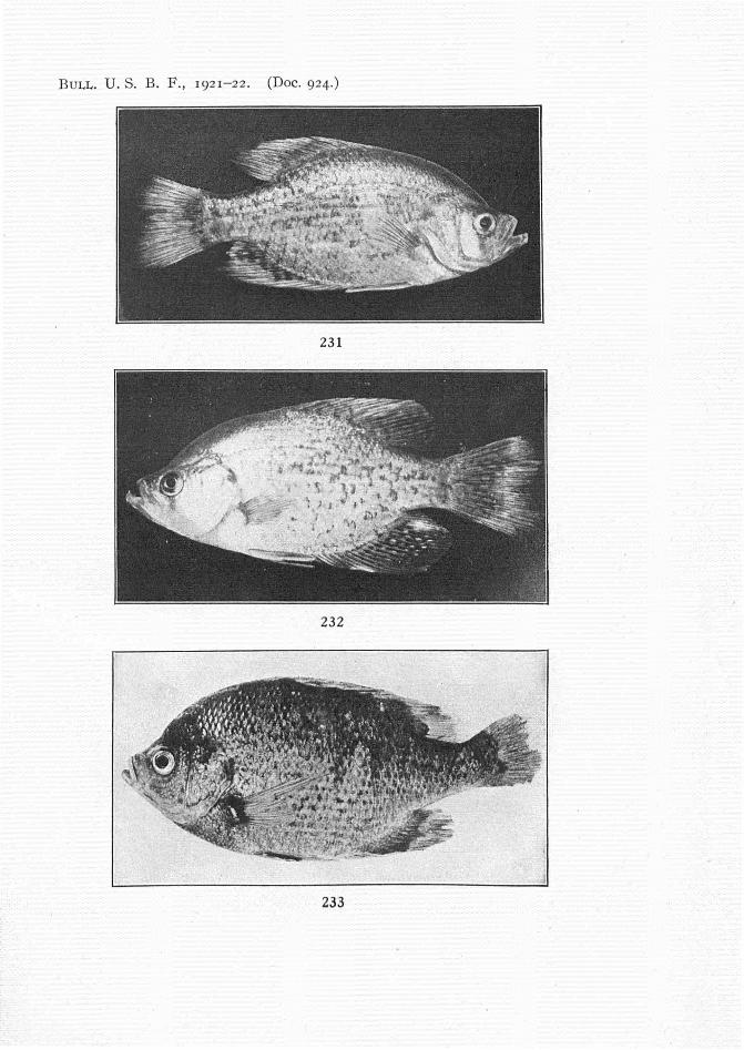
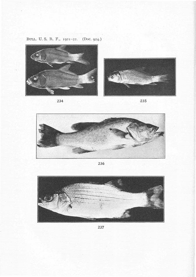
The lesions may occur on any part of the body, but in the majority of cases firstappear on the fins, especially the caudals, and from there spread to adjoining portions ofthe body (figs. 231-237). In late stages of the disease the lesions may cover from one-halfto two-thirds of the body, while the fins become badly frayed, the caudal sometimes becoming worn to a mere stub (figs. 233 and 234). There'is considerable variation in thesite of the initial infection. As will be shown later the disease is usually the result ofinjuries and first makes its appearance in the injured region. There is also considerablevariation dependent on the species of fish infected. In the crappies, for instance, thedisease is usually confined to the fins and gills (figs. 231, 232, and 243) and only rarelydoes the infection spread to the body. Possibly this may be due to the fact that thesefishes are especially susceptible to the disease and die before there is time for the bacteriato become widely spread over the surface of the body.
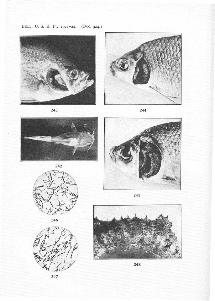
Lesions on the gills first appear as small, white patches (figs. 243 and 244). , Thesespread rapidly, and the fish usually dies within a few hours (fig. 245). . f.
On bullheads (A meiurus melas and A. nebulosus) the lesions have a somewhat differentappearance from those on scaled fishes. They usually first appear as numerous small,circular areas with sharp, distinct outlines (fig. 238). .
The centers of the lesions are dark blue overlaid by ~ whitish veil or cloudiness.Surrounding this is a well-defined zone about 5 mm. wide characterized by a distinctlyreddish tinge due to hyperemia. This region, like the central portion of the lesion, isoverlaid by a slight cloudiness. Later the lesions often become confluent and cover thegreater portion of the body (figs. 239-241). For some reason, in fingerling bullheadsthe disease more commonly starts on the caudal fin, as in other fishes, and the infectedarea gradually advances toward the anterior end, the entire posterior end of the body becoming a dirty white (fig. 242).
In many respects the lesions resemble those produced by Saprolegnia and, in fact,have usually been confused with them. A careful examination will, however, readilyenable one to distinguish between the two, since the bacterial lesions do not present thefuzzy appearance so characteristic of those infected with fungus. Of course they caneasily be distinguished by a microscopical examination, but this is usually not necessary.
In some instances a bacterial infection may be followed by an infection with Saprolegnia, but in such cases the fungus is of secondary importance. During the summer verylittle fungus has been observed at Fairport and infection with Saprolegnia appears to bedependent on the temperature of the water. When the temperature is high (above 75° F.)the bacteria develop rapidly and the fish die so quickly that the fungus does not havetime to develop to any appreciable extent. However, the bacteria are much more susceptible to any decrease in temperature than is the Saprolegnia, the result being that
BACTERIAl. DISEASE OF FRESH-WATER FISHES.
at temperatures below 75° F. the bacteria develop much more slowly and the fungusgrowth may become evident before the fish succumbs.
This is notably the case early in the fall. With the advent of cooler weather a considerable number of the diseased fish may show characteristic fungus growths in connection with the bacterial lesions. But even in these cases there can be little doubt that thelesions were primarily due to bacteria and that the Saprolegnia was a secondary invader.
Occasionally fish may die from the disease without showing any lesions on the bodyor fins. In such cases the gills are always badly infected, a large part of them beingentirely destroyed. In late stages of gill infection the fish often show characteristic signsof suffocation, such as swimming at the surface and gasping for breath. In the majorityof cases, fish with infected gills will also show lesions ~ the surface of the body or fins,but here again there is considerable variation in different species.
OCCURRENCE OF THE DISEASE.
I t seems probable that most species of fishes are liable to attack by this disease,although some species are much more susceptible than others. While it has been necessary to confine our observations to a comparatively small number of species the diseasehas been observed on the following fishes: The buffalofishes (Ictiobus bubalus and I.cyj>rinella), the sunfishes.(Lepomis incisor and L. humilis), the carp (CyPrinus carpio),the black basses (Micropterus salmoides and M. dolomieuj, the crappies (Pomoxis sparoides and P. annularis) , the warmouth (Chamobryttus gulosus) , the yellow perch (Pereaflavescens). the white bass (Roccus chrysops), the brook trout (Salvelinus jontinalis), theminnow. (Pimephales notatus), the channel catfish iLctalurus punctatus), and the bullheads (Ameiurus nebulosus and A. melas). From the above list it will be readily seen thatthe disease is widespread, and there is little reason to doubt that under favorable conditions it may occur on nearly all species of fresh-water fishes.
In one instance several tadpoles which were confined in a tank with a number ofinfected buffalofish developed well-defined lesions on the tail. The tadpoles were justbeginning to metamorphose, which may have made them more susceptible to infection.Attempts to inoculate adult frogs with the disease have proved unsuccessful.
So far it has not been possible to obtain any data regarding the geographical distribution of the disease. However, while on a visit to the St. Lawrence River at Ogdensburg, N. Y., during the first week in July, 1919. the writer found the bacteria in lesionson the smallmouth black bass and the common perch. In this case the fish did notappear to be seriously injured by the bacteria which were evidently growing very slowly.
CAUSE OF THE DISEASE.
The disease is produced by an apparently undescribed species of bacteria for whichI propose the name Bacillus columnaris. It has not yet been possible to grow the organism in pure culture, but its appearance is so characteristic that it is always easily recognizable if present in any numbers. It is a long, slender, flexible, rod-shaped organism.5 to 12J.t long and O·5J.t wide (figs. 246 and 247). The bacteria are very transparent andunless present in considerable numbers are difficult to distinguish on this account.They usually appear perfectly homogeneous, but rarely may exhibit a slightly granularstructure. The bacteria are motile and probably possess flagella, although owing to

BULLETIN OF THE BUREAU OF FISHERIES.
lack of facilities this has not been determined with certainty. They can often be seento move in a straight line for a considerable distance, the movement being accompaniedby a sinuous bending of the rod which may temporarily assume an S-shape. One of thecharacteristic movements is to turn one end slowly in a circle, the other end remainingstationary and forming a pivot on which the entire rod revolves. This movement isvery noticeable along the edge of scales or bits of infected tissue when placed on a slide.Large numbers of bacteria can usually be seen in such places with one end attached'while the free end moves back and forth in the manner described above.
Another characteristic is the formation of small chains of bacteria from one objectto another while on the slide. The chains are formed by the bacteria arranging themselves in rows joined end to e1l{/p the ends slightly overlapping. Usually these chainsconsist of two or three rows of bacteria arranged parallel to each other, but occasionallythere may be but a single row. Bacteria can be seen continually moving back andforth along these living chains in a very characteristic manner.
However, the most characteristic movement and the one which usually serves tomake the species easily recognizable is well shown when a little material scraped from alesion is placed on a slide in a drop of water. Possibly owing to the pressure' of thecover glass the bacteria soon collect in immense numbers on the edge of bits of infectedtissue, scales, etc. Here they form short columnlike masses, -each column being separated a short distance from its neighbor (figs. 248 and 249). The columns usually taperslightly toward the free end, which is ordinarily rounded but in rare cases may be pointed.In some cases the ends of the columns may be distinctly enlarged. In favorable casesa whole series of these columns may form along the edge of a scale. -These columnarmasses of bacteria are very characteristic, and it is apparently in this way that they freethemselves from the gelatinous matrix in which they are embedded while growing onthe fish. Along the sides and rounded ends of the columns can be seen bacteria withone end attached while the free end waves back and forth in the manner already described. Bacteria continually break loose from the columns while others swarm outto take their place. Occasionally short chains may be formed extending out from theends of the columns, the bacteria at the ends continually becoming free but often managing to work their way back along the chain after a time. When free, the bacteria atfirst exhibit a peculiar vibratory movement somewhat different from the Brownianmovement and which, when once seen, is easily recognized. After being free a shorttime they usually collect on the underside of the cover glass and become perfectlymotionless.
These characteristic swarming movements of the bacteria are no doubt continuallytaking place on the fish, at least in late stages of the disease, for it has been found thatbadly diseased fish are continually shedding bacteria in enormous numbers.
Owing to the fact that a variety of stains was not available the staining reactionsof the bacteria were not studied in detail. They stain readily with fuchsin and Giemsa'sstain, the stained preparations appearing perfectly homogeneous.
No evidence of sporulation has been observed, and that they do not form sporesis also indicated by the fact that the bacteria are easily killed by chemicals and drying.
. All attempts to grow the bacteria on artificial media have so far proved unsuccessful. During the summer of 1918 the writer made several attempts to isolate thebacteria, and these experiments were continued by Miss Mackenzie during the summer

BACTERIAL DISEASE OF FRESH-WATER FISHES.
of 1919. On account of the limited facilities at our disposal it was only possible to try,a few of the simpler media. Standard beef-broth agar and fish agar were tried, andalthough bacteria appeared in the plates in large numbers no trace of columnaris couldbe found. Several lots of media with a less acid reaction than the standard were tried,including one with the same reaction as the river water, but the results were all negative. Attempts to grow the bacteria on fish serum were equally unsuccessful. Possiblywith facilities for employing a wider range of media it may be possible to isolate the bacteria. However, the failure to isolate the bacteria did not prove so serious a handicapin the study of the disease as would ordinarily be the case. The bacteria can easily beprocured in large numbers from the lesions of infected fish, and their appearance is socharacteristic that they can be readily recognized.
While, of course, the failure to isolate the bacteria in pure culture has rendered itimpossible to demonstrate the cause of the disease beyond question, the evidence is soconclusive as to leave little room for doubt. The bacteria carr-always be found inabundance in the-lesions, and the disease can easily be produced in healthy fish byscraping off a few scales and applying a few bacteria from a diseased fish. In advancedstages of the disease there are usually large numbers of bacteria present in addition tocolumnaris, but it has been found that in small lesions columnaris is invariably enor->mously more abundant than any other organism, and in some cases when the lesionsfirst become visible there is a nearly pure culture of this species present.
Strong evidence for the causal relationship of columnaris to the disease can be seenin infected bullheads. The reader will recall that the lesions on these fish are characterized by an outer reddish zone about 5 mm. wide, while the center of the lesion hasa quite different appearance. When very small the entire lesion presents the sameappearance as the peripheral zone in later stages. As the lesions increase in size thecharacteristic blue center appears and enlarges with the growth of the lesion. It isobvious that the red zone is found only where the skin has been recently infected andthat the darker color is que to destructive changes produced by the bacteria. If a littlematerial is scraped from the darker central portion of the lesion it will be found thatconsiderable numbers of bacteria are present in addition to columnaris. On the otherhand, if material from the red zone is examined it will be found that nearly all the bacteria present are columnaris. Other species are so few as to be scarcely noticeable. Ofcourse the same thing is true of lesions on other species of fishes, but owing to the absenceof scales it is more striking in the case of the bullheads than elsewhere. The greatabundance of columnaris around the advancing edge of the lesions is so noticeable thatit soon became a matter of routine in our work to always procure the bacteria fromthis region. The smears from which the photomicrographs (figs. 246 and 247) weretaken were made from such material.
PATHOGENESIS.
As previously stated the bacteria grow only on the surface of the body or on thegills. No case has been observed where they had penetrated any distance into the tissues. When growing on the body they destroy the integument, the muscles sometimesbeing exposed in late stages of the disease. For obvious reasons the development ofthe lesions can be more easily studied in the case of scaleless fish, such as the common
266 BULI1ETIN OF THE BUREAU OF FISHERIES.
bullhead. Figure 250 is a cross section through a lesion which is just beginning to develop. The outer layer of the epidermis has begun to disintegrate, but the inner layeris not yet noticeably affected. Figure 251 shows a little later. stage. The epidermisis now entirely destroyed in one place at the right in the figure. On either side theepidermis is rapidly disintegrating. A somewhat later stage is shown in figure 252.The outer layer of the corium which has been exposed by the destruction of the epidermisis now beginning to show signs of disintegration. In late stages of the disease the coriummay be entirely destroyed in the center of the lesion, as shown in figure 253.
As previously described, the lesion grows rapidly outward in all directions from thecenter of infection. Figures 254 to 257 are from photomicrographs of sections throughthe margins of lesions and show the disintegration of the epidermis caused by the outward growth of the bacteria. Usually there is a noticeable hyperemia just underneaththe epidermis in this region. This is well shown in figures 255 to 257. The capillariesin the outer portion of the corium become gorged with blood; eventually their wallsdisintegrate and the blood fills the interstices of the corium and crowds between thecorium and the epidermis. Occasionally the blood corpuscles may even penetrate ashort distance into the epidermis (fig. 255). This is the cause of the reddish zone which
• is often quite distinct around the margin. Later as the epidermis is destroyed thecorpuscles also disintegrate. It is scarcely necessary to add that the hyperemic zoneadvances outward as the lesion enlarges. Usually the hyperemia is noticeable onlyunderneath that portion of the epidermis which is undergoing disintegration, but occasionally may occur before the overlying epidermis shows any change. No case hasbeen observed where the muscles showed any appreciable pathologic changes. Possiblythis may be due to the fact that the fish usually die by the time the lesions have developedto the stage shown in figure 253.
While the above account is based on a study of the lesions in bullheads the sameconditions are found in other fishes with some slight modifications due to the presenceof scales. In these fishes the scales become loosened and slough off as the integumentdisintegrates.
In all cases there is a thick, matted layer of bacteria covering the lesion. Thislayer is not shown in the figures since it invariably drops off during the treatment towhich the tissues are subjected in preparation for sectioning: Although the bacteriaoccur in enormous numbers in this superficial layer only a few scattered individuals canbe found among the disintegrating cells of the epidermis and corium.
When growing on the gills the bacteria produce much the same effects as on theintegument. The epithelium and blood vessels are entirely destroyed until only theskeletal parts are left. As would be expected gill infections are much more quickly fatalthan infections on the surface of the body.
METHODS OF INFECTION.
Since the bacteria grow only on the surface of the body and gills it is evident thatinfection can easily take place by their being carried in the water from one fish to another.As previously pointed out the bacteria are continually being set free from infected fish,especially in late stages of the disease. Where fish are crowded closely together, as inaquaria, the bacteria can thus readily pass from one individual to another. They are
BACTERIAL DISEASE OF FRES~-WATER FISHES:
also easily rubbed off onto the nets or hands when diseased fish are handled and then can. readily be transferred to healthy fish. We have several times infectedhealthy fish by
simply holding them in our hands after having previously handled diseased fish. Thereis also a possibility that the disease may be transmitted by predacious insects, but noexperiments have as yet been carried out along this line.
Undoubtedly the most important factors in the spread of the disease under ordinaryconditions are those which render the fish more susceptible to infection. All the evidenceat hand indicates that the bacteria rarely or never attack healthy, uninjured fish underordinary circumstances. If, however, a fish is injured or its vitality lowered in any wayit is liable to. contract the disease. Most outbreaks of the disease which have comeunder the writer's notice have occurred among fish. which had been recently handled.As is well known it is almost impossible to handle fish without some slight injury to thefins or body, and even a very slight injury is all that is necessary to enable the bacteria togain a foothold, and once started they are able to spread rapidly over the body. Duringwarm weather, when the temperature of the water at Fairport averages about 75 to80° F., an outbreak of the disease is almost certain to occur within 48 to 72 hours afterthe fish are handled. Well-defined lesions may sometimes appear in 24 hours, but thefish usually do not begin to die until after 48 hours. The greatest mortality usuallyoccurs on the third and fourth days, after which there is a gradual decrease until the diseasefinally disappears. If the fish are again handled, however, there is almost certain to beanother outbreak.
In a large percentage of cases the disease first makes its appearance on the fins,especially the caudal. This is, of course, readily explained since no other part of the bodyis so liable to be injured by the struggles of the fish when 'taken in a net. Of course, anyslight abrasion on the surface of the body, such as the removal of a few 'scales or even aslight injury to the epithelium, is equally liable to infection. I t has been found very easyto infect fish artificially by simply scraping away a few scales with a scalpel and applyinga small amount of material scraped from a diseased fish. Usually a characteristiclesionwill develop around the site of infection in 24 to 48 hours.
There is also evidence that in some cases lowered vitality unaccompanied by mechanical injuries may result in the fish becoming infected. This is probably the explanation of the epidemic which appeared in one of the ponds at Fairport in 'which youngbuffalofish were being held for a feeding experiment. These fish had not been handledfor some weeks previous to the outbreak of the epidemic. Just before the diseaseappeared the water had become noticeably foul and this, no doubt, so lowered the vitalityof the fish as to make them susceptible to infection. The writer has also been informedby fishermen that they had seen fish with the same disease in isolated ponds and sloughsalong the Mississippi River late in summer. At this time the water in these pondsbecomes very warm and stagnant, conditions which would favor the development of thebacteria while tending to lower the vitality of the fish.
Another important factor influencing the spread of the disease is the temperature ofthe water. As already pointed out columnar-is is very susceptible to temperature changesand grows rapidly only at a comparatively high temperature. The disease is distinctlya warm-weather disease, and so far as our observations go is not ordinarily of greatimportance when the temperature of the water is low. This was very noticeable at Fair-
85781°-22--9,
268 BULLETIN OF THE BUREAU OF FISHERIES.
port during September, 1919. With the advent of cooler weather there was a considerable decrease in the temperature of the water in the aquaria in which the experimentalfishwere kept. After the first week in September the ·average temperature of the waterdecreased from about 75 to 80° to about 70° F. and there was a notable decrease in theseverity of the disease. The percentage of infected fish decreased, the disease developedmore slowly, and' a considerable percentage of the infected fish recovered.
The writer does not, however, wish to give the impression that there is no dangerof an outbreak of the disease when the water temperature falls below 70° F. There issome evidence that an epidemic may occur at a comparatively low temperature, but itis probable that under such conditions the disease is much less severe and more easilycontrolled. That the bacteria may develop at lower temperatures is shown by anexperiment with brook trout. A number of fingerlings were shipped to Fairport fromthe hatchery at Manchester, Iowa, and placed in a trough supplied with running waterfrom a well. The temperature of the water averaged about 62° F. On September 13two badly diseased fish, one buffalofish and one bluegill, were placed in the trough amongthe trout. At a temperature of about 75° F. both fish would in all probability havedied in less than 24 hours. However, after being placed in the trough with the troutthe bluegill lived 48 hours and the buffalofish about 72 hours. One of the trout was founddying with the disease on September 17. Unfortunately, owing to the writer's departure from Fairport, it was impossible to continue the experiment longer.
During the first week in July, 1919, the writer found a slight infection on a yellowperch and one on a smallmouth black bass from the St. Lawrence River at Ogdensburg,N. Y. In each case the infection was in wounds on the side of the body. These fishwere in a tank with a large number of other fishes, and during the three days they wereunder observation none of the other fish contracted the disease. Furthermore, the lesionson the infected fish did not increase noticeably in size during this time and the fishshowed no ill effects from the disease. Since the temperature of the water averagedabout 65° F. it is probable that the failure of the disease to' develop rapidly and spreadto the other fishes in the tank was largely due to this fact. Many of the other fishesconfined in the tank had been more or less injured when captured, so there would 'seemto have been every opportunity for the disease to spread. However, in this case it wasimpossible to determine definitely whether the failure of the disease to develop was dueto the low temperature or to the fact that the bacteria may have belonged to a muchless virulent strain than that at Fairport. It has not yet been possible to carry out anydetailed experiments to determine the effect of temperature on the virulence and growthof the bacteria, but it is hoped to do so in the near future.
There is also evidence that, in general, fish are more likely to contract the diseasefrom fish of the same species than from one of another species. This means, of course,that physiological strains of bacteria may be developed on different species of fish. Asis well known such physiological strains are common among many species of bacteria.The strongest evidence of the occurrence of physiologically distinct strains in Bacilluscolumnaris was obtained in the course of some experiments with the black bullhead(Ameiurus melas). This species is ordinarily not very susceptible to the disease, andduring the summer of 1919 a number of these fish were kept in the same tank withinfected buffalofish and bluegill without any of them contracting the disease. Afterseveral weeks one of the bullhead was artificially inoculated with bacteria obtained
BACTERIAl. DISEASE OF FRESH-WATER FISHES.
from a diseased buffalofish. Inoculation was made as usual by scraping away the. epidermis from a small area on one side of the body and rubbing the bacteria into thewound. The bullhead developed a characteristic lesion, and within 3 or 4 days practically every bullhead in the tank became infected. The epidemic among the bullheadwas not confined to this tank but soon spread to bullhead in adjoining tanks. Thiscan be easily explained. On account of their apparent immunity to the disease noprecautions were taken to prevent the bacteria getting from one tank to another, andthey could easily have been transferred on the hands or nets. The epidemic developedwith remarkable rapidity, and within a few days nearly all the bullhead which had beenheld in the tanks from one to several weeks without showing any signs of the diseasewere dead or dying. Only by the use of control measures to be described later was theepidemic checked and a few fish saved. It was noticeable' that although the infectedbullhead were in several instances kept in the same tanks with buffalofish and bluegillthe disease did not spread among them nearly so rapidly as among bullhead, althoughordinarily both buffalofish and bluegill are more susceptible to the disease.
It has also been observed in making artificial inoculations that while it is usuallynot difficult to infect a fish with bacteria from another species a larger percentage ofpositive results is often obtained when the bacteria are taken from a fish of the samespecies. Further investigations along this line are greatly to be desired.
As intimated above, while most species of fresh-water fishes are liable to be attackedby the disease some species are much more susceptible than others. It is, of course, byno means easy to determine accurately the relative susceptibility of different species,but there can be no question that there is a great deal of variation in this respect. It ispossible to divide the fishes which have been most studied into two classes. The firstincludes those species which are very susceptible and are almost certain to contract thedisease in large numbers if handled during warm weather. In this class we would includethe buffalofishes (1ctiobus bubalus and cyprinella), the crappies (Pomoxis annularis andsparoides), and possibly the bluegill (Lepomis incisor). The second class includes thosefishes which are ordinarily only moderately susceptible and do not usually contract thedisease in such numbers, even in warm weather. Furthermore, a considerable percentage of the diseased fish may recover if kept under favorable conditions. In thisclass we would include the largemouth black bass (Micropterus salmoidesy, the channelcatfish (1ctalurus punctatus), the bullheads (Ameiurus nebulosus and melas), the sunfish(Lepomis humilis), the carp (Cyprinus carpio), the warmouth tChamobryttus gulosus),and the white bass (Roccus chrysops). Of.course itis not to be understood that all ofthe species in each class are equally susceptible, for that is certainly not the case, but inthe present state of our knowledge it does not seem advisable to attempt a more detailedanalysis of their relative susceptibility to the disease.
It is also noticeable that the young of any species are, in general, more susceptiblethan are the adults. This is especially true in the case of the carp, the young of whichare quite susceptible, while the adults are nearly immune.
At first it was assumed that the bacteria must be widely distributed in the water,since this would most readily explain the appearance of the disease when the fish arehandled, but we have no proof that this is the case, and there is some evidence that thebacteria do not live for any length of time off the fish. If this is true, we are forced toassume that they are able to live in small numbers on perfectly healthy fish without

BULLETIN OF THE BUREAU OF FISHERIES.
injuring them, but are capable of increasing enormously whenever proper conditionsarise. Such a state of affairs is not unknown as in the case of streptococci and pneumococci in man.
TREATMENT AND CONTROL OF THE DISEASE.
On account of the great economic importance of the disease special efforts havebeen made to develop effective methods of control. Early in the investigation itbecame evident that reliance must be placed chiefly on methods for preventing thespread of the disease rather than to attempt to cure fish already infected. This is dueto the fact that the bacteria, although living exclusively on the exterior of the fish, forma thick, matlike growth which protects the bacteria underneath from the effects ofchemicals. Furthermore, the bacteria soon make their way underneath the scaleswhich form an additional protection. To kill bacteria in such protected situationsrequires a comparatively strong solution.
A number of chemicals have been tried in the effort to find an effective control forthe disease, but only potassium permanganate and copper sulphate have been found tobe of any value. In all our experiments with chemicals we have first determined themaximum concentration of the chemical which the fish can stand without serious injury.Different species differ greatly in this respect, and unfortunately the fishes which areleast resistant to chemicals are often very susceptible to the disease. It was soon foundthat the buffalofishes are among the most susceptible species, both to chemicals and tothe disease, and it is believed that any treatment which will succeed with these fish willbe equally successful with most of our common fishes. Having ascertained the strengthof the solution which the more susceptible fishes could stand without injury, we thenattempted to determine the minimum concentration which will kill the bacteria whenfully exposed to the chemical. Since the bacteria can not be grown on culture media itis obviously impossible to determine with certainty when they are killed. It was found,however, that fairly accurate results could be obtained by placing the bacteria on a slideand then treating them with solutions of various strengths. The cessation of all movement by the bacteria was taken as evidence that they were seriously injured if notkilled,While, of course, there are serious objections to this method it was found in practice towork out remarkably well.
Of course the time element is important in the use of any chemical. Treatmentwith a very weak solution may be as effective as a much stronger solution if the time issufficiently increased. Temperature may also be an important factor, but we haveonly meager data as to its effects. With few exceptions our experiments have beencarried on with river water at a temperature of 75 to 80° F.
During the summer of 1918 an extensive series of experiments with potassiumpermanganate was undertaken. It was found to greatly weaken or kill the bacteriain solutions so dilute that most species of fish could be kept in them for some time without serious injury. In order to kill the bacteria it is necessary to treat the fish witha I to 50,000 solution for 10 to IS minutes. Weaker solutions have little effect. Buffalofishes can undergo this treatment without serious injury, while the black bass and sunfishescan stand a much stronger solution. A I to 20,000 solution was used on black bassfor the same length of time with good results. However, very few fishes can stand aI to 20,000 solution, and weaker solutions do not seem to penetrate far into the bacterial

BACTERIAL DISEASE OF FRESH-WATER FISHES. 27 1
mat covering the lesions. Furthermore, potassium pennanganate is expensive anddissolves slowly in water. For these reasons the potassium-pennanganate treatmentwas later abandoned in favor of copper sulphate.
A number of experiments with copper sulphate were made during the summer of1918, and it was found that a 1 to 3°,000 solution is more effective than a 1 to 50,000
solution of potassium permanganate and not so injurious to the fish. A solution ofthis strength will not, however, kill bacteria which are well protected, such as thosebeneath the, scales. Our experiments showed that normal fingerling buffalofish are notappreciably injured when placed in a 1 to 3°,000 solution for 30 minutes, but diseasedor weakened fish are not so resistant. Buffalofish over 1 year old are not injured by treatment with a 1 to 25,000 solution for 30 minutes, and black bass and bullheads canstand a 1 to 20,000 solution for the same length of time without injury. As a result ofthese experiments the following treatment was recommended and used with muchsuccess: The fish previous to the appearance of any lesions are placed in a 1 to 30,000
solution of copper sulphate for 20 minutes and are then removed at once to runningwater. If properly handled ·they rarely suffer any permanent injury. Wooden vesselsare preferable, and if it is necessary to use galvanized vessels they should be paintedto prevent chemical action between the copper sulphate and the metal sides of the vessel.A thin coating of melted paraffin over the inside of a galvanized tank serves the purposeadmirably. During the treatment some of the less resistant species of fishes may swimaround at the surface and show more or less signs of distress, but this does not necessarily indicate any serious injury.
It should be distinctly borne in mind that the treatment to be effective must begiven within afew hours after the fish have been handled and before any signs of infection appear. If the fish are to be confined in aquaria, two or three successive treatments at 12 to 24 hour intervals are advisable. When this treatment is properly usedit is believed it will effectually prevent any serious outbreak of the disease. It must berepeated, however, every time the fish are handled or subjected to injury in any way.
In an experimental test of this treatment 30 fingerling buffalofish were seinedfrom one pond. They were handled quite roughly, and 14 controls were placed in asmall aquarium without being treated. The remaining 16 fish were treated with a 1
to 30,000 solution of copper sulphate for 20 minutes and then placed in a small aquarium like that in which the controls were held. Throughout the experiment bothaquaria were supplied with running water, and the conditions in each were as nearlyidentical as it was possible to make them. Two of the treated fish died within 12 hours,probably as a result of mechanical injuries. At the end of 24 hours the fish were againtreated with a 1 to 3°,000 solution for 20 minutes. Twenty-four hours later two of thetreated fish were found dead, but a careful examination failed to disclose any signs ofBacillus columnaris. Since the remaining fish showed no signs of infection no treatment was given at this time, but 6 hours later. one fish showed a well-defined lesion atthe base of the tail. This fish was at once removed and the rest given the same treatment as before. Since no further signs of infection appeared in this lot in three daysthe experiment was discontinued. .
Among the controls 3 fish showed well-defined lesions after 24 hours, and at theend of 48 hours 11 out of the 14 were dead, while 2 of the remaining 3 had well-developed
272 BULLETIN OF THE BUREAU OF FISHERIES.
lesions on the tail and died a few hours later. The remaining control died the nextday with a large lesion on the side of the head. In short, there was a loss of 31U percent among the treated fish and 100 per cent among the controls. Four of the five fishwhich died among the treated lot were in all probability killed by the direct effects ofthe copper sulphate. A large number of experiments have shown that fish whenweakened in any way, as by handling or being kept in water deficient in oxygen, aremuch more susceptible to the injurious effects of the treatment than perfectly normalfish and are often killed by solutions which will not appreciably injure fish in good condition. It is this fact which has made it impossible to devise a treatment which willbe successful under all conditions. Of course it is evident that the fish which are soweakened as to be injured by the treatment are the very ones which are most liableto contract the disease if not treated.
It has not been practicable to conduct many experiments so completely undercontrol as this one, but we have in a number of instances successfully checked by thistreatment an outbreak of the disease among fish which were being kept in aquaria forother purposes. Another test of the treatment under somewhat different conditionsis of considerable interest: On July 20, 1919, in connection with a series of feedingexperiments which were being carried on at the Fairport station, a number ofg-year-oldbuffalo fish were seined from the pond in which they had been confined for sometime and distributed among four different ponds. This necessitated handling the fishrather roughly and transferring them some distance in a' galvanized tank. To makematters worse it was a very hot day and under ordinary circumstances there wouldundoubtedly have been considerable loss, especially since many of the fish were badlyrubbed during the transfer. Before being liberated in the ponds all the fish weretreated for 20 minutes with a I to 30,000 solution of copper sulphate. Of the 55I fishtreated only 4 died as a result of the transfer, and none of these showed any evidence ofthe disease. It is unfortunate that in this case it was not practicable to keep anycontrols for comparison with the treated fish. .
While the treatment described above has been very successful it has the greatdisadvantage of requiring considerable time, and this is a serious objection where manyfish are to be treated. During the summer of 1919 extensive experiments were carriedon with. a view to shortening the treatment. As a result a rapid treatment has beendevised which is believed to be even more effective than the longer treatment previouslyin use. Briefly, the new method consists in treating the fish with a I to 1,000 solutionof copper sulphate for one to two minutes. This treatment is so great an improvementon the earlier treatment with a I to 30,000 solution that in our later experiments weentirely discarded the latter.
When the fish are in good condition they can be placed in the I to 1,000 solutionfor two minutes without injury, but if they have been previously weakened in any waythey should not be exposed to the solution for more than one minute. Such hardyfishes as the black bass, sunfishes, and bullheads can be safely left in the solution forthree minutes if in good condition. In some cases it is advisable to repeat the treatmentafter 12 to 24 hours.
A number of experiments with the rapid treatment were carried out during the latterpart of the summer of 1919, but it will suffice to describe a few representative experiments.
BACTERIAL DISEASE OF FRESH-WATER FISHES. 273
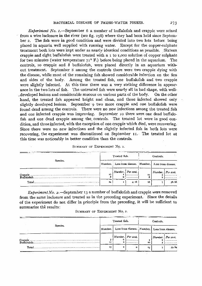
Experiment No. I.-September 6 a number of buffalofish and crappie were seinedfrom a wire inclosure in the river (see fig. 258) where they had been held since September 2. The fish were in good condition and were divided into two lots before beingplaced in aquaria well supplied with running water. Except for the copper-sulphatetreatment both lots were kept under as nearly identical conditions as possible. Sixteencrappie and eight buffalofish were treated with a I to'I,OOO solution of copper sulphatefor two minutes (water temperature 71° F.) before being placed in the aquarium. Thecontrols, 10 crappie and 8 buffalofish, were placed directly in an aquarium without treatment. September 8 among the controls there were two crappie dying withthe disease, while most of the remaining fish showed considerable infection on the finsand sides of the body. Among the treated fish, one buffalofish and two crappiewere slightly infected. At this time there was a very striking difference in appearance in the two lots of fish. The untreated fish were nearly all in bad shape, with well-
. developed lesions and considerable mucous on various parts of the body. On the otherhand, the treated fish appeared bright and clean, and those infected showed onlyslightly developed lesions. September' 9 two more crappie and one buffalofish werefound dead among the controls. There were no new infections among the treated fishand one infected crappie was improving. September 10 there were one dead buffalofish and one dead crappie among the, controls. The treated lot·were in good condition, and those infected, with the exception of one crappie which died, were recovering.Since there were no new infections and the slightly infected fish in both lots wererecovering, the experiment was discontinued on September 11. The treated lot atthis time was noticeably in better condition than the controls.
SUMMARY OF EXPSRIMENT No.!.
Treated fish. Controls.
Species.Number. Loss from disease. Number. Loss from disease.
Number. Per cent. Number. Per cent.Crappie ......................................................... 16 I .......... 10 5 ..........Buffalofish ...................................................... 8 0 .......... 8 2 ..........------------------
Total.. .................................. · .. ······ .. ······ . 24 I 4. t6 18 7 38.88
Experiment No. 2.-September 13 a number of buffalofish and crappie were removedfrom the same inclosure and treated as in the preceding experiment. Since the detailsof the experiment do not differ in principle from the preceding, it will be sufficient tosummarize the results:
SUMMARY OF EXPERIMENT NO.2.
Treated fish. Controls.
Species.Number. Loss from disease. Number. Loss from disease.
Number. Per cent, Number. Per cent.Crappie. . . . . . .. . . . .. .. .. . . .. . .. .. .. . . .. .. .. . . .. .. . .. . .. . .. .. . .. . 3 0 3 2 .Buffalofish.. . . .. . .. .. .. . . . .. .. . .. . . . . . . .. .. .. . . . .. .. . 12 0 to 5 .------------- ---
Total. ·· · .. ·· .. ··· ·· · 15 o o ra
274 BULLETIN OF THE BUREAU OF FISHERIES.
These two experiments indicate what the copper-sulphate treatment can accomplish when used under favorable conditions. If the fish have been greatly weakenedbefore being given the treatment, however, the results are far from encouraging.'
As pointed out, fish which have been seriously weakened by previous treatmentare often killed by the copper sulphate, and it is doubtful if any treatment can bedevised which can be safely used while the fish are in this condition. This is especiallytrue if the fish just previous to being treated have been confined for some time in asmall vessel where the oxygen supply is deficient. Several experiments were carriedout with fish seined from small ponds which had been isolated from the river when thewaters receded after the spring floods. These fish were transported in galvanizedwashtubs, often for a distance of 2 or 3 miles, and held in the tubs an hour or two beforereaching the station. A large number of fish were carried in each tub, and althoughattempts were made to keep the water aerated they were partially asphyxiated whenreaching the station. All our experiments in treating such fish were unsuccessful, the.great majority being killed by the copper sulphate. Possibly the fish could be successfully treated if first held in running water for several hours. The two followingexperiments are typical of results with these exhausted fish.
Experiment No. 3.-The fish were brought to the station at 5 p. m., September 10,
in a greatly weakened condition. Part were treated with 1 to 1,000 copper sulphatefor two minutes and then placed in an aquarium well supplied with running water.The remainder were placed in a similar aquarium without being treated. Most of thefish in the treated lot died within 24 hours. In the following summary all fish whichdied during the first 48 hours are tabulated as having died from the effects of the coppersulphate treatment or injuries sustained on their way to the laboratory. These fishshowed no well-developed bacterial lesions, and our previous experiments had shownthat fish rarely die from the effects of bacteria until more than 48 hours have elapsed.
SUMMARY OF EXPERIMENT NO.3.
Treated fish. Controls.
Species.Num- Loss from treat- Loss from disease. Num- Loss {rom injuries. Loss (rom disease.ber, ment and injuries. ber,
Number. Per cent. Number. Per cent. Number. Per cent. Number. Per cent.Crappie ..................... ,6 II .......... 0 .......... ar , .......... r ..........Buffalofish .................. 20 9 .......... 0 .......... '5 0 .......... 5 ..........Bluegill. .................... 3 , .......... a .......... 7 , .......... I ..........
----;-1--2-' I~----1------------
Total ................. 0 o 431 ::I 4· 65 7 ,6.27
This case is remarkable for the small number of infected fish among the controls.Possibly this can be explained by the fact that although the fish had been much weakened from lack of oxygen they had evidently been handled carefully and showed onlyslight mechanical injuries.
Experiment No. 4.-September II a number of buffalofish and bluegill were broughtto the laboratory from the same pond as in the preceding experiment and were treatedin the same way.
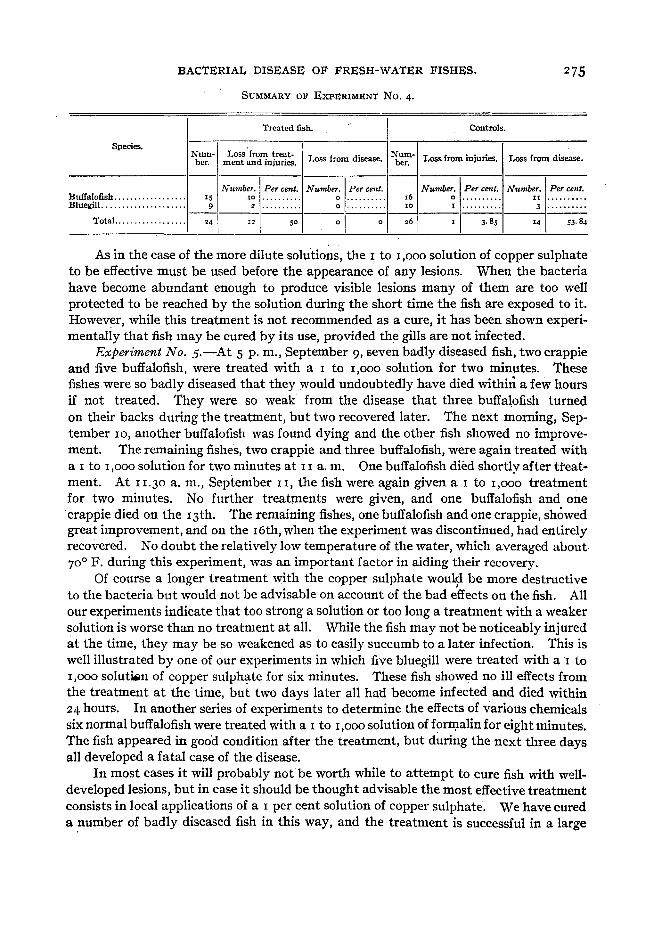
BACTERIAL DISEASE OF FRESH-WATER FISHES.
SUMMARY OF ExpnRIMENl' NO.4.
275
Treated fish..
Controls.
Species. -Num- Loss from treat- Loss from disease. Num- Loss from injuries. Loss from disease.ber, ment and injuries. ber,
Number. Per cent. Number. Per cent. Number. Per cent. Number. Per cent.Buffalofish .................. 'S 10 .......... 0 .......... 16 0 .......... II ..........Bluegill ..................... 9 2 .......... 0 .......... 10 I .......... 3 ..........----------------------------
Total.. ................ 24 I12 50 0 0 26 I 3· 85 14 53· 84
As in the case of the more dilute solutions, the I to 1,000 solution of copper sulphateto be effective must be used before the appearance of any lesions. When the bacteriahave become abundant enough to produce visible lesions many of them are too wellprotected to be reached by the solution during the short time the fish are exposed to it.However, while this treatment is not recommended as a cure, it has been shown experimentally that fish may be cured by its use, provided the gills are not infected.
Experiment No. 5.-At 5 p. m., September 9, seven badly diseased fish, two crappieand five buffalofish, were treated with a I to 1,000 solution for two minutes. Thesefishes were so badly diseased that they would undoubtedly have died within a few hoursif not treated. They were so weak from the disease that three buffalofish turnedon their backs during the treatment, but two recovered later. The next morning, September 10, another buffalo fish was found dying and the other fish showed no improvement. The remaining fishes, two crappie and three buffalofish, were again treated witha I to 1,000 solution for two minutes at I I a. m. One buffalofish died shortly after treatment. At 11.30 a. m., September II, the fish were again given a I to 1,000 treatmentfor two minutes. No further treatments were given, and one buffalofish and one.crappie died on the 13th. The remaining fishes, one buffalofish and one crappie, showedgreat improvement, and on the 16th, when the experiment was discontinued, had entirelyrecovered. No doubt the relatively low temperature of the water, which averaged about70° F. during this experiment, was an important factor in aiding their recovery.
Of course a longer treatment with the copper sulphate would be more destructiveto the bacteria but would notbe advisable on account of the bad effects on the fish. Allour experiments indicate that too strong a solution or too long a treatment with a weakersolution is worse than no treatment at all. While the fish may not be noticeably injuredat the time, they may be so weakened as to easily succumb to a later infection. This iswell illustrated by one of our experiments in which five bluegill were treated with ar to1,000 solutisn of copper sulphate for six minutes. These fish showed no ill effects fromthe treatment at the time, but two days later all hat! become infected and died within24 hours. In another series of experiments to determine the effects of various chemicalssix normal buffalofish were treated with a I to 1,000 solution of formalin for eight minutes.The fish appeared in good condition after the treatment, but during the next three daysall developed a fatal case of the disease.
In most cases it will probably notbe worth while to attempt to cure fish with welldeveloped lesions, but in case it should be thought advisable the most effective treatmentconsists in local applications of a I per cent solution of copper sulphate. We have cureda .number of badly diseased fish in this way, and the treatment is successful in a large
BULLETIN OF THE BUREAU OF FISHERIES.
percentage of cases, provided the gills are not affected. The solution can best be appliedby gently swabbing the lesion with a small piece of cotton which has been previouslydipped in the solution. Two or three applications at intervals of 6 to 12 hoursshould be sufficient. After each local application of the copper sulphate the fish shouldbe placed in a 1 to 1,000 solution for one minute.
Since, in most cases, the disease is primarily due to injuries or weakened vitalitythe old adage, "An ounce of prevention is worth a pound of cure," is peculiarly applicable in this case. Above everthing else, anyone handling fish should exercise thegreatest care to prevent injuring the fish in any way. Even the slightest injury, suchas the rubbing off of a few scales or even a small portion of the mucous covering, maylead to infection. When taken in a net or seine the delicate caudal fin is very easilyinjured by the struggles of the captured fish and every effort should be made to reducethe injury to a minimum. A large percentage of infections among fish taken in this wayfirst appear on the caudal fin, and undoubtedly injury to the fins is one of the mostcommon causes of infection.
Great care should be taken to prevent the spread of the disease through the useof infected nets or vessels. This can be easily prevented, since the bacteria are entirelydestroyed by thorough drying for several hours in direct sunlight.
Finally, in the case of fish confined in aquaria it is essential that all infected individuals be removed at once. As in the case of any contagious disease all contact ofhealthy with diseased individuals should be guarded against. It has been shown thatbacteria are continually leaving diseased fish and thus may. readily get on any healthyfish in the same aquarium.
It should be said in passing that this treatment is also very effective for ectoparasiticProtozoa. Experiments on fish infected with Costia, Chilodon, and Cyclochteta haveshown that these parasites are entirely destroyed by a single treatment with coppersulphate. In the case of fish infected with Ichthyophthirius the treatment is not sosuccessful, since the encysted stages are not affected. The exposed parasites are, however, entirely destroyed.
A number of other chemicals have been tried, but none of them has given encouraging results. Lysol and creolin were found to stop all movements of the bacteria infour to five minutes when diluted 1 to 5,000. The use of such strong solutions is, however, not practicable, since buffalofish andbullheads placed in a I to 5,000 solution for1 minute died within 24 hours. Formalin was also found to be of no value for the samereason.
Since sodium chloride is used- a great deal by fish-culturists for fungus a numberof infected fish were treated with solutions of various strengths. It was"soon foundthat the bacteria are not appreciably affected by solutions which seriously injure thefish,' and it 'is not believed that this treatment is of any value against the disease. Infact our experiments indicate that it may actually aggravate the disease by weakeningthe fish..
ECONOMIC IMPORTANCE OF THE DISEASE.
At the present time it is impossible to make any general statements regarding theimportance of this disease. So far, with the exception of two slightly infected fish atOgdensburg, N. Y., the disease has been recognized only at Fairport. Here it has caused
BACTERIAL DISEASE OF FRESH-WATER FISHES.
a very considerable mortality each summer for several years. This mortality for themost part has occurred in fishes which were being transferred from one pond to another,or to aquaria for experimental purposes. It was not until the past summer (1919) thatthe disease was shown to occur on fish in the Mississippi River as well as in the ponds.However, fish-culturists are well aware that is is impossible to handle many species offish during warm weather with any degree of success. Undoubtedly part of this mortality among fish which have been handled is due to the direct effects of the treatmentto which they have been subjected, but there is no question in the writer's mind that amuch larger part is directly due to this disease and only indirectly to the handling whichhas simply rendered the fish more susceptible to infection. We are convinced that agreat part of the mortality usually ascribed to fungus is in reality caused by Bacilluscolumnaris. In fact the writer is inclined to doubt if fungus is ever an important causeof fish mortality. In all probability Bacillus columnaris is widely distributed over thecountry and during warm weather, at least, is the most important agent in the destruction of fish which have been injured in any way.
Anglers are often advised to remove small fish from the hook and return them tothe water. In the light of our experience with the disease at Fairport it is doubtfulif many fish which have been handled in this way actually survive. Since the diseasewould not make its appearance until two or three days later, it is obvious that onlythrough carefully conducted experiments can their chance of survival be ascertained.An incident which occurred at Fairport during the summer of 1918 is very suggestivein this connection. A number of largemouth black bass were needed for some experimental work in mussel propagation. They were taken from one of the ponds on a hookand line and placed in a large tank supplied with running water. Within two or threedays nearly every fish had become infected with Bacillus columnaris. As previously mentioned, this species of fish is not very susceptible to the disease, and, furthermore, thefish in this case were several years old. As the reader will recall the,'~lder fish are muchless susceptible than the young.
During the last few years the Bureau of Fisheries has been rescuing large numbersof young fishes from the small pools and ponds in which they are imprisoned when thespring floods recede. These pools and ponds are widely scattered over the flood plainof the Mississippi, often only a few hundred yards from the river. In some instancesthey xp.ay be 2 or 3 miles from the main channel. As the waters recede these pondscontinually grow smaller and may eventually become entirely dry. They are oftencrowded with fish and the shallow waters exposed to the hot sun of July and Augustbecome 'heated to a relatively high temperature. These conditions combined with alimited supply of oxygen often result in the death of large numbers of fish. The conditions are such as to render the fish susceptible to infection by Bacillus columnaris, andfishermen have told the writer of having seen large numbers of diseased' fish in suchponds.
In transferring the fish to the river they must first be seined from the ponds, aprocess which. necessitates considerable rough handling, and then carried in galvanizedwashtubs (often under a broiling sun) to the river. It is obvious that such treatmentis likely to result in infection by Bacillus columnaris. That the fish when liberated in
278 BULLETIN OF THE BUREAU OF FISHERIES.
the river quickly swim away and are seen no more proves nothing. The disease wouldnot be evident for two or three days in any case.
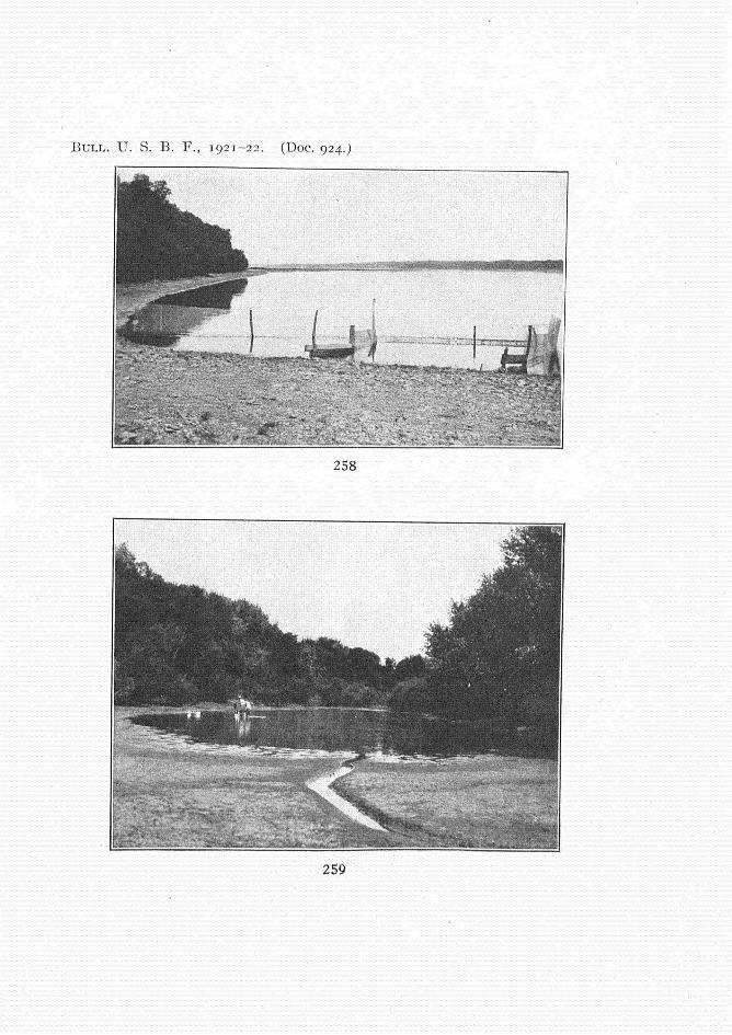
In order to determine the prevalence of the disease among rescued fishes a longseries of experiments will be required, but owing to the limited time at the writer'sdisposal only a few preliminary experiments have as yet been carried out. In theseexperiments a special effort was made to duplicate so far as possible the average conditiohs found in rescue work. The fish were seined from small, isolated ponds, one ofwhich is shown in figure 259, and then carried in washtubs to the river, where they wereplaced on a launch to be carried to the station. These fish were in the tubs from one-halfto one hour, which the writer was assured by the fishermen is somewhat less than theaverage time they are held in tubs in ordinary rescue work. On reaching the stationthe fish were at once placed in an inclosure surrounded by fine-meshed poultry wire.This inclosure (fig. 258) was constructed in shallow water along the river shore and wassufficiently large to obviate any danger of the fish being crowded. A small streamcarrying the overflow from the fishponds flowed into the inclosure, so there was a goodsupply of running water at all times. The cross partition shown in the figure was addedlater for some special experiments which are not considered in this paper. It is believedthat the fish held in this inclosure were in fully as favorable an environment as theywould have been if liberated in the river.
Experiment No. 6.-September 2, 1919, 42 buffalofish, 41 crappie, and 8 bluegillwere seined from the pond shown in figure 259. This pond was believed to be typicalof those from which fish are taken in rescue work. It was about 400 feet long by 100feet wide and quite shallow, being not over 3 or 4 feet in depth at the deepest part. Thebottom, as in most ponds of this kind, was composed of fine, soft mud. The temperature of the water at the time the fish were removed was 83° F. All the fishappeared in good condition when placed in the inclosure. The next day (Sept. 3)six crappie were found dead. Since no bacteria could be found on these fish they arebelieved to have died from injuries due to handling. September 4 one badly diseasedbuffalo fish was removed, and the next day there were two dead buffalofish, both withwell-developed lesions. The inclosure was seined on September 6 and all the fish removed.Only four dead fish were found, three buffalofish and one crappie. However, many ofthe living fish were badly diseased and in all probability would have died in a short time,but it was not thought best to leave them in the inclosure longer, since there wasgreat danger of the disease spreading from the infected to the healthy fish. It is notbelieved that arty of the fish which are classed as infected at this time had contractedthe disease in this way, since all were in advanced stages of the disease. The 'diseasedfishes included 19 buffalofish and 10 crappie. They were removed to aquaria well suppliedwith running water, but all died within 24 hours. It is believed justifiable to assumethat all the fish classed as diseased became infected as a result of the treatment to which'they had been subjected and would have eventually died if set free in the river.

BACTERIAL DISEASE OF FRESH-WATER FISHES.
SUMMARY 01" EXPSRIMSNT No.6.
279
Species. Number Fish lost from Fish lost from Total loss of fish.01fish. injuries, disease.
Number. Per cent. Number. Per cent. Number. Per cent,Buffalofish............................................ 4' 0 0 '5 59· 5' '5 59· 5'Crappie .............................................. 4' 6 '4.63 II .6.83 '7 4"46Bluegill. ............................................. 8 0 0 0 0 0 0
Experiment No. 7.--September 6, 1919,36 buffalofish and 3 crappie were seined fromthe same pond as in the preceding experiment and placed in the inclosure. The nextday eight buffalofish were found dead, evidentlyfrom the effects of injuries due to handling.All the fish were removed from the inclosure on September 9. There were 17 deadbuffalofish, 8 living badly diseased buffalofish, and 3 badly diseased crappie. Theremaining fish were in good condition.
SUMMARY OF EXPSRIMSNT NO.7.
-
Species. Number Fish lost from Fish lost from Total loss of fish.01fish. injuries. disease.
Number.! Per cent. Number. Per cent. Number.! Per cent.Buffalofish............................................ 36 8 22.22 '5 6g'44 33 9'· 67Crappie............................................... 3 o 0 3
.'00 3 '00
Experiment No. 8.--Since there was a possibility in the above experiments that thefish might have injured themselves on the poultry wire in trying to escape from theinclosure a fine-meshed seine was attached to stakes so as to form another inclosure bythe side of the one constructed of poultry netting.. September 10, 2 I buffalofish and 12
crappie were placed in this inclosure. These fish were taken from a different pond fromthat used in the preceding experiments, but conditions were much the same, exceptthat the pond was somewhat farther from the station.
SUMMARY 0.1' EXPERIMENT No.8.
Species, Number Fish lost lrom Fish lost lrom Total loss of fish.01fish. iniuries. disease.
N~'nber'! Pcr cent. NUmber'l Per cent. Number. [ Per cent.Bufialofish............................................ ar o .0 12 57. 14 I2 57· '4Crappie... , ........................................... I2 8 66.67 • ,6,67 10 83.33
While the above experiments are only preliminary and too much weight shouldnot be 'placed on the results, it is evident that the conditions are especially favorablefor the development of this disease among rescued fish and that in all probability itcauses a very considerable mortality during warm weather. Much of the rescue workis carried on in the fall and early winter, and with the low temperatures then prevailingthere is no doubt much less danger of infection by bacteria, but on this point we haveno data at present.
It is obvious that great care should be exercised to injure the fish as little as possible and that, whenever practicable, rescue operations should not be undertaken until

280 BULLETIN OF THE BUREAU OF 'FISHERIES.
cool weather. If the fish have not been greatly weakened by unfavorable conditions,especially lack of oxygen, treatment with 1 to 1,000 copper sulphate just before they areliberated in the river would doubtless cause a marked decrease in the loss from disease.Some experiments were attempted along this line in September, 1919, but owing to anaccident the results were of no value.
EXPLANATION OF FIGURES.
All figures, with the exception of figure 249, are from photographs by the author. Abbreviationsused are as follows: bl, blood; cor, corium; ep, epidermis; and mus, muscles.
FIGS. 231 and 232.--Crappie, Pomoxis sparoides, with dorsal, caudal, and anal fins infected. Notelesions just starting in three places on anal fin of fish in figure 232.
FIG. 233.-Bluegill, Lepomis incisor, with dorsal, caudal, and anal fins infected.FIGS. 234 and 235.-Firigerling buffalofish, Ictiobus bubalus, with caudal fins badly infected.FIG. 236.-Largemouth black bass, Micropterus salmoides, showing infection on dorsal, anal, and
caudal fins.FIG. 237.-White bass, Roccus chrysops, with infection developing on dorsal, anal, and caudal fins.FIG. 238.-Bullhead, Ameiurus melas, with a number of small lesions on dorsal surface of body.FIGS. 239 and 241.-Bullheads, Ameiurus melas, in late stages of the disease.FIG. 240.-Bullhead, Ameiurus melas, with nearly entire side of body covered with lesions.FIG. 242.-Small bullhead, Ameiurus melas, with posterior end of the body infected and another
lesion just back of pectoral fin.FIG. 243.-Head of crappie, Pomoxis sparoides, with operculum removed to show small lesion on
gill.FIG. 244.-Head of buffalofish, Ictiobus bubalus, with operculum removed to show a somewhat larger
lesion on gill than in figure 243.FIG. 245.-Head of buffalofish, lctiobus cyprinella, with operculum removed to show gills in late
stages of the disease.FIGS. 246 and 247.-Bacillus columnaris. From a dried smear stained with carbolfuchsin. X 800.FIG. 248.-Small portion of scale of bluegill to show formation of columns by bacteria along the
edge. Photomicrograph from preparation mounted in glycerin jelly and stained with eosin. Columnsare more slender and pointed than normal, due to shrinkage by the preserving fluid.
FIG. 249.-Showing formation of columns by bacteria along edge of a bit of infected tissue afterbeing removed to slide. Somewhat diagrammatic. X340.
FIGS. 250 to 257 are from cross sections through lesions in integument of bullhead, Ameiurus melas,FIG. 25o.-Lesion just beginning to develop. X 70.FIG. 25I.-A little later stage in development of lesion. The epidermis is entirely destroyed at
one place to the right in the figure. X 70.FIG. 252.-A somewhat later stage than figure 251. The corium is beginning to disintegrate where
the overlying epidermis has been destroyed. X 70.FIG. 253.-Cross-section through lesion in late stage of disease. The corium is entirely destroyed
at center of lesion. Less highly magnified than preceding figures.FIG. 254.-Showing disintegration of epidermis in early stage of development of lesion. X 108.FIGS. 255 to 257.-Sections through margin of well-developed lesions, the epidermis at the right
has been entirely destroyed. Note the blood corpuscles (bl) in the outer layer of the corlum and betweenthe corium and the epidermis. In figure 255 the blood has broken through into the epidermis for ashort distance. X 108.
FIG. 258.-Inclosure constructed of poultry netting in which fish were held in experiments 6, 7,and 8.
FIG. 259.-5mall pond from which fish were taken in experiments 6 and 7.
BULL. U. S. B. F ., 192 1-22. (Doc. 924.)
23 1
232
233
BULL. U. S. B . F ., 1921- 22. (Doc. 924.)
234 023 5
236
237

BULL. U. S. B. F " 1921- 22. (Doc. 924.)
238
239
240
241
BULL. U. S. B. F ., 1921- 22. (Doc. 924.)
243
242
246
247
244
245
248

BULL. U. S. B . F., 1921-22. (Doc. 9 24..)
249
250 252
251
ep
cor
253
- ,-e pcor

B ULL. U. S. B . F ., 1921 - 22 .
254
e p
cor
255
256
=--::-- bl
cor'
""';>---'jo,o~-.:r-j-- ep
~-T--b l
cor
257
B ULL. U. S. B. F ., 19 2 1- 22 . (Doc. 92 4 . )
258
259