A new antibiotic with potent activity targets...
10
OPEN ORIGINAL ARTICLE A new antibiotic with potent activity targets MscL Irene Iscla 1 , Robin Wray 1 , Paul Blount 1 , Jonah Larkins-Ford 2 , Annie L Conery 2 , Frederick M Ausubel 2 , Soumya Ramu 3 , Angela Kavanagh 3 , Johnny X Huang 3 , Mark A Blaskovich 3 , Matthew A Cooper 3 , Andres Obregon-Henao 4 , Ian Orme 4 , Edwin S Tjandra 5 , Uwe H Stroeher 6 , Melissa H Brown 6 , Cindy Macardle 7 , Nick van Holst 7 , Chee Ling Tong 8 , Ashley D Slattery 8 , Christopher T Gibson 8 , Colin L Raston 8 and Ramiz A Boulos 8 The growing problem of antibiotic-resistant bacteria is a major threat to human health. Paradoxically, new antibiotic discovery is declining, with most of the recently approved antibiotics corresponding to new uses for old antibiotics or structurally similar derivatives of known antibiotics. We used an in silico approach to design a new class of nontoxic antimicrobials for the bacteria-specific mechanosensitive ion channel of large conductance, MscL. One antimicrobial of this class, compound 10, is effective against methicillin-resistant Staphylococcus aureus with no cytotoxicity in human cell lines at the therapeutic concentrations. As predicted from in silico modeling, we show that the mechanism of action of compound 10 is at least partly dependent on interactions with MscL. Moreover we show that compound 10 cured a methicillin-resistant S. aureus infection in the model nematode Caenorhabditis elegans. Our work shows that compound 10, and other drugs that target MscL, are potentially important therapeutics against antibiotic-resistant bacterial infections. The Journal of Antibiotics (2015) 68, 453–462; doi:10.1038/ja.2015.4; published online 4 February 2015 INTRODUCTION The overprescription of antibiotics and failure of patients to complete antibiotic treatment regimens have contributed to the emergence of bacterial multi-drug resistance (MDR). At the same time, the large costs involved in developing new drugs, exacerbated by a complicated drug approval and patent process, 1 have caused a dearth in new antibiotic research with many pharmaceutical companies choosing to focus their efforts on more profitable, higher volume drugs. 2,3 As a result, fighting MDR bacterial infections in patients is becoming increasingly difficult with treatment options becoming very limited. 4,5 Furthermore, there are relatively few novel small molecules in the antibiotic development pipeline. 6 The mechanosensitive ion channel of large conductance (MscL) in bacteria is an attractive target for drug discovery because of its high level of conservation in bacterial species, and its absence from the human genome. Such level of conservation suggests that the channel has an important and conserved function, which has recently been highlighted as one of the top 20 targets for drug development. 7 In Escherichia coli, the transmembrane MscL channel consists of five identical subunits, each composed of 136 amino acids. 8,9 MscL has the largest pore size of any gated ion channel, estimated to be 28Å when fully open. 10,11 Mechanosensitive channels have evolved to sense mechanical tension on the membrane and convert it into an electrochemical response. As such, they act as gatekeepers, protecting bacterial cells against lysis following acute decrease in the osmotic environment. Moreover, these channels can act as entry points for drugs and other small molecules into bacterial cells. In this paper, we describe the in silico design of MscL ligands, which led to the discovery of a novel class of compounds with optimal binding to MscL. One of these ligands, 1,3,5-tris[(1E)-2′-(4″-benzoic acid)vinyl]benzene (referred to hereafter as 10, Ramizol), is an effective antimicrobial against methicillin-resistant Staphylococcus aur- eus (MRSA). 12 Using microscopic analysis and other techniques, we show that the mechanism of action of 10 in Gram-positive and Gram- negative bacteria involves its interaction with MscL. We also show that 10 exhibits in vivo efficacy in a Caenorhabditis elegans nematode infection model. Moreover, 10 exhibits low levels of toxicity in addition to being a potent antioxidant, 13 potentially providing an additional benefit by reducing bacterial-induced inflammation. RESULTS In silico design of ligands targeting MscL To explore the potential of MscL as a target for antibiotics, we developed a spatial map between the exposed oxygen atoms of amino acids, lining the gate of the MscL channel. This three-dimensional spatial map was used for the de novo design 14 of several potential 1 Department of Physiology, UT Southwestern Med Ctr, Dallas, TX, USA; 2 Department of Molecular Biology, Massachusetts General Hospital, Boston, MA, USA; 3 Institute for Molecular Bioscience, University of Queensland, St Lucia, QLD, Australia; 4 Department of Microbiology, Immunology, and Pathology, Colorado State University, Fort Collins, CO, USA; 5 School of Animal Biology, The University of Western Australia, Crawley, WA, Australia; 6 School of Biological Sciences, Flinders University, Bedford Park, SA, Australia; 7 Flinders Medical Science and Technology, Immunology, Allergy and Arthritis, Flinders University, Bedford Park, SA, Australia and 8 Centre for NanoScale Science and Technology, School of Chemical and Physical Sciences, Flinders University, Bedford Park, SA, Australia Correspondence: Paul Blount or Dr R Boulos, Centre for NanoScale Science and Technology, School of Chemical and Physical Sciences, Flinders University, Bedford Park, South Australia 5042, Australia. E-mail: [email protected] or ramiz.boulos@flinders.edu.au Received 19 September 2014; revised 27 November 2014; accepted 15 December 2014; published online 4 February 2015 The Journal of Antibiotics (2015) 68, 453–462 & 2015 Japan Antibiotics Research Association All rights reserved 0021-8820/15 www.nature.com/ja
-
Upload
nguyenhanh -
Category
Documents
-
view
215 -
download
0
Transcript of A new antibiotic with potent activity targets...

OPEN
ORIGINAL ARTICLE
A new antibiotic with potent activity targets MscL
Irene Iscla1, Robin Wray1, Paul Blount1, Jonah Larkins-Ford2, Annie L Conery2, Frederick M Ausubel2,Soumya Ramu3, Angela Kavanagh3, Johnny X Huang3, Mark A Blaskovich3, Matthew A Cooper3,Andres Obregon-Henao4, Ian Orme4, Edwin S Tjandra5, Uwe H Stroeher6, Melissa H Brown6,Cindy Macardle7, Nick van Holst7, Chee Ling Tong8, Ashley D Slattery8, Christopher T Gibson8,Colin L Raston8 and Ramiz A Boulos8
The growing problem of antibiotic-resistant bacteria is a major threat to human health. Paradoxically, new antibiotic discovery
is declining, with most of the recently approved antibiotics corresponding to new uses for old antibiotics or structurally similar
derivatives of known antibiotics. We used an in silico approach to design a new class of nontoxic antimicrobials for the
bacteria-specific mechanosensitive ion channel of large conductance, MscL. One antimicrobial of this class, compound 10, is
effective against methicillin-resistant Staphylococcus aureus with no cytotoxicity in human cell lines at the therapeutic
concentrations. As predicted from in silico modeling, we show that the mechanism of action of compound 10 is at least partly
dependent on interactions with MscL. Moreover we show that compound 10 cured a methicillin-resistant S. aureus infection in
the model nematode Caenorhabditis elegans. Our work shows that compound 10, and other drugs that target MscL, are
potentially important therapeutics against antibiotic-resistant bacterial infections.
The Journal of Antibiotics (2015) 68, 453–462; doi:10.1038/ja.2015.4; published online 4 February 2015
INTRODUCTION
The overprescription of antibiotics and failure of patients to completeantibiotic treatment regimens have contributed to the emergence ofbacterial multi-drug resistance (MDR). At the same time, the largecosts involved in developing new drugs, exacerbated by a complicateddrug approval and patent process,1 have caused a dearth in newantibiotic research with many pharmaceutical companies choosing tofocus their efforts on more profitable, higher volume drugs.2,3 As aresult, fighting MDR bacterial infections in patients is becomingincreasingly difficult with treatment options becoming very limited.4,5
Furthermore, there are relatively few novel small molecules in theantibiotic development pipeline.6
The mechanosensitive ion channel of large conductance (MscL) inbacteria is an attractive target for drug discovery because of its highlevel of conservation in bacterial species, and its absence from thehuman genome. Such level of conservation suggests that the channelhas an important and conserved function, which has recently beenhighlighted as one of the top 20 targets for drug development.7 InEscherichia coli, the transmembrane MscL channel consists of fiveidentical subunits, each composed of 136 amino acids.8,9 MscL has thelargest pore size of any gated ion channel, estimated to be 28 Å whenfully open.10,11 Mechanosensitive channels have evolved to sensemechanical tension on the membrane and convert it into an
electrochemical response. As such, they act as gatekeepers, protectingbacterial cells against lysis following acute decrease in the osmoticenvironment. Moreover, these channels can act as entry points fordrugs and other small molecules into bacterial cells.In this paper, we describe the in silico design of MscL ligands, which
led to the discovery of a novel class of compounds with optimalbinding to MscL. One of these ligands, 1,3,5-tris[(1E)-2′-(4″-benzoicacid)vinyl]benzene (referred to hereafter as 10, Ramizol), is aneffective antimicrobial against methicillin-resistant Staphylococcus aur-eus (MRSA).12 Using microscopic analysis and other techniques, weshow that the mechanism of action of 10 in Gram-positive and Gram-negative bacteria involves its interaction with MscL. We also show that10 exhibits in vivo efficacy in a Caenorhabditis elegans nematodeinfection model. Moreover, 10 exhibits low levels of toxicity inaddition to being a potent antioxidant,13 potentially providing anadditional benefit by reducing bacterial-induced inflammation.
RESULTS
In silico design of ligands targeting MscLTo explore the potential of MscL as a target for antibiotics, wedeveloped a spatial map between the exposed oxygen atoms of aminoacids, lining the gate of the MscL channel. This three-dimensionalspatial map was used for the de novo design14 of several potential
1Department of Physiology, UT Southwestern Med Ctr, Dallas, TX, USA; 2Department of Molecular Biology, Massachusetts General Hospital, Boston, MA, USA; 3Institute forMolecular Bioscience, University of Queensland, St Lucia, QLD, Australia; 4Department of Microbiology, Immunology, and Pathology, Colorado State University, Fort Collins, CO,USA; 5School of Animal Biology, The University of Western Australia, Crawley, WA, Australia; 6School of Biological Sciences, Flinders University, Bedford Park, SA, Australia;7Flinders Medical Science and Technology, Immunology, Allergy and Arthritis, Flinders University, Bedford Park, SA, Australia and 8Centre for NanoScale Science andTechnology, School of Chemical and Physical Sciences, Flinders University, Bedford Park, SA, AustraliaCorrespondence: Paul Blount or Dr R Boulos, Centre for NanoScale Science and Technology, School of Chemical and Physical Sciences, Flinders University, Bedford Park, SouthAustralia 5042, Australia.E-mail: [email protected] or [email protected] 19 September 2014; revised 27 November 2014; accepted 15 December 2014; published online 4 February 2015
The Journal of Antibiotics (2015) 68, 453–462& 2015 Japan Antibiotics Research Association All rights reserved 0021-8820/15www.nature.com/ja
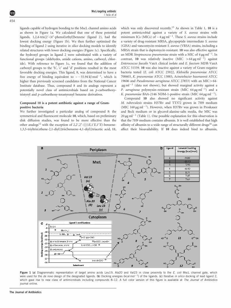
ligands capable of hydrogen bonding to the MscL channel amino acidsas shown in Figure 1a. We calculated that one of these potentialligands, 1,2,4-tris[2′-(4″-phenol)ethyl]benzene (ligand 2), had thelowest docking energy (Figure 1b). We then further optimized thebinding of ligand 2 using iterative in silico docking models to identifyrelated structures with lower docking energies (Figure 1c). Specifically,the hydroxyl groups in ligand 2 were substituted with a variety offunctional groups (aldehydes, amide cations, amino, carboxyl, chlor-ide). With reference to Figure 1c, we found that the addition ofcarboxyl groups to the ‘b’, ‘c’ and ‘d’ positions resulted in the mostfavorable docking energies. This ligand, 8, was determined to have afree energy of binding equivalent to ~− 55.94 kJ mol− 1, which ishigher than previously screened candidates from the National CancerInstitute database. Thus, compound 8 and its analogs represent apotentially novel class of antimicrobials based on p-carboethoxy-tristyryl and p-carboethoxy-terastyrenyl benzene derivatives.
Compound 10 is a potent antibiotic against a range of Gram-positive bacteriaWe further investigated a particular analog of compound 8: thesymmetrical and fluorescent molecule 10, which, based on preliminarydisk diffusion studies, was found to be more effective than theother analogs12 with the exception of 2,2′,2″-{[(1E,1′E,1″E)-benzene-1,3,5-triyltris(ethene-2,1-diyl)]tris(benzene-4,1-diyl)}triacetic acid, 11,
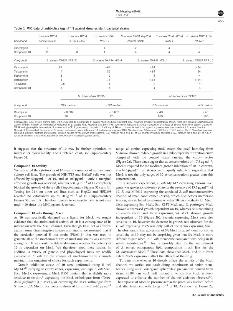
which was only discovered recently.15 As shown in Table 1, 10 is apotent antimicrobial against a variety of S. aureus strains withminimum ICs (MICs) of ~ 4 μg ml− 1. These S. aureus strains includea variety of drug-resistant MRSA, glycopeptide intermediate S. aureus(GISA) and vancomycin-resistant S. aureus (VRSA) strains, including aMRSA strain that is daptomycin-resistant. 10 was also effective againstan MDR Streptococcus pneumoniae strain with a MIC of 4 μgml− 1. Incontrast, 10 was relatively inactive (MIC 464 μg ml− 1) againstEnterococcus faecalis VanA clinical isolate and E. faecium MDR-VanAATCC 51559. 10 was also inactive against a variety of Gram-negativebacteria tested (E. coli ATCC 25922, Klebsiella pneumoniae ATCC700603, K. pneumoniae ATCC 13883, Acinetobacter baumannii ATCC19606 and Pseudomonas aeruginosa ATCC 27853) with an MIC464-μgml− 1 (data not shown), but showed marginal activity against aP. aeruginosa polymyxin–resistant strain (MIC 64 μg ml− 1) and aK. pneumoniae BAA-2146 NDM-1-positive strain (MIC 64 μgml− 1).Compound 10 also showed no significant activity against
M. tuberculosis strains H37Rv and TT372 grown in 7H9 medium(MIC 160 μg ml− 1). However, when H37Rv was grown in Proskauerand Beck medium or in glycerol-alanine-salts media, the MIC was20 μg ml− 1 (Table 1). One possible explanation for this observation isthat the 7H9 medium contains albumin. It is well established that highaffinity of albumin to a wide range of structurally different drugs16 canaffect their bioavailability. If 10 does indeed bind to albumin,
Figure 1 (a) Diagrammatic representation of target amino acids Leu19, Ala20 and Val23 in close proximity to the E. coli MscL channel gate, whichwere used for the de novo design of the designated ligands. (b) Docking energies (kcal mol−1) of the ligands. (c) Iterative in silico docking of lead ligand 2,which gave rise to new class of antimicrobials including compounds 8–12. A full color version of this figure is available at The Journal of Antibioticsjournal online.
MscL-targeting antibioticI Iscla et al
454
The Journal of Antibiotics
it suggests that the structure of 10 may be further optimized toincrease its bioavailability. For a detailed chart, see SupplementaryFigure S1.
Compound 10 toxicityWe measured the cytotoxicity of 10 against a number of human tissueculture cell lines. The growth of NIH/3T3 and HaCaT cells was notaffected by 50 μg ml− 1 of 10, and at 100 μgml− 1 only a marginaleffect on growth was observed, whereas 500 μg ml− 1 of 10 completelyblocked the growth of these cells (Supplementary Figures S2a and b).Testing for 24 h on other cell lines such as HepG2 and HEK293revealed no cytotoxicity up to 50 μg ml− 1 of 10 (SupplementaryFigures S2c and d). Therefore toxicity to eukaryotic cells is not seenuntil ~ 10 times the MIC against S. aureus.
Compound 10 acts through MscLAs 10 was specifically designed as a ligand for MscL, we soughtevidence that the antimicrobial activity of 10 is a consequence of itsinteraction with the MscL channel. Even though 10 is not as effectiveagainst some Gram-negative species and strains, we reasoned that ifthe particular parental E. coli strain (FRAG-1) that was used togenerate all of the mechanosensitive channel null strains was sensitiveenough to 10, we should be able to determine whether the potency of10 is dependant on MscL. We therefore tested these strains. Inaddition, a variety of genetic and physiological tools are readilyavailable in E. coli for the analysis of mechanosensitive channelsmaking it the organism of choice for such experiments.Growth inhibition assays of 10 were performed using E. coli
MJF61217 carrying an empty vector, expressing wild-type E. coli MscL(Eco MscL), expressing a MscL K55T mutant that is slightly moresensitive to tension,9 expressing the MscL orthologues from Clostri-dium perfingens (CP MscL), or expressing the MscL orthologue fromS. aureus (SA MscL). For concentrations of 10 in the 7.5–10 μgml− 1
range, all strains expressing mscL except the mscL homolog fromS. aureus showed reduced growth in a pilot-experiment titration curvecompared with the control strain carrying the empty vector(Figure 2a). These data suggest that at concentrations of ~ 7.5 μgml− 1,MscL is required for the mediated growth inhibition of 10. In contrast,at ~ 16.5 μgml− 1, all strains were equally inhibited, suggesting thatMscL is not the only target of 10 at concentrations greater than thisconcentration.In a separate experiment, E. coli MJF612 expressing various mscL
genes was grown to stationary phase in the presence of 13.5 μgml− 1 of10. E. coli MJF612 expressing the unrelated E. coli mechanosensitivechannel of small conductance (MscS), which also detects membranetension, was included to examine whether 10 has specificity for MscL.Cells expressing Eco MscL, Eco K55T MscL and C. perfringens MscLshowed a decreased growth dependant on 10, whereas cells containingan empty vector and those expressing SA MscL showed growthindependent of 10 (Figure 2b). Bacteria expressing MscS were alsosensitive to 10, however the decrease in growth rate observed for theE. coli expressing MscS was only half of the strain expressing MscL.The observation that expression of SA MscL in E. coli does not confersensitivity to 10 may not be surprising given that SA MscL is moredifficult to gate when in E. coli membrane compared with being in itsnative membranes.18 This is possibly due to the requirementof S. aureus endogenous lipid composition much like for theM. tuberculosis MscL.19 These data show that MscL, and to a lesserextent MscS expression, affect the efficacy of the drug.To determine whether 10 directly affects the activity of the MscL
channel, we carried out patch-clamp experiments of native mem-branes using an E. coli ‘giant’ spheroplast preparation derived fromstrain PB104 (an mscL null mutant in which Eco MscL is over-expressed to enhance the number of channel activities observed20).The response of MscL to pressure across the patch was assessed beforeand after treatment with 25 μgml− 1 of 10. As shown in Figure 2c,
Table 1 MIC data of antibiotics (μgml−1) against drug-resistant bacterial strains
Compound
S. aureus MRSA
clinical isolate
S. aureus MRSA
ATCC 43300
S. aureus GISA
NRS 17
S. aureus MRSA DapRSA
clinical isolate
S. aureus GISA, MRSA
NRS 1
S. pneum MDR ATCC
700677
Vancomycin 1 1 4 2 4 1
Compound 10 8 8 4 4 4 4
Compound S. aureus NARSA VRS 3b S. aureus NARSA VRS 4 S. aureus NARSA VRS 1 S. aureus NARSA VRS 10
Vancomycin 64 464 464 464
Teicoplanin 4 32 464 64
Daptomycin 1 2 4 4
Dalbavancin 1 16 464 464
Telavancin 0.5 1 2 4
Compound 10 4 4 4 4
M. tuberculosis H37Rv M. tuberculosis TT372
Compound GAS medium P&B medium 7H9 medium 7H9 medium
Rifampicin o0.002 o0.002 o40 o40
Compound 10 20 20 160 160
Abbreviations: GAS, glycerol-alanine-salts; GISA, glycopeptide intermediate S. aureus; MDR, multi-drug resistant; MIC, minimum inhibitory concentration; MRSA, methicillin-resistant Staphylococcusaureus; NARSA, Network of Antimicrobial Resistance in S. aureus; P&B, Proskauer and Beck; VRS, vancomycin-resistant S. aureus. Comparison of efficacy of 10 and vancomycin against a panel ofMRSA and glycopeptide intermediate S. aureus, and MDR S. pneumonia, comparison of efficacy of 10 and commercial antibiotics against a panel of vancomycin-resistant S. aureus from theNetwork of Antimicrobial Resistance in S. aureus, and comparison of efficacy of 10 and rifampicin against MDR Mycobacterium tuberculosis H37RV and TT372 strains. The 7H9 medium containsoleic acid, albumin, dextrose and catalase, which is needed for the growth of the bacteria. GAS medium has a final pH of 6.6 and the Proskauer and Beck (P&B) medium has a final pH of 7.4. Afull color version of this table is available at The Journal of Antibiotics journal online.
MscL-targeting antibioticI Iscla et al
455
The Journal of Antibiotics
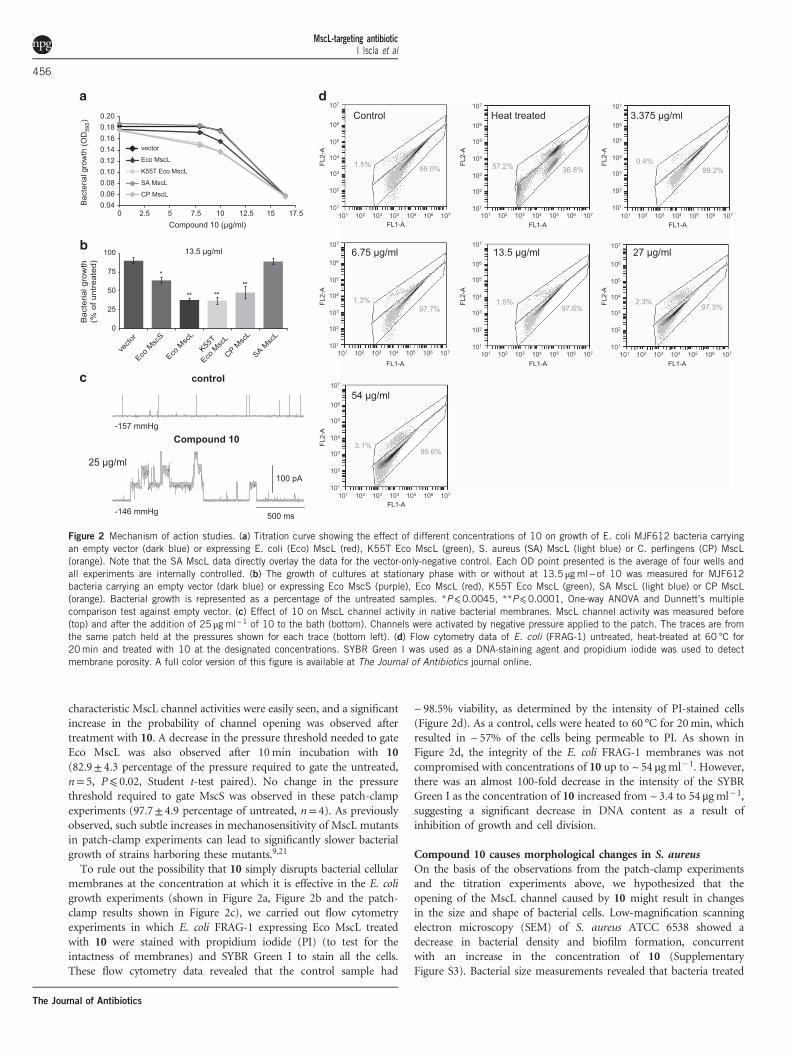
characteristic MscL channel activities were easily seen, and a significantincrease in the probability of channel opening was observed aftertreatment with 10. A decrease in the pressure threshold needed to gateEco MscL was also observed after 10min incubation with 10(82.9± 4.3 percentage of the pressure required to gate the untreated,n= 5, P⩽ 0.02, Student t-test paired). No change in the pressurethreshold required to gate MscS was observed in these patch-clampexperiments (97.7± 4.9 percentage of untreated, n= 4). As previouslyobserved, such subtle increases in mechanosensitivity of MscL mutantsin patch-clamp experiments can lead to significantly slower bacterialgrowth of strains harboring these mutants.9,21
To rule out the possibility that 10 simply disrupts bacterial cellularmembranes at the concentration at which it is effective in the E. coligrowth experiments (shown in Figure 2a, Figure 2b and the patch-clamp results shown in Figure 2c), we carried out flow cytometryexperiments in which E. coli FRAG-1 expressing Eco MscL treatedwith 10 were stained with propidium iodide (PI) (to test for theintactness of membranes) and SYBR Green I to stain all the cells.These flow cytometry data revealed that the control sample had
~ 98.5% viability, as determined by the intensity of PI-stained cells(Figure 2d). As a control, cells were heated to 60 °C for 20min, whichresulted in ~ 57% of the cells being permeable to PI. As shown inFigure 2d, the integrity of the E. coli FRAG-1 membranes was notcompromised with concentrations of 10 up to ~ 54 μg ml− 1. However,there was an almost 100-fold decrease in the intensity of the SYBRGreen I as the concentration of 10 increased from ~3.4 to 54 μg ml− 1,suggesting a significant decrease in DNA content as a result ofinhibition of growth and cell division.
Compound 10 causes morphological changes in S. aureusOn the basis of the observations from the patch-clamp experimentsand the titration experiments above, we hypothesized that theopening of the MscL channel caused by 10 might result in changesin the size and shape of bacterial cells. Low-magnification scanningelectron microscopy (SEM) of S. aureus ATCC 6538 showed adecrease in bacterial density and biofilm formation, concurrentwith an increase in the concentration of 10 (SupplementaryFigure S3). Bacterial size measurements revealed that bacteria treated
0 2.5 5 7.5 10 12.5 15 17.5
Bac
teria
l gro
wth
(OD
595)
Compound 10 (µg/ml)
Eco MscL
K55T Eco MscL
SA MscL
CP MscL
vector
0
25
50
75
100
vecto
r
Eco M
scS
Eco M
scL
K55T
Eco M
scL
CP Msc
L
SA Msc
L
**
*
**
Bac
teria
l gro
wth
(% o
f unt
reat
ed)
**
13.5 µg/ml
0.040.060.080.100.120.140.160.180.20
control
-157 mmHg
100 pA
500 ms
Ramizol®
-146 mmHg
Control Heat treated 3.375 g/ml
6.75 g/ml 13.5 g/ml 27 g/ml
54 g/ml
Compound 10
25 g/ml
Figure 2 Mechanism of action studies. (a) Titration curve showing the effect of different concentrations of 10 on growth of E. coli MJF612 bacteria carryingan empty vector (dark blue) or expressing E. coli (Eco) MscL (red), K55T Eco MscL (green), S. aureus (SA) MscL (light blue) or C. perfingens (CP) MscL(orange). Note that the SA MscL data directly overlay the data for the vector-only-negative control. Each OD point presented is the average of four wells andall experiments are internally controlled. (b) The growth of cultures at stationary phase with or without at 13.5 μgml− of 10 was measured for MJF612bacteria carrying an empty vector (dark blue) or expressing Eco MscS (purple), Eco MscL (red), K55T Eco MscL (green), SA MscL (light blue) or CP MscL(orange). Bacterial growth is represented as a percentage of the untreated samples. *P⩽0.0045, **P⩽0.0001, One-way ANOVA and Dunnett’s multiplecomparison test against empty vector. (c) Effect of 10 on MscL channel activity in native bacterial membranes. MscL channel activity was measured before(top) and after the addition of 25 μgml−1 of 10 to the bath (bottom). Channels were activated by negative pressure applied to the patch. The traces are fromthe same patch held at the pressures shown for each trace (bottom left). (d) Flow cytometry data of E. coli (FRAG-1) untreated, heat-treated at 60 °C for20min and treated with 10 at the designated concentrations. SYBR Green I was used as a DNA-staining agent and propidium iodide was used to detectmembrane porosity. A full color version of this figure is available at The Journal of Antibiotics journal online.
MscL-targeting antibioticI Iscla et al
456
The Journal of Antibiotics

with 0.5 μg ml− 1 were significantly wider than the control (Figure 3a).At concentrations of 10 40.5 μg ml− 1, there was a gradualbut significant reduction in the size of the bacteria. Moreover,untreated S. aureus showed a round and firm geometry with
distinct surface features, which become distorted with increasing drugconcentrations. Atomic force microscopy (AFM) also revealed astatistically significant change in morphology of the uppermost25 nm region of S. aureus (see Figure 3b for illustration). This region
Figure 3 Microscopic analysis of S. aureus ATCC 29213 treated with 10 at different concentrations. (a) SEM images and size measurements of S. aureuswith an inset showing the mean±95% confidence (n=12). Scale bar 1 μm and magnification ~×85 000. (b) The change in the ‘a’ parameter (representingbacteria curvature) is shown with representative 3D AFM images beneath (3 μm×3 μm×700 nm). The top 25 nm of an AFM scan was used as a basis for aparabolic equation fit y= ax2+bx+c to show the change in curvature after treatment with the drug. The inset shows the mean±95% confidence (n=10 for1×MBC and n=20 for other concentrations). A full color version of this figure is available at The Journal of Antibiotics journal online.
MscL-targeting antibioticI Iscla et al
457
The Journal of Antibiotics
was observed to be of narrow parabolic geometry in the controlsample, which then flattened as the drug concentration increased(Figure 3b). The AFM and SEM results support each other, as thelatter width measurement is inversely proportional to the measure-ment of the parabolic curvature parameter ‘a’. These morphologicalchanges are consistent with the spontaneous activation of MscL in thepresence of 10 leading to solute loss and osmolytes and consequently areduction in the size of S. aureus.
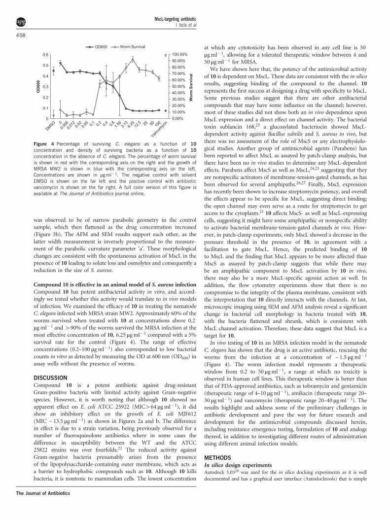
Compound 10 is effective in an animal model of S. aureus infectionCompound 10 has potent antibacterial activity in vitro, and accord-ingly we tested whether this activity would translate to in vivo modelsof infection. We examined the efficacy of 10 in treating the nematodeC. elegans infected with MRSA strain MW2. Approximately 60% of theworms survived when treated with 10 at concentrations above 0.2μg ml− 1 and 490% of the worms survived the MRSA infection at themost effective concentration of 10, 6.25 μgml− 1 compared with a 5%survival rate for the control (Figure 4). The range of effectiveconcentrations (0.2–100 μgml− 1) also corresponded to low bacterialcounts in vitro as detected by measuring the OD at 600 nm (OD600) inassay wells without the presence of worms.
DISCUSSION
Compound 10 is a potent antibiotic against drug-resistantGram-positive bacteria with limited activity against Gram-negativespecies. However, it is worth noting that although 10 showed noapparent effect on E. coli ATCC 25922 (MIC464 μg ml− 1), it didshow an inhibitory effect on the growth of E. coli MJF612(MIC ~13.5 μg ml− 1) as shown in Figures 2a and b. The differencein effect is due to a strain variation, being previously observed for anumber of fluoroquinolone antibiotics where in some cases thedifference in susceptibility between the WT and the ATCC25822 strains was over fourfolds.22 The reduced activity againstGram-negative bacteria presumably arises from the presenceof the lipopolysaccharide-containing outer membrane, which acts asa barrier to hydrophobic compounds such as 10. Although 10 killsbacteria, it is nontoxic to mammalian cells. The lowest concentration
at which any cytotoxicity has been observed in any cell line is 50μgml− 1, allowing for a tolerated therapeutic window between 4 and50 μg ml− 1 for MRSA.We have shown here that, the potency of the antimicrobial activity
of 10 is dependent on MscL. These data are consistent with the in silicoresults, suggesting binding of the compound to the channel. 10represents the first success at designing a drug with specificity to MscL.Some previous studies suggest that there are other antibacterialcompounds that may have some influence on the channel; however,most of these studies did not show both an in vivo dependence uponMscL expression and a direct effect on channel activity. The bacterialtoxin sublancin 168,23 a glucosylated bacteriocin showed MscL-dependent activity against Bacillus subtilis and S. aureus in vivo, butthere was no assessment of the role of MscS or any electrophysiolo-gical studies. Another group of antimicrobial agents (Parabens) hasbeen reported to affect MscL as assayed by patch-clamp analysis, butthere have been no in vivo studies to determine any MscL-dependenteffects. Parabens affect MscS as well as MscL,24,25 suggesting that theyare nonspecific activators of membrane-tension-gated channels, as hasbeen observed for several amphipaths.26,27 Finally, MscL expressionhas recently been shown to increase streptomycin potency, and overallthe effects appear to be specific for MscL, suggesting direct binding;the open channel may even serve as a route for streptomycin to getaccess to the cytoplasm.21 10 affects MscS- as well as MscL-expressingcells, suggesting it might have some amphipathic or nonspecific abilityto activate bacterial membrane-tension-gated channels in vivo. How-ever, in patch-clamp experiments, only MscL showed a decrease in thepressure threshold in the presence of 10, in agreement with afacilitation to gate MscL. Hence, the predicted binding of 10to MscL and the finding that MscL appears to be more affected thanMscS as assayed by patch-clamp suggests that while there maybe an amphipathic component to MscL activation by 10 in vivo,there may also be a more MscL-specific agonist action as well. Inaddition, the flow cytometry experiments show that there is nocompromise to the integrity of the plasma membrane, consistent withthe interpretation that 10 directly interacts with the channels. At last,microscopic imaging using SEM and AFM analysis reveal a significantchange in bacterial cell morphology in bacteria treated with 10,with the bacteria flattened and shrunk, which is consistent withMscL channel activation. Therefore, these data suggest that MscL is atarget for 10.In vivo testing of 10 in an MRSA infection model in the nematode
C. elegans has shown that the drug is an active antibiotic, rescuing theworms from the infection at a concentration of ~ 1.5 μgml− 1
(Figure 4). The worm infection model represents a therapeuticwindow from 0.2 to 50 μgml− 1, a range at which no toxicity isobserved in human cell lines. This therapeutic window is better thanthat of FDA-approved antibiotics, such as tobramycin and gentamicin(therapeutic range of 4–10 μgml− 1), amikacin (therapeutic range 20–30 μg ml− 1) and vancomycin (therapeutic range 20–40 μgml− 1). Theresults highlight and address some of the preliminary challenges inantibiotic development and pave the way for future research anddevelopment for the antimicrobial compounds discussed herein,including resistance emergence testing, formulation of 10 and analogsthereof, in addition to investigating different routes of administrationusing different animal infection models.
METHODS
In silico design experimentsAutodock 3.0528 was used for the in silico docking experiments as it is welldocumented and has a graphical user interface (Autodocktools) that is simple
0.00%10.00%20.00%30.00%40.00%50.00%60.00%70.00%80.00%90.00%100.00%
0
0.1
0.2
0.3
0.4
0.5
0.6
Wor
m S
urvi
val
OD
600
OD600 Worm Survival
Figure 4 Percentage of surviving C. elegans as a function of 10
concentration and density of surviving bacteria as a function of 10
concentration in the absence of C. elegans. The percentage of worm survivalis shown in red with the corresponding axis on the right and the growth ofMRSA MW2 is shown in blue with the corresponding axis on the left.Concentrations are shown in μgml−1. The negative control with solventDMSO is shown on the far left and the positive control with antibioticvancomycin is shown on the far right. A full color version of this figure isavailable at The Journal of Antibiotics journal online.
MscL-targeting antibioticI Iscla et al
458
The Journal of Antibiotics

to use. The atomic coordinates of the MscL protein from E. coli were obtainedfrom a homology structure designed previously.29 In this structure, the amino-acid residues of M. tuberculosis have been replaced with those of E. coli and thecoordinates of the amino acids left unaltered. The Eco MscL structure used is atruncated version of MscL and has only 95 amino-acid residues (Met12 toGlu107), compared with the 136 amino-acid residues representing the wholeprotein. Past experiments suggest that the rest of the protein is not significantfor the activity of MscL and therefore removing it was advantageous by savingcomputer power and significantly reducing the computational time. TheProtein Data Bank file of the Eco MscL was loaded in Autodocktools andthe water molecules removed. Polar hydrogens were added to the proteins andthe charges and solvation parameters were added to the atoms of themacromolecule, and the file saved in Protein Data Bank file format withcharges and solvation parameters included.The ligands were built and saved in Brookhaven format30 and Autodocktools
was used to prepare the ligands for docking. The rigid root of the ligand wasdefined automatically and the maximum number of rotatable bonds wasallowed. The number of active torsions was set to the number of rotatablebonds and the toggle activity of torsions allowed to move most atoms. Thepartial atomic charges were calculated31 using the AM1 Hamiltonian, and thegeometry of the ligand was optimized. These were entered into the Protein DataBank file replacing the charges generated using Autodocktools.The grid size and center used for the Autodock calculation (see
Supplementary Table S1 for parameters used for docking) was restricted tothe amino-acid residues near Ala20, as it has been shown that this is whereparabens and eriochrome cyanine bind.24 To narrow down the search for targetamino acids, the amino acids that surround the docked ligands weredetermined. The amino-acid residues surrounding the ligands for most ofthe dockings ranged from Leu19 to Lys31. Hydrogen acceptor groups (namelyoxygen atoms) were targeted in the protein side chain. Depending on wherethey occur, a ligand with hydrogen donor groups was then designed that canH-bond with the oxygen atoms. This approach was taken given that there werea limited number of amino acids with oxygen atoms exposed to the inside ofthe pore. This identified amino acids, Leu19, Ala20, Val23, Gly26 and Ala27,with oxygen atoms exposed to the inside of the pore. The oxygen atoms inGly26 and Ala27 are slightly shielded by other amino acids. In addition, theypoint to the side rather than to the inside of the pore, and hence may notcontribute significantly to hydrogen bonding.It is also important to note that the geometry of these five amino acids is the
same in the five subunits, as long as no more than one amino acid is consideredto be in any subunit. Therefore, instead of visualizing these amino acids in onechain, they were visualized as an amino acid/subunit so that subunit 1 hasLeu19, subunit 2 has Ala20, subunit 3 has Val23, subunit 4 has Gly26 andsubunit 5 has Ala27.As parts of the protein pore are narrower than others, it was important to
design a pharmacophore that has a length less than the diameter of the pore atthe position of all five amino-acid residues. If this is the case, then any onepharmacophore will, at most, bind to four amino acids. Owing to the fact thatthe most accessible oxygen atoms are in the following amino acids: Leu19,Ala20 and Val23, particular attention was given to these amino acids.A de novo approach was undertaken to design a pharmacophore and
seven ligands were constructed with three hydroxyl groups each (Figure 1a).These ligands were then docked with the Eco MscL. The assumption beingthat the closer the hydroxyl groups of a ligand to the spatial dimensions of theketone groups in Eco MscL (Supplementary Table S2), the better the docking.This was not the case; nonetheless, the results showed exceptional dockingenergies for ligand 2, which was then used as a lead in an iterative dockingprocess.The hydroxyl groups in ligand 2 were replaced by various functional
groups and the effect on docking energy was noted. The functionalgroups include: aldehydes, amide cations, amino, carboxyl and chlorines(Figure 1c). The carboxyl groups were deprotonated and were added, one ata time, until the carboxyl groups replaced all the hydroxyl groups. The fourphenyl groups of ligand 2 were also replaced by cyclohexane groups. The parentcompound was also included (which did not include the hydroxyl groups).The best docking energy was observed with replacement of all hydroxyl
groups for carboxyl groups with an overall charge of − 3 on the molecule.
This ligand has an exceptional free energy of binding equivalent to ~− 55.94kJmol− 1, which is higher than that previously reported.6 MscL may not showan overall ion selectivity; however, the docking results show preferential bindingto the mostly hydrophobic deprotonated tri-acid species.
Compound 10 synthesisDrug synthesis was carried out using a procedure published previously32 withsome modifications. Ethyl acetate (20%) in hexane was used for monitoring theprogress of the Heck cross-coupling reaction using thin layer chromatographyand 20:80 ethyl acetate-dichloromethane for eluting the product using finesilica after tetrahydrofuran removal under vacuum. The product from thesaponification reaction was collected by filtration and eluted in fine silica usinga 20:80 methanol-tetrahydrofuran solvent system, followed by the addition ofdiethyl ether as an anti-solvent to wash the compound.
MIC assayAll compounds were prepared at 160 μgml− 1 in water from a stock solution of20mM of 10 in dimethyl sulfoxide (DMSO). The compounds, along withstandard antibiotics, were serially diluted twofold using Mueller Hinton broth(MHB) in 96-well plates (nonbinding surface, Corning 3641, Tewksbury, MA,USA) using MHB. Concentrations of standards ranged from 64 μg ml− 1 to0.03 μgml− 1, and concentrations of compounds ranged from 8 to 0.003μgml− 1, with final volumes of 50 μl per well. Gram-positive bacteria werecultured in MHB (Bacto Laboratories Pty Ltd, Mt Pritchard, NSW, Australia) at37 °C overnight. Mid-log phase bacterial cultures were diluted to the finalconcentration of 5× 10⁵ CFUml− 1 (in MHB) and used with the dilutedcompounds to be tested. Plates were covered and incubated at 37 °C for 24 h,and MICs designated as the lowest concentration that showed no visiblegrowth. Experiments were carried out in duplicate (n= 2), with vancomycin asa positive inhibitor control.
M. tuberculosis testingRifampicin and 10 were resuspended in DMSO and diluted to obtain a drugconcentration of 160 μgml− 1. Five microliter of this drug concentration(equivalent to 0.8 μg) was dispensed into a 96-well plate and serially twofolddiluted in DMSO (final drug volume 5 μl). Thereafter, frozen aliquots of thebacterial strains H37Rv and Victor strain TT372 were thawed and suspended at2× 105 bacteria per ml in 7H9 media supplemented with oleic acid, albumin,dextrose and catalases (final albumin concentration of 5 g l− 1). Two hundredmicroliter of these bacterial suspensions were added to each well in 96-wellplates, containing either compound (final drug concentration: 4 μgml− 1).Plates were incubated at 37 °C for 7 days, when 20 μl of Alamar Blue reagentwere added to each well. Plates were incubated for an additional 2 days at 37 °C,when absorbance was evaluated in a microtiter plate reader at 570 nm. Bacterialabsorbance was normalized to the blank absorbance and compared with thepositive control (untreated bacteria). Data were graphed as percent inhibition.A similar procedure was carried out for M. tuberculosis H37Rv grown inglycerol-alanine-salts medium and Proskauer and Beck medium at a pH 7.4. Atconcentration ⩾ 80 μgml− 1, 10 precipitated in glycerol-alanine-salts mediumpH 6.6. However, no precipitation was observed at lower concentrations (40–5 μgml− 1) where it also inhibited bacterial growth.
NIH/3T3 and HaCaT cytotoxicity assayHaCaT immortalized keratinocytes and NIH/3T3 fibroblasts were seeded as1.5 × 103 cells per well in a 96-well plate in 50 μl media (Dulbecco’s modifiedEagle’s medium/F12+GlutaMAX from Gibco (Mulgrave, VIC, Australia) with10% v/v foetal bovine serum and 1% v/v penicillin/streptomycin). Cells wereincubated for 2 h at 37 °C, 5% CO2 to allow cells to attach to the plates. 10 wasprepared in DMSO and diluted 20 times in culture media, giving a finalconcentration of 1mgml− 1 with 5% DMSO. Fifty microliter of each dilutionwas added into 50 μl of culture medium in triplicates to reach the finalconcentrations. The cells were incubated with the compounds overnight at37 °C, 5% CO2. After the incubation, MTS(3-(4,5-dimethylthiazol-2-yl)-5-(3-carboxymethoxyphenyl)-2-(4-sulfophenyl)-2H-tetrazolium, inner salt)(Promega, Australia, Alexandria, NSW, Australia) (60 μl) was added to eachwell. The plates were then incubated for 3 h at 37 °C, 5% CO2. Sixty microliter
MscL-targeting antibioticI Iscla et al
459
The Journal of Antibiotics

from each well was transferred to a new plate and the absorbance was then readat 490 nm using EnSpire 2300 Multimode Plate Reader. Results are presented asthe average percentage of control± s.d. for each set of duplicate wells. Thedifference between the experiment and the blank is recorded as normalizedmean. For each treatment, the experiment was carried out in triplicate for eachtime point, at 0, 24, 48 and 72 h. Time 0 starts after overnight incubation.
HepG2 and HEK293 cytotoxicity assay10 was prepared in DMSO at 20mM. It was diluted 200 times in culture media,giving a final concentration of 50 μgml− 1 with 0.5% DMSO. Hepatocellularcarcinoma (HepG2 ATCC HB-8065) and human embryonic kidney cells(HEK293 ATCC CRL-1573) cells were seeded as 1.5 ×10⁴ cells per well in a96-well plate in a final volume of 100 μl in Dulbecco’s modified Eagle’s medi(Gibco-Invitrogen, Waltham, MA, USA), in which 10% or 1% of foetal bovineserum was added. Cells were incubated for 24 h at 37 °C, 5% CO2 to allow cellsto attach. All tested compounds were diluted from 2.5mgml− 1 to 0.38 μgml− 1
in threefold dilutions. Then, 10 μl of each dilution was added into 90 μl ofculture medium in triplicates. Colistin and Tamoxifen were used as the controls.The cells were incubated with the compounds for 24 h at 37 °C, 5% CO2. Afterthe incubation, 3-(4,5-Dimethylthiazol-2-yl)-2,5-diphenyltetrazolium bromide(Invitrogen) was added to each well to a final concentration of 0.4mgml− 1. Theplates were then incubated for 2 h at 37 °C, 5% CO2. Medium was removed, andcrystals were resuspended in 60 μl of DMSO. The absorbance was then read at570 nm using a Polarstar Omega instrument. The data were then analyzed byPrism software. Results are presented as the average percentage of control± s.d.for each set of duplicate wells using the following equation: Percent viability=(ABSTEST−ABS5%DMSO/ABSUNTREATED−ABS5%DMSO)×100.
Strains and cell growthConstructs were inserted in the PB10d expression vector9,33–35 and the E. colistrain MJF612, which is null for MscL, MscS and MscS homologs (FRAG-1ΔmscL::cm, ΔmscS, ΔmscK::kan, ΔybdG::aprΔ)17 were used as a host for allexperiments. Unless stated otherwise, cells were grown in citrate-phosphatedefined medium consisting of (per liter: 8.57 g of Na2HPO4, 0.87 g of K2HPO4,1.34 g of citric acid, 1.0 g of NH4SO4, 0.001 g of thiamine, 0.1 g ofMg2SO4.7H2O, 0.002 g of (NH4)2SO4.FeSO4.6H2O) plus 100 μgml− 1
ampicillin.
In vivo growth inhibition experimentsE. coli strain MJF612 was used as a host for Eco MscL (MscL), K55T Eco MscL(K55T MscL), C. perfringens MscL, SA MscL and E.coli MscS (MscS) constructsin the PB10d expression vector. Overnight cultures were grown in citrate-phosphate defined medium pH 8 plus 100 μgml− 1 ampicillin with shaking at37 °C. The following day, cultures were diluted 1:40 in the same media andgrown for 30min before inducing expression with 1mM isopropyl β-D-1-thiogalactopyranoside. After 30min of induction, all cultures were adjusted toan OD600 of 0.08. The cultures were then diluted 1:3 into pre-warmed citrate-phosphate defined medium pH 8 containing 100 μgml− 1 ampicillin plus 10.Cultures were loaded in 96-well plates (190 μl per well), sealed with abreathable film, and incubated at 37 °C for 16 h without shaking. The OD595
of the cultures was measured using a Multiskan Ascent (Thermo Scientific Inc.,Waltham, MA, USA).
ElectrophysiologyE. coli giant spheroplasts were generated as previously described20 from PB104strain (ΔmscL::Cm)34 expressing Eco MscL. Patch-clamp experiments wereperformed in the inside–out configuration, at room temperature undersymmetrical conditions in a buffer at pH 6.0 (200mM KCl, 90mM MgCl2,10mM CaCl2 and 5mM HEPES). Patches were excised, and recordings wereperformed at 20 mV (for simplicity the patch traces openings are shownupward). Data were acquired at a sampling rate of 20 kHz with 10 kHz filtrationusing an AxoPatch 200B amplifier (Molecular Devices, Sunnyvale, CA, USA). Apiezoelectric pressure transducer (World Precision Instruments, Sarasota, FL,USA) was used to measure the pressure throughout the experiments. Aftertaking three separate pressure threshold measurements in regular patch buffer
for control, a 25 μgml− 1 solution of 10 was perfused to the bath and channelsensitivity within the same patch was measured subsequently every 10min.
Flow cytometry experimentsBacterial cultures were prepared from a single colony of E. coli FRAG-1 grownon Lysogeny Broth media. The single colony was added to 8.5 ml of filteredMHB (0.2 μM Minisart, Sartorius Stedim) to which 10 (3.375 μgml− 1 to 54μgml− 1 in doubling concentrations) was added. The cultures were incubatedovernight at 37 °C with shaking in 2ml volumes before analysis, at which pointSYBR Green I (10×final in MHB) and PI (10 μgml− 1 final in H2O) wereadded. Cells were incubated for 5 min, prior to analysis. The heat-treatedsample was exposed to 60 °C for 20min before adding SYBR Green I and PIand incubated for 5 min.An Accuri C6 Flow Cytometer was used for the flow cytometer experiments.
The fluidics rate was set to Medium (35 μl min− 1) and the threshold limit wasset on FL1 (530/30) to a value of 800. Samples were run and 30 000 eventscollected. Sample data were analyzed using the CFlow software. FL2-A (585/60)(Emmax 605 nm, PI) was plotted on the y axis versus FL1-A (530/30) (Emmax
521 nm, SYBR Green I) on the x axis. No gates were applied and fluorescencecompensation was not required.
Sample preparation for microscopyThe bacterial strain S. aureus ATCC 6538 was grown overnight at 37 °C onMuller–Hinton Agar (Oxoid, Thermo Fisher Scientific Inc., Waltham, MA,USA). The bacteria were harvested and the OD of bacteria suspended in MHBwas adjusted to ~ 0.5 MacFarlane units so as to give 5× 10⁷ CFUml− 1. Bacteriawere then aliquoted into 10ml tubes and various amounts of 10 at 1 mgml− 1
dissolved in DMSO were added to give a final concentration ranging from32 μgml− 1 to 0.5 μgml− 1 (twofold serial dilutions). Bacterial numbers wereenumerated using the Miles–Misra method in which serial dilutions of 10 μlsamples from each culture were spread onto nutrient agar in duplicate. Afterincubation at 37 °C for 24 h, the colonies were counted.S. aureus ATCC 6538 cultures were treated as above and incubated overnight.
Cultures were loaded into sterile disposable syringes and filtered through a0.2-μm isopore polycarbonate hydrophilic filter (Millipore, Billerica, MA,USA). Bacteria on filters were fixed in 4% paraformaldehyde/1.25% glutar-aldehyde in phosphate-buffered saline and 4% sucrose (pH 7.2) for 15min.The fixative was removed with a sterile pipette and the filters washed in awashing buffer composed of 4% sucrose in phosphate-buffered saline for5min. The washing buffer was then removed and the bacteria were post-fixedin 2% OsO4 in water for 30min. The OsO4 were then pipetted out into anosmium waste bottle and the bacteria were washed in a washing buffercomposed of 4% sucrose in phosphate-buffered saline for 5 min. The bacteriawere then dehydrated in four consecutive different concentrations of ethanol,70% ethanol (1 change of 10min), 90% ethanol (1 change of 10min), 95%ethanol (1 change of 10min) and 100% ethanol (3 changes of 10min). Afterremoving the ethanol, the bacteria were critical point dried in hexamethyldi-silazane (2 changes of 15min) and then mounted on a stub and platinumcoated (10 nm). Carbon paint was then applied to the edges of the stub makingcontact with the filter to improve conductivity and reduce sample charging.
Scanning electron microscopy analysisSEM was carried out using a FEG Quanta 450 microscope at an acceleratingvoltage of 3–7 Kv and a working distance of 10mm. The specimens wereobserved using a secondary electron detector under high-vacuum conditions.Images were captured at high definition at ~×85 000 magnification and a scanrate of 100ms per frame.
Atomic force microscopy analysisAFM images were acquired in air using a Bruker Dimension FastScan AFMwith a Nanoscope V controller, operating in PeakForce Tapping mode. BrukerScanAsyst Air probes with a nominal tip radius of 2 nm and nominal springconstant of 0.4 Nm− 1 were used. Imaging parameters including set-point, scanrate (1–2Hz) and feedback gains were adjusted to optimize image quality andminimize imaging force. Images were analyzed using the Bruker NanoscopeAnalysis software (version 1.4). The AFM scanner was calibrated in the x, y and
MscL-targeting antibioticI Iscla et al
460
The Journal of Antibiotics

z directions using silicon calibration grids (Bruker model numbers PG: 1 μmpitch, 110 nm depth and VGRP: 10 μm pitch, 180 nm depth).After acquiring the images, 20 bacteria per sample were chosen at random
and a cross-section was drawn through the apex of each bacteria. The shape ofthe uppermost 25 nm of each bacteria was analyzed by fitting the parabolicfunction y= ax2+bx+c, and extracting the coefficient ‘a’, which represents thecurvature of the parabola. Higher ‘a’ values describe a narrow apex geometry,whereas low ‘a’ values describe a flatter apex with lower curvature. The analysiswas restricted to the uppermost 25 nm in order to ensure that all of the sampleswere analyzed in the same manner, as many of the bacteria are embedded inorganic matter to varying degrees, which restricts the analysis region ineach case.
C. elegans infection modelThe C. elegans infection assay was carried out as previously described.36 A stocksolution of 10 in DMSO (10mgml− 1) was prepared. Dilution series consistingof 1:10 dilution (from 100 μgml− 1) and a 1:2 dilution (from 100 μgml− 1)were tested. To prepare a 1:10 dilution series, 5 μl of 100 μgml− 1 of 10 wasdiluted into 45 μl of liquid media with 1% DMSO. This was repeated for alldilutions. To each well, 20 μl of compound containing liquid media was addedfollowed by the addition of 35 μl of MRSA and 15 μl media containing 15worms. Vancomycin at 20 and 100 μgml− 1 were used as positive controls andDMSO was used as a negative control. After infection and co-incubation withcompound for 4 days, bacteria were washed out and the worms were stainedwith Sytox Orange dye and imaged. The ratio of stained to unstained wormarea was used to measure worm death. Prior to washing, an OD600
measurement was taken to assess the bacterial growth.Full methods and any associated references are available in the online version
of the paper at www.nature.com/nature.
CONFLICT OF INTERESTRAB declares ownership of the intellectual property licensed to Boulos &Cooper Pharmaceuticals Pty Ltd. The remaining authors declare no conflict ofinterest.
ACKNOWLEDGEMENTS
The in vivo growth inhibition experiments and the electrophysiologyexperiments were supported by Grant I-1420 of the Welch Foundation, GrantRP100146 from the Cancer Prevention & Research Institute of Texas, andGrants AI08080701 and GM061028 from the National Institutes of Health. TheC. elegans experiments were supported by grant P01 AI083214 from theNational Institutes of Health. II is supported by Grant 12SDG8740012 from theNational American Heart Association. The funding bodies had no role instudy design, data collection and analysis, decision to publish or preparation ofthe manuscript. The content is solely the responsibility of the authors anddoes not necessarily represent the official views of the National Institutes ofHealth. MAB, JXH, SR and AK were supported by a Wellcome TrustSeeding Drug Discovery Award (094977/Z/10/Z), and MAC by an NHMRCPrincipal Research Fellowship (APP1059354). UHS is supported by NHMRCProject Grant 535053. We gratefully acknowledge support of this workby the Australian Research Council and the Government of South Australia.RAB gratefully acknowledges supervision from Allan J McKinley during hisHonors year when the modeling work was undertaken. SEM analysis wascarried out at Adelaide Microscopy and AFM studies were carried out usingfacilities in the School of Chemical and Physical Sciences, Flinders University.Both of these microscopy facilities are supported by the Australian Microscopyand Microanalysis Research Facility (AMMRF). Flow Cytometry experimentswere carried out at Flow Cytometry Immunology facility at Flinders MedicalCentre. Ramizol is a Trademark fully registered in Australia.Author contributions: RW and II conducted the in vivo growth inhibition
studies and the electrophysiology, JlF and AlC carried out the C. elegansinfection work, SR, AK and JXH conducted the susceptibility testing of MDR S.aureus and S. pneumonia, and cytotoxicity of HEK293 and HepG2 cells, AOcarried out the susceptibility testing of M. tuberculosis, EST carried out thecytotoxicity studies on NIH/3T3 and HaCaT cells lines, UHS conductedexperiments on the effect of 10 on viable counts of S. aureus and supplied
cultures for microscopy, NVH carried out the flow cytometry experiments,
RAB carried out the in-silico studies, synthesis of 10 and prepared the samples
for microscopy, ClT and RAB carried out the scanning electron microscopy
analysis, ADS, CTG and RAB carried out the atomic force microscopy analysis,
II, PB, AlC, JLF, FMA, MAB, UHS, MHB, AO, CLR and RAB wrote the
manuscript, PB, FMA, IO, MHB, MAC, NVH, CM and RAB designed the
experiments, and RAB coordinated the research.
1 Katz, M. L., Mueller, L. V., Polyakov, M. & Weinstock, S. F. Where have all the antibioticpatents gone? Nat. Biotechnol. 24, 1529–1531 (2006).
2 Christoffersen, R. E. Antibiotics—an investment worth making? Nat. Biotechnol. 24,1512–1514 (2006).
3 Clardy, J., Fischbach, M. A. & Walsh, C. T. New antibiotics from bacterial naturalproducts. Nat. Biotechnol. 24, 1541–1550 (2006).
4 Prasad, S. & Smith, P. Meeting the threat of antibiotic resistance: building anew frontline defence. Australian Government Office of the Chief Scientist. http://www.chiefscientist.gov.au/wp-content/uploads/OPS7-antibioticsPRINT.pdf (2013).
5 Piddock, L. J. Antibiotic action: helping deliver acation plans and strategies. LancetInfect. Dis. 13, 1009–1011 (2013).
6 Butler, M. S. & Cooper, M. A. Antibiotics in the clinical pipeline in 2011. J. Antibiot.64, 413–425 (2011).
7 Barh, D. et al. A novel comparative genomics analysis for common drug and vaccinetargets in Corynebacterium pseudotuberculosis and other CMN group of humanpathogens. Chem. Biol. Drug. Des. 78, 73–84 (2011).
8 Boulos, R. A. Antimicrobial dyes and mechanosensitive channels. A. Van Leeuwenhoek104, 155–167 (2013).
9 Prole, D. L. & Taylor, C. W. Identification and analysis of putative homologues ofmechanosensitive channels in pathogenic protozoa. PLoS One 8, doi:10.1371/journal.pone.0066068 (2013).
10 Corry, B. et al. An improved open-channel structure of MscL determined from FRETconfocal microscopy and simulation. J. Gen. Physiol. 136, 483–494 (2010).
11 Wang, Y. et al. Single molecule FRET reveals pore size and opening mechanism of amechano-sensitive ion channel. eLife 3, doi:10.7554/eLife.01834 (2014).
12 McKinley, A. J., Riley, T. V., Lengkeek, N. A., Stewart, S. G. & Boulos, R. A.Antimicrobial Compounds. US20120329871 A1 (2012).
13 James, E. et al. A novel antimicrobial reduces oxidative stress in cells. RSC Adv. 3,7277–7281 (2013).
14 DeGrado, W. F. & Summa, C. M. De novo design and structural characterization ofproteins and metalloproteins. Annu. Rev. Biochem. 68, 779–819 (1999).
15 Boulos, R. A. et al. Inspiration from old dyes: tris(stilbene) compounds as potent Gram-positive antibacterial agents. Chem. Eur. J. 19, 17980–17988 (2013).
16 Quinlan, G. J., Martin, G. S. & Evans, T. W. Albumin: biochemical properties andtherapeutic potential. Hepatology 41, 1211–1219 (2005).
17 Schumann, U. et al. YbdG in Escherichia coli is a threshold-setting mechanosensitivechannel with MscM activity. Proc. Natl Acad. Sci. 107, 12664–12669 (2010).
18 Yang, L. M., Zhong, D. & Blount, P. Chimeras reveal a single lipid-interface residue thatcontrols MscL channel kinetics as well as mechanosensitivity. Cell. Rep. 3,520–527 (2013).
19 Zhong, D. & Blount, P. Phosphatidylinositol is crucial for the mechanosensitivity ofMycobacterium tuberculosis MscL. Biochemistry 52, 5415–5120 (2013).
20 Blount, P., Sukharev, S. I., Moe, P. C., Martinac, B. & Kung, C. MechanosensitiveChannels of Bacteria, vol. 294, 458–482. Academic Press: San Diego, CA, USA, 1999.
21 Iscla, I., Wray, R., Wei, S., Posner, B. & Blount, P. Streptomycin potency is dependenton MscL channel expression. Nat. Commun. 5, doi:10.1038/ncomms5891 (2014).
22 Schedletzky, H., Wiedemann, B. & Heisig, P. The effect of moxifloxacin on its targettopoisomerases from Escherichia coli and Staphylococcus aures. J. Antimicrob. Che-mother. 43, 31–37 (1999).
23 Kouwen, T. R. et al. The large mechanosensitive channel MscL determines bacterialsusceptibility to the bacteriocin sublancin 168. Antimicrob. Agents Chemother. 53,4702–4711 (2009).
24 Nguyen, T., Clare, B., Hool, L. C. & Martinac, B. The effects of parabens on themechanosensitive channels of E. coli. Eur. Biophys. J. 34, 389–395 (2005).
25 Kamaraju, K. & Sukharev, S. I. The membrane lateral pressure-perturbing capacity ofparabens and their effects on the mechanosensitive channel directly correlate withhydrophobicity. Biochemistry 47, 10540–10550 (2008).
26 Martinac, B., Adler, J. & Kung, C. Mechanosensitive ion channels of E. coli activated byamphipaths. Nature 348, 261–263 (1990).
27 Perozo, E., Kloda, A., Cortes, D. M. & Martinac, B. Physical principles underlying thetransduction of bilayer deformation forces during the mechanosensitive channel gating.Nat. Struc. Mol. Biol. 9, 696–703 (2002).
28 Morris, G. M. et al. Automated docking using a Lamarckian genetic algorithm and anemperical binding free energy function. J. Comput. Chem. 19, 1639–1662 (1998).
29 Nguyen, T., Clare, B., Guo, W. & Martinac, B. The effects of parabens on themechanosensitive channels of E. coli. Eur. Biophys. J. 34, 389–395 (2005).
30 Spartan '02, Wavefunction, Inc.: Irvine, CA, USA, 2002.31 Gaussian 98, Gaussian, Inc.: Pittsburgh, PA, USA, 1998.32 Lengkeek, N. A. et al. The synthesis of fluorescent DNA intercalator precursors through
efficient multiple heck reactions. Aust. J. Chem. 64, 316–323 (2011).
MscL-targeting antibioticI Iscla et al
461
The Journal of Antibiotics

33 Moe, P. C., Levin, G. & Blount, P. Correlating a protein structure with function of abacterial mechanosensitive channel. J. Biol. Chem. 275, 31121–31127 (2000).
34 Blount, P., Sukharev, S. I., Schroeder, M. J., Nagle, S. K. & Kung, C. Single residuesubstitution that change the gating properties of a mechanosensitive channel inEscherichia coli. Proc. Natl Acad. Sci. USA 93, 1652–11657 (1996).
35 Blount, P. et al. Membrane topology and multimeric structure of a mechanosensitivechannel protein of Escherichia coli. EMBO J. 15, 4798–4805 (1996).
36 Rajamuthiah, R. et al. Whole animal automated platform for drug discovery againstmulti-drug resistant Staphylococcus aureus. PLoS ONE 9, doi:10.1371/journal.pone.0089189 (2014).
This work is licensed under a Creative CommonsAttribution-NonCommercial-NoDerivs 3.0 Unported
License. The images or other third party material in this article areincluded in the article’s Creative Commons license, unless indicatedotherwise in the credit line; if the material is not included under theCreative Commons license, users will need to obtain permission fromthe license holder to reproduce the material. To view a copy of thislicense, visit http://creativecommons.org/licenses/by-nc-nd/3.0/
Supplementary Information accompanies the paper on The Journal of Antibiotics website (http://www.nature.com/ja)
MscL-targeting antibioticI Iscla et al
462
The Journal of Antibiotics