A Modified Single Mini-Incision Complete Urinary Tract ... fileThe ureter on one side was first...
6
Clinical Study A Modified Single Mini-Incision Complete Urinary Tract Exenteration for Urothelial Carcinoma in Dialysis Patients I-Hsuan Chen, 1,2 Jen-Tai Lin, 1 Jeng-Yu Tsai, 1 Tony Wu, 1,3 and Chia-Cheng Yu 1,2,3,4 1 Division of Urology, Department of Surgery, Kaohsiung Veterans General Hospital, 386 Ta-Chung 1st Road, Kaohsiung 813, Taiwan 2 Division of Urology, Department of Surgery, Tri-Service General Hospital, National Defense Medical Center, Taipei 114, Taiwan 3 School of Medicine, National Yang-Ming University, Taipei 112, Taiwan 4 Department of Pharmacy, Tajen University, Pingtung 90741, Taiwan Correspondence should be addressed to Chia-Cheng Yu; [email protected] Received 24 April 2014; Revised 22 June 2014; Accepted 23 June 2014; Published 11 August 2014 Academic Editor: Li-Jen Wang Copyright © 2014 I-Hsuan Chen et al. is is an open access article distributed under the Creative Commons Attribution License, which permits unrestricted use, distribution, and reproduction in any medium, provided the original work is properly cited. Objective. To present our experience with single mini-incision complete urinary tract exenteration (CUTE) for female dialysis patients suffering from urothelial carcinoma (UC). Patients and Methods. Institutional review board approval was obtained. From 2005 through 2012, 14 female dialysis patients with UC underwent single mini-incision CUTE, in combination with radical hysterectomy and bilateral salpingo-oophorectomy. All were placed in the modified dorsal lithotomy position without repositioning. An infraumbilical midline mini-incision was made. Bilateral nephroureterectomy was first performed entirely extraperitoneally, followed by radical cystectomy with removal of the uterus and ovaries transperitoneally. Results. All procedures were done successfully without major complications. e median operative time was 242.5 minutes, and estimated blood loss was 500 mL. e median time to oral intake was 2 postoperative days; the median hospital stay was 11 days. Ten patients remained cancer-free at a median follow-up of 46.5 months; six patients were confirmed as having preoperatively undetectable UC or renal cell carcinoma, even aſter reviewing preoperative computed tomography. Conclusions. is modified technique provides a time- saving complete urinary tract extirpation to eliminate preoperatively undetectable malignancy, reduce metachronous recurrences, and avert perioperative complications associated with pneumoperitoneum and repositioning. Good cancer control and early convalescence can mutually be achieved in experienced hands. 1. Introduction In Taiwan, there is an increased risk for urothelial carcinoma (UC) in patients of end-stage renal disease (ESRD), with the incidence ranging from 0.89% to 2.1%, especially women aged 50 years or younger [1]. On account of its high recurrence rate and rapidly progressive behavior among dialysis patients, total urinary tract exenteration is a recommended treatment modality to reduce the incidence of metachronous multi- centric UC [2]. Besides, lack of suitable imaging studies for follow-up of the upper tract and the probability of morbidity related to stepwise urinary tract extirpation support the more aggressive surgical strategy for treating high-risk patients with ESRD. Conventionally, complete urinary tract exenteration (CUTE) is performed via a long midline incision extending from the xiphoid process to the pubic symphysis or a bilateral flank approach followed by a midline infraumbilical laparotomy incision. Long operative time and high surgical risks were of great concern at that time. With the advent of laparoscopic techniques and the experience of open surgery, we develop a single mini-incision unilateral nephroureterec- tomy with bladder cuff excision, via an infraumbilical midline incision, for upper tract UC. By applying this method to synchronous bilateral nephroureterectomy, CUTE, through a single mini-incision approach, in single session can be the treatment option of choice as if there are indications for radi- cal cystectomy in female patients receiving dialysis. Removal of gynecological organs may be undertaken simultaneously in peri- and postmenopausal patients. In the present study we describe our modified technique in treating female dialysis patients with urothelial carcinoma. Hindawi Publishing Corporation BioMed Research International Volume 2014, Article ID 649642, 6 pages http://dx.doi.org/10.1155/2014/649642
-
Upload
trinhxuyen -
Category
Documents
-
view
214 -
download
0
Transcript of A Modified Single Mini-Incision Complete Urinary Tract ... fileThe ureter on one side was first...

Clinical StudyA Modified Single Mini-Incision Complete Urinary TractExenteration for Urothelial Carcinoma in Dialysis Patients
I-Hsuan Chen,1,2 Jen-Tai Lin,1 Jeng-Yu Tsai,1 Tony Wu,1,3 and Chia-Cheng Yu1,2,3,4
1 Division of Urology, Department of Surgery, Kaohsiung Veterans General Hospital, 386 Ta-Chung 1st Road, Kaohsiung 813, Taiwan2Division of Urology, Department of Surgery, Tri-Service General Hospital, National Defense Medical Center, Taipei 114, Taiwan3 School of Medicine, National Yang-Ming University, Taipei 112, Taiwan4Department of Pharmacy, Tajen University, Pingtung 90741, Taiwan
Correspondence should be addressed to Chia-Cheng Yu; [email protected]
Received 24 April 2014; Revised 22 June 2014; Accepted 23 June 2014; Published 11 August 2014
Academic Editor: Li-Jen Wang
Copyright © 2014 I-Hsuan Chen et al. This is an open access article distributed under the Creative Commons Attribution License,which permits unrestricted use, distribution, and reproduction in any medium, provided the original work is properly cited.
Objective. To present our experience with single mini-incision complete urinary tract exenteration (CUTE) for female dialysispatients suffering from urothelial carcinoma (UC). Patients and Methods. Institutional review board approval was obtained.From 2005 through 2012, 14 female dialysis patients with UC underwent single mini-incision CUTE, in combination withradical hysterectomy and bilateral salpingo-oophorectomy. All were placed in the modified dorsal lithotomy position withoutrepositioning. An infraumbilical midline mini-incision was made. Bilateral nephroureterectomy was first performed entirelyextraperitoneally, followed by radical cystectomy with removal of the uterus and ovaries transperitoneally. Results. All procedureswere done successfully without major complications. The median operative time was 242.5 minutes, and estimated blood loss was500 mL. The median time to oral intake was 2 postoperative days; the median hospital stay was 11 days. Ten patients remainedcancer-free at a median follow-up of 46.5 months; six patients were confirmed as having preoperatively undetectable UC or renalcell carcinoma, even after reviewing preoperative computed tomography. Conclusions. This modified technique provides a time-saving complete urinary tract extirpation to eliminate preoperatively undetectable malignancy, reduce metachronous recurrences,and avert perioperative complications associated with pneumoperitoneum and repositioning. Good cancer control and earlyconvalescence can mutually be achieved in experienced hands.
1. Introduction
In Taiwan, there is an increased risk for urothelial carcinoma(UC) in patients of end-stage renal disease (ESRD), with theincidence ranging from0.89% to 2.1%, especiallywomen aged50 years or younger [1]. On account of its high recurrencerate and rapidly progressive behavior among dialysis patients,total urinary tract exenteration is a recommended treatmentmodality to reduce the incidence of metachronous multi-centric UC [2]. Besides, lack of suitable imaging studies forfollow-up of the upper tract and the probability of morbidityrelated to stepwise urinary tract extirpation support themoreaggressive surgical strategy for treating high-risk patientswith ESRD.
Conventionally, complete urinary tract exenteration(CUTE) is performed via a long midline incision extending
from the xiphoid process to the pubic symphysis or abilateral flank approach followed by a midline infraumbilicallaparotomy incision. Long operative time and high surgicalrisks were of great concern at that time. With the advent oflaparoscopic techniques and the experience of open surgery,we develop a single mini-incision unilateral nephroureterec-tomywith bladder cuff excision, via an infraumbilicalmidlineincision, for upper tract UC. By applying this method tosynchronous bilateral nephroureterectomy, CUTE, througha single mini-incision approach, in single session can be thetreatment option of choice as if there are indications for radi-cal cystectomy in female patients receiving dialysis. Removalof gynecological organs may be undertaken simultaneouslyin peri- and postmenopausal patients.
In the present study we describe our modified techniquein treating female dialysis patients with urothelial carcinoma.
Hindawi Publishing CorporationBioMed Research InternationalVolume 2014, Article ID 649642, 6 pageshttp://dx.doi.org/10.1155/2014/649642

2 BioMed Research International
Figure 1: Illustration of two cord-like structures passing forwardsin the peritoneal fold and entering the inguinal canal by the internalring, considered to be the round ligament or spermatic cord in thefemale and the male, respectively.
Throughout the whole course, patient repositioning or rotat-ing the operating table with cuff inflation is not needed; thespecimens were extracted en bloc from the infraumbilicalmidline incision. We also compare our results with those ofsimilar studies.
2. Patients and Methods
From 2005 through 2012, a total of 14 female dialysis patientsunderwent single mini-incision CUTE, radical hysterectomy,and bilateral salpingo-oophorectomy at our institution. Tenof them had multifocal UC (9 synchronous upper tract andbladder, 1 bilateral upper tract); 3 patients had organ-confinedbladder UC (clinical stage T1, T2, or carcinoma in situ)with left-sided hydronephrosis; the remaining one only hadurinary bladderUC.No lymphnode or distantmetastasis wasnoted preoperatively.
2.1. Extraperitoneal Preparation. All patients were placedin the modified dorsal lithotomy position with both legssupported in stirrups. An infraumbilical midline incision,about 10 cm in length, was made. The posterior rectus fasciawas carefully dissected to gain access to the extraperitonealspace, which was then created from the pubic symphysiscephalad using blunt dissection and sweeping method. Onceadequate working space was obtained with visualization ofthe psoas muscle, bilateral round ligaments attached to theperitoneum were identified, ligated, and divided (Figure 1).The retroperitoneal space could be widely explored using aBookwalter retractor system (Codman, USA), in companywith two customized long right-angle retractors (blade 15or 23 cm). One was used to lift the abdominal wall up andthe other sweeping the peritoneum and its contents medially(Figure 2).
2.2. Bilateral Nephroureterectomy. The ureter on one sidewas first identified, and ureteral skeletonization was per-formed cephalad using electrocautery toward the renal pelvis.After exploring the lower pole of the ipsilateral kidney, a
Figure 2: Illustration ofPeritonealmobilization. After division of theround ligament, two customized long right-angle retractors (upper2 blades), as well as a Bookwalter retractor system (lower 2 blades),are used to push the peritoneum medially and anteriorly, therebyexposing the retroperitoneum.
Figure 3: Intraoperative image of Perirenal dissection. A circum-ferential dissection of the kidney was performed along the planebetween the renal capsule and the perinephric fat, with assistanceof LigaSure.
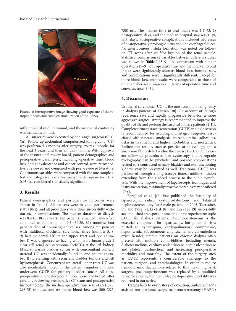
circumferential dissection was performed manually alongthe plane between the renal capsule and the perinephric fat(Figure 3). Besides, bipolar electrocautery would be used tofacilitate dissecting cephalad from the lower pole of the kid-ney toward the upper pole, by dividing any side-wall attach-ments. As the kidney was completely mobilized (Figure 4),the renal pedicle was isolated, ligated, and divided en blocwith Endo-GIA staplers (US Surgical Corporation, Norwalk,Connecticut), and the kidney was retrieved through themidline incision. The same procedure was repeated on theother side.
2.3. Radical Cystectomy. Radical cystectomy for women tra-ditionally includes removal of the uterus, bilateral fallop-ian tubes, ovaries, and part of the vagina. After bilateralextraperitoneal nephroureterectomy, transperitoneal cystec-tomy, radical hysterectomy, bilateral salpingo-oophorectomy,and pelvic lymph node dissection were accomplished usingstandard open surgical techniques. Frozen section pathol-ogy of the bladder neck was performed to ensure a safemargin.The entire specimens were en bloc retrieved from the
BioMed Research International 3
Figure 4: Intraoperative image showing good exposure of the re-troperitoneum and complete mobilization of the kidney.
infraumbilical midline wound, and the urothelial continuitywas maintained intact.
All surgeries were executed by one single surgeon (C. C.Yu). Follow-up abdominal computerized tomography (CT)was performed 3 months after surgery, every 6 months forthe next 3 years, and then annually for life. With approvalof the institutional review board, patient demographics andperioperative parameters, including operative time, bloodloss, and convalescence and cancer control, were retrospec-tively reviewed and compared with peer-reviewed literature.Continuous variables were compared with the one sample 𝑡-test and categorical variables using the chi-square test. 𝑃 <0.05 was considered statistically significant.
3. Results
Patient demographics and perioperative outcomes wereshown in Table 1. All patients were in good performancestatus (0-1), and all procedures were done successfully with-out major complications. The median duration of dialysiswas 8.5 (6, 10.75) years. Ten patients remained cancer-freeat a median follow-up of 46.5 (30.25, 87) months; fourpatients died of nonmalignant causes. Among ten patientswith multifocal urothelial carcinoma, three (number 5, 8,9) had incidental UC in the upper tract and one (num-ber 1) was diagnosed as having a 1mm Furhman grade Iclear cell renal cell carcinoma (ccRCC) at the left kidney.Muscle-invasive bladder cancer with concomitant bilateralureteral UC was incidentally found in one patient (num-ber 11) presenting with recurrent bladder tumors and lefthydronephrosis. Concurrent unilateral upper tract UC wasalso incidentally noted in the patient (number 13), whounderwent CUTE for primary bladder cancer. All thesepreoperatively undetectable tumors were confirmed aftercarefully reviewing preoperative CT scans and postoperativehistopathology. The median operative time was 242.5 (187.5,268.75) minutes, and estimated blood loss was 500 (325,
750) mL. The median time to oral intake was 2 (1.75, 2)postoperative days, and the median hospital stay was 11 (9,13.5) days. Postoperative complications included two casesof postoperatively prolonged ileus and one esophageal ulcer.No arteriovenous fistula formation was noted on follow-up CT scans after en bloc ligation of the renal pedicle.Statistical comparison of variables between different studieswas shown in Table 2 [3–9]. In comparison with similaroperations [7–9], our operative time and the interval to oralintake were significantly shorter; blood loss, hospital stay,and complications were insignificantly different. Except formore blood loss, our results were comparable to those ofother smaller-scale surgeries in terms of operative time andconvalescence [3–6].
4. Discussion
Urothelial carcinoma (UC) is the most common malignancyin dialysis patients of Taiwan [10]. On account of its highrecurrence rate and rapidly progressive behavior, a moreaggressive surgical strategy is recommended to improve thequality of life and prolong the survival of these patients [2, 11].Complete urinary tract exenteration (CUTE) in single sessionis recommended for avoiding multistaged surgeries, asso-ciated with repeated analgesia, intraabdominal adhesions,delay in treatment, and higher morbidities and mortalities.Bothersome results, such as positive urine cytology and asuspicious filling defect within the urinary tract, and unpleas-ant follow-up procedures, like cystoscopy and retrogradepyelography, can be precluded and possible complicationsrelated to a contracted urinary bladder and nonfunctioningkidneys may be prevented as well. Traditional CUTE wasperformed through a long transperitoneal midline incisionextending from the xiphoid process to the pubic symph-ysis. With the improvement of laparoscopic techniques andinstrumentation,minimally invasive therapiesmay be offered[7–9].
Berglund et al. [12] first published the feasibility oflaparoscopic radical cystoprostatectomy and bilateralnephroureterectomy for 2 male patients in 2005. Thereafter,Ou and Yang [7], Li et al. [8], and Lin et al. [9] successfullyaccomplished transperitoneoscopic or retroperitoneoscopicCUTE for dialysis patients. Pneumoperitoneum is theessential component for laparoscopy, but potential risksrelated to hypercapnia, cardiopulmonary compromise,hypothermia, subcutaneous emphysema, and air embolismexist. Besides, uremic patients on chronic dialysis oftenpresent with multiple comorbidities, including anemia,diabetes mellitus, cardiovascular disease, peptic ulcer diseaseand platelet dysfunction, and increasing perioperativemorbidity and mortality. The extent of the surgery suchas CUTE represents a considerable challenge to thepatient, surgeon, and anesthesiologist. In order to reducehemodynamic fluctuations related to this major high-risksurgery, pneumoperitoneum was replaced by a modifiedretractor system, and no 90-day postoperative mortality wasreported in our series.
Tracing back to our history of evolution, unilateral hand-assisted retroperitoneoscopic nephroureterectomy (HARN)
4 BioMed Research International
Table 1: Patient demographics and perioperative parameters.
Pt Age(y)
Durationof HD (yr) BMI (kg/m2) ECOG Clinically surgical
indication Hydronephrosis Pathologic tumor location(T stage)a Comorbidity
1 63 9 21.4 0 UB & LU Left UB(is) & LU(1)LK ccRCC(1a) HTN
2 50 14 21.7 0 UB & RU Right UB(1) & RU(3) HTN
3 61 8 25.9 1 UB & RU∗∗ Right UB(a) & RU(a)HTN, HCC, cirrhosis,
hepatitis B & C,SHPT
4 75 6 31.5 0 UB & LULK Left UB(a) & LU(a)
LK(a) None
5 63 17 17.1 1 UB & LK Bilateral UB(1) & LK(3)LU(a)
HTN, hepatitis C,pulmonary TB
6 52 4 31.8 0 UB & RK No UB(1) & RK(1) HTN, DM7 67 1 20.4 1 BK & RU Right BK(a) & RU(1) HTN, CHF
8 80 6 17.0 1 UB & LU∗∗ Bilateral UB(2a) & LU(a)RK(a) & RU(a)
HTN, DM,moderate MR
9 58 9 21.3 1 UB & RU No UB(is) & RU(1)LU(is) SHPT
10 70 2 22.6 1 UB & RU∗∗ Right UB(1) & RU(1) HTN, old CVA,parkinsonism
11 57 8 22.2 1 UB∗∗ Left UB(2a)LU(is) & RU(3) SHPT
12 57 13 17.3 1 UB∗∗ Left UB(1) Pulmonary TB
13 53 10 21.5 0 UB No UB(1)LU(a) HTN, hepatitis B
14 48 11∗ 21.2 1 UB Left UB(1) HTN, hepatitis C,SHPT, peritonitis∗∗∗
HD, hemodialysis; BMI, body mass index; ECOG, eastern cooperative oncology group performance status; UB, urinary bladder; LU, left ureter; RU, rightureter; LK, left kidney; RK, right kidney; BK, bilateral kidney; ccRCC, clear cell renal cell carcinoma; HTN, hypertension; HCC, hepatocellular carcinoma;SHPT, secondary hyperparathyroidism; TB, tuberculosis; DM, diabetes mellitus; CHF, congestive heart failure; CVA, cardiovascular accident.aAll patients had high-grade urothelial carcinoma.∗Peritoneal dialysis.∗∗Surgical indication was recurrent urothelial cancer.∗∗∗Continuous ambulatory peritoneal dialysis (CAPD) related sclerosing peritonitis.
Table 2: Comparison of outcomes from concurrent upper and lower urinary tract surgery.
Author Number Extent ofsurgery
Age(y)
Operative time(min)a
Blood loss(mL)a
Hospital stay(d)a
Time to intake(hr)a
Complication(%)b
El-Galley et al., 2011 [3] 36 BN N/A 222 175∗∗∗ 3.0∗∗∗ N/A 22.2Chueh et al., 2002 [4] 7 BNU 51.6 294∗∗ 218∗∗ 8.8∗ 39.0 14.3Tai et al., 2009 [5] 33 BNU 52.4 309∗∗ 226∗∗ 10.2 58.0 12.1Ou and Yang, 2011 [6] 13 BNU 60.0 215 216∗∗ 13.8 60.0 7.7Ou and Yang, 2011 [7] 10 CUTE 57.6 328∗∗∗ 628 14.7∗ 62.4∗ 10.0Li et al., 2009 [8] 5 CUTE 58.0 492∗∗∗ 378 12.2 72.0∗∗ 80.0Lin et al., 2011 [9] 5 CUTE 66.6 397∗∗∗ 532 10.8 91.2∗∗∗ 20.0Present study 14 CUTE 61.0 237.5 560.7 12.1 48.0 21.4BN, bilateral nephrectomy; BNU, bilateral nephroureterectomy; CUTE, complete urinary tract exenteration.aOne sample 𝑡-test.bChi-square test.∗
𝑃 < 0.05, ∗∗𝑃 < 0.01, ∗∗∗𝑃 < 0.001 for compared to present study.N/A, not available.
BioMed Research International 5
was performed via a Gibson incision at our institution. Basedon accumulated experiences and skills, unilateral extraperi-toneal nephroureterectomy could be practiced through aparamedian incision [13]. During this period, a camera portwas required to insert a laparoscope and ensure the securityof en bloc ligation of renal vessels; an extended paramedianincision could immediately be made as if major bleedingneeded to be treated. After becoming progressively proficientin surgical anatomy of the retroperitoneal space, better expo-sure could be attained and the renal hilum might be directlyvisualized without the need of laparoscopy. Additionally,palpitation of the renal pedicle, digital dissection, and retrac-tion were utilized to collaborate with perirenal dissection.In consideration of postoperative analgesia and functionalrecovery, an infraumbilical midline incision was attempted toaccomplish unilateral extraperitoneal nephroureterectomy,which had been done by one single surgeon inmore than 200patients.
For the purpose of good exposure of the retroperitoneum,two important surgical steps are addressed. First, division ofthe round ligament or spermatic cord facilitates mobilizationof the peritoneum and its contents [14, 15]. Anatomically,there are two cord-like structures passing forwards in theperitoneal fold and entering the inguinal canal by the internalring, considered to be the round ligament or spermaticcord in the female and the male, respectively. Mass ligationof these structures can separate the peritoneum off theiliac vessels and the psoas muscle is exposed laterally andposteriorly to help in identifying and skeletonizing the uretercephalad. Second, along with a Bookwalter retractor system,two customized long right-angle retractors, of which theblade is 15 or 23 cm, are used to push the peritoneummedially and anteriorly. With traction on the peritoneum,the intraperitoneal contents may be naturally retracted andprotected, lowering the risk of adjacent organ interference orinjury, even in the peritoneal dialysis patient with a historyof sclerosing peritonitis; a wide space may be provided forureterolysis, perirenal dissection, and isolation of the renalpedicle.
On the other hand, in order to complete laparoscopicbilateral upper urinary tract surgery in single session, uti-lizing gravity to maneuver the bowel is of great concern.Some experts alternated inflatable air tourniquet cuffs or gelrolls on each side of the patient’s back, or tilted the table tofacilitate displacing the bowel by gravity [4, 9, 16, 17], thussaving the time of repositioning and redraping. All patientsin our series were operated in the modified dorsal lithotomyposition, with both arms outstretched, throughout the wholeprocedure. Plenty of time could be preserved, attributing tono change of patient’s position. This could explain why ouroperative time was significantly shorter compared to thoseof published studies [7–9]. By virtue of minimally invasivesurgical techniques with laparoscopic instruments, as well astactile feedback and direct three-dimensional visualization,the method presented here permitted a faster upper urinarytract extirpation.
As for shorter interval to oral intake, it might be ascribedto shorter operative duration; other perioperative parametersappeared to be similar. Owing to very low premiums of
the National Health Insurance in Taiwan, patients usuallywould not be discharged until being fully recovered. That iswhy our hospital stay could not be shortened. Comparingother studies with regard to synchronous bilateral renalsurgery [3–6], there was significantly more blood loss inour series. The amount of bleeding might be correlatedwith the extent of surgery. Radical hysterectomy and bilat-eral salpingo-oophorectomywere simultaneously performed,partly explaining why the blood loss was significantly higherthan that of other smaller-scale surgeries. Nevertheless, oursurgical time and postoperative convalescence were stillcomparable.
With regard to oncologic control, 10 patients remainedcancer-free at a follow-up of at least 19 months, the longestbeing 105 months. This might be attributed to the broadenedindication for CUTE at our institute, including primarybladder cancer in uremic patients.
Preoperatively undetectable malignancy, such as smallRCC or superficial UC, in the upper tract could be enbloc removed in single session. Six patients were postop-eratively confirmed as having concurrent UC or ccRCC inour series, even after carefully reviewing preoperative CTscans. Although no other study is available to substantiatethis finding, it is confirmed that an aggressive surgicalapproach should be executed to treat UC in uremic patientsregarding its notorious behavior. However, there are stillcontraindications to our techniques, including advancedupper tract tumors and lymph node metastasis identifiedpreoperatively, due to difficulty in attaining negative surgicalmargins and removing enough lymph nodes. In anotheraspect, it should be concerned that the risk of rupturedcollecting system exists, especially in patients with moderateor severe hydronephrosis. There was no aforementionedcomplication encountered in our patients because most ofthem had localized upper tract tumors in atrophic kidneys.
The present study was limited by its retrospective natureand small case number. The amount of analgesia and qualityof life were not evaluated as well postoperatively. Despitegood exposure of the retroperitoneal space, it is still aconfined space to perform perirenal dissection, even inhand-assisted retroperitoneoscopic surgery. Without pro-ficient anatomical knowledge of the retroperitoneal spaceand sophisticated minimally invasive surgical skills, peri-toneal violation might be encountered, thereby causing thebowel to protrude through the peritoneal defect and hinderaccess to the surgical field. Reproducibility may be an issue,but, in experienced hands, it can be done efficiently andsafely with minimal morbidity. There were still two casesof postoperatively prolonged ileus, and it might be relatedto the transperitoneal approach for radical cystectomy. Aprospective randomized study with a larger sample and long-term follow-up would be needed to demonstrate the possiblebenefit more conclusively.
The concept of peritoneal mobilization promotes oursurgical evolution, leading to better exposure of the retroperi-toneum. Even though peritoneal violation is present, thecustomized long right-angle retractors andmoist laparotomypads may be utilized to overcome this obstacle. Our modi-fied technique provides an expanded indication for CUTE.
6 BioMed Research International
It is recommended that this method be implemented indialysis patients with multifocal UC in which upper tracttumors are localized without lymph node metastasis or thosewith primary bladder cancer eligible for radical cystectomy,thus eliminating the preoperatively undetectable tumors andreducing the likelihood of metachronous malignancy. Gyne-cologic organs can simultaneously be removed to achievebetter oncological control. Without pneumoperitoneum andrepositioning, it can be performed via an infraumbilical mid-line mini-incision, and the entire specimen can be extracteden bloc with intact urothelial continuity.
5. Conclusions
This modified single mini-incision CUTE provides a time-saving method for dialysis patients with UC, eliminat-ing preoperatively undetectable malignancy and reduc-ing metachronous recurrences; perioperative complicationsassociatedwith pneumoperitoneum and repositioning can beaverted as well. Good cancer control and early convalescencemay both be achieved in experienced hands.
Conflict of Interests
The authors declare that there is no conflict of interestsregarding the publication of this paper.
References
[1] S. M. Wang, M. N. Lai, P. C. Chen, and J. D. Wang, “Increasedrisk of urothelial cancer in young and middle aged patientswith end-stage renal disease,” Journal of the Formosan MedicalAssociation, 2013.
[2] C. F. Wu, J. J. Shee, D. R. Ho, W. C. Chen, and C. S. Chen,“Different treatment strategies for end stage renal diseasein patients with transitional cell carcinoma,” The Journal ofUrology, vol. 171, no. 1, pp. 126–129, 2004.
[3] R. El-Galley, S. Safavy, J. E. Busby, and J. Colli, “Outcomeof hand assisted laparoscopic bilateral native nephrectomy intransplant recipients,”The Journal of Urology, vol. 185, no. 3, pp.1021–1025, 2011.
[4] S. Chueh, J. Chen,W.Hsu,M.Hsieh, andM. Lai, “Hand assistedlaparoscopic bilateral nephroureterectomy in 1 session withoutrepositioning patients is facilitated by alternating inflationcuffs,”The Journal of Urology, vol. 167, no. 1, pp. 44–47, 2002.
[5] H. C. Tai, M. K. Lai, S. D. Chung, K. H. Huang, S. C. Chueh,andH. J. Yu, “Intermediate-termoncological outcomes of hand-assisted laparoscopic versus open bilateral nephroureterectomyfor dialysis and kidney transplant patients with upper urinarytract urothelial carcinoma,” Journal of Endourology, vol. 23, no.7, pp. 1139–1144, 2009.
[6] C. H. Ou and W. H. Yang, “Bilateral hand-assisted retroperi-toneoscopic nephroureterectomy (HARN) in the spread-eagleposition for dialysis patients-Low midline HARN in a com-pletely supine position,”Urology, vol. 77, no. 2, pp. 363–367, 2011.
[7] C. Ou and W. Yang, “Complete urinary tract exenteration fora dialysis patient with urothelial cancer: lower midline andextraperitoneal approach in a supine position,” Urology, vol. 77,no. 5, pp. 1244–1247, 2011.
[8] C. C. Li, H. S. Wang, W. J. Wu et al., “Laparoscopic com-plete urinary tract exenteration with the specimen withdrawntransvaginally,” BJU International, vol. 104, no. 11, pp. 82–86,2009.
[9] V. C. Lin, K. C. Hung, M. Chen et al., “Single-session laparo-scopic total urinary tract exenteration without repositioning formultifocal urothelial carcinoma in dialysis-dependent patients,”Urology, vol. 77, no. 1, pp. 98–103, 2011.
[10] C. H. Chang, C. M. Yang, and A. H. Yang, “Renal diagnosisof chronic hemodialysis patients with urinary tract transitionalcell carcinoma in Taiwan,”Cancer, vol. 109, no. 8, pp. 1487–1492,2007.
[11] B. P. Jiaan, C. C. Yu, Y. H. Lee, and J. K. Huang, “Uraemia withconcomitant urothelial cancer,” British Journal of Urology, vol.72, no. 4, pp. 458–461, 1993.
[12] R. K. Berglund, S. F. Matin, M. Desai, J. Kaouk, and I. S.Gill, “Laparoscopic radical cystoprostatectomy with bilateralnephroureterectomy: initial report,” BJU International, vol. 97,no. 1, pp. 37–41, 2006.
[13] A. N. Tessler, F. Yuvienco, and E. Farcon, “Paramedianextraperitoneal incision for total nephroureterectomy,”Urology,vol. 5, no. 3, pp. 397–398, 1975.
[14] M. C. Huang, K. L. Wang, H. S. Chen, Y. C. Yang, and T. H.Su, “Amodified suspension technique for better exposure of theretroperitoneal space during laparoscopic lymphadenectomy,”Journal of the American Association of Gynecologic Laparo-scopists, vol. 9, no. 4, pp. 531–535, 2002.
[15] A. Zomorrodi andA. Buhluli, “Viable testis after retroperitonealmass cord ligation in internal ring of inguinal canal in 15kidney recipients: five years of experience,” TransplantationProceedings, vol. 40, no. 1, pp. 208–209, 2008.
[16] M. C. Lipke, V. Bargman, M. Milgrom, and C. P. Sundaram,“Limitations of laparoscopy for bilateral nephrectomy for auto-somal dominant polycystic kidney disease,” The Journal ofUrology, vol. 177, no. 2, pp. 627–631, 2007.
[17] C. H. Ou and W. H. Yang, “Hand assisted retroperitoneo-scopic nephroureterectomy with the patient spread-eagled: anapproach through a completely supine position,”The Journal ofUrology, vol. 180, no. 5, pp. 1918–1922, 2008.