A Membrane-Associated Neuraminidase in Entamoeba ...iai.asm.org/content/55/1/181.full.pdf ·...
6
INFECTION AND IMMUNITY, Jan. 1987, p. 181-186 0019-9567/87/010181-06$02.00/0 Copyright ©3 1987, American Society for Microbiology A Membrane-Associated Neuraminidase in Entamoeba histolytica Trophozoites IFEANYI A. UDEZULU AND GORDON J. LEITCH* Department of Physiology, Morehouse School of Medicine, Atlanta, Georgia 30310-1495 Received 17 July 1986/Accepted 14 October 1986 Trophozoites of the parasitic amoeba Entamoeba histolytica HM-1:IMSS possess a surface neuraminidase capable of liberating N-acetylneuraminic acid (NANA) from N-acetylneuramin-lactose (aL2->3 or a2-6) or mucin in their medium. The neuraminidase was found to be membrane associated, with more than 50% of the yield being recovered in the plasma membrane fraction. The neuraminidase specific activity of the plasma membrane fraction was six times that of internal membrane fraction enzyme. The optimum pH and temperature for this enzyme were 6.7 and 37°C, respectively. Neuraminidase activity was inhibited by ethylene glycol-bis(,I-aminoethyl ether)-N,N,N',N'-tetraacetic acid, and the optimum Ca2+ concentration was 2 mM. The microfilament disruptor cytochalasin D (30 ,ug/ml) inhibited motility and neuraminidase activity of intact Entamoeba trophozoites. The cytochalasin D-induced loss of surface neuraminidase activity was explained in part by a redistribution of enzyme with a loss of plasma membrane enzyme and an increase in intracellular membrane enzyme. A qualitatively similar cytochalasin D effect was observed with two other membrane- associated enzymes, calcium-regulated ATPase and acid phosphatase. Membrane-associated enzyme was minimally affected by Triton X-100 and saponin. An N-acetylneuraminic acid aldolase, optimum pH, 7.4, was found in trophozoite homogenate supernatant fractions. NANA and NANA-containing compounds stimulated trophozoite-directed motility. This motility stimulation by NANA-containing compounds did not apparently require prior release of free NANA by the trophozoite surface neuraminidase. Entamoeba neuraminidase is one of a series of enzymes that may modify the mucus blanket and target cell surface and thereby play a role in the pathogenesis of amebiasis. Entamoeba histolytica is a parasitic amoeba in humans. Trophozoites of this parasite may remain in a commensal form, primarily in the large bowel lumen, or may invade the intestinal mucosa to produce signs and symptoms of intesti- nal amebiasis (17). The invasive process involves attachment of trophozoites to host mucosal epithelial cell surface mole- cules via some amoeba surface lectin (14, 25), killing of the host cell (11), and phagocytosis (30); alternatively, it may involve the trophozoites penetrating between cells (29), perhaps at the site where normal epithelial cell sloughing occurs (20). Whatever the signals or intestinal lumen envi- ronmental factors involved in the transition of trophozoites from commensal to invasive organisms, the amoebae must first penetrate the host's mucus blanket. This is probably not easily done, as Entamoeba trophozoite motility has been shown to be greatly reduced when the amoeba is associated with intestinal mucus (15). The physical and chemical properties of the mucus gel would be expected to play a role in determining whether or not a given Entamoeba trophozoite could gain access to the mucosal epithelium. We previously found that N-acetylneur- aminic acid (NANA) stimulates Entamoeba motility, whereas L-fucose inhibits it (15). This is probably significant in determining how an Entamoeba trophozoite behaves in or adjacent to the mucus blanket, as these two molecules are major mucin carbohydrate end groups (27). This observa- tion, together with the fact that the NANA content of a mucin gel is a major determinant of the charge characteris- tics of the gel (9), led us to investigate the possibility of Entamoeba trophozoites possessing a neuraminidase and * Corresponding author. whether the motility effects of NANA required the activity of such an enzyme. MATERIALS AND METHODS Trophozoites. Trophozoites of the pathogenic strain of E. histolytica, HM-1:IMSS, were cultivated axenically in Dia- mond TP-S-1 medium (7). The trophozoites were grown in capped 10-ml tubes and harvested in late growth phase by chilling of the tubes in ice water for 5 min, followed by centrifugation at 500 x g for 2.5 min. Amoebae were washed in NaCl solutions buffered with 20 mM piperazine-N,N'- bis(2-ethanesulfonic acid) (PIPES) to pH 6.7, with an osmolarity of 344 mOsm. Motility. Trophozoite directed motility was measured at 37°C as previously described (15). Briefly, this method counts the number of times any part of a trophozoite, usually a pseudopodium, intersects a side of a 1/16-mm hemacytom- eter grid square in 5 min. Trophozoites were harvested and washed three times in PIPES (20 mM)-buffered saline, pH 6.7, 344 mOsm. They were resuspended in the test solution (this saline solution contained the solute to be tested for motility stimulation), and the motility of representative trophozoites was immediately measured. All reagents were purchased from Sigma Chemical Co., St. Louis, Mo. Several concentrations of NANA (type IV) and equivalent NANA concentrations of N-acetylneuramin-lactose (from bovine colostrum, primarily oa2-*3) and mucin (type I) were used. In experiments in which cytochalasin D was used, this agent was first solubilized in dimethyl sulfoxide (DMSO) and added to the NaCl solution to a final DMSO concentration of 0.5%. The viability of trophozoites incubated in solutions containing 30 ,ug of cytochalasin D per ml for 60 min 181 Vol. 55, No. 1 on June 16, 2018 by guest http://iai.asm.org/ Downloaded from
Transcript of A Membrane-Associated Neuraminidase in Entamoeba ...iai.asm.org/content/55/1/181.full.pdf ·...

INFECTION AND IMMUNITY, Jan. 1987, p. 181-1860019-9567/87/010181-06$02.00/0Copyright ©3 1987, American Society for Microbiology
A Membrane-Associated Neuraminidase in Entamoebahistolytica Trophozoites
IFEANYI A. UDEZULU AND GORDON J. LEITCH*
Department of Physiology, Morehouse School of Medicine, Atlanta, Georgia 30310-1495
Received 17 July 1986/Accepted 14 October 1986
Trophozoites of the parasitic amoeba Entamoeba histolytica HM-1:IMSS possess a surface neuraminidasecapable of liberating N-acetylneuraminic acid (NANA) from N-acetylneuramin-lactose (aL2->3 or a2-6) ormucin in their medium. The neuraminidase was found to be membrane associated, with more than 50% of theyield being recovered in the plasma membrane fraction. The neuraminidase specific activity of the plasmamembrane fraction was six times that of internal membrane fraction enzyme. The optimum pH andtemperature for this enzyme were 6.7 and 37°C, respectively. Neuraminidase activity was inhibited by ethyleneglycol-bis(,I-aminoethyl ether)-N,N,N',N'-tetraacetic acid, and the optimum Ca2+ concentration was 2 mM.The microfilament disruptor cytochalasin D (30 ,ug/ml) inhibited motility and neuraminidase activity of intactEntamoeba trophozoites. The cytochalasin D-induced loss of surface neuraminidase activity was explained inpart by a redistribution of enzyme with a loss of plasma membrane enzyme and an increase in intracellularmembrane enzyme. A qualitatively similar cytochalasin D effect was observed with two other membrane-associated enzymes, calcium-regulated ATPase and acid phosphatase. Membrane-associated enzyme wasminimally affected by Triton X-100 and saponin. An N-acetylneuraminic acid aldolase, optimum pH, 7.4, wasfound in trophozoite homogenate supernatant fractions. NANA and NANA-containing compounds stimulatedtrophozoite-directed motility. This motility stimulation by NANA-containing compounds did not apparentlyrequire prior release of free NANA by the trophozoite surface neuraminidase. Entamoeba neuraminidase is oneof a series of enzymes that may modify the mucus blanket and target cell surface and thereby play a role in thepathogenesis of amebiasis.
Entamoeba histolytica is a parasitic amoeba in humans.Trophozoites of this parasite may remain in a commensalform, primarily in the large bowel lumen, or may invade theintestinal mucosa to produce signs and symptoms of intesti-nal amebiasis (17). The invasive process involves attachmentof trophozoites to host mucosal epithelial cell surface mole-cules via some amoeba surface lectin (14, 25), killing of thehost cell (11), and phagocytosis (30); alternatively, it mayinvolve the trophozoites penetrating between cells (29),perhaps at the site where normal epithelial cell sloughingoccurs (20). Whatever the signals or intestinal lumen envi-ronmental factors involved in the transition of trophozoitesfrom commensal to invasive organisms, the amoebae mustfirst penetrate the host's mucus blanket. This is probably noteasily done, as Entamoeba trophozoite motility has beenshown to be greatly reduced when the amoeba is associatedwith intestinal mucus (15).The physical and chemical properties of the mucus gel
would be expected to play a role in determining whether ornot a given Entamoeba trophozoite could gain access to themucosal epithelium. We previously found that N-acetylneur-aminic acid (NANA) stimulates Entamoeba motility,whereas L-fucose inhibits it (15). This is probably significantin determining how an Entamoeba trophozoite behaves in oradjacent to the mucus blanket, as these two molecules aremajor mucin carbohydrate end groups (27). This observa-tion, together with the fact that the NANA content of amucin gel is a major determinant of the charge characteris-tics of the gel (9), led us to investigate the possibility ofEntamoeba trophozoites possessing a neuraminidase and
* Corresponding author.
whether the motility effects of NANA required the activityof such an enzyme.
MATERIALS AND METHODSTrophozoites. Trophozoites of the pathogenic strain of E.
histolytica, HM-1:IMSS, were cultivated axenically in Dia-mond TP-S-1 medium (7). The trophozoites were grown incapped 10-ml tubes and harvested in late growth phase bychilling of the tubes in ice water for 5 min, followed bycentrifugation at 500 x g for 2.5 min. Amoebae were washedin NaCl solutions buffered with 20 mM piperazine-N,N'-bis(2-ethanesulfonic acid) (PIPES) to pH 6.7, with anosmolarity of 344 mOsm.
Motility. Trophozoite directed motility was measured at37°C as previously described (15). Briefly, this methodcounts the number of times any part of a trophozoite, usuallya pseudopodium, intersects a side of a 1/16-mm hemacytom-eter grid square in 5 min. Trophozoites were harvested andwashed three times in PIPES (20 mM)-buffered saline, pH6.7, 344 mOsm. They were resuspended in the test solution(this saline solution contained the solute to be tested formotility stimulation), and the motility of representativetrophozoites was immediately measured. All reagents were
purchased from Sigma Chemical Co., St. Louis, Mo. Severalconcentrations of NANA (type IV) and equivalent NANAconcentrations of N-acetylneuramin-lactose (from bovinecolostrum, primarily oa2-*3) and mucin (type I) were used. Inexperiments in which cytochalasin D was used, this agentwas first solubilized in dimethyl sulfoxide (DMSO) andadded to the NaCl solution to a final DMSO concentration of0.5%. The viability of trophozoites incubated in solutionscontaining 30 ,ug of cytochalasin D per ml for 60 min
181
Vol. 55, No. 1
on June 16, 2018 by guesthttp://iai.asm
.org/D
ownloaded from

182 UDEZULU AND LEITCH
25
20
15
10
5
0
E-1.4P-" IVE- .4.,'(Z) Q
2 (3)00w
W (2)E- .6
b-4 r-0 '-N &ao W
x .=II'L -
0E-
25
20
15
10
5
O -
25
20
15
10
NANA,* .8mM
MUCIN
0 5 15 30 45 60
TIME, minutes
FIG. 1. E. histolytica trophozoite motility measured in a PIPES-buffered saline solution (pH 6.7, 344 mOsm) containing NANA,N-acetylneuramin-lactose (NAN-lactose), or mucin. The mucinconcentration is given as equivalent to its NANA concentration.Motility was scored as the number of times the lines of a 1/16-mm-square hemacytometer grid was intersected by any part of a tropho-zoite in a 5-min period. The values are means SEM.
remained at or above 95%, provided the DMSO concentra-tion was not greater than 0.5%.Enzyme assays. Neuraminidase was assayed by measuring
the release of NANA from an appropriate substrate: N-acetylneuramin-lactose (from bovine colostrum, primarilya2-*3, or human colostrum, primarily a2--6) and mucin(Sigma; type I). Free NANA was determined by thethiobarbituric acid assay of Warren (31). When the NANAcontent of mucin samples was to be determined, the sampleswere first hydrolyzed in 0.2 N H2SO4 at 800C for 1 h toliberate sialic acid before its measurement.When trophozoite membrane neuraminidase was to be
measured, the membrane sample was incubated for 30 min in150 mM NaCI-2 mM CaCIz-20 mM PIPES (pH 6.7)-0.2 mM
N-acetylneuramin-lactose (bovine), with a final membraneprotein concentration of 0.01 to 0.04 mg/ml.Entamoeba membrane calcium-regulated ATPase (Ca-
ATPase) was measured by the method of McLaughlin andMuller (19) with a final calcium concentration of 55 ,uM, a pHof 8.8, and incubation for 10 min at 30°C. Pi liberated fromthe ATP substrate was measured by the method of McLaugh-lin and Meerovitch (18).Membrane p-nitrophenylphosphatase (acid phosphatase)
was measured by the method of Serrano et al. (26) withincubation at 37°C for 2 min and an incubation medium pH of5.0.NANA aldolase was measured in homogenates of washed
trophozoites by an adaptation of the method of Barnett et al.(3). Samples were incubated in 100 mM potassium phosphatebuffer for 30 min at 37°C. After color development, trichlo-roacetic acid was added to blanks, standards, and samples toa final concentration of 4%. Calcium carbonate was thenadded to excess, and the A585 of the filtrate was measured.Sample protein concentrations were measured by themethod of Bradford (4) with bovine serum albumin as astandard.Entamoeba membrane isolation. Entamoeba trophozoites
were washed and suspended in phosphate-buffered salinecontaining 1 mg of concanavalin A per ml for 5 min. Celllysis and membrane fraction isolation were performed by themethod of Aley et al. (2). This method yields three mem-brane fractions referred to as plasma membranes, internalvesiculated membranes, and internal nonvesiculated mem-branes (including debris). Before any enzyme assay, thesemembrane fractions were exhaustively dialyzed against 10mM Tris hydrochloride, pH 6.7, at 4°C.
Statistical analyses were performed by paired Student ttests.
RESULTS
Figure 1 illustrates the effects of NANA, N-acetylneur-amin-lactose (bovine), and mucin on the motility ofEntamoeba trophozoites. Each point represents the meanmotility value of 10 trophozoites (2 trophozoites measured infive replicate experiments). The mucin concentrations aregiven as equivalent NANA concentrations. The motilitystimulation seen with NANA was the same as that seen withan equivalent concentration of N-acetylneuramin-lactose.Equivalent concentrations of lactose did not stimulate mo-tility. Mucin, at equivalent NANA concentrations of up to0.08 mM, similarly stimulated Entamoeba trophozoite mo-tility. At higher concentrations of mucin, motility decreaseduntil, by an equivalent NANA concentration of 0.56 mM,motility was completely inhibited.When intact Entamoeba trophozoites were incubated in
unbuffered NaCl, pH 6.7, containing N-acetylneuramin-lactose (of bovine or human colostrum origin) or mucin, freeNANA was detected in the medium. No free NANA couldbe detected in the medium of trophozoites suspended in theNaCl solution alone, suggesting the existence of anEntamoeba surface neuraminidase.
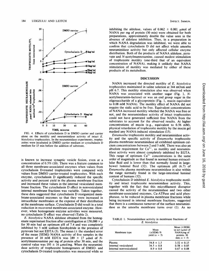
Figure 2 illustrates the effects of varying the mediumcalcium concentration on trophozoite motility, the 30-minneuraminidase activity of intact trophozoites, and theneuraminidase specific activity of the isolated Entamoebaplasma membrane fraction. Removing medium and cellsurface calcium with 0.2 mM ethylene glycol-bis(p-aminoethyl ether)-N,N,N',N'-tetraacetic acid (EGTA) sig-nificantly inhibited motility and neuraminidase activity when
INFECT. IMMUN.
.08mM
.02mM0 0- .56mM
on June 16, 2018 by guesthttp://iai.asm
.org/D
ownloaded from
E. HISTOL YTICA NEURAMINIDASE 183
compared with Ca2l-free medium alone. Motility and bothintact trophozoite and Entamoeba plasma membrane neura-minidase activities exhibited optimum medium calcium con-centrations. Whereas these three optimum concentrationswere not identical, they were all in the range of 2 to 5 mM.The optimum pHs for trophozoite homogenate and
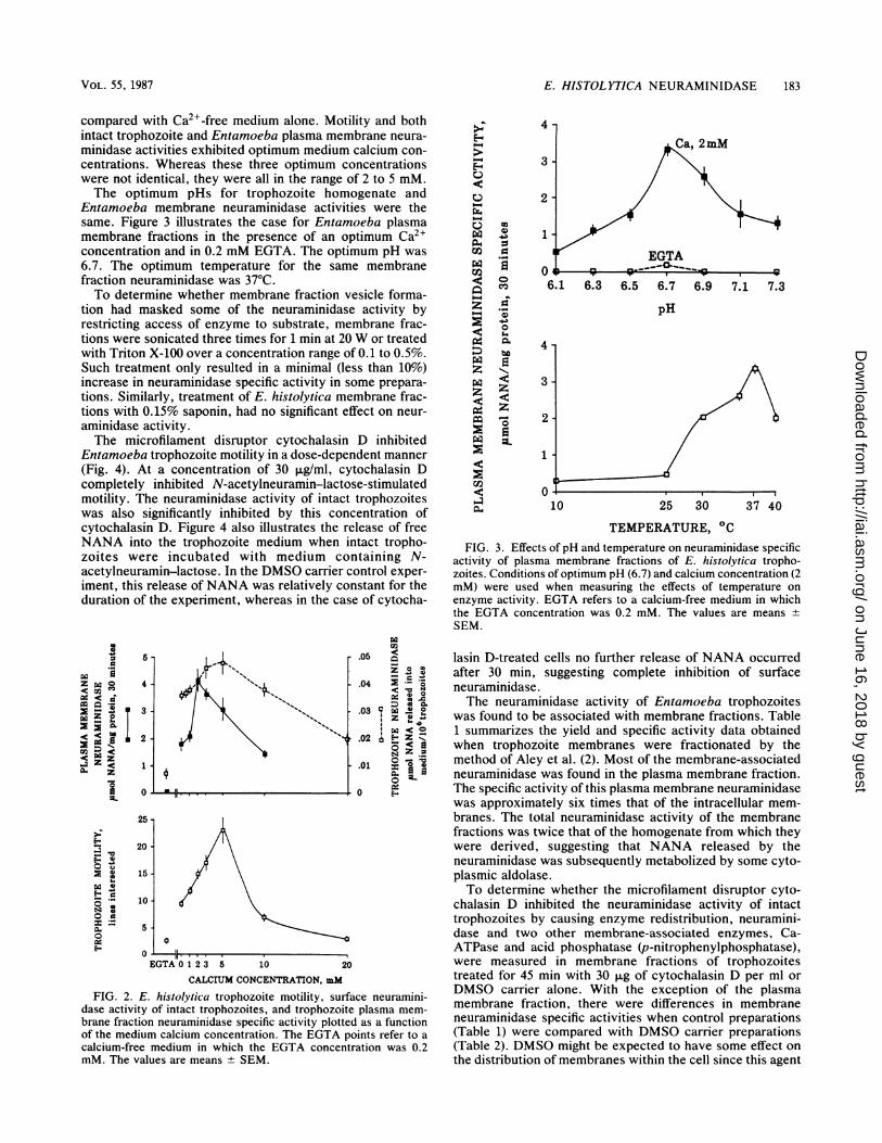
Entamoeba membrane neuraminidase activities were thesame. Figure 3 illustrates the case for Entamoeba plasmamembrane fractions in the presence of an optimum Ca2+concentration and in 0.2 mM EGTA. The optimum pH was6.7. The optimum temperature for the same membranefraction neuraminidase was 37°C.To determine whether membrane fraction vesicle forma-
tion had masked some of the neuraminidase activity byrestricting access of enzyme to substrate, membrane frac-tions were sonicated three times for 1 min at 20 W or treatedwith Triton X-100 over a concentration range of 0.1 to 0.5%.Such treatment only resulted in a minimal (less than 10%)increase in neuraminidase specific activity in some prepara-tions. Similarly, treatment of E. histolytica membrane frac-tions with 0.15% saponin, had no significant effect on neur-aminidase activity.The microfilament disruptor cytochalasin D inhibited
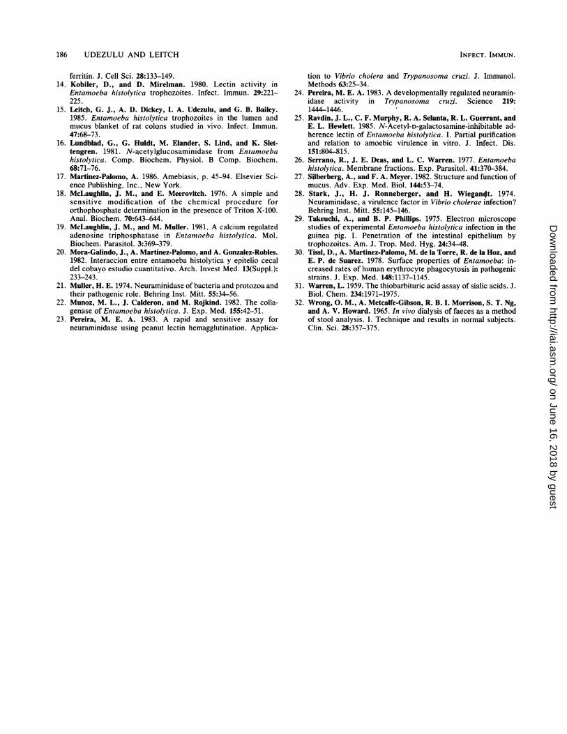
Entamoeba trophozoite motility in a dose-dependent manner(Fig. 4). At a concentration of 30 ,ug/ml, cytochalasin Dcompletely inhibited N-acetylneuramin-lactose-stimulatedmotility. The neuraminidase activity of intact trophozoiteswas also significantly inhibited by this concentration ofcytochalasin D. Figure 4 also illustrates the release of freeNANA into the trophozoite medium when intact tropho-zoites were incubated with medium containing N-acetylneuramin-lactose. In the DMSO carrier control exper-iment, this release of NANA was relatively constant for theduration of the experiment, whereas in the case of cytocha-
Oa
cv~S.-X X C6.< -IC te
X X i
.< 04 zzA$ z
25 -
E-
N4 0E- .0,0
O 10
:=
0.
N-
20 -
15 -
10 -
5-
0
N
.05 CZ ° Cv
.04 2 *-*o
:D a X..03 v X 0
Da < o.02 6 E- Z
N Z =
.01 a
0 E-
EGTA0123 5 10 20
CALCIUM CONCENTRATION, mM
FIG. 2. E. histolytica trophozoite motility, surface neuramini-dase activity of intact trophozoites, and trophozoite plasma mem-brane fraction neuraminidase specific activity plotted as a functionof the medium calcium concentration. The EGTA points refer to acalcium-free medium in which the EGTA concentration was 0.2mM. The values are means SEM.
Ez
z
E-v
¢0-
¢M
u¢qA:
¢Da¢
4
3
2
0
.40
._q
z
0
CID
a
-6-:t
1
0 -
6.1
4
3
2
1
0
6.3 6.5 6.7
pH
10
6.9 7.1 7.3
25 30 37 40
TEMPERATURE, 0CFIG. 3. Effects of pH and temperature on neuraminidase specific
activity of plasma membrane fractions of E. histolytica tropho-zoites. Conditions of optimum pH (6.7) and calcium concentration (2mM) were used when measuring the effects of temperature onenzyme activity. EGTA refers to a calcium-free medium in whichthe EGTA concentration was 0.2 mM. The values are means ±SEM.
lasin D-treated cells no further release of NANA occurredafter 30 min, suggesting complete inhibition of surfaceneuraminidase.The neuraminidase activity of Entamoeba trophozoites
was found to be associated with membrane fractions. Table1 summarizes the yield and specific activity data obtainedwhen trophozoite membranes were fractionated by themethod of Aley et al. (2). Most of the membrane-associatedneuraminidase was found in the plasma membrane fraction.The specific activity of this plasma membrane neuraminidasewas approximately six times that of the intracellular mem-branes. The total neuraminidase activity of the membranefractions was twice that of the homogenate from which theywere derived, suggesting that NANA released by theneuraminidase was subsequently metabolized by some cyto-plasmic aldolase.To determine whether the microfilament disruptor cyto-
chalasin D inhibited the neuraminidase activity of intacttrophozoites by causing enzyme redistribution, neuramini-dase and two other membrane-associated enzymes, Ca-ATPase and acid phosphatase (p-nitrophenylphosphatase),were measured in membrane fractions of trophozoitestreated for 45 min with 30 ,ug of cytochalasin D per ml orDMSO carrier alone. With the exception of the plasmamembrane fraction, there were differences in membraneneuraminidase specific activities when control preparations(Table 1) were compared with DMSO carrier preparations(Table 2). DMSO might be expected to have some effect onthe distribution of membranes within the cell since this agent
VOL. 55, 1987
0JL-
on June 16, 2018 by guesthttp://iai.asm
.org/D
ownloaded from
184 UDEZULU AND LEITCH
inhibiting the aldolase, values of 0.062 ± 0.001 ,umol ofNANA per mg of protein (30 min) were obtained for bothpreparations, approximately double the value seen in theabsence of aldolase inhibition. Thus, in a preparation inwhich NANA degradation was inhibited, we were able toconfirm that cytochalasin D did not affect whole amoebaneuraminidase activity but only affected cellular enzymedistribution. Both of the products of NANA aldolase, pyru-vate and N-acetylmannosamine, caused modest stimulationof trophozoite motility (one-third that of an equivalentconcentration of NANA), making it unlikely that NANAstimulation of motility was mediated by either of theseproducts of its metabolism.
zEz
0
N0
.0
.* o
o0 Aen.9*0
c) 0a I..
F4< 0
z 1-I.._
z_ lo 4)
.0
.01
.0
8 -
DMSO carrier, '
06 - ,X'
14
'2 - 'Cytochalasin D 30pzg/ml
A'00 5 15 30 45 60
TIME, minutesFIG. 4. Effects of cytochalasin D in DMSO carrier and carrier
alone on the motility and neuraminidase activity of intact E.histolytica trophozoites. In the neuraminidase experiments, tropho-zoites were incubated in DMSO carrier medium or cytochalasin Dmedium for 15 min before the addition of substrate.
is known to increase synaptic vesicle fusion, even at aconcentration of 0.5% (10). There was a feature common toall three membrane-associated enzymes when values fromcytochalasin D-treated trophozoites were compared withvalues from DMSO carrier-treated trophozoites. With eachenzyme, cytochalasin D significantly reduced the specificactivity and percent yield in the plasma membrane fractionand increased these values in the internal vesiculated mem-brane fraction. The cytochalasin D effect in nonvesiculatedinternal membrane fractions was variable. Taken together,these data suggested that cytochalasin D reorganized mem-brane-associated enzymes so that they were increased inintracellular membranes at the expense of their distributionat the membrane surface. Cytochalasin D did result in a totalreduction in recovered membrane-associated enzyme. How-ever, when homogenate enzyme activities were measured,no cytochalasin D effect was observed (Table 2).
E. histolytica NANA aldolase obtained from the homog-enate supernatant fraction after centrifugation at 150,000 x gfor 30 min had an optimum pH of 7.4 and was completelyinhibited by 5 mM sodium borohydride in the presence ofpyruvate but not EDTA (3). The mean (+ the standard errorof the mean [SEM]) NANA activity of five samples in thepresence of 10 mM EDTA was 189 17 ,umol of N-acetylmannosamine per mg of protein after 30 min, and thecontrol value was 193 ± 16 pumolImg. When the neuramini-dase activity of trophozoite homogenates of DMSO- andcytochalasin D-treated trophozoites was measured while so
DISCUSSIONNANA increased the directed motility of E. histolytica
trophozoites maintained in saline solution at 344 mOsm andpH 6.7. This motility stimulation also was observed whenNANA was associated with another sugar (Fig. 1, N-acetylneuramin-lactose) or was an end group sugar in theoligosaccharide of a glycoprotein (Fig. 1, mucin equivalentto 0.08 mM NANA). The motility effect of NANA did notrequire the sialic acid to be free. Equivalent concentrationsofNANA increased motility whether the NANA was free ornot, and the neuraminidase activity of intact trophozoitescould not have generated sufficient free NANA from thesubstrates to account for the observed results. At higherconcentrations of mucin (e.g., equivalent to 0.56 mM),physical retardation of trophozoite motility by the mucin gelmasked any NANA-induced stimulation (15).Entamoeba trophozoite motility and neuraminidase activ-
ity and the specific activity of Entamoeba trophozoiteplasma membrane neuraminidase all exhibited optimum cal-cium concentrations between 2 and 5 mM. There was also anabsolute requirement for Ca2+, as motility and neuramin-idase activity were almost completely inhibited by EGTA.This range of optimum Ca2+ concentrations is the sameorder of magnitude as that found in normal human extracel-lular fluid and is lower than that normally found in large-bowel luminal fluid (32). The optimum pH (6.7) ofEntamoeba plasma membrane neuraminidase is also withinthe range normally found in the large-intestinal luminalcontent of humans (32).
Cytochalasin D inhibited E. histolytica trophozoite motil-ity and intact trophozoite neuraminidase activity. This,together with the fact that this microfilament disruptorcaused the activity of the neuraminidase and two othermembrane-associated enzymes, Ca-ATPase and acid phos-phatase, to be reduced in plasma membrane fractions whilebeing increased in internal membrane fractions, suggestedthat there is a continuous turnover of the surface neuramini-dase as the amoeba membrane turns over (1). As no
TABLE 1. Neuraminidase activity in membrane fractions ofE. histolytica
Mean (±SEM)Mean sp act (,umol of
Membrane type (±SEM) % NANA/mg ofyield (n = 5) protein, 30
min; n = 5)
Plasma 54.8 ± 1.5 3.52 ± 0.15Internal vesiculated 34.5 ± 0.8 0.58 ± 0.05Internal nonvesiculated 8.9 ± 0.8 0.67 ± 0.08
20
E-o
0~-4
0.1N t
15
10
5
0
INFECT. IMMUN.
on June 16, 2018 by guesthttp://iai.asm
.org/D
ownloaded from

E. HISTOLYTICA NEURAMINIDASE 185
TABLE 2. Effect of cytochalasin D (30 jig/ml) on specific activities of E. histolytica membrane-associated and homogenate enzymes
Mean (±SEM) activity (n = 5) with:Enzyme
DMSO carrier Cytochalasin D Difference (P)
Plasma membranesNeuraminidase (,umol of NANA/mg of protein, 30 min) 3.326 ± 0.185 0.458 ± 0.027 <0.001Ca-ATPase (,imol of Pi/mg of protein, 10 min) 394.1 ± 24.2 165.4 + 2.7 <0.001p-Nitrophenylphosphatase (,umol/mg of protein, 2 min) 20.30 + 1.23 10.51 ± 0.30 <0.001
Internal vesiculated membranesNeuraminidase (,imol of NANA/mg of protein, 30 min) 0.095 ± 0.019 0.125 ± 0.023 <0.05Ca-ATPase (,umol of Pi/mg of protein, 10 min) 104.9 ± 3.7 147.0 ± 2.7 <0.01p-Nitrophenylphosphatase (,umol/mg of protein, 2 min) 1.63 ± 0.08 2.10 ± 0.05 <0.01
Internal nonvesiculated membranesNeuraminidase (,umol of NANA/mg of protein, 30 min) 0.256 ± 0.048 0.168 ± 0.032 >0.05Ca-ATPase (,mol of Pi/mg of protein, 10 min) 188.8 ± 22.8 149.6 ± 11.9 >0.05p-Nitrophenylphosphatase (p.mol/mg of protein, 2 min) 11.08 ± 1.04 8.93 ± 0.63 <0.05
Whole cell homogenateNeuraminidase (,umol of NANA/mg of protein, 30 min) 0.031 ± 0.001 0.032 ± 0.001 >0.05Ca-ATPase (p.mol of Pi/mg of protein, 10 min) 35,2 ± 0.09 37.7 ± 1.5 >0.05p-Nitrophenylphosphatase (,umol/mg of protein, 2 min) 1.88 + 0.04 1.85 ± 0.02 >0.05
neuraminidase activity was recovered from intact trophozo-ite supernatant solutions, neuraminidase must be exportedto the surface of the amoeba and then returned to theintracellular membrane pool by pinocytosis. Presumably,cytochalasin D inhibited this export of enzyme to the cellsurface more than it inhibited enzyme uptake into intracel-lular vesicles. Cytochalasin D had qualitatively, but notquantitatively, the same effect on the three membrane-associated enzymes studied.Neuraminidases are widely distributed in bacteria and
eucaryotic cells (21). In pathogenic organisms, attemptshave been made to determine whether neuraminidase is apathogenic factor, e.g., contributing to bacterial adherenceor mucus degradation in the case of enteropathogens. In thecase of Vibrio cholerae, these attempts have been inconclu-sive (28), and although there is an apparent correlationbetween V. cholerae strain neuraminidase activity and theseverity of the clinical signs and symptoms of cholera, manyother known enteropathogens lack the enzyme (12).Other parasitic protozoa also possess neuraminidase ac-
tivity. Notable among these is Trypanosoma cruzi, whichvaries its neuraminidase activity with its life cycle (24). As isthe case of E. histolytica, the neuraminidase in T. cruzi isfound on the trypomastigote surface, has an optimum pH ofapproximately 6.5, and is calcium dependent (23).
Most, but not all, organisms that possess a neuraminidasepossess an NANA aldolase (21). E. histolytica, however,possesses both a membrane neuraminidase and a solublealdolase (type 1 [21]). Entamoeba trophozoites probably donot contain NANA on their cell surface. In mammalian cells,NANA carboxyl groups make a significant contribution tothe negative surface charge of the cell surface (6). Inprotozoa, e.g., Naegleria gruberi (13) and Trypanosomalewisi (8), the surface negative charge is unaffected byneuraminidase. However, in Amoeba proteus, neuramini-dase inhibits phagocytosis (5), which may indicate the pres-ence of NANA in this amoeba.Whereas no role has yet been demonstrated for E.
histolytica surface neuraminidase in the pathogenesis ofamebiasis, it may be that this enzyme is one of a series ofcalcium-activated, membrane-associated enzymes (19), oneof several that may be involved in the destruction of mucin,
cell surface glycoproteins, and extracellular matrix duringamebic invasion (16, 22).
ACKNOWLEDGMENTS
This work was supported by Public Health Service grant RR-8006from the General Research Support Branch, Division of ResearchResources, National Institutes of Health, Bethesda, Md. I.A.U. wasa recipient of a Nigerian Government Scholarship while a student inthe Department of Biology, Atlanta University, Atlanta, Ga.
LITERATURE CITED1. Aley, S. B., Z. A. Cohn, and W. A. Scott. 1984. Endocytosis in
Entamoeba histolytica. Evidence for a unique non-acidifiedcompartment. J. Exp. Med. 160:724-737.
2. Aley, S. B., W. A. Scott, and Z. A. Cohn. 1980. Plasmamembrane of Entamoeba histolytica. J. Exp. Med. 152:391-404.
3. Barnett, j. E. G., D. L. Corina, and G. Rasool. 1971. Studies onN-acetylneuraminic acid aldolase. Biochem. J. 125:275-284.
4. Bradford, M. M. 1976. A rapid and sensitive method for thequantitation of microgram quantities of protein utilizing theprinciple of protein dye binding. Anal. Biochem. 72:248-254.
5. Chatterjee, S., and P. K. Ray. 1975. Pinocytic activity inneuraminidase treated amoeba. Indian J. Exp. Biol. 13:294-295.
6. Cook, G. M. W., D. H. Heard, and G. V. F. Seaman. 1961. Sialicacids and the electrokinetic charge of the human erythrocyte.Nature (London) 191:44 47.
7. Diamond, L. S. 1968. Techniques of axenic cultivation ofEntamoeba histolytica Schaudinn, 1903 and E. histolytica-likeamebae. J. Parsitol. 54:1047-1056.
8. Dwyer, D. M. 1975. Cell surface saccharides of Trypanosomalewisi. I. Polycation-induced cell agglutination and fine structurecytochemistry. J. Cell Sci. 19:621-644.
9. Forstner, G., A. Wesley, and J. Forstner. 1982. Clinical aspectsof gastrointestinal mucus. Adv. Exp. Med. Biol. 144:199-224.
10. Geron, N., and H. Meiri. 1985. The fusogenic substancedimethyl sulfoxide enhances exocytosis in motor nerve endings.Biochim. Biophys. Acta 819:258-262.
11. Jarumilinta, R., and F. Kradolfer. 1965. The toxic effect ofEntamoeba histolytica on leucocytes. Am. J. Trop. Med. Hyg.58:375-381.
12. Kabir, S., N. Ahmad, and S. Ali. 1984. Neuraminidase produc-tion by Vibrio cholerae 01 and other diarrheagenic bacteria.Infect. Immun. 44:747-749.
13. King, C. A., and T. M. Preston. 1977. Studies on anionic sites onthe surface of the amoeba Naegleria gruberi using cationized
VOL. 55, 1987
on June 16, 2018 by guesthttp://iai.asm
.org/D
ownloaded from
186 UDEZULU AND LEITCH
ferritin. J. Cell Sci. 28:133-149.14. Kobiler, D., and D. Mirelman. 1980. Lectin activity in
Entamoeba histolytica trophozoites. Infect. Immun. 29:221-225.
15. Leitch, G. J., A. D. Dickey, I. A. Udezulu, and G. B. Bailey.1985. Entamoeba histolytica trophozoites in the lumen andmucus blanket of rat colQns studied in vivo. Infect. Immun.47:68-73.
16. Lundblad, G., G. Huldt, M. Elander, S. Lind, and K. Slet-tengren. 1981. N-acetylglucosaminidase from Entamoebahistolytica. Comp. Biochem. Physiol. B Comp. Biochem.68:71-76.
17. Martinez-Palomo, A. 1986. Amebiasis, p. 45-94. Elsevier Sci-ence Publishing, Inc., New York.
18. McLaughlin, J. M., and E. Meerovitch. 1976. A simple andsensitive modification of the chemical procedure fororthophosphate determination in the presence of Triton X-100.Anal. Biochem. 70:643-644.
19. McLaughlin, J. M., and M. Muller. 1981. A calcium regulatedadenosine triphosphatase in Entamoeba histolytica. Mol.Biochem. Parasitol. 3:369-379.
20. Mora-Galindo, J., A. Martinez-Palomo, and A. Gonzalez-Robles.1982. Interaccion entre entamoeba histolytica y epitelio cecaldel cobayo estudio cuantitativo. Arch. Invest Med. 13(Suppl.):233-243.
21. Muller, H. E. 1974. Neuraminidase of bacteria and protozoa andtheir pathogenic role. Behring Inst. Mitt. 55:34-56.
22. Munoz, M. L., J. Calderon, and M. Rojkind. 1982. The colla-genase of Entamoeba histolytica. J. Exp. Med. 155:42-51.
23. Pereira, M, E. A. 1983. A rapid and sensitive assay forneuraminidase using peanut lectin hemagglutination. Applica-
tion to Vibrio cholera and Trypanosoma cruzi. J. Immunol.Methods 63:25-34.
24. Pereira, M. E. A. 1983. A developmentally regulated neuramin-idase activity in Trypanosoma cruzi. Science 219:1444 1446.
25. Ravdin, J. L., C. F. Murphy, R. A. Selanta, R. L. Guerrant, andE. L. Hewlett. 1985. N-Acetyl-D-galactosamine-inhibitable ad-herence lectin of Entamoeba histolytica. I. Partial purificationand relation to amoebic virulence in vitro. J. Infect. Dis.151:804-815.
26. Serrano, R., J. E. Deas, and L. C. Warren. 1977. Entamoebahistolytica. Membrane fractions. Exp. Parasitol. 41:370-384.
27. Silberberg, A., and F. A. Meyer. 1982. Structure and function ofmucus. Adv. Exp. Med. Biol. 144:53-74.
28. Stark, J., H. J. Ronneberger, and H. Wiegan4t. 1974.Neuraminidase, a virulence factor in Vibrio cholerae infection?Behring Inst. Mitt. 55:145-146.
29. Takeuchi, A., and B. P. Phillips. 1975. Electron microscopestudies of experimental Entamoeba histolytica infection in theguinea pig. I. Penetration of the intestinal epithelium bytrophozoites. Am. J, Trop. Med. Hyg. 24:34-48.
30. Tissl, D., A. Martinez-Palomo, M. de la Torre, R. de la Hoz, andE. P. de Suarez. 1978. Surface properties of Entamoeba: in-creased rates of human erythrocyte phagocytosis in pathogenicstrains. J. Exp. Med. 148:1137-1145.
31. Warren, L. 1959. The thiobarbituric acid assay of sialic acids. J.Biol. Chem. 234:1971-1975.
32. Wrong, 0. M., A. Metcalfe-Gibson, R. B. I. Morrison, S. T. Ng,and A. V. Howard. 1965. In vivo dialysis of faeces as a methodof stool analysis. I. Technique and results in normal subjects.Clin. Sci. 28:357-375.
INFECT. IMMUN.
on June 16, 2018 by guesthttp://iai.asm
.org/D
ownloaded from