A large deletion spanning PITX2 and PANCR in a Chinese ...
9
Axenfeld-Rieger syndrome (ARS) is a rare disease primarily characterized by abnormal development of the ante- rior segment of the eye, such as iris hypoplasia, corectopia, polycoria, posterior embryotoxon, and corneal abnormalities [ 1- 3]. Around 50% of patients with ARS develop glaucoma, which leads to blindness without appropriate intervention. Extraocular symptoms, such as dental anomalies, redundant periumbilical skin, craniofacial abnormalities, heart defects, hearing loss, and so on, can also be observed in all or some of the patients with ARS [ 1,2]. ARS is inherited in an autosomal dominant manner. Mutations in the major pathogenic genes PITX2 (OMIM 601542) and FOXC1 (OMIM 601090) account for about 40–70% of the patients with ARS [2,4]. In addition to the common intragenic mutations (missense, nonsense, splicing, and small deletion/insertion), large deletions affecting the entire or part of the coding and upstream regulatory regions of PITX2 and FOXC1 have also been reported [ 5- 7]. Another two genes ( CYP1B1-Gene ID: 1545, OMIM: 601771 [8] and PRDM5- Gene ID: 11107, OMIM: 614161 [9]) and one locus (13q14 [ 10]) have also been implicated with ARS. However, the underlying genetic causes in at least 30% of patients with ARS remain unclear. Genotype–phenotype studies indicate that mutations in PITX2 are more commonly associated with additional systemic symptoms such as dental, umbilical, and cranio- facial abnormalities. Meanwhile, patients with mutations in FOXC1 often show ocular symptoms alone or combined with heart and hearing defects [ 4,6, 11 ]. However, recent studies also reported that mutations in PITX2 can result in dental anomalies [ 12] and heart diseases [ 13- 16] without the classical symptoms of ARS. We report a four-generation family with ARS from Hubei province, China. The clinical features of the affected individuals are summarized. With the help of whole-exome sequencing (WES), quantitative PCR (qPCR), and long-range PCR, a genomic deletion spanning PITX2 and PANCR (Gene ID: 110231149, OMIM: 617286) was identified. Molecular Vision 2020; 26:670-678 <http://www.molvis.org/molvis/v26/670> Received 11 January 2020 | Accepted 2 October 2020 | Published 4 October 2020 © 2020 Molecular Vision 670 A large deletion spanning PITX2 and PANCR in a Chinese family with Axenfeld-Rieger syndrome Yayun Qin, 1 Pang Gao, 1 Shanshan Yu, 1 Jingzhen Li, 1 Yuwen Huang, 1 Danna Jia, 1 Zhaohui Tang, 1 Pengcheng Li, 2 Fei Liu, 1 Mugen Liu 1 1 Key Laboratory of Molecular Biophysics of Ministry of Education, College of Life Science and Technology, Huazhong University of Science and Technology, Wuhan, P.R. China; 2 Department of Ophthalmology, Union Hospital, Tongji Medical College, Huazhong University of Science and Technology, Wuhan, P.R. China Purpose: To identify the genetic cause in a four-generation Chinese family with Axenfeld-Rieger syndrome (ARS). Methods: The family members received clinical examinations of the eye, tooth, periumbilical skin, and heart. Sanger sequencing and whole-exome sequencing (WES) were performed to screen potential mutations. The genomic deletion region around the PITX2 gene was estimated from single nucleotide polymorphism (SNP) data from WES and then confirmed with “quantitative PCR (qPCR) using a set of primers. The DNA breakpoint was further identified with long-range PCR and Sanger sequencing. Results: Symptoms including anterior segment dysplasia of the eye (iris dysplasia, multiple pupils, and posterior embryo- toxon), dental dysplasia, and periumbilical skin redundancy were present in all of the affected individuals. Three of them had glaucoma. Corneal abnormalities (inferior sclerocornea, corneal endothelial dystrophy, and central corneal scar) were seen in most of the affected individuals. Cataract, limited eye movement, electrocardiographic abnormalities, intel- lectual disability, and recurrent miscarriages were observed in some of the affected individuals. No mutations in the cod- ing and exon-intron adjacent regions of the PITX2 and FOXC1 genes were identified with Sanger sequencing. According to the SNP data from WES, we suspected that there might be a deletion region (at most 1.6 Mb) around the PITX2 gene. With the use of qPCR and long-range PCR, we identified a 53,840 bp deletion (chr4: 111,535,454–111,588,933) spanning PITX2 and PANCR. The genomic deletion cosegregated with the major ARS symptoms observed in the family members. Conclusions: With the help of WES, qPCR, and long-range PCR, we identified a genomic deletion encompassing PITX2 and the adjacent noncoding gene PANCR in a Chinese family with ARS. The clinical features of the affected individuals are reported. This work may broaden understanding of the phenotypic and mutational spectrums related to ARS. Correspondence to: Fei Liu, Key Laboratory of Molecular Biophysics of Ministry of Education, College of Life Science and Technology, Huazhong University of Science and Technology, Wuhan 430074, P.R. China; [email protected]
Transcript of A large deletion spanning PITX2 and PANCR in a Chinese ...

Axenfeld-Rieger syndrome (ARS) is a rare disease primarily characterized by abnormal development of the ante-rior segment of the eye, such as iris hypoplasia, corectopia, polycoria, posterior embryotoxon, and corneal abnormalities [1-3]. Around 50% of patients with ARS develop glaucoma, which leads to blindness without appropriate intervention. Extraocular symptoms, such as dental anomalies, redundant periumbilical skin, craniofacial abnormalities, heart defects, hearing loss, and so on, can also be observed in all or some of the patients with ARS [1,2].
ARS is inherited in an autosomal dominant manner. Mutations in the major pathogenic genes PITX2 (OMIM 601542) and FOXC1 (OMIM 601090) account for about 40–70% of the patients with ARS [2,4]. In addition to the common intragenic mutations (missense, nonsense, splicing, and small deletion/insertion), large deletions affecting the entire or part of the coding and upstream regulatory regions
of PITX2 and FOXC1 have also been reported [5-7]. Another two genes (CYP1B1-Gene ID: 1545, OMIM: 601771 [8] and PRDM5-Gene ID: 11107, OMIM: 614161 [9]) and one locus (13q14 [10]) have also been implicated with ARS. However, the underlying genetic causes in at least 30% of patients with ARS remain unclear.
Genotype–phenotype studies indicate that mutations in PITX2 are more commonly associated with additional systemic symptoms such as dental, umbilical, and cranio-facial abnormalities. Meanwhile, patients with mutations in FOXC1 often show ocular symptoms alone or combined with heart and hearing defects [4,6,11]. However, recent studies also reported that mutations in PITX2 can result in dental anomalies [12] and heart diseases [13-16] without the classical symptoms of ARS.
We report a four-generation family with ARS from Hubei province, China. The clinical features of the affected individuals are summarized. With the help of whole-exome sequencing (WES), quantitative PCR (qPCR), and long-range PCR, a genomic deletion spanning PITX2 and PANCR (Gene ID: 110231149, OMIM: 617286) was identified.
Molecular Vision 2020; 26:670-678 <http://www.molvis.org/molvis/v26/670>Received 11 January 2020 | Accepted 2 October 2020 | Published 4 October 2020
© 2020 Molecular Vision
670
A large deletion spanning PITX2 and PANCR in a Chinese family with Axenfeld-Rieger syndrome
Yayun Qin,1 Pang Gao,1 Shanshan Yu,1 Jingzhen Li,1 Yuwen Huang,1 Danna Jia,1 Zhaohui Tang,1 Pengcheng Li,2 Fei Liu,1 Mugen Liu1
1Key Laboratory of Molecular Biophysics of Ministry of Education, College of Life Science and Technology, Huazhong University of Science and Technology, Wuhan, P.R. China; 2Department of Ophthalmology, Union Hospital, Tongji Medical College, Huazhong University of Science and Technology, Wuhan, P.R. China
Purpose: To identify the genetic cause in a four-generation Chinese family with Axenfeld-Rieger syndrome (ARS).Methods: The family members received clinical examinations of the eye, tooth, periumbilical skin, and heart. Sanger sequencing and whole-exome sequencing (WES) were performed to screen potential mutations. The genomic deletion region around the PITX2 gene was estimated from single nucleotide polymorphism (SNP) data from WES and then confirmed with “quantitative PCR (qPCR) using a set of primers. The DNA breakpoint was further identified with long-range PCR and Sanger sequencing.Results: Symptoms including anterior segment dysplasia of the eye (iris dysplasia, multiple pupils, and posterior embryo-toxon), dental dysplasia, and periumbilical skin redundancy were present in all of the affected individuals. Three of them had glaucoma. Corneal abnormalities (inferior sclerocornea, corneal endothelial dystrophy, and central corneal scar) were seen in most of the affected individuals. Cataract, limited eye movement, electrocardiographic abnormalities, intel-lectual disability, and recurrent miscarriages were observed in some of the affected individuals. No mutations in the cod-ing and exon-intron adjacent regions of the PITX2 and FOXC1 genes were identified with Sanger sequencing. According to the SNP data from WES, we suspected that there might be a deletion region (at most 1.6 Mb) around the PITX2 gene. With the use of qPCR and long-range PCR, we identified a 53,840 bp deletion (chr4: 111,535,454–111,588,933) spanning PITX2 and PANCR. The genomic deletion cosegregated with the major ARS symptoms observed in the family members.Conclusions: With the help of WES, qPCR, and long-range PCR, we identified a genomic deletion encompassing PITX2 and the adjacent noncoding gene PANCR in a Chinese family with ARS. The clinical features of the affected individuals are reported. This work may broaden understanding of the phenotypic and mutational spectrums related to ARS.
Correspondence to: Fei Liu, Key Laboratory of Molecular Biophysics of Ministry of Education, College of Life Science and Technology, Huazhong University of Science and Technology, Wuhan 430074, P.R. China; [email protected]
Molecular Vision 2020; 26:670-678 <http://www.molvis.org/molvis/v26/670> © 2020 Molecular Vision
671
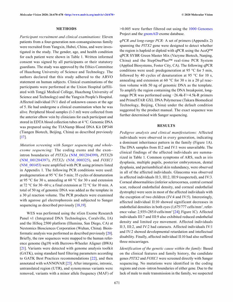
METHODS
Participant recruitment and clinical examinations: Eleven patients from a four-generation non-consanguineous family were recruited from Yangxin, Hubei, China, and were inves-tigated in the study. The gender, age, and health condition for each patient were shown in Table 1. Written informed consent was signed by all participants or their statutory guardians. The study was approved by the Ethics Committee of Huazhong University of Science and Technology. The authors declared that this study adhered to the ARVO statement on human subjects. Clinical examinations of the participants were performed at the Union Hospital (affili-ated with Tongji Medical College, Huazhong University of Science and Technology) and the Yangxin People’s Hospital. Affected individual IV:1 died of unknown causes at the age of 5. He had undergone a clinical examination when he was alive. Peripheral blood samples (1-3 ml) were collected from the anterior elbow vein by clinicians for each participant and stored in EDTA blood collection tubes at 4 °C. Genomic DNA was prepared using the TIANamp Blood DNA Kit DP348 (Tiangen Biotech, Beijing, China) as described previously [17].
Mutation screening with Sanger sequencing and whole-exome sequencing: The coding exons and the exon–intron boundaries of PITX2a (NM_001204399), PITX2b (NM_001204397), PITX2c (NM_000325), and FOXC1 (NM_001453) were amplified with PCR using primers listed in Appendix 1. The following PCR conditions were used: predegeneration at 95 °C for 5 min; 35 cycles of denaturation at 95 °C for 30 s, annealing at 60 °C for 30 s and extension at 72 °C for 30–60 s; a final extension at 72 °C for 10 min. A total of 50 ng of genomic DNA was added as the template in a 30 μl reaction volume. The PCR products were examined with agarose gel electrophoresis and subjected to Sanger sequencing as described previously [18,19].
WES was performed using the xGen Exome Research Panel v1 (Integrated DNA Technologies, Coralville, IA) and the HiSeq 2500 platform (Illumina, San Diego, CA) at Nextomics Biosciences Corporation (Wuhan, China). Bioin-formatic analysis was performed as described previously [20]. Briefly, the raw sequences were mapped to the human refer-ence genome (hg19) with Burrows-Wheeler Aligner (BWA) [21]. Variants were detected with genome analysis toolkit (GATK), using standard hard filtering parameters according to GATK Best Practices recommendations [22], and then annotated with wANNOVAR [23]. After intergenic, intronic, untranslated region (UTR), and synonymous variants were removed, variants with a minor allele frequency (MAF) of
>0.005 were further filtered out using the 1000 Genomes Project and the gnomAD exome database.
qPCR and long-range PCR: A set of primers (Appendix 2) spanning the PITX2 gene were designed to detect whether the region is haploid or diploid with qPCR using the AceQ™ qPCR SYBR Green Master Mix (Vazyme Biotech, Nanjing, China) and the StepOnePlus™ real-time PCR System (Applied Biosystems, Foster City, CA). The following qPCR conditions were used: predegeneration at 95 °C for 5 min, followed by 40 cycles of denaturation at 95 °C for 10 s, annealing and extension at 60 °C for 30 s in a 20 μl reac-tion volume with 50 ng of genomic DNA as the template. To amplify the region containing the DNA breakpoint, long-range PCR was performed using −26.4k-F/+4.5k-R primers and PrimeSTAR GXL DNA Polymerase (Takara Biomedical Technology, Beijing, China) under the default condition suggested by the product manual. The exact sequence was further determined with Sanger sequencing.
RESULTS
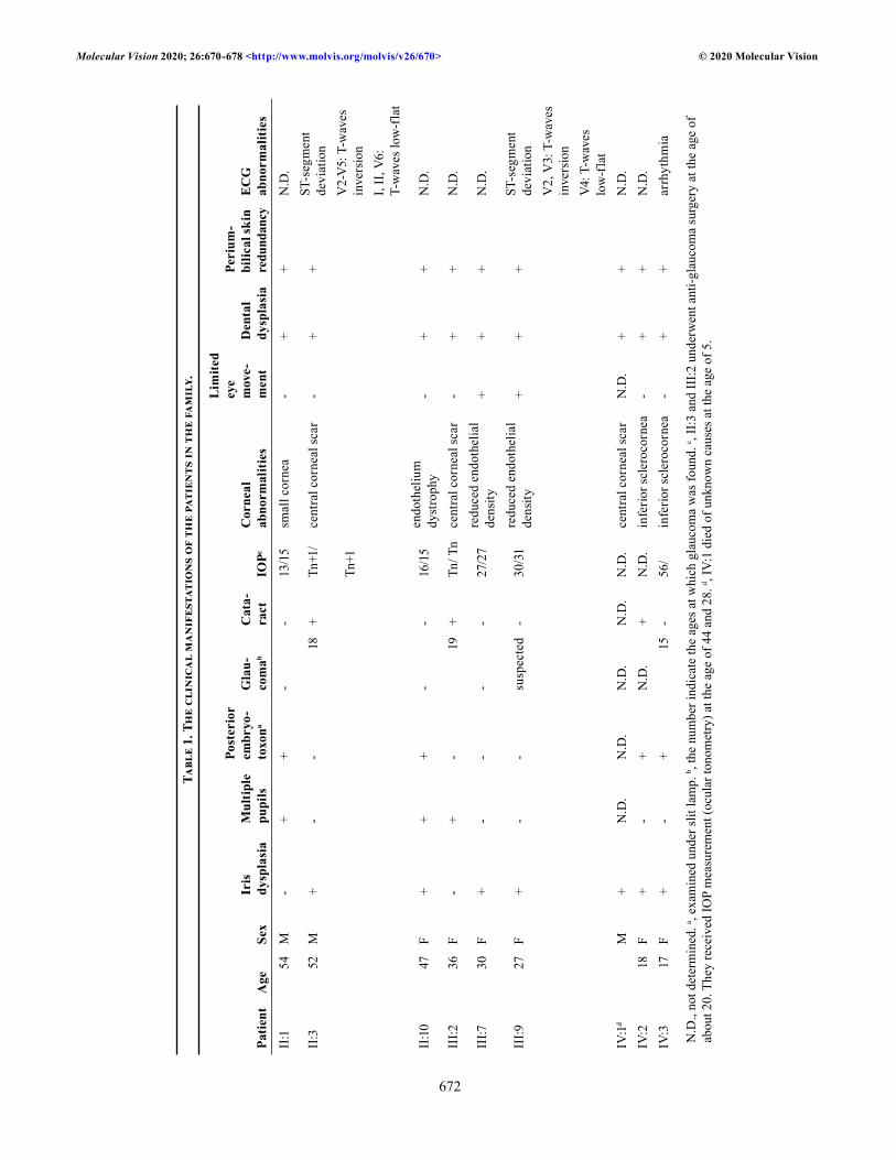
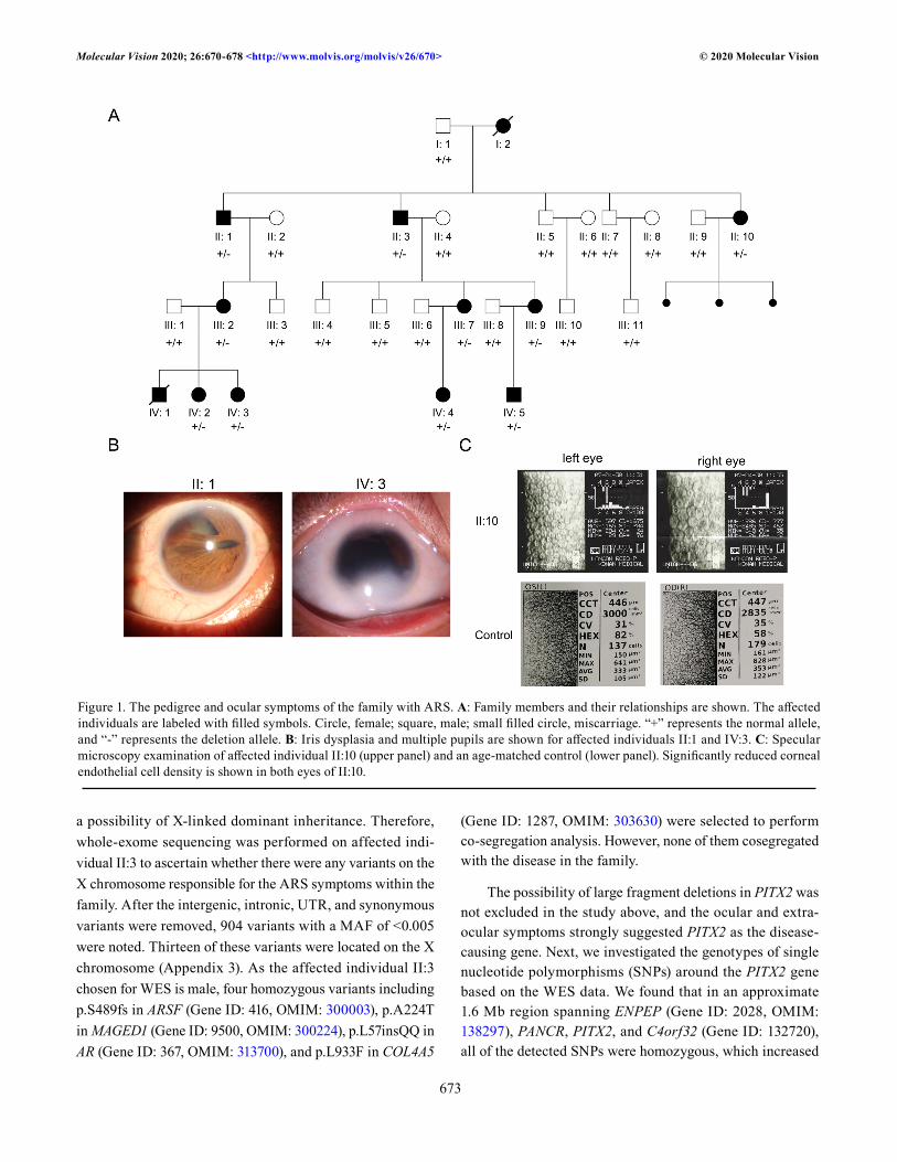
Pedigree analysis and clinical manifestations: Affected individuals were observed in every generation, indicating a dominant inheritance pattern in the family (Figure 1A). The DNA samples from II:2 and IV:1 were unavailable. The clinical findings of the affected individuals are summa-rized in Table 1. Common symptoms of ARS, such as iris dysplasia, multiple pupils, posterior embryotoxon, dental dysplasia, and periumbilical skin redundancy, were observed in all of the affected individuals. Glaucoma was observed in affected individuals II:3, III:2, III:9 (suspected), and IV:3. Corneal abnormalities (inferior sclerocornea, central corneal scar, reduced endothelial density, and corneal endothelial dystrophy) were seen in most of the affected individuals with the exception of two children (IV:4 and IV:5). Interestingly, affected individual II:10 showed significant decreases in endothelial densities in both eyes (1,675/777 cells/mm2, refer-ence value: 2,935±285.0 cells/mm2 [24]; Figure 1C). Affected individuals III:7 and III:9 also exhibited reduced endothelial density and limited eye movement. Affected individuals II:3, III:2, and IV:2 had cataracts. Affected individuals IV:1 and IV:2 showed developmental retardation and intellectual disability. Finally, affected individual II:10 had also suffered three miscarriages.
Identification of the genetic cause within the family: Based on the clinical features and family history, the candidate genes PITX2 and FOXC1 were screened directly with Sanger sequencing. No mutations were identified in the coding regions and exon–intron boundaries of either gene. Due to the lack of male to male transmission in the family, we suspected
Molecular Vision 2020; 26:670-678 <http://www.molvis.org/molvis/v26/670> © 2020 Molecular Vision
672
Tabl
e 1.
The
cli
nica
l m
anif
esta
tion
s of
the
pat
ient
s in
the
fam
ily.
Patie
ntA
geSe
xIr
is
dysp
lasi
aM
ultip
le
pupi
ls
Post
erio
r em
bryo
-to
xona
Gla
u-co
mab
Cat
a-ra
ctIO
PcC
orne
al
abno
rmal
ities
Lim
ited
eye
mov
e-m
ent
Den
tal
dysp
lasi
a
Peri
um-
bilic
al sk
in
redu
ndan
cyE
CG
ab
norm
aliti
es
II:1
54M
-+
+-
-13
/15
smal
l cor
nea
-+
+N
.D.
II:3
52M
+-
-18
+Tn
+1/
cent
ral c
orne
al sc
ar-
++
ST-s
egm
ent
devi
atio
n
Tn+1
V2-
V5:
T-w
aves
in
vers
ion
I, II
, V6:
T-
wav
es lo
w-f
lat
II:1
047
F+
++
--
16/1
5en
doth
eliu
m
dyst
roph
y-
++
N.D
.
III:2
36F
-+
-19
+Tn
/ Tn
cent
ral c
orne
al sc
ar-
++
N.D
.
III:7
30F
+-
--
-27
/27
redu
ced
endo
thel
ial
dens
ity+
++
N.D
.
III:9
27F
+-
-su
spec
ted
-30
/31
redu
ced
endo
thel
ial
dens
ity+
++
ST-s
egm
ent
devi
atio
nV
2, V
3: T
-wav
es
inve
rsio
nV4
: T-w
aves
lo
w-f
lat
IV:1
dM
+N
.D.
N.D
.N
.D.
N.D
.N
.D.
cent
ral c
orne
al sc
arN
.D.
++
N.D
.IV
:218
F+
-+
N.D
.+
N.D
.in
ferio
r scl
eroc
orne
a-
++
N.D
.IV
:317
F+
-+
15-
56/
infe
rior s
cler
ocor
nea
-+
+ar
rhyt
hmia
N.D
., no
t det
erm
ined
. a , ex
amin
ed u
nder
slit
lam
p. b ,
the
num
ber i
ndic
ate
the
ages
at w
hich
gla
ucom
a w
as fo
und.
c , II
:3 a
nd II
I:2 u
nder
wen
t ant
i-gla
ucom
a su
rger
y at
the
age
of
abou
t 20.
The
y re
ceiv
ed IO
P m
easu
rem
ent (
ocul
ar to
nom
etry
) at t
he a
ge o
f 44
and
28. d ,
IV:1
die
d of
unk
now
n ca
uses
at t
he a
ge o
f 5.
Molecular Vision 2020; 26:670-678 <http://www.molvis.org/molvis/v26/670> © 2020 Molecular Vision
673
a possibility of X-linked dominant inheritance. Therefore, whole-exome sequencing was performed on affected indi-vidual II:3 to ascertain whether there were any variants on the X chromosome responsible for the ARS symptoms within the family. After the intergenic, intronic, UTR, and synonymous variants were removed, 904 variants with a MAF of <0.005 were noted. Thirteen of these variants were located on the X chromosome (Appendix 3). As the affected individual II:3 chosen for WES is male, four homozygous variants including p.S489fs in ARSF (Gene ID: 416, OMIM: 300003), p.A224T in MAGED1 (Gene ID: 9500, OMIM: 300224), p.L57insQQ in AR (Gene ID: 367, OMIM: 313700), and p.L933F in COL4A5
(Gene ID: 1287, OMIM: 303630) were selected to perform co-segregation analysis. However, none of them cosegregated with the disease in the family.
The possibility of large fragment deletions in PITX2 was not excluded in the study above, and the ocular and extra-ocular symptoms strongly suggested PITX2 as the disease-causing gene. Next, we investigated the genotypes of single nucleotide polymorphisms (SNPs) around the PITX2 gene based on the WES data. We found that in an approximate 1.6 Mb region spanning ENPEP (Gene ID: 2028, OMIM: 138297), PANCR, PITX2, and C4orf32 (Gene ID: 132720), all of the detected SNPs were homozygous, which increased
Figure 1. The pedigree and ocular symptoms of the family with ARS. A: Family members and their relationships are shown. The affected individuals are labeled with filled symbols. Circle, female; square, male; small filled circle, miscarriage. “+” represents the normal allele, and “-” represents the deletion allele. B: Iris dysplasia and multiple pupils are shown for affected individuals II:1 and IV:3. C: Specular microscopy examination of affected individual II:10 (upper panel) and an age-matched control (lower panel). Significantly reduced corneal endothelial cell density is shown in both eyes of II:10.

Molecular Vision 2020; 26:670-678 <http://www.molvis.org/molvis/v26/670> © 2020 Molecular Vision
674
the possibility of the haploid status of the whole or part of the PITX2 gene.
To confirm the deletion and identify the exact DNA breakpoint, we performed qPCR analysis on DNA from indi-viduals III:5 (unaffected) and III:9 (affected) using primers covering the exons and the 5′- and 3′- regions of PITX2 (Appendix 2). All of the examined exons were haploid in the affected individual (Figure 2A), confirming the deletion of the whole PITX2 gene. The deletion range was identified by extending the qPCR primers upstream and downstream of PITX2. The DNA breakpoint was located between −26.4 kb to −23.3 kb upstream of PITX2 and +2.5 kb to +4.5 kb downstream of PITX2. Using the −26.4k-F/+4.5k-R primer pair, an approximate 2 kb DNA fragment was amplified only in affected individual III:9 (Figure 2B). Sanger sequencing of this fragment uncovered a 53,840 bp deletion (chr4:
111,535,454–111,588,933; Figure 2C), which contained the whole PITX2 gene and the first exon of the noncoding gene PANCR. The deletion exactly cosegregated with the disease in the family, as detected with PCR amplification using the −26.4k-F/+4.5k-R primer pair (Appendix 4).
Electrocardiographic abnormalities in affected individuals: Several studies have shown that PITX2 plays an important role in the development and maintenance of the heart [25-28]. The adjacent lncRNA PANCR regulates the expression of PITX2c, the cardiac-specific transcript of PITX2 [29]. In addition, mutations in PITX2 have been associated with a wide range of heart defects, such as atrial fibrillation [14,16], tetralogy of Fallot [13,30], and congenital atrial septal defects [31]. However, no signs of heart disease were reported by any of the affected family members. To investigate any poten-tial asymptomatic cardiac anomalies, affected (II:3, III:9,
Figure 2. Identification of the large genomic deletion spanning PITX2 in the family. A: Confirming and mapping the deleted region around PITX2 with quantitative PCR (qPCR). The deletion was positioned between 26.4 kb upstream and 4.5 kb downstream of the PITX2 gene. B: The DNA fragment containing the breakpoint was amplified with long-range PCR using the −26.4k-F/+4.5k-R primers. C: The sequencing result of the DNA fragment above shows an approximate 50 kb deletion spanning the first exon of PANCR and the entire PITX2 gene.

Molecular Vision 2020; 26:670-678 <http://www.molvis.org/molvis/v26/670> © 2020 Molecular Vision
675
and IV:3) and unaffected (III:5) individuals received color Doppler echocardiography and electrocardiography tests. No obvious changes in the heart structure were observed in affected individuals II:3 and III:9. Affected individual IV:3 showed left ventricular false tendon and arrhythmia. The electrocardiograph of IV:3 also demonstrated the presence of arrhythmia and ST-T abnormalities (Figure 3, Table 1). Furthermore, affected individuals II:3 and III:9 also presented ST-T abnormalities with normal sinus rhythm (Figure 3, Table 1). These changes were not detected in unaffected individual III:5.
DISCUSSION
PITX2 encodes a homeobox transcription factor that plays an essential role in the development of many organs [32-34]. Complete deletion of Pitx2 in mice causes embryonic lethality and multiorgan malformations, including ARS-related
anomalies and heart defects [35,36]. Heterozygous Pitx2-null mice show the ocular features of ARS and glaucoma [37], and are susceptible to atrial arrhythmias [28]. Doubtlessly, PITX2 is important for normal heart function. In this study, indi-viduals with ARS showed no obvious heart structural symp-toms or defects. However, the ECG revealed irregularities in cardiac electrophysiology in some of the affected individuals. A patient with ARS with a mutation in PITX2 was previously diagnosed with Wolf-Parkinson-White (WPW) syndrome [3]. WPW syndrome is a form of heart disease characterized by tachycardia and other changes in ECG. More than half of individuals with WPW syndrome are asymptomatic, but they have a low risk of undergoing cardiac arrest or sudden cardiac death [38]. To a certain extent, the cardiac features of patients examined in this family may belong to the category of WPW syndrome. Nevertheless, compared with the eye, tooth, and periumbilical skin abnormalities, heart defects are rarely reported in patients with mutations in PITX2.
Figure 3. The electrocardiographic abnormalities of the affected individuals in the family. IV:3 shows arrhythmia (indicated by the horizontal arrows). II:3, III:9, and IV:3 show ST-T abnormalities (indicated by arrows).
Molecular Vision 2020; 26:670-678 <http://www.molvis.org/molvis/v26/670> © 2020 Molecular Vision
676
Other unusual features, such as limited eye movement, corneal abnormalities, cataract, recurrent spontaneous miscarriages, and intellectual disability, were observed in certain patients in the family. Pitx2 is required for the devel-opment and maintenance of extraocular muscles [39], which may explain the presence of limited eye movement in the two patients in this study. Most of the patients (except the two young children) in this family showed corneal abnormali-ties, which are thought to be infrequent in patients with ARS according to previous reports [1,2]. Developmental retarda-tion and intellectual disability have also been reported in patients with mutations in PITX2 by other researchers [6,40]. Recurrent spontaneous miscarriages have not previously been reported in individuals with mutations in PITX2. It is as yet not possible to determine whether these unusual symptoms are associated with ARS, or perhaps caused by other under-lying factors.
In summary, the clinical features of the affected indi-viduals in a four-generation Chinese family with ARS were reported. A 53,840 bp genomic deletion spanning PITX2 and PANCR was identified to be responsible for the disease in this particular family. This study widens our understanding of the phenotypic and mutational spectrum of PITX2-asso-ciated Axenfeld-Rieger syndrome. This work suggests that patients with mutations in PITX2, even with no signs of heart problems, should undergo an ECG to assess the risk of heart dysfunction and arrythmias.
APPENDIX 1. PRIMERS USED FOR MUTATION SCREENING IN PITX2 AND FOXC1.
To access the data, click or select the words “Appendix 1.”
APPENDIX 2. PRIMERS USED FOR QPCR IN THE DETECTION OF PITX2 DELETION.
To access the data, click or select the words “Appendix 2.”
APPENDIX 3. DATA SETS.
To access the data, click or select the words “Appendix 3.” Candidate variants on the X chromosome with a MAF under 0.005.
APPENDIX 4. CO-SEGREGATION ANALYSIS OF THE PITX2 LARGE DELETION FOR ALL PARTICIPANTS IN THE FAMILY BY PCR USING THE −26.4K-F/+4.5K-R PRIMER PAIR.
To access the data, click or select the words “Appendix 4.”
ACKNOWLEDGMENTS
The authors are grateful to the family members for partici-pating in this study. We thank Dr. Min Ming and Dr. Jiang-feng Yuan (Yangxin People’s Hospital, Hubei, China) for their kind help in the clinical examinations of some family members. We also thank Dr Steven Patterson (Department of Biologic and Biomedical Sciences, Glasgow Caledonian University, Glasgow, Scotland) for English proofreading. This study was supported by grants from the National Natural Science Foundation of China (No.31601026, No.81670890, No.31801041, and No.31871260). To whom correspondence should be sent: Dr. Fei Liu ([email protected]) or Dr. Pengcheng Li ([email protected]).
REFERENCES1. Seifi M, Walter MA. Axenfeld-Rieger syndrome. Clin Genet
2018; 93:1123-30. [PMID: 28972279].
2. Chang TC, Summers CG, Schimmenti LA, Grajewski AL. Axenfeld-Rieger syndrome: new perspectives. Br J Ophthalmol 2012; 96:318-22. [PMID: 22199394].
3. Hendee KE, Sorokina EA, Muheisen SS, Reis LM, Tyler RC, Markovic V, Cuturilo G, Link BA, Semina EV. PITX2 deficiency and associated human disease: insights from the zebrafish model. Hum Mol Genet 2018; 27:1675-95. [PMID: 29506241].
4. Tümer Z, Bach-Holm D. Axenfeld-Rieger syndrome and spec-trum of PITX2 and FOXC1 mutations. Eur J Hum Genet 2009; 17:1527-39. [PMID: 19513095].
5. D’haene B, Meire F, Claerhout I, Kroes HY, Plomp A, Arens YH, de Ravel T, Casteels I, De Jaegere S, Hooghe S, Wuyts W, van den Ende J, Roulez F, Veenstra-Knol HE, Oldenburg RA, Giltay J, Verheij JB, de Faber JT, Menten B, De Paepe A, Kestelyn P, Leroy BP, De Baere E. Expanding the spectrum of FOXC1 and PITX2 mutations and copy number changes in patients with anterior segment malformations. Invest Ophthalmol Vis Sci 2011; 52:324-33. [PMID: 20881294].
6. Reis LM, Tyler RC, Volkmann Kloss BA, Schilter KF, Levin AV, Lowry RB, Zwijnenburg PJ, Stroh E, Broeckel U, Murray JC, Semina EV. PITX2 and FOXC1 spectrum of mutations in ocular syndromes. Eur J Hum Genet 2012; 20:1224-33. [PMID: 22569110].
7. Protas ME, Weh E, Footz T, Kasberger J, Baraban SC, Levin AV, Katz LJ, Ritch R, Walter MA, Semina EV, Gould DB. Mutations of conserved non-coding elements of PITX2 in patients with ocular dysgenesis and developmental glaucoma. Hum Mol Genet 2017; 26:3630-8. [PMID: 28911203].
8. Tanwar M, Dada T, Dada R. Axenfeld-Rieger Syndrome Asso-ciated with Congenital Glaucoma and Cytochrome P4501B1 Gene Mutations. Case Rep Med 2010; 2010:212656-[PMID: 20827438].
9. Micheal S, Siddiqui SN, Zafar SN, Venselaar H, Qamar R, Khan MI, den Hollander AI. Whole exome sequencing

Molecular Vision 2020; 26:670-678 <http://www.molvis.org/molvis/v26/670> © 2020 Molecular Vision
677
identifies a heterozygous missense variant in the PRDM5 gene in a family with Axenfeld-Rieger syndrome. Neuroge-netics 2016; 17:17-23. [PMID: 26489929].
10. Phillips JC, del Bono EA, Haines JL, Pralea AM, Cohen JS, Greff LJ, Wiggs JL. A second locus for Rieger syndrome maps to chromosome 13q14. Am J Hum Genet 1996; 59:613-9. [PMID: 8751862].
11. Strungaru MH, Dinu I, Walter MA. Genotype-phenotype correlations in Axenfeld-Rieger malformation and glau-coma patients with FOXC1 and PITX2 mutations. Invest Ophthalmol Vis Sci 2007; 48:228-37. [PMID: 17197537].
12. Intarak N, Theerapanon T, Ittiwut C, Suphapeetiporn K, Porn-taveetus T, Shotelersuk V. A novel PITX2 mutation in non-syndromic orodental anomalies. Oral Dis 2018; 24:611-8. [PMID: 29121437].
13. Vande Perre P, Zazo Seco C, Patat O, Bouneau L, Vigouroux A, Bourgeois D, El Hout S, Chassaing N, Calvas P. 4q25 microdeletion encompassing PITX2: A patient presenting with tetralogy of Fallot and dental anomalies without ocular features. Eur J Med Genet 2018; 61:72-8. [PMID: 29100920].
14. Mechakra A, Footz T, Walter M, Aranega A, Hernandez-Torres F, Morel E, Millat G, Yang YQ, Chahine M, Chevalier P, Christe G. A Novel PITX2c Gain-of-Function Mutation, p.Met207Val, in Patients With Familial Atrial Fibrillation. Am J Cardiol 2019; 123:787-93. [PMID: 30558760].
15. Wei D, Gong XH, Qiu G, Wang J, Yang YQ. Novel PITX2c loss-of-function mutations associated with complex congen-ital heart disease. Int J Mol Med 2014; 33:1201-8. [PMID: 24604414].
16. Wang J, Zhang DF, Sun YM, Yang YQ. A novel PITX2c loss-of-function mutation associated with familial atrial fibrilla-tion. Eur J Med Genet 2014; 57:25-31. [PMID: 24333117].
17. Liu F, Li P, Liu Y, Li W, Wong F, Du R, Wang L, Li C, Jiang F, Tang Z, Liu M. Novel compound heterozygous mutations in MYO7A in a Chinese family with Usher syndrome type 1. Mol Vis 2013; 19:695-701. [PMID: 23559863].
18. Tang Z, Wang Z, Wang Z, Ke T, Wang QK, Liu M. Novel compound heterozygous mutations in CERKL cause auto-somal recessive retinitis pigmentosa in a nonconsanguineous Chinese family. Arch Ophthalmol 2009; 127:1077-8. [PMID: 19667359].
19. Zhang S, Wang L, Hao Y, Wang P, Hao P, Yin K, Wang QK, Liu M. T14484C and T14502C in the mitochondrial ND6 gene are associated with Leber’s hereditary optic neuropathy in a Chinese family. Mitochondrion 2008; 8:205-10. [PMID: 18440284].
20. Gao M, Zhang S, Liu C, Qin Y, Archacki S, Jin L, Wang Y, Liu F, Chen J, Liu Y, Wang J, Huang M, Liao S, Tang Z, Guo AY, Jiang F, Liu M. Whole exome sequencing identifies a novel NRL mutation in a Chinese family with autosomal dominant retinitis pigmentosa. Mol Vis 2016; 22:234-42. [PMID: 27081294].
21. Li H, Durbin R. Fast and accurate long-read alignment with Burrows-Wheeler transform. Bioinformatics 2010; 26:589-95. [PMID: 20080505].
22. Van der Auwera GA, Carneiro MO, Hartl C, Poplin R, Del Angel G, Levy-Moonshine A, Jordan T, Shakir K, Roazen D, Thibault J, Banks E, Garimella KV, Altshuler D, Gabriel S, DePristo MA. From FastQ data to high confidence variant calls: the Genome Analysis Toolkit best practices pipeline. Curr Protoc Bioinformatics 2013; 43:11-[PMID: 25431634].
23. Yang H, Wang K. Genomic variant annotation and prioritiza-tion with ANNOVAR and wANNOVAR. Nat Protoc 2015; 10:1556-66. [PMID: 26379229].
24. Yunliang S, Yuqiang H, Ying-Peng L, Ming-Zhi Z, Lam DS, Rao SK. Corneal endothelial cell density and morphology in healthy Chinese eyes. Cornea 2007; 26:130-2. [PMID: 17251798].
25. Nadadur RD, Broman MT, Boukens B, Mazurek SR, Yang X, van den Boogaard M, Bekeny J, Gadek M, Ward T, Zhang M, Qiao Y, Martin JF, Seidman CE, Seidman J, Christof-fels V, Efimov IR, McNally EM, Weber CR, Moskowitz IP. Pitx2 modulates a Tbx5-dependent gene regulatory network to maintain atrial rhythm. Sci Transl Med 2016; 8:354ra115-[PMID: 27582060].
26. Kirchhof P, Kahr PC, Kaese S, Piccini I, Vokshi I, Scheld HH, Rotering H, Fortmueller L, Laakmann S, Verheule S, Schotten U, Fabritz L, Brown NA. PITX2c is expressed in the adult left atrium, and reducing Pitx2c expression promotes atrial fibrillation inducibility and complex changes in gene expression. Circ Cardiovasc Genet 2011; 4:123-33. [PMID: 21282332].
27. Chinchilla A, Daimi H, Lozano-Velasco E, Dominguez JN, Caballero R, Delpon E, Tamargo J, Cinca J, Hove-Madsen L, Aranega AE, Franco D. PITX2 insufficiency leads to atrial electrical and structural remodeling linked to arrhyth-mogenesis. Circ Cardiovasc Genet 2011; 4:269-79. [PMID: 21511879].
28. Wang J, Klysik E, Sood S, Johnson RL, Wehrens XH, Martin JF. Pitx2 prevents susceptibility to atrial arrhythmias by inhibiting left-sided pacemaker specification. Proc Natl Acad Sci USA 2010; 107:9753-8. [PMID: 20457925].
29. Gore-Panter SR, Hsu J, Barnard J, Moravec CS, Van Wagoner DR, Chung MK, Smith JD. PANCR, the PITX2 Adjacent Noncoding RNA, Is Expressed in Human Left Atria and Regulates PITX2c Expression. Circ Arrhythm Electro-physiol 2016; 9:e003197-[PMID: 26783232].
30. Sun YM, Wang J, Qiu XB, Yuan F, Xu YJ, Li RG, Qu XK, Huang RT, Xue S, Yang YQ. PITX2 loss-of-function muta-tion contributes to tetralogy of Fallot. Gene 2016; 577:258-64. [PMID: 26657035].
31. Yuan F, Zhao L, Wang J, Zhang W, Li X, Qiu XB, Li RG, Xu YJ, Xu L, Qu XK, Fang WY, Yang YQ. PITX2c loss-of-function mutations responsible for congenital atrial septal defects. Int J Med Sci 2013; 10:1422-9. [PMID: 23983605].

Molecular Vision 2020; 26:670-678 <http://www.molvis.org/molvis/v26/670> © 2020 Molecular Vision
678
32. Franco D, Sedmera D, Lozano-Velasco E. Multiple Roles of Pitx2 in Cardiac Development and Disease. J Cardiovasc Dev Dis. 2017; 4.
33. Waite MR, Martin DM. Axial level-specific regulation of neuronal development: lessons from PITX2. J Neurosci Res 2015; 93:195-8. [PMID: 25124216].
34. Hernandez-Torres F, Rodriguez-Outeirino L, Franco D, Aranega AE. Pitx2 in Embryonic and Adult Myogenesis. Front Cell Dev Biol 2017; 5:46-[PMID: 28507987].
35. Lin CR, Kioussi C, O’Connell S, Briata P, Szeto D, Liu F, Izpisua-Belmonte JC, Rosenfeld MG. Pitx2 regulates lung asymmetry, cardiac positioning and pituitary and tooth morphogenesis. Nature 1999; 401:279-82. [PMID: 10499586].
36. Lu MF, Pressman C, Dyer R, Johnson RL, Martin JF. Func-tion of Rieger syndrome gene in left-right asymmetry and craniofacial development. Nature 1999; 401:276-8. [PMID: 10499585].
37. Chen L, Gage PJ. Heterozygous Pitx2 Null Mice Accurately Recapitulate the Ocular Features of Axenfeld-Rieger Syndrome and Congenital Glaucoma. Invest Ophthalmol Vis Sci 2016; 57:5023-30. [PMID: 27654429].
38. Pappone C, Vicedomini G, Manguso F, Saviano M, Baldi M, Pappone A, Ciaccio C, Giannelli L, Ionescu B, Petretta A, Vitale R, Cuko A, Calovic Z, Fundaliotis A, Moscatiello M, Tavazzi L, Santinelli V. Wolff-Parkinson-White syndrome in the era of catheter ablation: insights from a registry study of 2169 patients. Circulation 2014; 130:811-9. [PMID: 25052405].
39. Diehl AG, Zareparsi S, Qian M, Khanna R, Angeles R, Gage PJ. Extraocular muscle morphogenesis and gene expression are regulated by Pitx2 gene dose. Invest Ophthalmol Vis Sci 2006; 47:1785-93. [PMID: 16638982].
40. Titheradge H, Togneri F, McMullan D, Brueton L, Lim D, Williams D. Axenfeld-Rieger syndrome: further clinical and array delineation of four unrelated patients with a 4q25 microdeletion. Am J Med Genet A 2014; 164A:1695-701. [PMID: 24715413].
Articles are provided courtesy of Emory University and the Zhongshan Ophthalmic Center, Sun Yat-sen University, P.R. China. The print version of this article was created on 4 October 2020. This reflects all typographical corrections and errata to the article through that date. Details of any changes may be found in the online version of the article.