PROTEIN METABOLISM: PROTEIN TURNOVER; GENERAL WAYS OF AMINO ACIDS METABOLISM

Protein Catabolism
A Introduction toProtein Structure and Metabolism
with emphasis onDeamination and Gluconeogenesis
By Noel Ways
N C
3CH
OH
C
HOH
H
H
O
OH
C
3CH
OH
CO O
OH
OH
NH3
UREA
+

RNH2 C
H
CO
OHBasic Amino Acid
Radical Group
Hydrogen
Acid GroupAmino Group
H
NH2 C
H
CO
OH
Glycine
3CH
NH2 C
H
CO
OH
Alanine
2CH
NH2 C
H
OH
CO
OH
Serine
3CH
OH
NH2 C
H
OH
CO
OHThreonine
CO
OH
C
NH
2CH2CH
NH2 C
H
2CH
2
2
NH
NH
Arginine
Amino Acids and Protein Structure
When covalently bonding two amino acids together by a dehydration synthesis a peptide bond is formed. Additional amino acids may be added to lengthen the chain. As the linear sequence of amino acids expands, the polymer is called the primary structure.
Note: Values arerounded for purposesof this illustrationA protein is a polymer of amino acids that has a complex and unique structure that will impart to it an important physiological or structural function. As such, proteins are not a preferred energy source, even though they yield the same energy as carbohydrate, 4 kcal/gm. When there is energy deprivation or stress that demands a quick and ready source of energy, or there is simply too much protein in the diet, proteins will be used as an energy source. When this happens, amino acids are modified in such a way that they can enter glycolysis or the Krebs cycle where ATP or glucose can be produced. As mentioned, proteins are polymers of amino acids. Characteristic to all amino acids (the monomer of proteins) is a structure that consists of an amino group ( - NH2 ), a hydrogen ( - H ), and an acid group ( - COOH); all covalently bonded to a central carbon. Looking at the basic structure of an amino acid at right, one notes an additional "R" group. The "R" stands for radical, and it is here that the variations between different amino acids occur. There are 20 different types of amino acids necessary for protein synthesis, and each amino acid will differ only in it’s "R" group. Six examples are shown below.
H
NH2 C
H
CO
OHGlycine
3CH
OH
NH2 C
H
OH
CO
OH
Threonine
H O2
H
NH C
H H+
-
C
O
Glycine
3CH
OH
N C
H
OH
CO
OHThreonine
PeptideBond
Page 2

As the linear primary structure increases in size, it begins to take on a secondary structure, held together by hydrogen bonds. The secondary structure includes characteristic pleating and helical structural formations, held together by hydrogen bonds.
As the primary and secondary structure continue to grow, a complex irregular geometry develops as the protein repeatedly folds on itself. This level of structural organization is called the tertiary structure. It is the unique configuration of the tertiary structure that frequently gives a protein its unique properties and functions.
The last level of structural organization that occasionally occurs is the quaternary structure. Here, more than one protein are bonded together, and it is this structural relationship between adjoining proteins that gives this level of structural organization particular functions unique to themselves.
The primary Structure could also be represented this way:
OH
C C
H
N
H
Glycine
OH
C C
H
N
H
Glycine
3CH
N C
H
H O
C
Alanine
Peptide Bond
3CH
N C
H
H O
C
Alanine
2CH
C
H
OH
N
H O
C
Serine
N
H O
C
3CH
OH
C
H
OH
Threonine
N
H O
C
CH
OH
C
H
OH
Threonine
N
H O
C
3CH
OH
C
H
OH
Threonine
N
H O
C
3CH
OH
C
H
OH
Threonine
N
H O
C
C
NH
2CH2CH
C
H
2CH
2
2
NH
NH
Arginine
N
H O
C
C
NH
2CH2CH
C
H
2CH
2
2
NH
NH
Arginine
Page 3
A T G A Ar A G S T T A T
+-
Hydrogen bonds due tothe attractive force betweenhydrogen (+) and oxygen (-).
Helix Pleating
Example of a primary structure:

The catabolic breakdown of complex proteins begins in the stomach where parietal cells of the gastric pits secrete hydrogen ions creating an extraordinarily low pH. This pH disrupts and breaks the hydrogen bonds, resulting in the unraveling of the proteins. An example of how the low pH does this is seen below. In a sense, the hydrogen (H+) compete with the hydrogen of the amino acids for the negatively charged oxygen. Since the oxygen is now associated with hydrogen in solution, and not the hydrogen of an adjacent strand of the protein, the bond is broken. The overwhelming number of hydrogen obliterates the secondary, tertiary, and quaternary structures.
Once the complex structure of the protein has been destroyed, the linear sequence (primary structure) is exposed.
+
-+
++ +
+++
++
+++
-
Hydrogen Bondbetween hydrogen (+)
and oxygen (-)
Hydrogen Bonds arebroken by competing forhydrogen from the low pH
Low pH
Page 4
Catabolism of Protein

HCl Pepsin
HClPepsinogen
PeptidesPeptidases Amino
AcidsProtein
DenaturedComplex Protein
Page 5
As the acid is denaturing the proteins, the chief cells of the gastric pits are secreting an inactive protein-digesting enzyme called pepsinogen. The inactive form is important so that the enzyme does not digest the cells that are making it. At the same time, parietal cells are secreting hydrochloric acid (HCl). Once both the pepsinogen and the acid are in the lumen of the stomach, the HCl converts the pepinogen into the active protein-digesting enzyme, pepsin.
The pepsin, now hydrolyzes (digests) the primary structure at specific loci, breaking the protein up into small amino acid chains called peptides. The peptides will proceed into the small intestine where digestion will be completed.
Hydrochloric acid, therefore, has two functions that must work concurrently. First, the acid will disrupt hydrogen bonding thereby denaturing the protein to the primary structure to provide easy access to the protein digesting enzymes. Secondly, the inactive pepsinogen must be activated, and this occurs in the presence of HCl. The result is protein fragments called peptides ("short" chains of amino acids). The breakdown of peptides into amino acids is discussed on the next page.
Low pHPepsinogen (inactive) Pepsin (active)
Pepsin
Protein Peptides

OH
C C
HGlycine
OH
C C
H
N
H
Glycine
3CH
N C
H
H O
C
Alanine
Peptide Bond
2CH
C
H
OH
N
H O
C
Serine
N
H O
C
3CH
OH
C
H
OH
Threonine
N
H
C
3CH
OH
C
HOH
O
OH
Threonine
N
H O
C
3CH
OH
C
H
OH
Threonine
N
H O
C
C
NH
2CH2CH
C
H
2CH
2
2
NH
NH
Arginine
N
H O
C
C
NH
2CH2CH
C
H
2CH
2
2
NH
NH
Arginine
COH
OOH
C C
H
N
H
Glycine
2CH
C
H
OH
N
H O
C
Serine
N
H O
C
3CH
OH
C
H
OH
Threonine
N
H O
C
3CH
OH
C
H
OH
Threonine
2NH
N
H
C
NH
2CH2CH
C
H
2CH
2NH
NH
Arginine
N C
3CH
OH
C
HOH
H
H
O
OH
Threonine
C
3CH
OH
CO O
OH
OH
H2O
+
GLYCOLYSIS orCITRIC ACID CYCLE
Once the peptides enter into the lumen of the small intestine, protein catabolism proceeds as proteases continue to break the peptides into smaller segments. Finally, peptidases then break off individual amino acids.
Note that after deamination the molecule that remains is a carbohydrate that can be further catabolized, providing energy for cellular functions. Furthermore, during this metabolic pathway, NADH is produced that will deliver high-energy electrons to the electron transport
Protein Segment
H2O
NAD+NADH
NH3H+
In liver
UREA
To KIDNEYS(for elimination)
TOE.T.C.
Page 6
Carbohydrate
Once the amino acids (the monomers) are liberated, they are themselves catabolized. The first step in this process is deamination: the removal of the nitrogen containing amine group. Deamination results in
the production of ammonia (NH3). This highly toxic chemical will then be detoxified in the liver by converting it to urea. The urea will be eliminated by the kidneys.
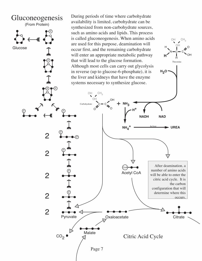
Gluconeogenesis
Citric Acid Cycle
P
P
P P
PP
P
P
P
P
P
Glucose
Pyruvate
2
2
2
2
2
CoA
Acetyl CoA
Oxaloacetate Citrate
CO2Malate
Carbohydrate
N C
3CH
OH
C
HOH
H
H
O
OH
Threonine
C
3CH
OH
CO O
OH
OH
H2O
NADNADH
NH3
NH4+
H++
UREAIn liver
After deamination, a number of amino acids will be able to enter the
citric acid cycle. It is the carbon
configuration that will determine where this
occurs.
During periods of time where carbohydrate availability is limited, carbohydrate can be synthesized from non-carbohydrate sources, such as amino acids and lipids. This process is called gluconeogenesis. When amino acids are used for this purpose, deamination will occur first, and the remaining carbohydrate will enter an appropriate metabolic pathway that will lead to the glucose formation. Although most cells can carry out glycolysis in reverse (up to glucose-6-phosphate), it is the liver and kidneys that have the enzyme systems necessary to synthesize glucose.
(From Protein)
Page 7