A hydrogen-bonding triad stabilizes the chemical ... · PDF fileA hydrogen-bonding triad...
13
Research Paper 153 A hydrogen-bonding triad stabilizes the chemical transition state of a group I ribozyme Scott A Strobe1 and Lori Ortoleva-Donnelly Background: The group I intron is an RNA enzyme capable of efficiently catalyzing phosphoryl-transfer reactions. Functional groups that stabilize the chemical transition state of the cleavage reaction have been identified, but they are all located within either the 5’-exon (Pl) helix or the guanosine cofactor, which are the substrates of the reaction. Functional groups within the ribozyme active site are also expected to assist in transition-state stabilization, and their role must be explored to understand the chemical basis of group I intron catalysis. Results: Using nucleotide analog interference mapping and site-specific functional group substitution experiments, we demonstrate that the 2’-OH at A207, a highly conserved nucleotide in the ribozyme active site, specifically stabilizes the chemical transition state by -2 kcal mol-‘. The A207 2’-OH only makes its contribution when the U(-1) 2’-OH immediately adjacent to the scissile phosphate is present, suggesting that the 2’-OHS of A207 and U(-1) interact during the chemical step. Conclusions: These data support a model in which the 3’-oxyanion leaving group of the transesterification reaction is stabilized by a hydrogen-bonding triad consisting of the 2’-OH groups of U(-1) and A207 and the exocyclic amine of G22. Because all three nucleotides occur within highly conserved non-canonical base pairings, this stabilization mechanism is likely to occur throughout group I introns. Although this mechanism utilizes functional groups distinctive of RNA enzymes, it is analogous to the transition states of some protein enzymes that perform similar phosphoryl-transfer reactions. Address: Department of Molecular Biophysics and Biochemistry, Yale University, 260 Whitney Avenue, New Haven, CT 06520-8114, USA. Correspondence: Scott A Strobe1 E-mail: [email protected] Key words: interference suppression, nucleotide analog interference mapping, phosphoryl transfer, reaction mechanism, self-splicing intron Received: 1 December 1998 Revisions requested: 30 December 1998 Revisions received: 7 January 1999 Accepted: 13 January 1999 Published: 17 February 1999 Chemistry & Biology March 1999, 6:153-l 65 http://biomednet.com/elecref/1074552100600153 Q Elsevier Science Ltd ISSN 1074-5521 Introduction Understanding the chemical mechanism of catalysis by RIXA enzymes is a subject of particular interest because the nucleotide building blocks that comprise ribozymes are fundamentally different from those of protein enzymes, yet both macromolecules efficiently catalyze phosphoryl- transfer reactions [ 11. This raises an intriguing question: do these two very different classes of macromolecular biocata- lysts utilize similar strategies to catalyze phosphate transes- terification? Although the reaction mechanisms of several protein enzymes that promote phosphoryl-transfer reac- tions have been elucidated [Z], the chemical basis of ribozyme catalysis must be further characterized before a comparison of transition-state mechanisms of protein and RNA enzymes can be completed. The Tetrahymena group I intron catalyzes two phosphate- transesteritication reactions in the course of self-splicing (Figures 1,2) [3]. It promotes these reactions with a rate enhancement -lO1l-fold over the uncatalyzed rate and with much greater specificity [4]. Both the first and second steps of splicing involve nucleophilic attack by a 3’-OH on a phosphodiester linkage, which displaces a 3’-oxyanion leaving group from the scissile phosphate, and both reac- tions are catalyzed within the same active site [S]. Alternative constructs of the group I intron have been developed to independently explore both steps of splicing (Figure 2) [6,7]. The L-21 ScaI form of the intron lacks the 5 and 3’ exons as well as the terminal guanosine, and it has been used to study the first step of splicing (Figure 2a) [6,8]. This enzyme binds exogenous guanosine and an oligonucleotide substrate analogous to the S’-3’ ligated exons. The second step of splicing has been investigated using the L-21 G414 form of the intron (Figure 2b) [7,9]. It retains the terminal guanosine (G414), which can attack the oligonucleotide substrate and transfer the 3’ exon onto the end of the intron in a reaction analogous to the reverse of the second step of splicing. For both forms of the intron, the binding of the oligonucleotide substrate occurs in two distinct steps [lO,ll]. First? the oligonucleoCde substrate base pairs with the internal guide sequence (IGS) to form the Pl or substrate helix in an open complex (Figures 1.2). This helix docks into the ribozyme active site to form the closed complex where the substrate is cleaved by guanosine. Experiments to elucidate the chemical basis of group I intron catalysis have focused primarily upon metal ions and the role of particular functional groups located within the two substrates of the ribozyme reaction, that is, the
Transcript of A hydrogen-bonding triad stabilizes the chemical ... · PDF fileA hydrogen-bonding triad...

Research Paper 153
A hydrogen-bonding triad stabilizes the chemical transition state of a group I ribozyme Scott A Strobe1 and Lori Ortoleva-Donnelly
Background: The group I intron is an RNA enzyme capable of efficiently
catalyzing phosphoryl-transfer reactions. Functional groups that stabilize the
chemical transition state of the cleavage reaction have been identified, but they
are all located within either the 5’-exon (Pl) helix or the guanosine cofactor,
which are the substrates of the reaction. Functional groups within the ribozyme
active site are also expected to assist in transition-state stabilization, and their role
must be explored to understand the chemical basis of group I intron catalysis.
Results: Using nucleotide analog interference mapping and site-specific
functional group substitution experiments, we demonstrate that the 2’-OH at
A207, a highly conserved nucleotide in the ribozyme active site, specifically
stabilizes the chemical transition state by -2 kcal mol-‘. The A207 2’-OH only
makes its contribution when the U(-1) 2’-OH immediately adjacent to the
scissile phosphate is present, suggesting that the 2’-OHS of A207 and U(-1)
interact during the chemical step.
Conclusions: These data support a model in which the 3’-oxyanion leaving
group of the transesterification reaction is stabilized by a hydrogen-bonding
triad consisting of the 2’-OH groups of U(-1) and A207 and the exocyclic
amine of G22. Because all three nucleotides occur within highly conserved
non-canonical base pairings, this stabilization mechanism is likely to occur
throughout group I introns. Although this mechanism utilizes functional groups
distinctive of RNA enzymes, it is analogous to the transition states of some
protein enzymes that perform similar phosphoryl-transfer reactions.
Address: Department of Molecular Biophysics and
Biochemistry, Yale University, 260 Whitney Avenue, New Haven, CT 06520-8114, USA.
Correspondence: Scott A Strobe1 E-mail: [email protected]
Key words: interference suppression, nucleotide
analog interference mapping, phosphoryl transfer, reaction mechanism, self-splicing intron
Received: 1 December 1998
Revisions requested: 30 December 1998 Revisions received: 7 January 1999 Accepted: 13 January 1999
Published: 17 February 1999
Chemistry & Biology March 1999, 6:153-l 65
http://biomednet.com/elecref/1074552100600153
Q Elsevier Science Ltd ISSN 1074-5521
Introduction Understanding the chemical mechanism of catalysis by RIXA enzymes is a subject of particular interest because the nucleotide building blocks that comprise ribozymes are fundamentally different from those of protein enzymes, yet both macromolecules efficiently catalyze phosphoryl- transfer reactions [ 11. This raises an intriguing question: do these two very different classes of macromolecular biocata- lysts utilize similar strategies to catalyze phosphate transes- terification? Although the reaction mechanisms of several protein enzymes that promote phosphoryl-transfer reac- tions have been elucidated [Z], the chemical basis of ribozyme catalysis must be further characterized before a comparison of transition-state mechanisms of protein and RNA enzymes can be completed.
The Tetrahymena group I intron catalyzes two phosphate- transesteritication reactions in the course of self-splicing (Figures 1,2) [3]. It promotes these reactions with a rate enhancement -lO1l-fold over the uncatalyzed rate and with much greater specificity [4]. Both the first and second steps of splicing involve nucleophilic attack by a 3’-OH on a phosphodiester linkage, which displaces a 3’-oxyanion leaving group from the scissile phosphate, and both reac- tions are catalyzed within the same active site [S].
Alternative constructs of the group I intron have been developed to independently explore both steps of splicing (Figure 2) [6,7]. The L-21 ScaI form of the intron lacks the 5 and 3’ exons as well as the terminal guanosine, and it has been used to study the first step of splicing (Figure 2a) [6,8]. This enzyme binds exogenous guanosine and an oligonucleotide substrate analogous to the S’-3’ ligated exons. The second step of splicing has been investigated using the L-21 G414 form of the intron (Figure 2b) [7,9]. It retains the terminal guanosine (G414), which can attack the oligonucleotide substrate and transfer the 3’ exon onto
the end of the intron in a reaction analogous to the reverse of the second step of splicing. For both forms of the intron, the binding of the oligonucleotide substrate occurs in two distinct steps [lO,ll]. First? the oligonucleoCde substrate base pairs with the internal guide sequence (IGS) to form the Pl or substrate helix in an open complex (Figures 1.2). This helix docks into the ribozyme active site to form the closed complex where the substrate is cleaved by guanosine.
Experiments to elucidate the chemical basis of group I intron catalysis have focused primarily upon metal ions and the role of particular functional groups located within the two substrates of the ribozyme reaction, that is, the

154 Chemistry & Biology 1999, Vol 6 No 3
Figure 1
Chemistry & Biology
The secondary structure of the Tetrahymena group I ribozyme with the GU wobble pair in Pl (green), the sheared A.A pairs in J4/5 (orange
and blue), and the terminal guanosine (red) highlighted.
guanosine cofactor and the Pl duplex comprised of the 5’-exon and the IGS [l&21]. The experiments demonstrace
Figure 2
that the group I intron is a metalloenzyme because it
requires at least three magnesium ions to stabilize the chemical steps of splicing ([Z--26]; S. Shan, A. Yoshida, J.A. Piccirilli and D. Herschlag. personal communication). One metal ion activates the 3’-OH of G for nucleophilic attack
[25]. A second interacts with the 2’-OH of G, which might improve the nucleophilicity of the 3’-OH ([26]; S. Shan, A. Yoshida, J.A. Piccirilli and D. Herschlag. personal communication). A third metal ion stabilizes the 3’-OH leaving group of L(-1), which is the terminal nucleotide of the 5’-exon. The two metal ions that coordinate the nucle- ophile and the leaving group provide a large fraction of the energy required for catalysis, as each enhances the reaction rate by as much as lO.OOO-fold [24,25].
In addition to the metal ions, specific functional groups within the substrate helix contribute to transition-state sta- bilization. Six 2’-OH groups in the Pl helix make ground- state tertiary interactions that provide intrinsic binding energy for the transition state [21,27-311. In addition to these interactions, two functional groups within the highly conserved GZ.U(-1) wobble pair adjacent to the 5’-splice site selectively stabilize the chemical step of group I intron splicing. These groups are the Z-OH of U(-1) and the N2 exocyclic amine of G22 [16,20]. The U(-1) Z’-OH is imme- diately adjacent to the 3’-oxyanion leaving group of the same nucleotide and accelerates the reaction by more than lOOO-fold (-5 kcal mol-I) (Figure 3). Functional group sub- stitution experiments suggested that this hydroxyl group makes its contribution by donating a hydrogen bond to the 3’-oxyanion and that this hydrogen bond only forms during
(a)
-3 FFFVP ‘4 GGGAGG
Undecked Docked
L-21 G414
I
Undecked Docked Chemistry & Biology
(a) A reaction scheme for the L-21 Seal reaction, which is analogous to the first step
of intron splicing and uses exogenous guanosine as the nticleophile. (b) A reaction
scheme for the 3’-exon ligation reaction
performed by the L-2 1 G414 form of the group I intron. The reaction is analogous to
the reverse of the second step of intron splicing and uses the terminal nucleotide of
the intron, G414, as the nucleophile.

Research Paper RNA catalytic triad Strobe1 and Ortoleva-Donnelly 155
Figure 3
Chemstry & Blolog:
A model of the tertiary hydrogen-bonding interactlons between the G,U pair of Pl (green) and the two consecutively stacked sheared A.A
pairs of the J4/5 region (orange and blue).
the transition state [16]. Substitution of this Z-OH has no effect on the binding of the substrate into the active site [16,27,28]. The second important functional group is the G22 exocyclic amine, which acts as a primary determinant of 5’-splice site selection and is the only sequence-specific contact between Pl and the catalytic core [19,20]. The G22 amine makes a substantial contribution to ground-state helix docking (-2.0 kcalmol-i), but it also makes a contri- bution to the chemical transition state (-1 kcal mol-I).
Experiments involving site-specific substitution of these two important functional groups indicated that there is a small amount of transition-state energy (<O.S kcal mol-i) attributable to an interaction between them [18,20]. Within a wobble pair, however, these groups are not sufficiently close to form a direct hydrogen-bonding interaction that could account for this energy. A crystal structure of a model duplex containing a G.17 pair demonstrated that an ordered water molecule can bridge between the I: 2’-OH and G NZ amino groups [32]. This led to the proposal that a water molecule or 2’-hydroxyl group could participate in the chemistry of the group I intron by bridging between the catalytically important groups of the wobble pair [1X,32], but the existence of this putative mediating functional group had not been proven.
We recently reported a model for the ground-state docking interactions between the 54/S region of the ribozyme active
site and the G&?.U(-1) pair (Figure 1) [33]. In this model the G+U pair utilizes its 2’-OH and N2 amine to make four hydrogen bonds with the convex minor groove surface created by two consecutively stacked sheared A-A pairs in the highly conserved 5315 internal loop (Figure 3). This interaction places the 2’-OH of nucleotide A207 between the two functional groups known to contribute to chem- istry. It occupies approximately the same position as the water molecule observed within the crystal structure of a G.U wobble pair [32]. Based upon its location within the G.II wobble pair, it seems plausible that the 2’-OH of A207 also contributes to the chemical step of splicing. In this report, we demonstrate that there is a hydrogen-bonding triad between the exocyclic amine of G22 and the 2’-OHS of C(-1) and A207, and that this network of interactions selectively stabilizes the chemical transition state of the TeW&menrr group I intron.
Results Nucleotide analog interference mapping The Pl-J4/5 ground state docking model led to the hypoth- esis that the 2’-OH of A207 might participate directly in the chemistry of group I intron splicing (Figure 3) [33]. Our initial experiments to demonstrate such a role involved the LWZ of nucleotide analog interference mapping (MAIM). a chemogenetic method that makes it possible to rapidly identify the chemical groups responsible for RNA function (Figure 4) [33,35]. NAILI involves random low lev.el incor- poration of an a-phosphorothioate-tagged nucleotide analog into the intron by T7 RNA polymerase and selection of the resulting transcripts for splicing activity. The active introns are treated with iodine to cleave the phosphoroth- ioate linkage and the fragments are analyzed using poly- acrylamide gel electrophoresis (PAGE) [36]. I f incorpora- tion of a nucleotide analog at a particular site impairs activity. it can be rapidly identified by disappearance of a specific band within the RNA sequencing ladder (Figure 4). The L-21 G414 form of the Tetrahymena group 1 intron is particularly amenable to this experimental approach because it can transfer the 3’-end of a radiolabeled oligonucleotide substrate onto its 3’ end, thus marking the active introns in the population with a radioactive tag (Figure 2b) (351.
Previous experiments using dAaS [5’-O-(1-thio)-2’-deoxy- adenosine monophosphate] and FANS [5’-O-( I-thio)-2’- deoxy-2’-fluoroadenosine monophosphate]. in which the 2’-OH of A is replaced with a proton and with a fluorine, respectively, revealed an interference pattern at A207 that was essential for modeling the Pl-J4/5 interaction [33]. Strong interference was observed with dAaS at A207, but “AaS incorporation at A207 resulted in slight enhancement of activity. Because the highly electronega- tive 2’-fluoro group can still accept a hydrogen bond, such an interference pattern argued that the 2’-OH of A207 is a hydrogen-bond acceptor. Interference-suppression experi- ments supported this prediction by demonstrating that the

156 Chemistry & Biology 1999, Vol 6 No 3
Figure 4 Figure 5
l Active variant i I,
inactive variant
IL A-A-L$-A-A-G~,, ILA--A-~-54-A-~4,, ES
L t
Ribozyme reaction F
No reaction
A-A-;$-A-A-G4w’ Ribozyme not end-labeled
1 12 Cleavage
FA-A-A--G~~*
-8 - ‘7
I I
I c____,
-- Inactive
Chemistry & Biology
Nucleotide analog interference mapping (NAIM) using the 3’-exon ligation reaction as the selection step.
A207 Z-OH accepts a hydrogen bond from the G22 amino group (inosine substitution at position 22 partially sup- pressed dAcxS interference at A207) [33]. There was still no direct data to demonstrate that the 2’-OH groups of A207 and U(-1) interact, although their proximity in the 0 model (-3.0-3.5 A) implied that such an interaction was structurally reasonable with a modest realignment of the nucleotides (Figure 3).
Interference mapping at A207 when the chemical step is
limiting
I f 4207 bridges between L(-1) and G22 in the chemical step, we expected that it would act as a hydrogen-bond donor to the L(-1) 2’-OH. “AaS should therefore show interference under conditions when chemistry is the limit- ing step in the reaction. This was not observed in the results reported previously; but chose experiments were performed using a substrate with a 2’-deoxy substitution at the cleavage site. Although chemistry is limiting under those conditions, the substrate lacks the U(-1) Z’-OH that is expected to participate in the critical interaction. We repeated the experiments using a substrate containing the U(-1) Z-OH, but again failed to detect FANS interference at A207 despite testing several reaction conditions, includ- ing low pH and low Mg’+ concentrations (data not shown). This can be explained by the fact that for cleavage of an all ribose substrate by a population of ribozyme variants that are each present at very low concentration, substrate binding rather than chemistry is the limiting step of the reaction [8,37]. Functional groups that moderately disrupt
(a) FAcxS effect
(No interference)
Closed
\
(b)
8
”
r _....____ -.i’eel -fmFAaS effect
,I\’ (Interference)
‘pen u \ j-dG25or122 Closed
\
Destabilization
of docked
conformation
E+rS + E.rS =5 E.rS
Open Closed
Ligation
Chemistry & Biology ~
Free-energy profiles for substrate binding, docking and chemical transformation for (a) the wild-type and (b) the dG25 and 122- substituted ribozymes.
chemistry would not be expected to show interference under such conditions (Figure 5a. green).
The kinetic profile of the reaction predicts that “AaS will only cause interference at A207 if the experiments are per- formed using a substrate with a ribose at the cleavage site under conditions in which chemistry is limiting. To achieve this condition, we prepared ribozymes that were impaired in their ability to dock the Pl helix into the ribozyme active site [19,30]. Because destabilization of the docked ground- state complex equally disrupts the chemical transition state. the energetic barrier for chemistry is raised to the point that it approaches the energetic barrier for substrate binding (Figure Sb, blue). LJpon reaching the docked complex. these impaired ribozymes are more likely to dissociate than the wild-type enzyme. Dissociation is even more favored if the energy of the chemical transition is further increased by loss of a particular functional group that contributes to the chemical srep (Figure 5b, red).
To determine the role of the A207 Z’-OH using NAIXI we prepared two sets of ribozymes impaired in their ability to dock the Pl helix. Each set of ribozymes lacks a specific

Research Paper RNA catalytic triad Strobe1 and Ortoleva-Donnelly 157
functional group important for Pl helix docking into a different region of the active site [30,38]. One set of ribozymes contained an inosine at position 22 (122), which lacks the exocyclic amine important for Pl docking into J4/5 [19,33]. The second set of ribozymes contained a 2’-deoxy substitution at G25, which lacks a Z-OH impor- tant for Pl helix docking into the single-stranded segment 58/7,[30,39]. Both RNAs were transcribed with either AC& or “AaS randomly incorporated throughout the length of the transcript to test for interference at A207 and other positions in the RNA. A total of four enzymes were pre- pared: 122 AaS, 122 FAlcGs, &25 AcGS and dG25 “ilaS.
The substituted L-21 G414 ribozymes were assayed for interference using a 3’-end-labeled substrate with a ribose at the cleavage site (rld45S; CdCdCUCrTAAAAA). The 2’-deoxynucleotide substitutions at -4 and -5 were in- cluded to eliminate miscleavage of the substrate due to the 122 and dG25 substitutions [11,20,40]. Following the 3’-exon ligation reaction, the RNAs were treated with iodine, and the cleavage products resolved using PAGE to determine the effect of “AaS substitution at A207 and other positions within the ribozyme.
In contrast to the previous experiments, where either chemistry was not rate-limiting or the substrate lacked a 2’-OH at the cleavage site, both the I22 and dG25 ribozymes displayed significant ‘AaS interference at A207 (Figure 6a). Neither of these enzymes showed AaS inter- ference at this position, indicating that the effect is specifi- cally due to the 2’-fluoro substitution and not a phosphorothioate effect. Furthermore, no other changes in the interference pattern were detected in comparison with the results published previously, which used a substrate with a 2’-deoxy nucleotide at the cleavage site [33,41]. The fact that FAaS interference is seen with both the 122 and dG25 ribozymes argues that it is not dependent upon the specific modification introduced to destabilize the docked ground state of the reaction. These data support a mecha- nism in which a hydrogen-bond donor at the Z-OH position of AZ07 is important for ribozyme function, but only under conditions in which chemistry is the rate-limiting step.
A207 interference suppression by 2’.deoxy substitution at W-1) To determine how the A207 Z-OH participates in the chemical step, we performed an interference suppression experiment. NAIS is an extension of NAIM that employs the logic of genetics to identify specific hydrogen-bonding partners within an RNA tertiary structure [33]. In princi- ple, if an interaction is disrupted by deletion or alteration of one functional group in an interacting pair, then no sig- nificant energetic penalty will result from deletion or alter- ation of the second functional group. The suppression of an interference effect can be detected by the reappear- ance of a specific band on the sequencing gel (Figure 4).
The transition-state model predicts that the 2’-OH of A207 donates a hydrogen bond to the 2’-OH of U(-1). On the basis of this model, deletion of the hydroxyl group at the cleavage site should eliminate any positive contribution made by the hydroxyl group at A207. This would be observed experimentally as A207 ‘AaS interference sup- pression resulting from elimination of the U(-1) 2’-OH. To test this possibility, the 122 and dG2.5 ribozymes were again assayed for interference using a substrate with a 2’-deoxy substitution at the cleavage site (d145S; CdCdCIJCdTAA- AAA). In contrast to the results described above, the sub- strate with a 2’-deoxynucleotide did not shovv interference at A207 with ‘AaS (Figure 6). This is true in spite of the fact that d14SS is more than lOOO-fold less reactive than rlS, and thus the reaction conditions were significantly more selective than those used for the all-ribose substrate. Furthermore, the site of interference suppression was spe- cific, in that ‘AaS interference at A218 (Figure 6) and other control sites within the intron were not suppressed by the Z’-deoxy substitution at the cleavage site (data not shown). “AaS interference suppression at A207 was observed for both the I22 and the u’G25 ribozymes, which confirms that the effect is independent of the type of substitution used co destabilize the docked ground-state conformation. These data argue that the 2’-OH groups at A207 and U(-1) inter- act during the chemical transition state? most likely via a direct hydrogen bond.
Characterization of A207-substituted ribozymes Interference and suppression data provide only qualitative evidence that the 2’-OH of AZ07 participates in the chem- istry of group I intron splicing. To measure the A207 Z-OH contribution directly, we prepared L-21 ScaI ribozymes with a specific Z’-deoxy or 2’-fluoro substitution at A207 and measured kinetic and thermodynamic constants for the site-specifically substituted ribozymes. The L-21 ScaI form of the ribozyme binds an oligonucleotide substrate and uses exogenous guanosine to cleave at the 5’-splice site (Figure 2a) [6,8]. This reaction is analogous to the first. rather than the second, step of splicing, so these experi- ments make it possible to determine if the A207 2’-OH is also important for the first step of intron splicing. The A207-substituted ribozymes were prepared semisyntheti- tally by a three-piece ligation of two truncated RNA tran- scripts and a 9-mer synthetic oligoribonucleotide contain- ing either adenosine (rA), 2’-deoxyadenosine (dA) or 2’-deoxy-2’-fluoroadenosine (“A) at A207 to produce ribo- zymes ir.A2~?7, dAZO7 and FKO7, respectively. The overall yield of the ligation reaction was only about 2% despite extensive efforts to optimize the ligation conditions (see the Materials and methods section).
The PI helix docking model predicts that the A207 2’-OH accepts a hydrogen bond from the N2 amine of G22 in the ground state [33] (Figure 3). This should be manifest as a reduction in oligonucleotide substrate affinity by the dr1207

158 Chemistry & Biology 1999, Vol 6 No 3
Figure 6
(
aS
A206 *A207
b)
a)
?i bozyme
Ribose substrate
dG25 122
Deoxy substrate
dG25 122
’ A FA’ ‘A FA ’ ’ A FA’ ‘A FA ’
1 2 3 4 5 6 7 8
Ribose substrate Deoxy substrate
dG25 122 dG25 122
l----lr----- Ir-----
A207 A218 A207 A218 A207 A218 A207 A218
1
A206 A207 *
(a) NAIM for the dG25 and /22-substituted
ribozymes with the analogs AaS and FANS using the substrates rl d45S (ribose
substrate) or dl45S (deoxy substrate). The cleavage band for A207 is marked with an
asterisk. Note that the band is missing in the FANS lanes with the ribose substrate (lanes 2
and 4). but is present in the FAaS lanes with the deoxy substrate (lanes 6 and 8).
(b) Histogram depicting the magnitude of
FAaS interference at A207 and A21 8 for the
dG25- and /22-substituted ribozymes.
ribozvme, whereas FA207 would not show the effect becabse it still has a hydrogen-bond acceptor available for the GZZ amino group. To test this prediction, we mea- sured the equilibrium dissociation constants of the rilZO7, dA207 and FA2O7 ribozymes for the oligonucleotides rlP (GGCCCUCrT) and dlP (CCCUCdT; Table 1) [42]. rilO7 bound both oligonucleotides with the same affinity as the fully transcribed L-21 ScaI RNA, which demon- strates that there are no unexpected effects resulting from ribozyme preparation by three-piece ligation. Consistent with the Pl docking model (Figure 3), dAZO7 showed a modest 0.4-0.8 kcalmol-’ loss of binding energy for both substrates, whereas FA2O7 was at least as stable as the all- ribose enzyme. The magnitude of the dAZO7 effect is con- sistent with that expected for deletion of this functional group, because the G2Z amine contributes 2 kcal mol-’ and it interacts with both the 2’-OH and N3 of AZ07. whereas the 2’-deoxy substitution at AZ07 only eliminates one of these interactions [19,33]. The observation that the d.4207 substitution is less destabilizing with a ribonu- cleotide than with a deoxynucleotide at the cleavage site (0.4 compared with 0.8 kcal mol-I, respectively, Table 1) is further evidence against a ground-state hydrogen bond between the A207 and U-l Z-OH groups. The lack of an effect from Z’-fluoro substitution quantitatively confirms
that the Z-OH of A207 acts as a hydrogen-bond acceptor in the ground state as predicted in the Pl docking model (Figure 3) [33].
We further characterized the A207-substituted ribozymes kinetically to determine if there is any effect on the chem- ical step. We initially measured the second-order rate con- stant (k,,,/K,)” for cleavage of the all-ribose substrate rlS (GGCCCUCrT,M), which under single-turnover con- ditions represents the rate of reaction for Es (substrate
fully bound to the ribozyme, but the G-binding site unoc- cupied) with free G. For the wild-type ribozyme under these conditions, (k,,,/K,,,)” is limited by a combination of
Table 1
Equilibrium dissociation constants for oligonucleotide binding by the A207-substituted L-21 Seal ribozymes.
Ribozvme K, CnM) MG K&M) MG
rlP kcal mol-l dlP kcal mol-’
Wild-type 7.lfl.l - 10.9 kO.9 -
rA207 8.8 +_ 2.3 0.1 13.2* 1.1 0.1
dA207 14.6 i 1 .O 0.4 42.2 in 3.1 0.8 FA207 6.8 + 4.0 0.0 10.5 f 5.1 0.0

Research Paper RNA catalytic triad Strobe1 and Ortoleva-Donnelly 159
Table 2
Kinetic characterization of the A207-substituted ribozymes.
Ribozyme
Wild-type
rA207 dA207 FA207
&,,,&,)G (1 04M-‘min-‘) K,,
14f2
13*3 1.1 0.42 f 0.05 3.3
0.20 + 0.02 7.0
$5
90
170130
130*10
k,,,(min-‘1 rlS
15
0.68 * 0.04
0.44 + 0.02
MG kcal mol-’
1.9
2.1
kchemhW) MG dlS kcal mol-’
0.0044 f 0.0002 -
0.0043 * 0.0002 0.0 0.0028 + 0.0002 0.3
0.0038 * 0.0003 0.1
guanosine binding and chemistry [8,43]. Although ~4207 reacted at a rate equivalent to that of the wild-type RlXA. both the dA207 and FAZ07 ribozymes were more than 30-fold less efficient at substrate cleavage (Table 2).
Because this observed decrease in the cleavage rate might be caused by a reduction in G affinity (K,” effect) or a reduction in the rate of chemistry (kcar effect), we measured kobs at several guanosine concentrations and calculated k,,, and K, for the reaction (Figure 7, Table 2). Under these conditions K, is equal to the K, for guanosine binding and
Figure 7
(a) 0.700
0.600 f
0.500 dA207
7 n -2z
dii- fA207
t , 10 100 1000 10000
GMP concentration (kM)
0.005
0.004
0.003
0.002
0.001
n ”
Ribo substrate Deoxy substrate -
Chemistry & Biolog Y
(a) A plot of kobs versus [GMPI for the cleavage of rl S by the dA207- and FA207-substituted ribozymes. (b) A histogram of kat values for the cleavage of oligonucleotide substrates rl S (ribo substrate) and dl S (deoxy substrate) by wildetype (wt) and A207-substituted ribozymes. Note that the scale on they axis differs for the two substrates.
the kc,, is equal to kchrmT the rate of the chemical step [20,43,44]. The kinetic data indicate that KmG for the dA307 and FA207 ribozymes is less than twofold higher than the wild-type ribozyme, which argues against a direct participa- tion of the A207 2’-OH in G binding (Table 2). In contrast, k,,, of rlS cleavage was 22- and 34-fold lower for &I207 and %‘07, respectively. This represents at least a 2 kcalmol-’ loss in transition-state stabilization energy specific to the chemical step. Because the effect is observed with both the 2’-fluoro and 2’-deoxy substitutions. it argues that the 2’-OH of A207 donates a hydrogen bond in the chemical step.
The interference-suppression experiments suggested that the A207 2’-OH interacts with the 2’-OH of 11(-l) during the chemical step. To determine if the A207 substitutions still have an effect on chemistry when there is a ?‘-deoxy group at the cleavage site, we measured k,,, for the sub- strate dlS at saturating enzyme and saturating guanosine ConcentraCons (Figure 7, Table 2). Under these condi- tions, k,,, is again equal to the rate of chemistry (kchem). As expected, all the ribozymes cleaved the single Y-deoxy- substituted substrate more than lOOO-fold slower than they cleaved the all-ribose substrate: but there was no addi- tional loss of chemical transition-state energy resulting from modification of the 2’-OH at A207 (kchem for dA107 and FL4ZO7 was not even twofold less than that of the wild- type ribozyme). Together these data suggest that the 2’-OH of A207 contributes about 2 kcalmol-l to chemistry by donating a hydrogen bond to the Z-OH of U(-1).
Discussion Model of the group I ribozyme chemical transition state
Previous models of the group I intron chemical transition state have proposed that the 2’-OH adjacent to the scissile phosphate interacts with a metal ion, a water molecule or a specific functional group within the catalytic core of the ribozyme during the chemical step [18,32]. The interfer- ence and single-site substitution experiments described here support a model in which the Z-OH of U(-1) accepts a hydrogen bond from the 2’-OH of ‘4207, a functional group located within the ribozyme active site. The A207 2’-OH represents the first functional group within rhe cat- alytic core of the group I that has been shown to partici- pate directly in the stabilization of the chemical transition

160 Chemistry & Biology 1999, Vol 6 No 3
Figure 8
Model of the Tetrabymena group I intron chemical transition state involving a hydrogen-bonding triad between the 2’-OH groups of
U(-1) and A207, and the exocyclic amine of G22.
state. In addition to forming a transition-state hydrogen bond with U(-I), the A207 ?-OH also makes a ground- state hydrogen bond that is most likely retained in the chemical step with the GZ2 N2 amino group. This series of interactions therefore defines a hydrogen-bonding triad between the functional groups of three nucleotides: U(-1), A207 and G22. All three of the nucleotides in the network are at least 99% conserved among group I introns [45]. Each nucleotide is located within a noncanonical base pair that is also highly conserved, including the GZZ.U(-1) wobble pair at the cleavage site and the A207.Al13 sheared pair in the active site. This suggests that the hydrogen-bonding triad is likely to be a conserved catalytic component of almost all group I introns.
Although these biochemical experiments cannot com- pletely rule out the possibility that a water molecule or metal ion might mediate the interaction between these groups, all of the tertiary contacts between Pl and the 54/S region can be accommodated with reasonable distances and geometries by invoking direct hydrogen bonds between the functional groups (Figure 3). The G.U docking model into 54/S is based upon an intermolecular interaction within the P4-P6 crystal structure, and the distances and geometries within that structure are inconsistent with water- or metal-mediated interactions [33,46]. Further- more, the biochemical experiments define which func- tional groups act as hydrogen-bond donors or acceptors. and this relationship can be properly maintained within the triad model without invoking a metal ion. The sim- plest model based upon the biochemical and structural data is therefore that the functional groups in the triad make direct hydrogen-bonding interactions.
A model for the Tetrahynena group I ribozyme transition state that incorporates these data with previous studies is shown in Figure 8 [47]. In addition to stabilization by a metal ion, the 3’-oxyanion is activated by the hydrogen- bonding triad. Previous studies on the pH profile of the reaction suggested that the CJ(-1) Z-OH does not donate a proton to the leaving group (general acid mechanism), but rather that it stabilizes the developing negative charge by a hydrogen-bonding mechanism [16]. This network of inter- actions involving the Z-OH of A207 might increase the ability of the U(-1) ?-OH to serve as a hydrogen-bond donor by lowering the pK, of the hydroxyl group, or it might make an entropic contribution by restricting the rota- tional freedom of the U(-1) Z-OH for hydrogen bonding to the 3’-oxyanion [18,32]. Because RNA lacks functional groups with pK, values near neutrality, a hydrogen-bonding network might provide a viable alternative to a general acid mechanism by dissipating a negative charge across several functional groups.
An alternative set of interactions appears to activate the 3’-nucleophile of G (Figure 8; [26,48]; S. Shan, A. Yoshida, J.A. Piccirilli and D. Herschlag, personal communication). In addition to direct metal-ion coordination, the 3’-oxy- anion nucleophile is likely to accept a hydrogen bond from the adjoining Z-OH group of G. This conclusion is based upon the observation that 2’-deoxyguanosine is a competi- tive inhibitor of the splicing reaction [48]. The ?-OH of G is, in turn, coordinated to a third metal in the active site [26] (S. Shan. A. Yoshida, J.A. Piccirilli and D. Herschlag, personal communication). This implies that on the G side of the transesterification reaction a metal ion might take the place of the hydrogen-bonding network.
The interference and single-site substitution data indicate that the same hydrogen-bonding network stabilizes both the first and second steps of splicing, consistent with the observation that both reactions are catalyzed within the same active site [S]. The forward and the reverse of the second step of splicing are energetically equivalent with an equilibrium constant of -1 [9]. This implies that the hydrogen-bonding triad also stabilizes the deprotonated form of the 3’-oxyanion nucleophile during the exon-liga- tion step of splicing. The triad is therefore predicted to be responsible for stabilizing the leaving group during the first step of splicing and for activating the nucleophile during the second step of splicing.
Based upon the ground state docking model for the Pl interaction with the J4/5 region? it is intriguing to spec- ulate on the magnitude of the conformational change required for the ribozyme to form the catalytic tertiary interaction in the chemical transition state. Because ground- state docking interactions were predictive of a transition- state tertiary contact, it argues against a large conforma- tional change within the J4/5 region of the active site [33].
Research Paper RNA catalytic triad Strobe1 and Ortoleva-Donnelly 161
Instead, small changes in the conformation of the ,4207 or C(-1) ribose sugars might alone be sufficient to align these groups for chemistry. Although it remains possible that larger conformational rearrangements are required outside of the J4/5 region in order to bring the catalytic metals into proper alignment for chemistry, the Pl-J4/5 interaction appears to be largely pre-ordered within the docked ground-state complex.
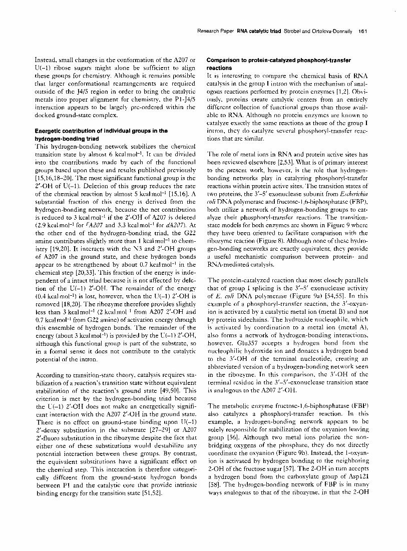
Energetic contribution of individual groups in the hydrogen-bonding triad This hydrogen-bonding network stabilizes the chemical transition state by almost 6 kcalmol-‘. It can be divided into the contributions made by each of the functional groups based upon these and results published previously [15,16,18-203. The most significant functional group is the Z-OH of U(-1). Deletion of this group reduces the rate of the chemical reaction by almost 5 kcalmol-i [15,16]. A substantial fraction of this energy is derived from the hydrogen-bonding network, because the net contribution is reduced to 3 kcalmol-’ if the Z-OH of A207 is deleted (2.9 kcal mol-l for FAZ07 and 3.3 kcal mol-’ for dAZU7). At the other end of the hydrogen-bonding triad, the G22 amine contributes slightly more than 1 kcal mol-i to chem- istry [19,20]. It interacts with the N3 and ?-OH groups of A207 in the ground state, and these hydrogen bonds appear to be strengthened by about 0.7 kcalmol-l in the chemical step [20,33]. This fraction of the energy is inde- pendent of a intact triad because it is not affected by dele- tion of the U(-1) 2’-OH. The remainder of the energy (0.4 kcalmolI1) is lost, however, when the U(-1) 2’-OH is removed [ 18,201. The ribozyme therefore provides slightly less than 3 kcal mol-1 (2 kcalmol-i from A207 2’-OH and 0.7 kcal mol-i from G22 amine) of activation energy through this ensemble of hydrogen bonds. The remainder of the energy (about 3 kcalmol-l) is provided by the U(-1) 2’-OH, although this functional group is part of the substrate, so in a formal sense it does not contribute to the catalytic potential of the intron.
According to transition-state theory, catalysis requires sta- bilization of a reaction’s transition state without equivalent stabilization of the reaction’s ground state [49,501. This criterion is met by the hydrogen-bonding triad because the U(-1) 2’-OH does not make an energetically signifi- cant interaction with the A207 2’-OH in the ground state. There is no effect on ground-state binding upon U(-1) 2’-deoxy substitution in the substrate [27-291 or A207 2’-fluoro substitution in the ribozyme despite the fact that either one of these substitutions would destabilize any potential interaction between these groups. By contrast, the equivalent substitutions have a significant effect on the chemical step. This interaction is therefore categori- cally different from the ground-state hydrogen bonds between Pl and the catalytic core that provide intrinsic binding energy for the transition state (51,521.
Comparison to protein-catalyzed phosphoryl-transfer reactions It is interesting to compare the chemical basis of RNA catalysis in the group I intron with the mechanism of anal- ogous reactions performed by protein enzymes [ 1,2]. Obvi- ously, proteins create catalytic centers from an entirely different collection of functional groups than those avail- able to RNA. Although no protein enzymes are known to catalyze exactly the same reactions as those of the group I intron, they do catalyze several phosphor+-transfer reac- tions that are similar.
The role of metal ions in RNA and protein active sites has been reviewed elsewhere [2,53]. What is of primary interest to the present work, however, is the role that hpdrogen- bonding networks play in catalyzing phosphor-$-transfer reactions within protein active sites. The transition states of two proteins, the 3’-5’ exonuclease subunit from Eschetichia CO/~ DNA polymerase and fructose-l ,6-biphosphatase (FBP), both utilize a network of hydrogen-bonding groups to cat- alyze their phosphoryl-transfer reactions. The transition- state models for both enzymes are shown in Figure 9 where they have been oriented to facilitate comparison with the ribozyme reaction (Figure 8). Although none of these hydro- gen-bonding networks are exactly equivalent, they provide a useful mechanistic comparison between protein- and RNA-mediated catalysis.
The protein-catalyzed reaction that most closely parallels that of group I splicing is the 3’-5’ exonuclease activity of E. coli DNA polymerase (Figure 9a) [54,55]. In this example of a phosphoryl-transfer reaction, the 3’-oxyan- ion is activated by a catalytic metal ion (metal B) and not by protein sidechains. The hydroxide nucleophile, which is activated by coordination to a metal ion (metal A), also forms a network of hydrogen-bonding interactions, however. Glu357 accepts a hydrogen bond from the nucleophilic hydroxide ion and donates a hydrogen bond to the 3’-OH of the terminal nucleotide, creating an abbreviated version of a hydrogen-bonding network seen in the ribozyme. In this comparison, the 3’-OH of the terminal residue in the 3’-5’-exonuclease transition state is analogous to the A207 2’-OH.
The metabolic enzyme fructose-1,6-biphosphatase (FBP) also catalyzes a phosphoryl-transfer reaction. In this example, a hydrogen-bonding network appears to be solely responsible for stabilization of the oxyanion leaving group [56]. Although two metal ions polarize the non- bridging oxygens of the phosphate, they do not directly coordinate the oxyanion (Figure 9b). Instead, the l-oxyan- ion is activated by hydrogen bonding to the neighboring 2-OH of the fructose sugar [57]. The 2-OH in turn accepts a hydrogen bond from the carboxylate group of Asp121 [58]. The hydrogen-bonding network of FBP is in many ways analogous to that of the ribozyme, in that the 2-OH

162 Chemistry & Biology 1999, Vol 6 No 3
Figure 9
(a)
Vr497
Transition-state models for phosphoryl- transfer reactions catalyzed by (a) the 3’-5’
exonuclease domain of E. co/i DNA
polymerase and (b) fructose-l ,6-
-6
0
Gkia Chemistry & Biology
of the fructose is equivalent to the 2’-OH of I~!(-1) in the 5’-exon, and Asp121 plays a role similar to that of the A207 2’-OH. A corollary to the G22 exocyclic amine has not been proposed within FRP.
In these two examples of protein-mediated catalysis, the anion leaving group is stabilized by an ensemble of hydrogen bonds rather than by direct protonation by a
protein sidechain (general acid mechanism). The obser- vation that an RN.4 enzyme employs a catalytic strategy analogous to that of proteins reinforces the importance of hydrogen-bonding networks in transition-state stabil- ization. Such networks might be particularly important for RNA catalysts as they lack titratable functional groups near neutral pH. This result suggests that despite the chemical differences between protein and RIKA residues, the functional groups of both macromolecules can be arranged to create mechanistically comparable active sites.
Significance The mechanism of RNA-mediated catalysis is an issue of significant biochemical interest, particularly in com- parison with the catalytic strategies employed by proteins that perform similar phosphoryl-transfer reactions. In this work, we demonstrate that the transition state of the Tetrahymena group I ribozyme is, in part, stabilized by an extended network of hydrogen bonds that help stabil- ize the 3’-oxyanion leaving group. This hydrogen- bonding triad includes two functional groups within the substrate helix (Pl), and one within the ribozyme active site that bridges between them. The 2’-OH of A207, which is located in the highly conserved 5415 segment, is the first group identified in the intron active site that
specifically stabilizes the chemical transition state. The hydrogen-bonding network provides almost 3 kcalmol-1 of chemical potential to the reaction, and stabilizes both steps of group I intron splicing. Because all three nucleo- tides [G22, A207 and UC-l)] are highly conserved, and all three are located within conserved noncanonical base pairs, this mechanism for transition-state stabilization is likely to be employed throughout the group I family of self-splicing introns. Based upon comparison with the ground-state model for Pl helix docking, minor struc- tural rearrangements within the 5415 region appear to be sufficient for the ribozyme to progress from the docked ground state to the chemical transition state of the reac- tion. The hydrogen-bonding network in the ribozyme transition state has mechanistic parallels to that of the protein enzymes 3’-5’ exonuclease and fructose-16 biphosphatase, which employ similar networks to stabi- lize their chemical transition states. This observation suggests that, despite the chemical differences between protein and RNA residues, the functional groups of both macromolecules can be arranged to create mechanisti- cally comparable active sites.
Materials and methods Materials The phosphorothioate-tagged nucleoside triphosphates were purchased
from Amersham (ATPaS and dATPctS) or synthesized as previously described previously (FATPaS) [59]. The oligonucleotide substrates
used in the ribozyme reactions and the nine-base oligonucleotides used
for preparation of the rA207 and dA207 ribozymes were synthesized by
Dharmacon Inc. using P/-ACE chemistry, and deprotected according to
manufacturer’s recommendation [60]. No further purification was neces-
sary prior to use. The 17-mer oligonucleotides used to introduce site-
specific substitutions in the IGS and the 9-mer oligonucleotide containing
the single 2’-deoxy-2’-fluoroadenosine substitution at A207 were pre-
pared by 2’.TBDMS chemistry, deprotected and purified as described
Research Paper RNA catalytic triad Strobe1 and Ortoleva-Donnelly 163
previously. The phosphoramidite of 2’-deoxy-2’-fluoroadenosine neces-
sary to prepare the FA207 ribozyme was obtained as a generous gift
from Isis Pharmaceuticals.
Preparation of 122 and dG25 ribozymes containing AaS and
FAaS The L-21 G414 ribozymes were prepared semisynthetically by ligating
a 17-mer oligonucleotide containing either an 122 or dG25 substitu-
tion onto the 5’-end of a truncated L-38 G414 transcript [30]. The L-38 G414 RNA was transcribed in vitro from the Earl cut plasmid
pUCL-38G414 . AoS or FAaS substitutions (5% incorporation) were introduced randomly into the RNA by adding 5’-O-(l-thio)adenosine
triphosphate or 5’-C-(I-thio)-2’-deoxy-2’-fluoroadenosine triphosphate
in the transcription reaction as described previously [59]. FAaS incor-
poration required the use of the Y639F mutant T7 RNA polymerase,
which efficiently incorporates nucleotides modified at the 2’ position
[59,61 I. GMP (5 mM, 1 mM NTPs) was included in the transcription reaction to create a free 5’-phosphate for ligation onto the 17-mer
oligonucleotide. The RNAs were purified by PAGE (6% acrylamide,
7 M urea), eluted from the gel into TE (10 mM Tris-HCI, 0.1 mM EDTA,
pi-l 7.5) desalted and concentrated. The ligation reaction was per- formed using 1 nmole of L-38 G414 RNA, 2 nmoles of oligoribonu-
cleotide, 2 nmoles of DNA splint oligonucleotide, and T4 DNA ligase
(100 units) as previously described for preparation of semisynthetic
L-21 Seal RNAs [30]. The four ligated L-21 G414 RNAs (122 AaS, 122
FAaS, dG25 AC&, or dG25 FAaS) were purified by PAGE (5% acry
lamide, 7 M urea), eluted into TE, ethanol precipitated, redissolved in
TE and stored at -2O’C.
Interference mapping and interference suppression The oligonucleotide substrates d145S (5’CdCdCUCdTAAAAAS)
and rldC45S (5’~CdCdCUClJAAAAA-3’) were radiolabeled at their
3’-ends with yeast poly-A-polymerase and s2P-a-cordycepin [62]. Both
oligonucleotides were purified by PAGE (10% acrylamide, nondenatur-
ing) and eluted into TE. For ease of synthesis, dl45S has dT instead of
dU incorporated at the cleavage site. Previous studies have shown that
the additional 5-methyl group has no effect on substrate binding or
chemistry [19,20].
To test for interference at A207 in the context of a ribose at the cleavage
site, the four semisynthetic L-21 G414 RNAs (100 nM) were preincu-
bated at 5O’C in 10 pl of 50 mM MES, pH 5.0, 3 mM MgCI,, and 1 mM Mn(OAc), for 10 min. The RNA solutions were combined with an equal
volume of radiolabeled rl d45S (-10 nM) dissolved in the same buffer,
incubated for 2 min and quenched by the addition of two volumes of stop
solution (8 M urea, 20 mM EDTA, pH 8.0, 0.010/o xylene cyanol, 0.01%
bromophenol blue). The reactions were split in half, and 100 mM iodine in ethanol (1 I1 0 volume) was added to one portion of each reaction. The
other portion was left untreated to control for background degradation of
the RNA. All the RNAs were heated to 9O’C for 1 min and the cleavage products resolved by 5% denaturing PAGE.
Interference suppression at A207 resulting from a 2’-deoxyribose sub-
stitution at the cleavage site was investigated by performing the reac- tions in 50 mM HEPES pH 7.0, 3 mM MgCI, and 1 mM Mn(OAc), with
the oligonucleotide substrate dl45S, which contains a P’-deoxyribose
substitution at the cleavage site. The d145S substrate reacts signifi-
cantly slower than rld45S, which necessitates using higher pH and
longer reaction times to reach a comparable level of reactivity. The ribozymes were preincubated at 5O’C for 10 min, combined with the
substrate and incubated at 50°C for 60 min. The reactions were treated
with iodine as described above. The gels from both experiments were dried and the intensity of the cleavage products quantitated by Phos-
phorlmager analysis. The extent of analog incorporation at each posi-
tion in the transcript was obtained by 5’-end-labeling the original unselected RNA transcripts with rs2P-ATP and T4 polynucleotide
kinase [59]. The 5’.end-labeled RNAs were cleaved with iodine and the
cleavage products resolved by PAGE.
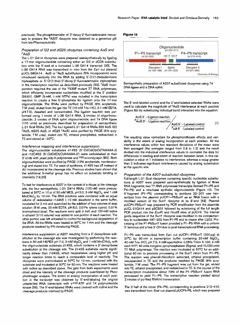
Figure 10
Oligonucleotide (nt 203-211)
Pl-P5 transcript P4-PQ transcript
5 3’
Disruptor I Splint Disruptor II (nt 150-188) (i-d 191-223) (nt 251-290)
Chemistry & B&g
Semisynthetic preparation of A207substituted ribozymes using T4 DNA ligase and a DNA splint.
The 5’-end-labeled control and the 3’-end-labeled selected RNAs were
used to calculate the magnitude of FAaS interference at each position (Figure 6b) by substituting individual band intensities into the equation:
AaS 3’ - Ligation reaction
FAaS 3’ - Ligation reaction
/
AoS 5’- Labeled control
FaA 5’ - Labeled control
(1)
The resulting value normalizes for phosphorothioate effects and vari- ability in the extent of analog incorporation at each position. All the
interference values within two standard deviations of the mean were
then averaged (the averages ranged from 0.8 to 1.2) and the result divided into the individual interference values to normalize the data for
differences in loading and extent of reaction between lanes. In this cal- culation a value of 1 indicates no interference, whereas a value greater
than 2 indicates significant interference caused by analog substitution at the specific site.
Preparation of the A207-substituted ribozymes Full-length L-21 Seal ribozymes containing specific nucleotide substitu- tions at A207 were prepared semi-synthetically by ligation of three RNA fragments, two T7 RNA polymerase transcripts (termed Pl -P5 and
P4-P9) and a nine-base synthetic oligonucleotide (Figure 10). The
5’-fragment (Pl-P5) corresponding to positions 22-202 was tran-
scribed from the plasmid pUCPl-P5SunY as an RNA fusion with a modified version of the SunY ribozyme at its 3’-end 1631. Plasmid
pUCPl-P5SunY was prepared by PCR amplification from the plasmids pUCL-21G414 and pSC931 followed by subcloning of the full length
PCR product into the EcoRl and /+ndlll sites of pUCl9. The internal guide sequence of the SunY ribozyme was modified to be complemen-
tary to nucleotides 197-202 from Pl -P5 and to cleave after U202. Pro- cessing of the Pl -PSSunY fusion provided a Pl -P5 RNA with a uniform
3’ terminus and a free 3’sOH due to post-transcriptional RNA processing.
Pl-P5 was transcribed from Earl cut pUCPl-P5SunY (200 kg) at 37°C for 90 min in transcription buffer containing 20 mM MgCI,,
40 mM Tris-HCI, pH 7.5,4 mM spermidine, 0.05% Triton X-l 00,4 mM each NTP, 40 units inorganic pyrophosphatase (Sigma), and 15,000 units
T7 RNA polymerase. The reaction was incubated at 50°C for an addi- tional 60 min to promote processing of the SunY intron from Pl-P5.
The reaction was phenol/chloroform extracted, ethanol precipitated, resuspended in TE and the products resolved by PAGE (6% acry
lamide, 7 M urea). The Pl-P5 fragment was cut from the gel, eluted into TE, ethanol precipitated, and redissolved in TE. In the course of the
transcription incubations about 70% of the Pl-PSSunY fusion RNA processed to yield Pl-P5. The transcription reaction yielded about 2 nmoles of purified RNA/ml transcription.
The 3’-half of the intron (P4-F9), corresponding to positions 212-410, was transcribed from Seal cut plasmid pUCPCP9, which was prepared
164 Chemistry & Biology 1999, Vol 6 No 3
by PCR amplification, followed by cloning into the EcoRl and HindIll
sites of pUCl9 to create a DNA template with the T7 promoter adjacent to intron position G212. P4-P9 RNA was transcribed at 37°C for
120 min in a reaction containing 15 mM MgCI,, 40 mM Tris-HCI, pH 7.5,
4 mM spermidine, 0.05% Triton X-l 00, 2 mM each NTP, 50 units inor- ganic pyrophosphatase, 25,000 units T7 RNA polymerase, and 20 mM
GMP to incorporate a 5’-phosphate for use in ligation. The RNA was puri- fied as described for Pl -P5, yielding about 3 nmoles of purified RNA/ml
transcription. The three 9-mer synthetic oligonucleotides (B’CCUAAC-
CAC-33 corresponding to positions 203-211 were 5’.phosphorylated with ATP using PNK to provide a 5’.phosphate for ligation.
The Pl -P5 (4 nmoles), P4-P9 (4 nmoles) and the 9-mer oligonucleo-
tide (8 nmoles) were mixed with 4 nmoles of a 33-nt DNA splint (com-
plementary to positions 191-223) and two DNA disruptor oligonucleo-
tides [complementary to positions 251-290 (6 nmoles) and 150-l 88 (8 nmoles)] in a 400 ul reaction containing 25 mM Tris-HCI, pH 7.0
and 25 mM NaCl (Figure 10). The RNA and DNA segments were annealed by heating to 90“C for 2 min and cooling the reaction slowly
to room temperature. The ligation reaction was initiated by the addition
of ATP (0.5 mM), fresh DlT (4 mM), 50 mM Tris-HCI, pH 7.5, 10 mM MgCI,, and 800 Weiss units of T4 DNA ligase in a final reaction
volume of 600 pl. The ligation reactions were incubated at 25°C for 4 h, phenol-chloroform extracted, ethanol precipitated. The full-length
L-21 Seal RNAs were purified using PAGE (6% acrylamide, 7 M urea) and concentrated as described for the Pl -P5 RNA.
Although ligation of the P4-P9 fragment to the 9-mer oligonucleotide was reasonably efficient (50%) ligation of the Pl-P5 fragment to the
oligonucleotide proceeded very inefficiently (5% yield). The overall liga-
tion efficiency for the full-length material was about 2%. This significantly limited the number of experiments that could be performed with the
A207 substituted ribozymes, though it was possible to generate a suffi- cient amount of material for the basic characterizations described above.
A total of three RNAs were prepared in this manner. rA207 contained a
ribose at A207 and was chemically equivalent to the wild-type L-21 Seal RNA. dA207 and FA207 contained a single 2’.deoxynucleotide or
2’-deoxy-2’-flouronucleotide substitution at A207, respectively.
Thermodynamic and kinetic characterization of A207-substituted ribozymes Equilibrium dissociation constants (K,) for the ribozyme 5’-exon complex were measured at 40°C using native gel mobility shift analysis
under conditions of excess ribozyme and a trace concentration (I K&O)
of 5’-32P-labeled 5’-exon analog rl P (GGCCCUCrT) or dl P (CCCUCdT) [64]. Each ribozyme was prefolded at 50°C for 20 min in 50 mM
Tris-HCI (pH 7.5), 10 mM NaCI, 0.1 mM EDTA, 4 mM MgCI,. The solu-
tion was cooled to 40°C and combined with an equal volume of the 5’.exon oligonucleotide analog dissolved in the same buffer plus 6%
glycerol and 0.1% xylene cyanol. The solution was incubated for 30 min at 40°C and the bound and free 5’-exon analog fractions separated by
PAGE (8% acrylamide, nondenaturing, 4 mM MgCI,, 100 mM Tris- HEPES pH 7.5). The fraction of oligonucleotide bound (9) at each
ribozyme concentration, [El, was quantitated by Phosphorlmager analy-
sis. The K,, was calculated by the nonlinear least squares fit of the
equation 9 = [E]/([E] + K,). The values reported in Table 1 are an average of at least three independent experimental trials.
Measurement of (k,,,/K,)o, k,,, and K, o for the single turnover cleav-
age of the oligonucleotide substrates rlS (GGCCCUCrTAAAAA) or
dlS (CCCUCdTAAAAA) by the L-21 Seal form of the Tetrahymena ribozyme followed previously described methods [8,20,43,44]. The tran- scribed L-21 Seal RNA or its A207 substituted variants were incubated
in reaction buffer (50 nM ribozyme, 50 mM HEPES pH 7.0, 10 mM
MgCI,) at 50°C for 20 min, cooled to 30°C, and the reaction was initi- ated by the addition of an equal volume of the same buffered solution
containing 5’-32Pslabeled oligonucleotide (~1 nM) and GMP (1 pM to 5 mM), prewarmed to 3O’C. Portions of the reaction were removed at
various times and quenched on ice with 2 volumes of 7.5 M urea,
20 mM EDTA, 0.1% xylene cyanol, 0.1% bromophenol blue. Reaction
products were resolved from the unreacted substrate by denaturing 20% PAGE. The fraction reacted at each time point measured by
Phosphorlmager analysis, and the reaction rate (k& at each GMP concentration was calculated using an exponential decay with an end-
point. (kcat/K,)o at low GMP concentration was calculated as a linear
fit to the plot of kobs verses GMP concentration [8]. The rate constants k,,r and K,o for each enzyme were calculated by plotting kohs versus
GMP concentration and fitting to the equation kabs=br[GMP]/ (K,o + [GMP]) [44]. The values reported in Table 2 are the combina-
tion of at least three independent experimental trials. No increase in
kobs was observed upon doubling the ribozyme concentrations at satu- rating GMP concentration.
Acknowledgements We thank R. Sousa for the clone of the Y639F mutant T7 RNA DOIV-
merase, B. Ross of Isis Pharmaceuticals Inc. for the gift of 2’.deoxy-2’ flu- oroadenosine, both as a nucleoside and as a protected phosphoramidite, J. Doudna for plasmid pSC931, and A. Szewczak for assistance with mol- ecular modeling. We also thank members of the Strobe1 lab for critical comments on the manuscript. This work was supported by NIH grant GM54839, a Beckman Young investigator Award, and a Searle Scholar award. S.A.S. is supported by a Junior Faculty Research Award from the American Cancer Society.
References 1.
2.
Narlikar, G.J. & Herschlag, D. (1997). Mechanistic aspects of enzymatic catalysis: lessons from comparison of RNA and protein enzymes. Annu. Rev. Biochem. 66, 19-59. Strater, N., Lipscomb, W.N., Klabunde, T. & Krebs, B. (1996). Two- metal ion catalysis in enzymatic acvl- and phosphorvl-transfer
3.
4.
5.
6.
7.
8.
9.
10.
11.
12.
13.
14.
15.
16.
reactions. Ang&v. Chem. Inf. Ed. kg/. 36, 2Oi4-2.055. Cech, T.R. (1990). Self-splicing of group I introns. Ann. Rev. Biochem. 59, 543-568. - - Cech, T.R. (1987). The chemistry of self-splicing RNA and RNA enzymes. Science 236, 1532-l 539. Been, M.D. & Perrotta, A.T. (1991). Group I intron self-splicing with adenosine: evidence for a single nucleoside-binding site. Science 252,434.438. Zaug, A.J., Grosshans, C.A. & Cech, T.R. (1988). Sequence-specific endoribonuclease activity of the Terrahymena ribozyme: enhanced cleavage of certain oligonucleotide substrates that form mismatched ribozyme-substrate complexes. Biochemistry 27, 8924-8931. Beaudrv. A.A. & Jovce. G.F. (1992). Directed evolution of an RNA enzyme. Science 257,.635-641. Herschlac, D. & Cech, T.R. (1990). Catalvsis of RNA cleavage bv the Tetrahymena thermophila ribozyme. 1. Kinetic description ofthe- reaction of an RNA substrate complementary to the active site. Biochemistry 29, 10159-l 0171. Mei, R. & Herschlag, D. (1996). Mechanistic investigations of a ribozyme derived from the Tetrahymena group I intron. Insights into catalysis and the second step of self-splicing. Biochemistry 36, 5796-5809. Bevilacqua, P.C., Kierzek, R., Johnson, K.A. &Turner, D.H. (1992). Dynamics of ribozyme binding of substrate revealed by fluorescence- detected stopped-flow methods. Science 258, 1355-l 358. Herschlag, D. (1992). Evidence for processivity and two-step binding of the RNA substrate from studies of Jl12 mutants of the Tetrahymena ribozyme. Biochemistry 31~ 1386-l 399. Bass, B.L. & Cech, T.R. (1984). Specific interaction between the self- splicing RNA of Tetrahymena and its guanosine substrate: implications for biological catalysis by RNA. Nature 308, 820-826. Moran, S., Kierzek, R. & Turner, D.H. (1993). Binding of guanosine and 3’ splice site analogues to a group I ribozyme: interactions with functional groups of guanosine and with additional nucleotides. Biochemistry 32, 52476256. Li, Y. &Turner, D.H. (1997). Effects of Mg2+ and the 2’.OH of guanosine on steps required for substrate binding and reactivity with the Tetrahymena ribozyme reveal several local folding transitions. Biochemistry 36, 11 131-l 1139. Herschlag, D. & Cech, T.R. (1990). DNA cleavage catalysed by the ribozyme from Tetrahymena. Nature 344, 405-409. Herschlag, D., Eckstein, F. & Cech, T.R. (1993). The importance of being ribose at the cleavage site in the Tefrahymena ribozyme reaction. Biochemistry 32, 8312-8321.

Research Paper RNA catalytic triad Strobe1 and Ortoleva-Donnelly 165
17.
18.
19.
20.
21.
22.
23.
24.
25.
26.
27.
28.
29.
30.
31.
32.
33.
34
35.
36.
37.
38.
39.
40.
41.
42.
Herschlag, D., Eckstein, F. & Cech, T.R. (1993). Contributions of 2’. hydroxyl groups of the RNA substrate to binding and catalysis by the Tefrahymena ribozyme. An energetic picture of an active site composed of RNA. Biochemistry 32, 8299-8311. Knitt, D.S., Narlikar, G.J. & Herschlag, D. (1994). Dissection of the role of the conserved GU pair in group I RNA self-splicing. Biochemistry 33, 13864-l 3879. Strobel, S.A. & Cech, T.R. (1995). Minor groove recognition of the conserved G,U pair at the Tetrahymena ribozyme reaction site. Science 267, 675-679. Strobel, S.A. & Cech, T.R. (1996). Exocyclic amine of the conserved GU pair at the cleavage site of the Tetrahymena ribozyme contributes to ti’splice site selection and transition state stabilization. Biochemistry 35, 1201-l 211. Narlikar, G.J., Khosla, M., Usman, N. & Herschlag, D. (1997). Quantitating tertiary binding energies of 2’.OH groups on the Pl duplex of the Tetrahymena ribozyme: Intrinsic binding energy in an RNA enzyme. Biochemistry 36, 2465-2477. Pyle, A.M. (1993). Ribozymes: a distinct class of metalloenzymes. Science 261, 709-714. Grosshans, C.A. & Cech, T.R. (1989). Metal ion requirements for sequence-specific endoribonuclease activity of the Tetrahymena ribozyme. Biochemistry 28, 6688-6694. Piccirilli, J.A., Vyle, J.S., Caruthers, M.H. & Cech, T.R. (1993). Metal ion catalysis in the Tetrahymena ribozyme reaction. Nature 362, 85-88. Weinstein, L.B., Jones, B.C.N.M., Cosstick, R. & Cech, T.R. (1997). A second catalytic metal ion in a group I ribozyme. Nature 388, 805-608. Sjoegren, A.S., Pettersson, E., Sjoeberg, B.M. & Stroemberg, R. (1997). Metal ion interaction with cosubstrate in self-splicing of group I introns. Nucleic Acids Res. 25, 648-653. Pyle, A.M. & Cech, T.R. (1991). Ribozyme recognition of RNA by tertiary interactions with specific ribose 2’.OH groups. Nature 350, 628-631. Bevilacqua, P.C. &Turner, D.H. (1991). Comparison of binding of mixed ribose-deoxyribose analogues of CUCU to a ribozyme and to GGAGAA by equilibrium dialysis: evidence for ribozyme specific interactions with 2’ OH groups. Biochemistry 30, 10632-l 0640. Herschlag, D., Eckstein, F. & Cech, T.R. (1993). Contributions of 2’.hydroxyl groups of the RNA substrate to binding and catalysis by the Tetrahymena ribozyme. An energetic picture of an active site composed of RNA. Biochemistry 32, 8299-8311. Strobel, S.A. & Cech, T.R. (1993). Tertiary interactions with the internal guide sequence mediate docking of the Pl helix into the catalytic core of the Tetrahymena ribozyme. Biochemistry 32, 13593-l 3604. Narlikar, G.J., Gopalakrishnan, V., McConnell, T.S., Usman, N. & Herschlag, D. (1995). Use of binding energy by an RNA enzyme for catalysis by positioning and substrate destabilization. Proc. Nat/ Acad. SC;. USA 92, 3668-3672. Holbrook, S.R., Cheong, C., Tinoco, I. &Kim, S.-H. (1991). Crystal structure of an RNA double helix incorporating a track of non- Watson-Crick base pairs. Nature 353, 579-581. Strobel, S.A., Ortoleva-Donnelly, L., Ryder, S.P., Cate, J.H. & Moncoeur, E. (1998). Complementary sets of noncanonical base pairs mediate RNA helix packing in the group I intron active site. Nat. Struct. Biol. 5, 60-66. Gaur, R.K. & Krupp, G. (1993). Modification interference approach to detect ribose moieties important for the optimal activity of a ribozyme. Nucleic Acids Res. 21, 21-26. Strobel, S.A. & Shetty, K. (1997). Defining the chemical groups essential for Tetrahymena group I intron function by nucleotide analog interference mapping. Proc. Nat/ Acad. Sci. USA 94, 2903-2908. Gish, G. & Eckstein, F. (1988). DNA and RNA sequence determination based on phosphorothioate chemistry. Science 240, 1520-l 522. Young, B., Herschlag, D. & Cech, T.R. (1991). Mutations in a nonconserved sequence of the Tetrahymena ribozyme increase activity and specificity. Cell 67, 1007-l 019. Moore, M.J. &Sharp, P.A. (1992). Site-specific modification of pre- mRNA: the 2’-hydroxyl groups at the splice sites. Science 256,992-997. Szewczak, A.A., Ortoleva-Donnelly, L., Ryder, BP., Moncouer, E. & Strobe& S.A. (1998). A minor groove triple helix in the active site of the Tetrahymena group I intron. Nat. Struct. Biol. 5, 1037-l 042. Strobel, S.A. & Cech, T.R. (1994). Translocation of an RNA duplex on a ribozyme. Nat, Struct, Biol. 1, 13-l 7. Ortoleva-Donnelly, L., Kronman, M. & Strobel, S.A. (1998). Identifying sites of RNA tertiary structure by Nucleotide Analog Interference Mapping with @-methylguanosine. Biochemistry 37, 12933-l 2942. Pyle, A.M., McSwiggen, J.A. & Cech, T.R. (1990). Direct measurement of oligonucleotide substrate binding to wild-type and mutant ribozymes from Tetrahymena. Proc. Nat/ Acad. Sci. USA 87, 8187-8191.
43.
44.
45.
46.
47.
48.
49.
50.
51.
52.
53.
54.
55.
56.
57.
58.
59.
60.
61.
62.
63.
64.
Herschlag, D., Piccirilli, J.A. & Cech, T.R. (1991). Ribozyme-catalyzed and nonenzymatic reactions of phosphate diesters: rate effects upon substitution of sulfur for a nonbridging phosphoryl oxygen atom. Biochemistry 30, 4844-4054. McConnell, T.S., Cech, T.R. & Herschlag, D. (1993). Guanosine binding to the Tetrahymena ribozyme: thermodynamic coupling with oligonucleotide binding. froc. Nat/ Acad. Sci. USA 90, 8362-8366. Damberger, S.H. & Gutell, R.R. (1994). A comparative database of group I intron structures. Nucleic Acids Res. 22, 3508-3510. Cate, J.H., et al., & Doudna, J.A. (1996). Crystal structure of a group I ribozyme domain: principles of RNA packing. Science 273, 1678-l 685. Cech, T.R., Herschlag, D., Piccirilli, J.A. & Pyle, A.M. (1992). RNA catalysis by a group I ribozyme. 1 Biol. Chem. 267, 17479-l 7482. Bass, B.L. & Cech, T.R. (1986). Ribozyme inhibitors: deoxyguanosine and dideoxyguanosine are competitive inhibitors of self-splicing of the Tetrahymena ribosomal ribonucleic acid precursor. Biochemistry 25, 4473-4477. Lienhard, G.E. (1973). Enzymatic catalysis and transition-state theory. Science 180, 149-180. Jencks, W.P. (1975). Binding energy, specificity, and enzymic catalysis: the Circe effect. Adv. Enzymol. 43, 219-410. Narlikar, G.J., Gopalakrishnan, V., McConnell, T.S., Usman, N. & Herschlag, D. (1995). Use of binding energy by an RNA enzyme for catalysis by positioning and substrate destabilization. Proc. Nat/ Acad. Sci. USA 92, 3668-3672. Narlikar, G.J. & Herschlag, D. (1998). Direct demonstration of the catalytic role of binding interaction in an enzymatic reaction. Biochemistry 37, 9902-9911. Steitz, T.A. & Steitz, J.A. (1993). A general two-metal-ion mechanism for catalytic RNA. Proc. Nat/ Acad. Sci. USA 90, 6498-6502. Beese, L.S. & Steitz, T.A. (1991). Structural basis for the 3’-5’ exonuclease activity of Escherichia co/i DNA polymerase I: a two metal ion mechanism. EM60 J. 10, 25-33. Derbyshire, V., Grindley, N.D.F. &Joyce, C.M. (1991). The 3’-5’ exonuclease of DNA polymerase I of Escherichia co/i: contribution of each amino acid at the active site to the reaction. EMBO J. 10, 17-24. Zhang, Y., Liang, J.-Y., Huang, S., Ke, H. & Lipscomb, W.N. (1993). Crystallographic studies of the catalytic mechanism of the neutral form of fructose-i ,6-bisphosphatase. Biochemisfry 32, 1844-l 857. Marcus, C.J. (1976). Inhibition of bovine hepatic fructose-l ,6- diphosphatase by substrate analogs. 1. Biol. Chem. 251, 2963. El-Maghrabi, M.R., Gidh-Jain, M., Austin, L.R. & Pilkis. S.J. (1993). Isolation of a human liver fructose-l ,6-bisphosphatase cDNA and expression of the protein in Escherichia co/i. Role of Asp-l 18 and Asp-l 21 in catalysis. J. Biol. Chem. 268, 9466-9472. Ortoleva-Donnelly, L., Szewczak, A.A., Gutell, R.R. & Strobel, S.A. (1998). The chemical basis of adenosine conservation throughout the Tetrahymena ribozyme. RNA 4, 498519. Scaringe, S.A., Wincott, F.E. & Caruthers, M.H. (1996). Novel RNA synthesis method using 5’-0-silyl-2’-O-orthoester protecting groups. J. Am. Chem. Sot. 120,11820-l 1821. Sousa, R. & Padilla, R. (1995). A mutant T7 RNA polymerase as a DNA polymerase. EMBO J. 14, 4609-4621. Lingner, J. & Keller, W. (1993). 3’-end labeling of RNA with recombinant yeast poly(A) polymerase. Nucleic Acids Res. 21, 2917-2920. Doudna, J.A. & Szostak, J.W. (1989). Miniribozymes, small derivatives of the sunyintron, are catalytically active. Mol. Cell. Biol. 9,5480-5483. Pyle, A.M., Moran, S., Strobel, S.A., Chapman, T., Turner, D.H. & Cech, T.R. (1994). Replacement of the Conserved GU with a G-C pair at the cleavage site of the Tetrahymena ribozyme decreases binding, reactivity, and fidelity. Biochemistry 33, 13856-l 3863.
Because Chemistry & Biology operates a ‘Continuous
Publication System’ for Research Papers, this paper has been
published via the internet before being printed. The paper can
be accessed from http://biomednet.com/cbiology/cmb - for
further information, see the explanation on the contents pages.