A HISTOPATHOLOGICAL AND HISTOCHEMICAL STUDY OF …
103
A HISTOPATHOLOGICAL AND HISTOCHEMICAL STUDY OF CHOLECYSTITIS SUBMITTED FOR M.D. IN PATHOLOGY THE TAMILNADU DR.MGR MEDICAL UNIVERSITY DEPARTMENT OF PATHOLOGY PSG INSTITUTE OF MEDICAL SCIENCE & RESEARCH PEELAMEDU, COIMBATORE – 641 004. TAMILNADU, INDIA. MARCH 2008
Transcript of A HISTOPATHOLOGICAL AND HISTOCHEMICAL STUDY OF …

1
A HISTOPATHOLOGICAL AND
HISTOCHEMICAL STUDY OF CHOLECYSTITIS
SUBMITTED FOR
M.D. IN PATHOLOGY
THE TAMILNADU DR.MGR MEDICAL UNIVERSITY
DEPARTMENT OF PATHOLOGY PSG INSTITUTE OF MEDICAL SCIENCE & RESEARCH
PEELAMEDU, COIMBATORE – 641 004.
TAMILNADU, INDIA.
MARCH 2008

2
CONTENTS
Page No.
CERTIFICATE
ACKNOWLEDGMENT
1. INTRODUCTION 1
2. AIMS AND OBJECTIVES 3
3. REVIEW OF LITERATURE 4
4. MATERIAL 18
5. METHODS 19
6. RESULTS 27
7. DISCUSSION 40
8. SUMMARY AND CONCLUSION 47
9. APPENDIX I
10. BIBLIOGRAPHY
11. MASTER CHART

3
ACKNOWLEDGMENT
I record my gratitude to Dr.V.Nirmala, Professor, Department of
Pathology, PSG IMS&R, for her valuable guidance and advice.
I sincerely thank Dr.Prasanna N.Kumar, Professor and HOD,
Department of Pathology and Dr.S.Ramalingam, Principal, PSG
IMS&R for their constant encouragement and logistic support.
I express my sincere gratitude to my Professors.
Dr.AmmuSivaraman, AlameluJayaram and Associate Professors
Dr.S.Santhakumari, Dr.S.Vanitha, Dr.T.N.Subba Rao for their kind
gesture and invaluable support.
I express my sincere gratitude to Dr.Suma Pillai, Dr.Nithilavalli,
Dr.S.Saraswathy, Dr.V.Sandhya who have encouraged me all along in
this project.
I wish to earnestly acknowledge my colleagues, Dr.Uma Maheswari,
Dr.Aysha Ali, Dr.Ramganesh who extended their support throughout
the study period.
My heartful thanks to Dr.Rajeev Roy who has very kindly extended
his constant help and assistance in completing this research work.
4
At this juncture, I deem it, fit to thank all the technical staffs of
Department of Pathology, PSGIMS&R, especially Mrs.Angeline Mary,
Mrs.Suceela Stanley, Mrs.Mangaiarkarasi and Mrs.Gomathi.
I am deeply greatful to Dr.Ananth, HOD of Biochemistry and
Dr.Jeyasingh, HOD of Surgery who has extended their invaluable
support and assistance for making my venture fruitful.
My special thanks to Mrs.Uma, Department of Pharmacology for her
valuable support extended to me.
I acknowledge with thanks, the encouragement, support and
inspiration given to me by my family members throughout the
preparation of this thesis.
Dr.P.Anupama

5
CERTIFICATE
This is certify that the dissertation work entitled “A
HISTOPATHOLOGICAL AND HISTOCHEMICAL STUDY OF
CHOLECYSTITIS” submitted by Dr.P.Anupama is the work done by
her during the period of study in this department from June 2005 to
March 2008. This work has been done under my direct supervision and
guidance.
Dr.V.Nirmala, Professor of Pathology, PSG IMS & R, Coimbatore.
Dr.Prasanna N.Kumar, Dr.S.Ramalingam, Professor and HOD, Principal, Department of Pathology, PSG IMS & R, PSG IMS & R, Coimbatore. Coimbatore.

6
INTRODUCTION

1
INTRODUCTION
Gallstones are a major cause of morbidity throughout the world,
necessitating hospitalization and cholecystectomy. Gallbladder is known
to play crucial role in the formation of gallstones. Therefore,
understanding the interaction between gallbladder mucosa and bile is an
important step towards understanding the pathogenesis of gallstone
disease.
Cholesterol saturated ‘lithogenic’ bile, originating from the liver
and considered an important factor in gallstone formation, has been found
in healthy individuals1. Lithogenic bile therefore cannot be the only factor
involved in the process. Other factors such as supersaturation of bile with
calcium2, gallbladder mucus, prostaglandins and functional failure of
electrolyte absorption by gallbladder mucosa also may influence gallstone
formation.
Biliary calcium can reduce the solubility of cholesterol3 rendering
the bile lithogenic. Apart from being a critical initiating factor, calcium is
physically incorporated into gallstones as well.

2
Gallbladder mucus has long been recognized as an important factor
contributing to gall stone development4, 5. The implication of mucin in
gallstone formation has been widely studied. Hypersecretion of mucus
occurs during gallstone formation in humans 6,7,8,9, and experimental
animals10. Apart from forming the nucleus for calculus, the mucins form a
structural component of gallstones as shown by histochemical studies on
calculi11, 12. Calcium and prostaglandins can stimulate mucus secretion by
gallbladder mucosa 13,14,15
Normal human gallbladder contains predominantly sulphated acid
mucin16. It is this sulphated mucin content that is increased in gallstone
disease. Metaplastic and neoplastic gallbladder epithelium on the other
hand shows an increase in sialomucins and decrease in sulphomucins.
Several studies have suggested progression from metaplasia, through
dysplasia, to adenocarcinoma of gallbladder17. The existence of such a
pathway has not been definitely proven.
A study correlating gallbladder mucin histochemistry with
morphology would help one understand the role of mucins in gallstone
disease and carcinogenesis. Further, correlating the above with the
chemical composition of stones could lead to the identification of a high
risk group, with possible therapeutic implications.

1
AIMS AND OBJECTIVES

3
AIMS AND OBJECTIVES
1. Qualitative and quantitative assessment of gallbladder mucins in
chronic calculous cholecystitis.
2. Correlating the mucin histochemistry and morphology of gallbladder in
chronic calculous cholecystitis with each other and with the chemical
composition of gallstones.

1
REVIEW Of LITERATURE

4
REVIEW OF LITERATURE
The gallbladder is one of the most frequently received specimens in
surgical pathology laboratory, on which the pathologist routinely
documents the presence of gallstones and inflammation. The formation of
gallstones is closely linked to bile and mucosal epithelial interaction18.
Gallbladder is a pear shaped sac that lies attached to the posterior
aspect of the right lobe of liver. It measures about 10 cms in length and 3-
4 cms in width and is divided into fundus, body and neck. The wall of
gallbladder consists of mucosa, muscularis propria and perimuscular
connective tissue. There is no muscularis mucosae and sub mucosa. The
mucosa is lined by columnar epithelium with lightly eosinophilic
cytoplasm and basally located nuclei. Only the neck part has true glands
that are tubuloalveolar mucous type 19. Metaplasia is not seen in normal
gallbladder, but is common in cholelithiasis and cholecystitis. Metaplasia
can be gastric or intestinal in type. Gastric metaplasia in turn is either
foveolar (gastric foveolar type surface epithelium) or antral (gastric antral
type glands). Intestinal metaplasia is characterized by columnar cells of
intestinal type interspersed with goblet cells20; Endocrine and Paneth cells
may be present20.

5
Rokitansky Aschoff sinuses represent herniation of mucosal
epithelium into lamina propria, smooth muscle or perimuscular
connective tissue and are a common feature of chronic cholecystitis21.
The gallbladder concentrates, stores and releases bile.
Approximately 800 – 1000ml of bile flows daily into the gallbladder from
the liver. 40 –70ml of bile can be stored in the gallbladder 22.
The proposed pathogenesis of gallstones, based on human studies
and experimental animal models, involves steps such as bile saturation,
nucleation, precipitation of cholesterol monohydrate crystals and growth
to stone sized aggregates. Gallstone containing bile is characterized by
supersaturation with cholesterol and rapid in vitro nucleation of
cholesterol crystals23,24.
Gallstones vary considerably in chemical composition, the basic
constituents being cholesterol, calcium bilurubinate and calcium
carbonate either alone or in combination 25.

6
Figure 1. Multiple Small Cholesterol Gallstones
Cholesterol gallstones (75%-80%) include, pure cholesterol stones
(10%) and mixed stones (10%). Pure cholesterol stones are yellow white,
round to ovoid and smooth; have a crystalline or a laminated cut surface
and measures up to 4cms in diameter. Mixed stones are those which have
lower cholesterol content. Depending on the proportion of calcium
carbonate, bilirubin and phosphates, mixed stones are laminated grey –
white to black. They are usually smaller, multiple and faceted, may have
laminated cut surface and a dark core. More than 80% of cholesterol
stones lack calcium carbonate and are hence radiolucent .

7
Figure 2. Pigment Stones
Pigment stones (15%-25%) include brown and black stones. They
are more common in Asia. By definition, they contain <25% - 30%
cholesterol. They form as a result of increased unconjugated bilirubin in
the bile, which then forms insoluble calcium salts. Black stones are black
or deep brown, relatively small resist crushing, have an irregular shiny
surface and on fracturing have a glass like, featureless appearance. They
arise in sterile bile. Many black stones (50%-75%) contain enough
calcium carbonates and phosphates to render them radio opaque. Brown
stones are much softer than black ones, and have a rough, flaky
appearance, and at times may be greasy appearing. They form in the
context of biliary stasis and infection. Brown stones are usually
radiolucent 26,27.

8
Before the appearance of gallstones there is always the formation
of “biliary sludge” containing mucus gel, hydrophobic bile pigments,
cholesterol, lecithin lipid crystals and cholesterol monohydrate crystals.
The cholesterol crystal nucleation seems to occur in the mucus gel on the
epithelial surface.
Mucin secretion by gallbladder and formation of biliary sludge (of
which mucus gel is a crucial component) are important factors in the
pathogenesis of gallstone disease and its sequelae 28,29,30,.
MUCINS
Mucins are the chemical components of the secretion delivered by
certain types of epithelial and connective tissue cells. The original term
mucin was coined by Carpenter as early as in the year 1846 31. Reid and
Clamp in 1978 suggested glycoconjugates as general term, which could
be subdivided into ‘proteoglycans’ and ‘glycoproteins’ 32.

9
STRUCTURE OF MUCIN:
Figure 3. Structure of Mucin
Mucin glycoproteins are large, complex molecules consisting of a
peptide backbone and numerous oligosaccharide side chains, which
represents the products of mucin genes and glycols transferase genes
respectively. Mucin molecules are among the largest molecule in nature,
ranging in size from 3 - 23 million Daltons. Electron microscopy reveals
them to be linear, flexible threads.
Oligosaccharide side chain

10
Free hexose groups are often available, together with certain acidic
moieties, the presence of which will markedly influence histochemical
reactivity. The different mucins may be present as a single type within a
given tissue unit, or more usually as a mixture of different types. The
synthesis of mucin is initiated in the rough endoplasmic reticulum of the
producing cells and is completed in the golgi apparatus. Sulphation of the
hexosamine molecule occurs in the golgi region 8,33.
Scheme of an easy method to classify the mucin like a proteins the
only two classical techniques.8
AB-PAS HID-AB
Neutral mucins Red Negative
Sulphomucins Red-blue Brown-black
Sialomucins Red-blue Blue
Sulpho-sialomucins Red-blue Brown-blue
AB-PAS, sequential staining with AB(2.5) (blue) and PAS (red);
HID-AB, sequential staining with HID(brown to black) and AB(2.5)
(blue).
C

11
The different types of mucins which can be distinguished
histochemically are as follows 34:
ACID MUCINS
1. Strongly sulphated
a. connective tissue
b. epithelial
2. Weakly sulphated
Sulphated; histochemically atypical
3. Carboxylated; sialomucin
a. enzyme – labile (N-acetyl form)
b. enzyme – Resistant (N-acetyl O-acetyl form)
4. Sulfated sialomucin
5. Carboxylated; nonsulphated uronic acid (Hyaluronic
acid)
NEUTRAL MUCINS
There are no subdivisions to this group

12
STUDIES ON MUCINS IN GALL STONE DISEASE Role of gallbladder mucin as a nucleating factor and as a component
of calculi and alterations in these mucins in non-neoplastic, preneoplastic
and neoplastic diseases of the gallbladder have received wide attention
among workers. Review of literature in this field revealed many studies
connecting mucins with gallstone diseases, which could be grouped into
sections as shown below:
1) Biochemical studies on gallbladder bile.
2) Studies on gallstones.
3) Mucin histochemistry of non neoplastic gallbladder mucosa
4) Mucin histochemistry of neoplastic gallbladder mucosa
5) Electron microscopic molecular and animal experimental studies.
BIOCHEMICAL STUDIES ON BILE:
The major acid mucins secreted by gallbladder mucosa are
sulphomucin 35. Increase in the mucin content of bile in patients with
gallstone disease, as against controls has been shown by many workers
using biochemical techniques 9,35,36.

13
Harvey et al however found no difference in the mucus content of bile,
between cases with and without gallstones. Their study consisted of
isolating gallbladder mucus by sepharose gel filtration followed by
ultracentrifugation 37.
Levy et al studied model bile and demonstrated the accelerating
effect of mucins on nucleation of cholesterol monohydrate crystals, an
early step in lithogenesis 30.
STUDIES ON GALLSTONES:
Womack et al as early as in the year 1963, demonstrated the
presence of mucopolysaccharides in gallstones. Whole stones were
sectioned and stained for mucins 11.
Subsequently other workers also showed presence of mucin as a
structural component of cholesterol stone matrix 38 . The amount of
mucins in cholesterol stones was found to be less, compared to pigment
stones . It was also shown that the mucins in pigment stones are mostly
sulphated. The bridging action of sulphomucins promoting solidification
of the mucus gel during stone formation was suggested 12.

14
MUCIN HISTOCHEMISTRY OF NON NEOPLASTIC
GALLBLADDER MUCOSA:
Normal gallbladder epithelium contains sulphated acid mucins39,
with very small quantities of nonsulphated mucins. In recent years there
has been widespread use of mucin histochemistry as an aid to the
diagnosis of diseases. Particular attention has been paid to alterations in
the relative amounts of sulphated and nonsulphated mucins present within
epithelial cells. Many techniques are available for this purpose; the most
widely used being the High Iron Diamine – Alcian Blue (HID-AB)
technique.
Qualitative changes in mucin occur in metaplastic gallbladder
mucosa16,19,20,40. In cholecystitis without metaplasia no qualitative
alterations in mucins have been observed, though the quantity of
sulphomucin increases16. Presence of sulphated mucins in the surface
epithelium and neck glands of gallbladder and nonsulphated acid mucins
and neutral mucins in the goblet cells of intestinal metaplasia and in
metaplastic gastric epithelium have been confirmed by studies on large
number of cases 20

15
Madrid et al studied epithelial mucins of gallbladder using
conventional techniques and demonstrated the presence of sulphated and
carboxylated mucins, the former predominating 40. Further localization of
sulphomucins to the surface epithelium, sialomucins to the mid portion
and either sulphomucins or PAS positive, probably neutral, mucins to the
deeper glands was demonstrated in one study 41
MUCIN HISTOCHEMISTRY AND NEOPLASTIC GALLBLADDER
MUCOSA:
The precise relationship between gallbladder cancer and its
precursors remain ill defined. Several studies have shown that invasive
carcinoma is preceded by dysplasia, but progression from cholelithiasis to
dysplasia has not been proven.
It has been suggested that antral metaplasia may be the
pathogenetic link in this context 42,43,44,45. There are also several studies
that suggest a progression from intestinal metaplasia through dysplasia, to
adenocarcinoma of the gallbladder 46,47,48,49 . These studies point out the
increased incidence of intestinal metaplasia in specimens with carcinoma,
presence of intestinal metaplasia in the vicinity of carcinoma and within
the carcinoma48 . Intestinal metaplasia therefore is considered to represent

16
a neoplastic transformation or a predisposition to neoplastic
transformation.
The importance of extensive sampling in the detection of intestinal
metaplasia was emphasized by Durate et al 49.
Histochemical and immunohistochemical studies have shown
similarities between pyloric metaplasia, intestinal metaplasia and
carcinoma of gallbladder. Presence of endocrine cells in pyloric
metaplasia suggests that these glands are an integral component of
intestinal metaplasia. It is therefore possible that intestinal and gastric
metaplasia arise from stem endodermal cells that differentiate into either
direction43,44,45.
Immunohistochemical studies an normal, metaplastic and
dysplastic gallbladder mucosa have shown a difference in mucin
expression in these groups. Normal epithelium had mucin phenotypes
similar to gastric pyloric mucosa (MUC5), while metaplastic and
dysplastic epithelium expressed colonic/intestinal mucin phenotype
(MUC2), 50.
17
ELECTRON MICROSCOPIC, MOLECULAR AND ANIMAL
EXPERIMENTAL STUDIES:
Apart from HID-AB and PAS staining, electron microscopy51,52,
lectin histochemistry 40 and immunohistochemistry41,42,53 have been
applied to the study of mammalian gallbladder mucins. Lee induced
different types of stones in experimental animals along with
histochemical staining of epithelium for mucins and biochemical analysis
of the mucins 28,29. In this study comprising composition of stones with
histochemical and biochemical analysis of mucins, no qualitative changes
were observed, despite a quantitative increase in mucin.
Experimental studies have been performed on ground squirrel with
application of bile biochemistry, mucosal morphology and mucin
histochemistry 52,54,55,56 . Mucin hypersecretion was found to be an
initiating event in gallstone formation. Histochemically, as in most other
studies, sulphomucins predominated.
More sophisticated techniques such as mucin gene expression in
cell lines however have shown on altered mucin core peptide in
cholecystitis 57. Mucin core proteins, it was found, altered with increasing
degree of inflammation.

18
MATERIAL

18
MATERIAL
A total number of 40 specimens were selected from
gallbladders with clinical and histopathological diagnosis of chronic
calculous cholecystitis received in the department of pathology PSG
INSTITUTE OF MEDICAL SCIENCES AND RESEARCH during the
period of 2005 to 2007. Criteria for selection were 1) Histopathological
confirmation of chronic calculous cholecystitis. 2) Presence of calculi
accompanying the specimen. 3) Availability of sufficient mucosa and
well preserved lining epithelium in sections. 4) Availability of
corresponding paraffin blocks. While 36 specimens fulfilled the above
criteria, 4 cases had only biliary sand in the container. 3 gallbladders
resected for choledochal cysts, were taken as controls.

19
METHODS

19
METHODS
Haematoxylin & Eosin stained sections of the selected specimens
were screened and one to two sections with adequate amount of well
preserved mucosa with lining epithelium were chosen. The corresponding
paraffin blocks were selected and isolated. Four sections were cut on each
of the selected blocks for special stains. A proforma was prepared for
assessment as shown in Appendix I. The slides were assessed according
to the proforma.
HAEMATOXYLIN & EOSIN STAIN
Sections stained with Haematoxylin & Eosin were assessed for the
intensity of inflammation and degree of fibrosis, which in turn were
graded as mild, moderate and severe (1+, 2+, 3+) (figures 4,5,6,7,8). The
number of Rokitansky Aschoff sinuses was indicated as many, few and
nil (figure 9). Gastric metaplasia and intestinal metaplasia were also noted
and indicated as present or absent (figures 10,11).

20
MUCIN HISTOCHEMISTRY
The following special stains for mucin were done with a view to
assess the quantity and quality of mucins in the superficial and deep parts
of gallbladder mucosa.
1. High Iron Diamine – Alcian Blue Stain (HID-AB)
2. Alcian Blue – Periodic Acid Schiff (AB-PAS)
HIGH IRON DIAMINE – ALCIAN BLUE STAIN (HID-AB) 58
REAGENTS:
N N dimethyl-meta –phenyline diamine dihydrochloride - 120 mg
N N dimethyl – para –phenyline diamine dihydrochloride - 20 mg
Distilled water - 50ml
Ferric chloride (60 % BDH SOLUTION) -1.4 ML
Dissolve the two diamine salts simultaneously in distilled water,
add to the ferric chloride solution and mix.

21
PROCEDURE
1. Dewax the positive control section and the test sections and bring to
distilled water.
2. Treat all the sections with diamine solutions for 24 hours.
3. Wash well in running water.
4. Counterstain with 1% AB (PH 2.5) in 3 % acetic acid for 5 minutes
wash and counterstain with 0.5 % aqueous neutral red for 2-3 min
wash in water.
5. Rinse in absolute alcohol.
6. Clear in xylene and mount.
RESULTS
Sulphated mucins – black/brown
Carboxylated mucins – blue

22
ALCIAN BLUE - PERIODIC ACID SCHIFF STAIN (AB-PAS) 59
REAGENTS
Solution A
Alcian blue - 1gm
3 per cent Acetic acid – 100ml
Solution B
Periodic acid solution
Periodic acid - 1gm
Distilled water 200 ml
Solution C
Basic fuschin 1gm
Potassium metabisulphite 2gm
Concentrated hydrochloric acid 2ml
Activated charcoal 2gm
Distilled water 200ml

23
PROCEDURE
1. Dewax section and bring to water
2. Alcian blue solution, 5 min
3. Wash in water, then in distilled water
4. 1% aqueous periodic acid, 5 min
5. Rinse well in distilled water.
6. Schiff’s reagent 15 min.
7. Wash in running tap water 5-10 min.
8. Stain nuclei lightly with haematoxylin solution.
9. Wash in water.
10. Rinse in absolute alcohol.
11. Clear in xylene and mount.
RESULTS
Acid mucins – blue
Neutral mucins – magenta

24
A scoring system was devised, based on the percentage positivity
of cells in each field under low power examination (10X), as shown
below:
75% -100 % - 5+
50% - 75% - 4+
25% - 50% - 3+
5% - 25% - 2+
0% - 5% - 1+
(Figures 12,13,14,15,16)
The values were tabulated and statistically assessed using ANOVA,
t-test, χ2 (Chi2) test.
Biochemical analysis of calculi, wherever available (36 samples)
was performed to determine the chemical composition of calculi. Gross
parameters such as external appearance, colour, number of stones and
weight were noted. The following biochemical procedures were carried
out on the 36 samples of calculi.

25
BIOCHEMICAL ANALYSIS 60
CHOLESTEROL
Wash the gallstones with water and dry. Powder the stone and heat
some with successive small portions of ether in a test tube by inserting the
tube in some warm water and filter. Dissolve a little of residue obtained
on evaporation of the ether in chloroform and add a little of mixture of
acetic anhydride and sulphuric acid (in the proportion of 10 ml to 0.1 ml).
A dark green colour develops rapidly.
PHOSPHATE AND CALCIUM OXALATE
Treat the remaining residue after ether extraction, with dilute
hydrochloric acid (25%). This dissolves the inorganic salts present. Filter
and test the filtrate for phosphate with molybdate. Make some of the
solution alkaline with ammonia and add acetic acid and ammonium
oxalate solution. If calcium is present a precipitate of calcium oxalate is
formed.

26
BILE PIGMENT
Test the precipitate remaining after treatment with hydrochloric acid
for bile pigments. Wash the material remaining on the filter paper and
extract with warm chloroform. Examine the chloroform extract for
bilirubin by means of diazo reagent. Change to pink colour indicates
presence of bile pigment.
The mucin histochemistry scores obtained were tabulated,
separately for the three grades of inflammation and fibrosis. An attempt
was made to correlate the score with degree of inflammation, fibrosis,
presence of metaplasia (intestinal and pyloric) and type of stone.
Statistical assessment of the results was also performed.

1

2

3

4

5

6

7

8

9

27
RESULTS

27
RESULTS
The three specimens of gallbladder used as a controls showed
predominantly Alcian Blue positive mucins in the surface and deep
mucosal epithelium. PAS positivity was seen in traces.
HID –AB stain showed strong HID positivity indicating
predominance of sulphomucins.( TABLE 1)
TABLE 1
AB-PAS HID-AB Techniques
Superficial Deep
AB 4.7 2.6 AB-PAS
PAS 0.06 0.03
HID 4.5 2.8 HID-AB
AB 0 0.03

28
INFLAMMATION AND MUCIN HISTOCHEMISTRY
Of the total 36 cases studied, 9 showed mild (Grade I)
inflammation, 16 cases showed moderate (Grade II) inflammation and 11
cases showed severe (Grade III) inflammation. The mean scores for
Alcian Blue, PAS and High Iron Diamine positive mucins in the three
groups were tabulated. (Table 2-5)
TABLE 2
AB – PAS STAIN
The mean scores for Alcian Blue positive (acid) mucins in the
superficial and deep mucosa, in the three grades of inflammation are
shown. There is progressive decrease in the mean scores for Alcian Blue
positive (acid) mucins in the superficial and deep mucosal epithelium,
with increasing grades of inflammation.
ALCIAN BLUE [ACID MUCIN]
MEAN SCORE INFLAMMATION
GRADE SUPERFICIAL DEEP
I (9 cases) 2.7 3.4
II (16 cases) 2.2 3.1
III (11 cases) 1.95 2.5

29
TABLE 3
AB –PAS STAIN
The mean scores for PAS positive mucins in the superficial and
deep mucosa are tabulated against grades of inflammation. The scores are
higher in Grade III inflammation, than in Grade 1 inflammation. Grade II
inflammation however shows random scores , not conforming to any
pattern.
PAS [ NEUTRAL MUCIN]
MEAN SCORE
INFLAMMATION
GRADE SUPERFICIAL DEEP
I (9 cases) 0.25 0.14
II (16 cases) 0.11 0.31
III (11 cases) 0.29 0.4

30
TABLE 4
HID –AB STAIN
The mean scores for HID positive sulphomucins is tabulated
against the three grades of inflammation.The scores are the lowest for
Grade III inflammation. A progressive decrease in HID scores is obvious
in the superficial mucosal epithelium.
HID – [SULPHOMUCIN]
MEAN SCORE
INFLAMMATION
GRADE SUPERFICIAL DEEP
I (9 cases) 3.9 3.6
II (16 cases) 2.5 3.9
III (11 cases) 2.1 2.8

31
HID
- AB
STA
IN IN
FLAM
MAT
ION
G
RAD
E
11
16
9M
ILD
MO
DE
RA
TE
SE
VE
RE

27
3.9
3.6
2.5
3.9
2.1
2.8
12
3G
RAD
E O
F IN
FLAM
MAT
ION
SULP
HO
MU
CIN
MEA
N S
CO
RE
SU
PE
RFI
CIA
L
DE
EP

32
TABLE 5
HID –AB STAIN
Alcian Blue scores are tabulated against the three grades of
inflammation. Here, unlike the HID scores, the highest values are seen in
Grade III inflammation .
ALCIAN BLUE – [SIALOMUCIN]
MEAN SCORE
INFLAMMATION
GRADE SUPERFICIAL DEEP
I (9 cases) 0.14 0.19
II (16 cases) 0.062 0.13
III (11 cases) 0.3 0.56

33
0.14
0.19
0.06
2
0.13
0.3
0.56
12
3G
RAD
E O
F IN
FLAM
MAT
ION
SIAL
OM
UC
IN M
EAN
SC
OR
E
SU
PE
RFI
CIA
L
DE
EP

33
INFLAMMATION AND METAPLASIA
TABLE 6
The table shows the number of cases with intestinal and Gastric
metaplasia and their percentage, in the three grades of inflammation.
Metaplastic epithelium is characterized by presence of PAS positive
mucins and / or goblets cells containing AB positive mucins in AB –
PAS stains sections (figures 17,18,19,20,21). Intestinal metaplasia has the
highest incidence in Grade I inflammation, and is lowest in Grade III
inflammation .The reverse is seen with gastric metaplasia, which has the
highest incidence in Grade III Inflammation, and a considerably lower
incidence in Grade I inflammation .
INFLAMMATION GRADE
TYPE OF METAPLASIA I
(9 cases)
II
(16 cases)
III
(11 cases)
TOTAL
36
IM
(INTESTINALMETAPLASIA)
5
(56%)
3
(19%)
5
(22%)
13
GM
(GASTRIC METAPLASIA)
1
(11%)
6
(38%)
5
(46%)
12

34
MET
APLA
SIA
19%
22%
56%
38%
46%
11%
12
3
GR
ADE
OF
INFL
AMM
ATIO
N
INCIDENCE
INTE
STI
NA
L M
TEA
PLA
SIA
GA
STR
IC M
ETA
PLA
SIA

34
COMPOSITION OF CALCULI AND GRADE OF INFLAMMATION
TABLE 7
Of the 36 cases with inflammation 3 did not have calculi suitable
for biochemical analysis. The results tabulated in table VII are on the
remaining 33 cases. There were 13 cases with cholesterol calculi and 20
with pigment calculi. Distribution of the two types of calculi among the
three grades of inflammation is shown. The presence of pigment stones
appears to correlate with severity of inflammation (3/8 in Grade I
inflammation,9/11 in Grade III inflammation) as against cholesterol
stones(5/8 in Grade I inflammation, 2/11in Grade III inflammation).
GRADE OF
INFLAMMATION
PIGMENT
STONE
CHOLESTEROL
STONE
I (8 cases) 3 5
II (14 cases) 8 6
III (11 cases) 9 2
TOTAL - 33 TOTAL - 20 TOTAL - 13

35
35
86
92
12
3
GR
ADE
OF
INFL
AMM
ATIO
N
CO
MPO
SITI
ON
OF
CAL
CU
LI A
ND
GR
ADE
OF
INFL
AMM
ATIO
N
CH
OLE
STE
RO
L ST
ON
E
PIG
MEN
T ST
ON
E

35
COMPOSITION OF STONES AND MUCIN HISTOCHEMISTRY
TABLE 8
Mean scores of sialomucins in the superficial and deep mucosal
epithelium are higher in cases with pigment stones (0.85,0.72),when
compared with those having cholesterol stones (0.24,0.58)the number of
cases that show presence of sialomucins is low with both types of
calculi(5/20 cases with pigment stones and 6/13 cases with cholesterol
stones ).
PIGMENT
STONES
CHOLESTEROL
STONES
TOTAL
NUMBER
20 13
SIALOMUCINS
PRESENT IN
5 6
SUPERFICIAL DEEP SUPERFICIAL DEEP SIALOMUCIN
SCORE 0.85 0.72 0.24 0.58

36
COMPOSITION OF CALCULI AND METAPLASIA:
TABLE 9
It is shown that intestinal metaplasia is more or less equally
associated with pigment and cholesterol calculi(35% and 30%
incidence). Gastric metaplasia shows a considerably higher incidence
(40%) in association with pigment stones, when compared with
cholesterol stones (23%).
TYPES OF
METAPLASIA
PIGMENT
STONES
CHOLESTEROL
STONES
INTESTINAL
METAPLASIA
35% 30%
GASTRIC
METAPLASIA
40% 23%

37
35%
30%
40%
23%
CO
MP
OS
ITIO
N O
F C
ALC
ULI
AN
D M
ET
APLA
SIA
INTE
STIN
AL
ME
TAP
LAS
IA
GA
STR
IC
ME
TAP
LAS
IA
PIG
ME
NT
STO
NES
CH
OLE
STR
OL
STO
NE
S

37
FIBROSIS AND MUCIN HISTOCHEMISTRY
TABLE 10 & 11
AB- PAS STAIN
Out of the 40 cases, 7 showed no fibrosis. The mean scores for
remaining 33 cases are tabulated.
Alcian Blue scores (acid mucins) are slightly lower in Grade III
fibrosis ,compared with Grade I . Neutral mucins (PAS positive ) on the
other hand shows higher scores in Grade III as against Grade I fibrosis.
ALCIAN BLUE – [ACID
MUCIN]
MEAN SCORE
PAS – [NEUTRAL MUCIN]
MEAN SCORE
FIBROSIS
GRADE
SUPERFICIAL DEEP SUPERFICIAL DEEP
I (19 cases) 2.46 2.81 0.13 0.22
II (9 cases) 3.0 3.1 0.96 0.45
III (5 cases) 2.0 1.7 0.85 0.32

38
TABLE 12
HID-AB STAIN
No correlation is seen between HID scores (for sulphomucins) and
degree for fibrosis. Alcian Blue scores (for sialomucins) are the lowest
with Grade III fibrosis .
HID – [SULPHOMUCIN]
AB – [SIALO MUCIN]
FIBROSIS
GRADE SUPERFICIAL DEEP SUPERFICIAL DEEP
I (19 cases)
2.64
3.52
0.11
0.17
II (9 cases)
3.08
3.27
0.26
1.97
III (5 cases)
1.7
3.7
0
0.08

39
FIBROSIS AND STONE COMPOSITION
TABLE 13
GRADE OF
FIBROSIS
PIGMENT
STONE
CHOLESTEROL
STONE
NO
STONES
TOTAL
I 10 6 3 19
II 5 3 1 9
III 4 1 0 5
TOTAL (33) 23 13 4 33
Grade of fibrosis is correlated with type of calculus. Pigment
stones have a higher incidence in Grade III fibrosis (4/5) as against Grade
I fibrosis (10/19).

40
Statistically significant difference was seen in the sulphomucin
content between Grade I and Grade III inflammation (Table 4). No
statistical significance was detected in the other observations through
application of tests like ANOVA, t-test, χ2 (Chi2) tests. However the total
number of cases in each group was low, and more number of tissue
samples needs to be studied to confirm the significance of these results.
Rokitansky Aschoff sinuses showed no correlation with mucin
histochemistry .

41
DISCUSSION

41
DISCUSSION
Attempts have been made in the past to correlate gallbladder
morphology, mucin histochemistry, and composition of calculi in
gallstone disease 16, 20, 39 .Most of the previous studies however have
combined any two of the three aspects, that is, either morphology with
mucin histochemistry ,mucin histochemistry with composition of calculi
or composition of calculi with morphology. Few studies correlating all
the three with one another have been recorded in literature .Purpose of the
present study was to determine whether qualitative and / or quantitative
variations in gallbladder mucins occurs in chronic calculous cholecystitis
and whether the alterations ,if any, correlate with morphological changes
in the gallbladder and / or with the type of calculous present .
In short, an attempt has been made through this study, to correlate
gallbladder morphology, mucin histochemistry and composition of calculi
to one another , in chronic calculous cholecystitis specimens.
Gallbladders removed for choledochal cysts were the control in the
present study. The mucosal histology was normal and there was no
inflammation, fibrosis or metaplasia. AB-PAS and HID-AB staining of
sections showed predominantly HID positive sulphated acid mucins

42
throughout the mucosa,with traces of sialomucins and neutral mucins in
foci.
Normal gallbladder mucosa is known to contain predominantly
sulphomucins. Traces of sulphomucins and neutral mucins also may be
present 39.
Our observations on control samples conform to this well –
established normal pattern of mucin histochemistry of gallbladder
mucosa.
Our results indicate a decrease in intraepithelial total acid mucin
content in chronic calculous cholecystitis .Mucin depletion in mucosal
epithelial cells is well known in inflammatory conditions of the
gastrointestinal tract .In various forms of colitis presenting with mucus
diarrhoea or dysentery and showing active inflammation of the mucosa
,mucin depletion is a constant finding . Increased mucin secretion by
gallbladder mucosa during gallstone formation has been described in
literature 6,7,8,9,39 .Earlier investigators have shown that mucins , in
addition to being a structural component of gallstones 12, also play an
acceleratory role in lithogenesis 29.

43
Our observation of decreased intraepithelial mucins in inflamed
gallbladder mucosa is likely to be a reflection of increased secretion of
mucin into bile which is known to occur in calculous disease.
The decrease in the intraepithelial mucins in chronic cholecystitis ,
we found , was due to decrease in sulphomucin which is the predominant
type of mucin in gallbladder mucosa. Further, it was observed that cases
with severe inflammation showed the maximum decrease in sulphomucin.
This was associated with a concomitant increase in sialomucin scores and
a high incidence of gastric metaplasia. Intestinal metaplasia on the other
hand ,did not correlate with the degree of inflammation or sialomucin
content .
No qualitative changes in gallbladder epithelial mucins have been
observed in the earlier studies on chronic cholecystitis 37 . Sialomucins
are known to occur in traces in normal gallbladder mucosa and in
considerable quantities in metaplastic mucosa.
In the present study, an increase in sialomucins was observed in
gallbladders showing severe inflammation .Interestingly, it was in this
group that gastric metaplasia had the highest incidence (figure 22, 23).

44
It therefore follows that, sialomucins in significant quantities tend
to appear in the areas of gastric metaplasia in the gallbladder mucosa.
Their presence is not confined to the goblet cells of intestinal metaplasia
which had the lowest percentage of incidence in severe inflammation, in
the cases assessed figure (24,25).
It has been suggested that antral and intestinal metaplasia in the
gallbladder are histogenetically related having the same progenitor cell
,and could therefore be parts of a morphological spectrum 44 .Transition
from gastric to intestinal metaplasia is a likely possibility 17.
Pre - neoplastic role of intestinal metaplasia in the gallbladder and the
metaplasia → dysplasia → neoplasia sequence have received wide
attention among workers 11,17,61. The mucin profile changes with
progressive transformation to neoplasia, from normal with sulphomucin
predominating through metaplastic and dysplastic showing increasing
amounts of sialomucin, to full fledged neoplastic with sialomucins
predominating .
The high incidence of gastric metaplasia in severe inflammation and
its association with increased expression of sialomucins with the
concomitant reduction in sulphomucins would point, perhaps tentatively,

45
towards a role for gastric metaplasia in the proposed chain of events
stated above .
Basu et al studied the morphological changes in chronic calculous
cholecystitis in relation to the type of stones 62. They found that
inflammation was more severe with pigment calculi while fibrosis and
related complications were more frequent with cholesterol calculi.
The present study supports the association between pigment stones
and severe inflammation. Fibrosis also was more in cases with pigment
calculi in our study.
Mucins have been shown to be a structural component of
gallstones11,38. Histochemical studies carried out on calculi have
demonstrated presence of sulphomucins in them, especially in pigment
stones 12.However no correlation between mucin histochemistry of
mucosal epithelium and the type of stone has been recorded in literature.
In the present study, pigment stones were found more often in
association with severe inflammation, gastric metaplasia and increased
expression of sialomucins ,as against cholesterol stones.

46
We were unable to establish a statistical significance to the above
observations, as the number of cases studied was small, especially in the
sialomucin expressing group (even though the scores were high.)But the
scores and percentage values did show a distinct pattern indicating a
correlation between a severe inflammation, gastric metaplasia,
sialomucins and pigment calculi.(Refer tables 4 to 9 )
Considering the proposed pathway of Gastric metaplasia →
Intestinal metaplasia → Dysplasia → Adenocarcinoma of gallbladder and
the proven presence of sialomucins in considerable amounts in dysplastic
and neoplastic gallbladder mucosa. It is reasonable to speculate on a
pigment stone → severe inflammation → gastric metaplasia →
sialomucin link up, with possible transition to dysplasia, with or without
the intervention of intestinal metaplasia. Further studies on large series
are required to enable us to draw definite conclusions. If such a high risk
group emerges, it will be of significance from the preventive, prognostic
and therapeutic point of view.

47
SUMMARY AND CONCLUSIONS

47
SUMMARY AND CONCLUSIONS
I. The normal gallbladder epithelium contains sulphated acid
mucins with traces of neutral and sialomucins .The
sulphomucin content decreases in chronic calculus cholecystitis
II. In chronic calculus cholecystitis with severe (Grade III)
inflammation (as against mild inflammation) :
1) Total acid mucin content is decreased.
2) This decrease is due to HID positive (sulpho) mucin.
3) Neutral mucin and sialomucin contents are increased.
4) There is a higher incidence of Gastric metaplasia and
pigment stones .
III. Pigment gallstones tend to have an association with :
1) Severe inflammation
2) Higher degree of fibrosis
3) Gastric metaplasia
4) Presence of sialomucins

48
More number of cases need to be studied to see whether high risk
group consisting of pigment stones → severe inflammation → gastric
metaplasia → sialomucin emerges. If it does, will be of therapeutic and
prognostic significance.

49
APPENDIX I
49
APPENDIX I
PROFORMA
Name: Hosp. No.
Age :
Sex :
Clinical Diagnosis:
Clinical History:
Radiological findings:
Operative findings:
PATHOLOGICAL FINDINGS
A.GROSS :
Size:
Contacted/distended/neither:
Wall thickness:
Outer surface:
Mucosal lining:
Calculi-single/multiple:
Colour/ type:
Adherent liver:
Lymph node;
Any other:
B. MICROSCOPIC :
a) Mucosa-

50
Epithelium:
Ulceration:
Inflammatory cells:
Foam cells:
Lymphoid follicles:
Epithelial regeneration:
Intestinal metaplasia:
Pyloric metaplasia:
Dysplasia:
b) Rokitansky Aschoff sinuses
c) Muscle coat :
Inflammation
Fibrosis
Abscess
Any other
d) Perimuscular tissue :
Inflammation
Fibrosis
e) Diverticulosis
f) Adherent liver
g) Lymph node

51
C. Mucin Histochemistry :
AB-PAS
HID-AB
D. BIOCHEMICAL ANALYSIS-RESULT

52
BIBLIOGRAPHY

53
BIBLIOGRAPHY
1. Holzbach RT, Marsh M, Olszweski M, Holan K
Cholesterol solubility in bile. Evidence that supersaturated bile is
frequent in healthy man. J Clin Invest 1973; 52: 1467- 79
2. Shiffman ML, Moore EW.
Bile is supersaturated with calcium in most patients with cholesterol and
mixed gallstones (Abstract). Gastroenterology 1987; 92: A 1775
3. Neithercut WD
Effect of calcium, magnesium and sodium ions on in vitro nucleation of
human gall bladder bile. Gut 1989; 30: 665 – 70
4. Broomfield P, Chopra R, Shein Baum R et al.
Formation and prevention of lithogenic bile and gallstones during
weight loss (abstract). Gastroenterology 1987; 92:1721
5. La Mont JT, Smith BF, Moore JRL.
Role of gallbladder mucin in pathophysiology of gallstones. Hepatology
1984; 4 : 51 – 56

54
6. Shiffman ML, Sugerman H J, Kellum H J, Moore EW
Changes in gallbladder bile composition following gallstone formation
and weight reduction. Gastroenterology 1992; 103 : 214-221
7. Shalin S, Ahlberg J, Einarsson K, Henriksson R, Danielson A.
Quantitative ultrastructural studies of gallbladder epithelium in
gallstones free subjects and patients with gallstones. Gut 1990; 31:100 –
105
8. Madrid J F, Hernandez F, Ballesta J.
Characterisation of glycoproteins in the epithelial cells of human and
other mammalian gallbladder ,A Review pathol int .1996 apr;46:261-6
9. Swobodnik W, Wenk H, Janowitz P et al.
Total biliary protein, mucus glycoproteins, cyclic AMP and
apolipoprotein in the gall bladder bile of patients with cholesterol stones
and stone free controls. Scand J Gastroenterol 1991; 26:771-778
10. Pemsingh RS, Mac Pherson BR and Scott GW.
Mucus hypersecretion in the gallbladder epithelium of the ground
squirrel fed a lithogenic diet for the induction of cholesterol gallstones
Hepatology 1987; 7:1267-71

55
11. Womack N, Zeppa R, Irvine G.L
The anatomy of gallstones. Ann surgery 1963;157: 670-686
12. Maki T, Matushiro T, Suzuki N and Nakamura N.
Role of sulphated glycoprotein in gallstone formation
Surgery, gynaec, obstetrics. 1971 ; 132:846-854
13. Malet PF, Locke CL, Trotman BW, Soloway RD.
The calcium ionophore A23187 stimulates glycoprotein secretion by
the guinea pig gallbladder. Hepatology 1986; 6 : 569 – 73
14. Wahlin T, Thornell E, Jivegard L, Svanvik J.
Effects of intraluminal prostaglandins, E2 in vivo on secretory
behaviour and ultrastructural changes in mouse gallbladder
epithelium. Gastroenterology 1988; 95 : 1632 – 1635
15. La Morte WW, La Mont JT, Hale W, Booker ML, Scott TE, Turner
B. Gallbladder prostaglandins and lysophospholipids as mediators
of mucin secretion during cholelithiasis. Am J Physiol 1986;
251:701-9

56
16. Esterly JR, Spicer SS.
Mucin histochemistry of human gall bladder: changes in
adenocarcinoma, cystic fibrosis and cholecystitis. J Nat: cancer inst
1968; 40:1-11
17. Mukhopadhyay S, Landas SK.
Putative precursors of gallbladder dysplasia; A review of 400 routinely
resected specimens. Arch- path Lab Med 2004; 129:386-390.
18 . M.R Jacyna
Interactions between gallbladder bile and mucosa;relevance to
gallstone formation. Gut 1990;31:568-70
19. Frierson HF, The gross anatomy and histology of the gallbladder,
extra hepatic bile ducts, Vaterian system and minor papilla.
Am J Surg Path 1989;13:146-162.
20. Laitio M. Morphology and histochemistry of non tumorous
gallbladder epithelium. A series of 103 cases. Path.Res.Pract.1980 ;
167(2-4):335-345.

57
21. Elfving G, Crypts and ducts in Gall bladder wall. Acta
pathol,microbiol,scand 1960;49 (suppl)135:1-45.
22. Guyton AC.
The liver and biliary system .In: Guyton AC – Text book of medical
physiology.Philadelphia:W B Saunders ;1976:936-944
23. Johnston DE,Kaplan MM, Pathogenesis and treatment of gall stones.
N Engl J Med 1993;328: 412-421
24. Smith BF, Lamonte JT. The sequence of events in gall stones
formation. Lab invest 1987 ;56:125-126
25. Small DM. Gall stones .N Engl J Med 1968;279:588-92
26.Adsay N V, Gallbladder, Extrahepatic biliary tree, and ampulla In:
Sternberg’s Diagnostic surgical pathology 4th ed, Philadelphia,
Lippincott- Raven publishers,1779-1780.
27. Ostrow JD. The etiology of pigment gall stones
Hepatology .1984 ;4:215S-222S

58
28 . Lee SP,J . LaMonte T and Carey MC
Role of gall stone ,mucus hypersecretion in the evaluation of cholesterol
gall stone studies in the prairie dog. J.CLIN invest 1981 ;67: 1712- 1723
29. Lee SP, Hypersecretion of mucus glycoproteins by the gallbladder
epithelium in experimental cholelithiasis. J Pathol 1981; 134:199-207.
30. Levy PF, Smith BF and La Monte JT.
Human gallbladder mucin accelerates in vitro nucleation of cholesterol
in artificial bile. Gasteroenterology 1984;87:270-275
31. Mercer ,J . Personal communication ,In: theory and practice of
histological techniques,John D. Bancroft Alan stevens 4th ed 1978 ;175
32. Reid J and Clamp,JR.
The biochemical and histochemical nomenclature of mucus . British
medical bulletin 1978; 34: 1-8.
33. Kaliner M,Maromz,Patowc et al Human respiratory mucus
J Allergy clinic immunol 1984 ;73:318-23
59
34. Harry C.Cook Carbohydrates In: Theory and practice of
histological techniques Jhon D Bancroft ,Allan Stevens.4th ed ,
Churchill livingstone .NY 1996: 176
35. Matsushiro T ,Nemoto,T,Endo,M,Yosizawa,Z.
Glycoproteins and sulfated glycoproteins isolated from human bile.
Clin.chim.acta 1970; 30: 645
36 Shiffman ML, Shamburek R D, Schwartz CC ,Sugerman HJ,Kellum
JM,Moore EW, Gallbladder mucin, arachidionic acid and lipids in
patients who develop gallstones during weight reduction.
Gasteroenterology 1993;105:1200-1208
37. Harvey PRC, Rupar C, Gallinger S, Petrunka CN and Strasberg SM.
Quantitive and qualitive comparison of gallbladder mucus
glycoprotein from patients with and without gallstones. GUT 1986;
27:374-81
38. Smith BF and La Monte JT.
Identification of gallbladder mucin – bilirubin complex in human
cholesterol gallstone matrix. J Clin Invest 1985;76:439-445.

60
39. Hakkinen, I .,Laitio M, Epithelial glycoproteins of human gallbladder.
Arch Pathol.1970;90:137-142
40. Madrid JF ,Ballesta J, GaleraT, CastellsMT, Perez- Tomas R
Histochemistry of glycolconjugates in the gallbladder epithelium in
ten animal species. Histochemistry 1988; 88:519-24.
41. Carlie F, Lygidakis NJ, Crescenzi.A,Barsotti P,Natellis
C,CarotenutoF.Mucin producing cells and endocrine cells of
gallbladder epithelium in patients with uncomplicated
cholelithiasis. G Chir.1990;11:475-9
42. Tatematsu M ,Furihata C,Miki , K Ichinose M,Shirai T ,Tatematsu K,
ItoN. Complete and incomplete pyloric gland metaplasia of human
gallbladder. Arch.pathol (pubmed) 1984;108(11):917-21
43. Albores – Saavedra J, Alcantra – Vazquez A, Cruz - Ortiz H, Herrera
– Goepfert R. The precursor lesion of invasive gallbladder
carcinoma: hyperplasia, atypical hyperplasia and and carcinoma in
situ.Cancer 1980;45:919-927.

61
44. Albores - Saavedra J, Nadji M, Henson DE, Ziegels – Weissman J,
Mones JM. Intestinal metaplasia of the gallbladder: A morphologic
and immunocytochemical study. Human Path 1986;17:614-20
45. Roa I, Araya JC, Villaseca M et al
Preneoplastic lesions and gallbladder cancer; An estimate of the
period required for progression. Gasteroenterology 1996;111:232-236.
46. Laitio M, Histogenesis of epith. neoplasms of human gallbladder I :
Dysplasia. Pathol Res Pract 1983;178:51-56.
47 . Dowling GP, Kelly JK. The histiogenesis of adenocarcinoma of the
gallbladder. Cancer 1986;58:1702-1708
48. Kozuka S, Hachisuka K
Incidence by age and sex of intestinal metaplasia in gallbladder
Hum pathol.1984;15:779-784
49. Duarte I, Llanos O, Domke H, Harz C, ValdiviesoV
Metaplasia and precursor lesions of gallbladder carcinoma: frequency,
distribution and probability of detection in routine histologic samples.
Cancer 1993; 72:878-884.

62
50. Saski M ,YamatoT,NakanumaY ,HOSB,Kim
Expression of MUC2, MUC5AC and MUC6 apomucins in
carcinoma, dysplasia and nondysplastic epithelia of the gallbladder.
Am J physiol.1998;274:871-878
51. Gilloteaux J, Karkares, Don AQ, Sexton R.cholelithiasis induced in
Syrian hamster :Evidence for an vitro mucinous nucleating process
and down regulation of cholesterol 7α hydroxylase [CYPT] gene
by medroxyprogesterone, Micro Res Tech 1997; 39:56-70.
52. Pemsingh RS, Mac Pherson BR and Scott GW
Characterization of lipid accumulation in gallbladder mucosa of
ground squirrel fed on lithogenic diet. J.Pathol 1998; 154:173-180.
53. Tsutsumi Y, Nagura H, Osamura Y, Watanabe K, Yanaihara N
Histochemical studies of metaplastic lesion in the gallbladder. Jpn J
surg.1983;13:317-323
54. Pemsingh RS, Mac Pherson BR and Scott GW
Morphological observations on the gallbladder of ground squirrels fed
on a lithogenic diet. J.Pathol 1987; 152:127-135.

63
55. Mac Pherson BR, Pemsingh RS and Scott GW
Experimental cholelithiasis in the ground squirrel.
Lab invest 1987; 56:138-145.
56. Pemsingh RS, Mac Pherson BR.
Ground squirrel model for cholelithiasis. Role of epithelial
glycoproteins Micronc.res.Tech. 1997;39:39-55
57. HO S B, Shekels LL, Toribara NW,Gipson IK,Kim YS
Altered mucin core peptide expression in acute and chronic
cholecystitis. Cancer 1999; 86:2625-2631
58. Spicer S S. Diamine methods for differentiating mucopolysacchrides
Histochemically .Journal of Histochemistry and Cytochemistry,
1965;13:11
59. Mowry . Alcian Blue Techniques for the histochemical study of acidic
carbohydrates , journal of histochemistry and cytochemistry 1958 ;
6:82
60. Harlod Varley In : Practical Clinical Biochemistry. Stones. CBS
Publishers, 4th edition 1988: 722-23.
64
61. Gupta SC, Misrav, Singh PA, Roy A, Misra SP , Gupta AR.
Gallstones and carcinoma gallbladder.
Dig Dis Sci.2000;45:1061-1071
62. Basu R, Baig SJ , Biswas ,Das S, Baju R,Chattopadhya,YG
Histopathological changes in gall bladder mucus in cholelithiasis
correlation with chemical composition of gall stones.
Tropic gastroenterol 2002; 23: 25-27

65
MASTER CHART

66
MASTER CHART
INFLAMMATION - Grade 0
S.No Biopsy No. AB PAS HID AB Quantity Bio – Chemistry
Sup. 3.7
Sup. 0
Sup. 2.7
Sup. 0
1 2430/05
Deep 0
Deep 0
Deep 1.3
Deep 0
AB-PAS & HID-AB Along the borders to 1/3rd of the cytoplasm
Bile Pigment Stones
Sup. 3
Sup. 0
Sup. 3
Sup. 0
2 103/06
Deep 2.8
Deep 0
Deep 4.8
Deep 0.1
AB-PAS Along the borders to 1/3rd of the cytoplasm HID-AB Along the borders
Biliary Sands
Sup. 0
Sup. 0
Sup. 0
Sup. 0
3 1798/06
Deep 2.3
Deep 0
Deep 1.1
Deep 0
AB 2/3rd of the cytoplasm HID 2/3rd of the cytoplasm
Bile Pigment Stones
Sup. 0.3
Sup. 0
Sup. 0.4
Sup. 0
4 84/07
Deep 3.9
Deep 0
Deep 4.0
Deep 0
AB 2/3rd of the cytoplasm HID Along the borders to 1/3rd of the cytoplasm
Bile Pigment Stones

67
INFLAMMATION - Grade 1
S. No
Biopsy No. AB PAS HID AB Quantity Bio –
Chemistry Sup.
3 Sup.
0 Sup. 4.8
Sup. 0
1 2656/05
Deep 0.7
Deep 0
Deep 0.7
Deep 0
Sup. & Deep AB Along the borders Sup. & Deep HID Along the borders, to1/3rd of the cytoplasm
Bile Pigment Stones
Sup. 5
Sup. 0.1
Sup. 5
Sup. 0.2
2 2663/05
Deep 5
Deep 0.09
Deep 5
Deep 0.4
Sup. & Deep AB-PAS & HID-AB Along the borders 1/3rd of the cytoplasm
Cholesterol Stones
Sup. 1.4
Sup. 0.29
Sup. 1.62
Sup. 0
3 133/06
Deep 3.5
Deep 0.7
Deep 4.25
Deep 0.5
Sup. & Deep AB-PAS & HID-AB Along the borders 1/3rd to 2/3rd of the cytoplasm
Cholesterol Stones
Sup. 1.6
Sup. 0
Sup. 3.6
Sup. 0.1
4 260/06
Deep 4.9
Deep 0.1
Deep 4.8
Deep 0.2
Sup. & Deep AB-PAS & HID-AB Along the borders, to1/3rd of the cytoplasm
Bile Pigment Stones
Sup. 4.3
Sup. 1.8
Sup. 5
Sup. 0.7
5 365/06
Deep 4.3
Deep 0.3
Deep 5
Deep 0
Sup. & Deep AB-PAS & HID-AB Along the borders, to 2/3rd of the cytoplasm
Cholesterol Stones
Sup. 5
Sup. 00.7
Sup. 4.25
Sup. 0.3
6 2674/06
Deep 0.3
Deep 0.1
Deep 0.04
Deep 0.6
Sup. & Deep AB-PAS & HID-AB Along the borders, to1/3rd of the cytoplasm
Biliary Sands
Sup. 3.5
Sup. 0
Sup. 4.3
Sup. 0
7 5/07
Deep 4.8
Deep 0
Deep 4.9
Deep 0
Sup. AB Seen along the borders, to1/3rd of the cytoplasm Sup. & Deep HID – AB 1/3rd to 2/3rd of the cytoplasm
Bile Pigment Stones
Sup. 4.4
Sup. 0
Sup. 4.55
Sup. 0
8 161/07
Deep 5
Deep 0
Deep 4.61
Deep 0
Deep AB 2/3rd of the cytoplasm Sup. & Deep HID 2/3rd of the cytoplasm
Cholesterol Stones
Sup. 1.3
Sup. 0
Sup. 1.9
Sup. 0
9 327/07
Deep 2.8
Deep 0
Deep 2.6
Deep 0
Sup. & Deep AB-PAS & HID-AB Along the borders, to1/3rd of the cytoplasm
Cholesterol Stones

68
INFLAMMATION Grade 2
S. No
Biopsy No. AB PAS HID AB Quantity Bio –
Chemistry Sup. 0.7
Sup. 0
Sup. 0.6
Sup. 0
1 1282/05
Deep 2
Deep 0
Deep 3
Deep 0
Sup. & Deep AB Along the borders, to1/3rd of the cytoplasm Sup. & Deep HID Along the borders, to1/3rd of the cytoplasm
Bile Pigment Stones
Sup. 1.6
Sup. 0.3
Sup. 1.6
Sup. 0
2 2106/05
Deep 4.9
Deep 0
Deep 5
Deep 0
Sup. & Deep AB Along the borders, to1/3rd of the cytoplasm Sup. & Deep HID - AB Along the borders, to1/3rd of the cytoplasm
Cholesterol Stones
Sup. 2.3
Sup. 0.5
Sup. 3.3
Sup. 0
3 2529/05
Deep 1.7
Deep 0.8
Deep 4.1
Deep 0
Sup. & Deep AB - PAS Along the borders Sup. & Deep HID - AB Along the borders, to1/3rd of the cytoplasm
Bile Pigment Stones
Sup. 1.5
Sup. 0.2
Sup. 0.8
Sup. 0
4 2570/05
Deep 1.7
Deep 0
Deep 3.9
Deep 0
Sup. & Deep AB Along the borders, to1/3rd of the cytoplasm Sup. & Deep HID Along the borders, to1/3rd of the cytoplasm
Bile Pigment Stones
Sup. 0.3
Sup. 0
Sup. 0
Sup. 0
5 2497/05
Deep 1.2
Deep 2.2
Deep 0.9
Deep 0
Sup. & Deep AB Along the borders, to1/3rd of the cytoplasm Deep HID 1/3rd of the cytoplasm
Bile Pigment Stones
Sup. 4.8
Sup. 0
Sup. 4.8
Sup. 0
6 1212/06
Deep 5
Deep 0
Deep 5
Deep 0
Sup. & Deep AB Along the borders, to1/3rd of the cytoplasm Deep HID Along the borders, to 1/3rd of the cytoplasm
Cholesterol Stones
Sup. 3.8
Sup. 0
Sup. 5
Sup. 0
7 1261/06
Deep 3.8
Deep 0.09
Deep 4.75
Deep 0
Sup. & Deep AB – PAS Along the borders to 2/3rd of the cytoplasm Sup. & Deep HID Along the borders to 2/3rd of the cytoplasm
Cholesterol Stones
Sup. 1.1
Sup. 0
Sup. 2.5
Sup. 0
8 1440/06
Deep 4.1
Deep 0.8
Deep 4.1
Deep 0
Sup. & Deep AB Along the borders to 2/3rd of the cytoplasm Deep HID 2/3rd of the cytoplasm
Biliary Sands

69
INFLAMMATION Grade 2 (Conti..)
S. No
Biopsy No. AB PAS HID AB Quantity Bio –
Chemistry Sup. 0.75
Sup. 0
Sup. 1
Sup. 0
9 2069/06
Deep 3.25
Deep 0.5
Deep 5
Deep 0
Sup. & Deep AB 2/3rd of the cytoplasm Sup. & Deep HID 1/3rd to 2/3rd of the cytoplasm
Bile Pigment Stones
Sup. 3.5
Sup. 0
Sup. 1.75
Sup. 0
10 2119/06
Deep 0.4
Deep 0
Deep 3.5
Deep 0
Sup. & Deep AB 1/3rd to 2/3rd of the cytoplasm Sup. & Deep HID 2/3rd of the cytoplasm
Cholesterol Stones
Sup. 1.5
Sup. 0
Sup. 3.8
Sup. 0
11 2143/06
Deep 2.3
Deep 0
Deep 3.4
Deep 0
Sup. & Deep AB Along the borders, to1/3rd of the cytoplasm Sup. & Deep HID Along the borders, to1/3rd of the cytoplasm
Biliary Sands
Sup. 4
Sup. 0
Sup. 4.4
Sup. 0
12 2654/06
Deep 5
Deep 0
Deep 4.7
Deep 0
Sup. & Deep AB Along the borders, to1/3rd of the cytoplasm Sup. & Deep HID Along the borders, to1/3rd of the cytoplasm
Bile Pigment Stones
Sup. 2.2
Sup. 0
Sup. 3.5
Sup. 0
13 2765/06
Deep 4.7
Deep 0
Deep 5
Deep 0
Sup. & Deep AB Along the borders, to1/3rd of the cytoplasm Sup. & Deep HID Along the borders , to1/3rd of the cytoplasm
Bile Pigment Stones
Sup. 3.6
Sup. 0
Sup. 3.25
Sup. 0.1
14 2945/06
Deep 4.4
Deep 0
Deep 3.6
Deep 3.5
Sup. & Deep AB 1/3rd to 2/3rd of the cytoplasm Deep HID – AB 1/3rd to 2/3rd of the cytoplasm
Cholesterol Stones
Sup. 1.1
Sup. 0.7
Sup. 2.35
Sup. 0
15 3047/06
Deep 2.8
Deep 0.5
Deep 4.8
Deep 0.5
Sup. & Deep AB – PAS Along the borders to 1/3rd of the cytoplasm Sup. & Deep HID Along the borders to 2/3rd of the cytoplasm
Bile Pigment Stones
Sup. 2
Sup. 0
Sup. 2.75
Sup. 0
16 222/07
Deep 2.6
Deep 0
Deep 4
Deep 0
Sup. & Deep AB 1/3rd to 2/3rd of the cytoplasm Sup. & Deep HID 1/3rd to 2/3rd of the cytoplasm
Bile Pigment Stones

70
INFLAMMATION - Grade 3
S. No
Biopsy No. AB PAS HID AB Quantity Bio –
Chemistry Sup. 4.3
Sup. 0
Sup. 4.3
Sup. 0
1 1460/05
Deep 4.1
Deep 0
Deep 3.4
Deep 0
Sup. & Deep AB Along the borders, to1/3rd of the cytoplasm Sup. & Deep HID 1/3rd to 2/3rd of the cytoplasm
Bile Pigment Stones
Sup. 1.58
Sup. 0
Sup. 1.5
Sup. 0
2 1813/05
Deep 0
Deep 0
Deep 1.16
Deep 0
Sup. & Deep AB Along the borders Sup. & Deep HID Along the borders 1/3rd of the cytoplasm
Cholesterol Stones
Sup. 0.2
Sup. 1
Sup. 1.3
Sup. 0.2
3 2216/05
Deep 0.3
Deep 1.1
Deep 1.4
Deep 0.5
Sup. & Deep AB-PAS 1/3rd to 2/3rd of the cytoplasm Sup. & Deep HID-AB 1/3rd to 2/3rd of the cytoplasm
Cholesterol Stones
Sup. 0
Sup. 0
Sup. 0
Sup. 0
4 2634/05
Deep 1
Deep 0
Deep 0.8
Deep 0
Deep AB Along the borders Deep HID Along the borders
Bile Pigment Stones
Sup. 2.8
Sup. 0.3
Sup. 2.3
Sup. 1.5
5 1031/06
Deep 3.7
Deep 0.4
Deep 0.3
Deep 0.7
Sup. & Deep AB-PAS Along the borders to 1/3rd of the cytoplasm Sup. & Deep HID-AB 1/3rd to 2/3rd of the cytoplasm
Bile Pigment Stones
Sup. 2.68
Sup. 0.25
Sup. 3.4
Sup. 1.6
6 1120/06
Deep 3.3
Deep 0.74
Deep 3.6
Deep 1.1
Sup. & Deep AB-PAS 1/3rd to 2/3rd of the cytoplasm Sup. & Deep HID-AB 1/3rd to 2/3rd of the cytoplasm
Bile Pigment Stones
Sup. 5
Sup. 0
Sup. 2.6
Sup. 0
7 1254/06
Deep 4.25
Deep 0.6
Deep 5
Deep 0
Sup. AB-PAS 1/3rd to 2/3rd of the cytoplasm Sup. HID-AB 2/3rd of the cytoplasm
Bile Pigment Stones
Sup. 3.5
Sup. 1
Sup. 2
Sup. 0
8 3145/06
Deep 5
Deep 1.3
Deep 3.5
Deep 2.1
Sup. & Deep AB-PAS 2/3rd of the cytoplasm Sup. & Deep HID-AB 2/3rd of the cytoplasm
Bile Pigment Stones
Sup. 0
Sup. 0
Sup. 1
Sup. 0
9 3153/06
Deep 0.4
Deep 0.5
Deep 4.2
Deep 0.4
Deep AB-PAS Along the borders to 1/3rd of the cytoplasm Sup. & Deep HID-AB Along the borders to 1/3rd of the cytoplasm
Bile Pigment Stones
Sup. 0.7
Sup. 0
Sup. 2
Sup. 0
10 10/07
Deep 1.8
Deep 0.07
Deep 3.3
Deep 0
Sup. & Deep AB-PAS Along the borders to 1/3rd of the cytoplasm Sup. & Deep HID Along the borders
Bile Pigment Stones
Sup. 0.7
Sup. 0.1
Sup. 2.8
Sup. 0.07
11 66/07
Deep 3.8
Deep 0.2
Deep 3.6
Deep 0.07
Sup. & Deep AB-PAS Along the borders to 1/3rd of the cytoplasm Sup. & Deep HID-AB Along the borders to 1/3rd of the cytoplasm
Bile Pigment Stones

71
FIBROSIS - Grade 0
S.No Biopsy No. AB PAS HID AB Quantity Bio – Chemistry
Sup. 1.58
Sup. 0
Sup. 1.5
Sup. 0
1 1813/05
Deep 0
Deep 0
Deep 1.16
Deep 0
Sup. & Deep AB Along the borders Sup. & Deep HID Along the borders
Cholesterol Stones
Sup. 3
Sup. 0
Sup. 4.8
Sup. 0
2 2656/05
Deep 0.7
Deep 0
Deep 0.7
Deep 0
Sup. & Deep AB Along the borders Sup. & Deep HID Along the borders to 1/3rd of the cytoplasm
Bile Pigment Stones
Sup. 5
Sup. 0.1
Sup. 5
Sup. 0.2
3 2663/05
Deep 5
Deep 0.09
Deep 5
Deep 0.4
Sup. & Deep AB-PAS Along the borders to 1/3rd of the cytoplasm Sup. & Deep HID-AB Along the borders to 1/3rd of the cytoplasm
Cholesterol Stones
Sup. 1.4
Sup. 0.29
Sup. 1.62
Sup. 0
4 133/06
Deep 3.5
Deep 0.7
Deep 4.25
Deep 0.5
Sup. AB-PAS 1/3rd to 2/3rd of the cytoplasm Sup. & Deep HID-AB 1/3rd to 2/3rd of the cytoplasm
Cholesterol Stones
Sup. 2.2
Sup. 0
Sup. 3.5
Sup. 0
5 2765/06
Deep 4.7
Deep 0
Deep 5
Deep 0
Sup. & Deep AB Along the borders to 1/3rd of the cytoplasm Sup. & Deep HID Along the borders to 1/3rd of the cytoplasm
Bile Pigment Stones
Sup. 3.5
Sup. 1
Sup. 2
Sup. 0
6 3145/06
Deep 5
Deep 1.3
Deep 3.5
Deep 2.1
Sup. & Deep AB-PAS 2/3rd of the cytoplasm Sup. & Deep HID-AB 2/3rd of the cytoplasm
Bile Pigment Stones
Sup. 0.3
Sup. 0
Sup. 0.4
Sup. 0
7 84/07
Deep 3.9
Deep 0
Deep 4.0
Deep 0
Deep AB 2/3rd of the cytoplasm Sup. & Deep HID-AB Along the borders to 1/3rd of the cytoplasm
Bile Pigment Stones
72
FIBROSIS - Grade 1
S.No Biopsy No. AB PAS HID AB Quantity Bio – Chem.
Sup. 0.7
Sup. 0
Sup. 0.6
Sup. 0
1 1282/05
Deep 2
Deep 0
Deep 3
Deep 0
Sup. & Deep AB Along the borders, to1/3rd of the cytoplasm Sup. & Deep HID Along the borders, to1/3rd of the cytoplasm
Bile Pigment Stones
Sup. 1.6
Sup. 0.3
Sup. 1.6
Sup. 0
2 2106/05
Deep 4.9
Deep 0
Deep 5
Deep 0
Sup. & Deep AB Along the borders, to1/3rd of the cytoplasm Sup. & Deep HID – AB Along the borders, to1/3rd of the cytoplasm
Cholesterol Stones
Sup. 0.2
Sup. 1
Sup. 1.3
Sup. 0.2
3 2216/05
Deep 0.3
Deep 1.1
Deep 1.4
Deep 0.5
Sup. & Deep AB - PAS Along the borders Sup. & Deep HID - AB Along the borders, to1/3rd of the cytoplasm
Cholesterol Stones
Sup. 1.5
Sup. 0.2
Sup. 0.8
Sup. 0
4 2570/05
Deep 1.7
Deep 0
Deep 3.9
Deep 0
Sup. & Deep AB Along the borders, to1/3rd of the cytoplasm Sup. & Deep HID Along the borders, to1/3rd of the cytoplasm
Bile Pigment Stones
Sup. 3.7
Sup. 0
Sup. 2.7
Sup. 0
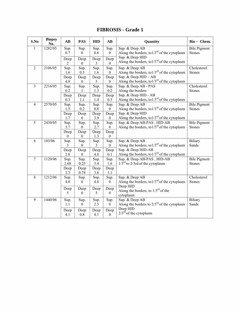
5 2430/05
Deep 0
Deep 0
Deep 1.3
Deep 0
Sup. & Deep AB-PAS , HID-AB Along the borders, to1/3rd of the cytoplasm
Bile Pigment Stones
Sup. 3
Sup. 0
Sup. 3
Sup. 0
6 103/06
Deep 2.8
Deep 0
Deep 4.8
Deep 0.1
Sup. & Deep AB Along the borders, to1/3rd of the cytoplasm Sup. & Deep HID-AB Along the borders, to1/3rd of the cytoplasm
Biliary Sands
Sup. 2.68
Sup. 0.25
Sup. 3.4
Sup. 1.6
7 1120/06
Deep 3.3
Deep 0.74
Deep 3.6
Deep 1.1
Sup. & Deep AB-PAS , HID-AB 1/3rd to 2/3rd of the cytoplasm
Bile Pigment Stones
Sup. 4.8
Sup. 0
Sup. 4.8
Sup. 0
8 1212/06
Deep 5
Deep 0
Deep 5
Deep 0
Sup. & Deep AB Along the borders, to1/3rd of the cytoplasm Deep HID Along the borders, to 1/3rd of the cytoplasm
Cholesterol Stones
Sup. 1.1
Sup. 0
Sup. 2.5
Sup. 0
9 1440/06
Deep 4.1
Deep 0.8
Deep 4.1
Deep 0
Sup. & Deep AB Along the borders to 2/3rd of the cytoplasm Deep HID 2/3rd of the cytoplasm
Biliary Sands

73
FIBROSIS - Grade 1 (Conti..)
S.No Biopsy No. AB PAS HID AB Quantity Bio – Chem.
Sup. 0
Sup. 0
Sup. 0
Sup. 0
10 1798/06
Deep 2.3
Deep 0
Deep 1.1
Deep 0
Deep AB 2/3rd of the cytoplasm Sup. & Deep HID 2/3rd of the cytoplasm
Bile Pigment Stones
Sup. 3.5
Sup. 0
Sup. 1.75
Sup. 0
11 2119/06
Deep 0.4
Deep 0
Deep 3.5
Deep 0
Sup. & Deep AB 1/3rd to 2/3rd of the cytoplasm Sup. & Deep HID 2/3rd of the cytoplasm
Cholesterol Stones
Sup. 4
Sup. 0
Sup. 4.4
Sup. 0
12 2654/06
Deep 5
Deep 0
Deep 4.7
Deep 0
Sup. & Deep AB Along the borders, to1/3rd of the cytoplasm Sup. & Deep HID Along the borders, to1/3rd of the cytoplasm
Bile Pigment Stones
Sup. 5
Sup. 0.07
Sup. 4.25
Sup. 0.3
13 2674/06
Deep 0.3
Deep 0.1
Deep 0.4
Deep 0.6
Sup. & Deep AB-PAS Along the borders, to1/3rd of the cytoplasm Sup. & Deep HID-AB Along the borders , to1/3rd of the cytoplasm
Biliary Sands
Sup. 3.6
Sup. 0
Sup. 3.25
Sup. 0.1
14 2945/06
Deep 4.4
Deep 0
Deep 3.6
Deep 3.5
Sup. & Deep AB 1/3rd to 2/3rd of the cytoplasm Deep HID – AB 1/3rd to 2/3rd of the cytoplasm
Cholesterol Stones
Sup. 1.1
Sup. 0.7
Sup. 2.35
Sup. 0
15 3047/06
Deep 2.8
Deep 0.5
Deep 4.8
Deep 0.5
Sup. & Deep AB – PAS Along the borders to 1/3rd of the cytoplasm Sup. & Deep HID Along the borders to 2/3rd of the cytoplasm
Bile Pigment Stones
Sup. 3.5
Sup. 0
Sup. 4.3
Sup. 0
16 5/07
Deep 4.8
Deep 0
Deep 4.9
Deep 0
Sup. & Deep AB Along the borders to 1/3rd of the cytoplasm Sup. & Deep HID 1/3rd to 2/3rd of the cytoplasm
Bile Pigment Stones
Sup. 0.7
Sup. 0
Sup. 2
Sup. 0
17 10/07
Deep 1.8
Deep 0.07
Deep 3.3
Deep 0
Sup. & Deep AB-PAS Along the borders to 1/3rd of the cytoplasm Sup. & Deep HID Along the borders to 1/3rd of the cytoplasm
Bile Pigment Stones
Sup. 4.4
Sup. 0
Sup. 4.55
Sup. 0
18 161/07
Deep 5
Deep 0
Deep 4.61
Deep 0
Deep AB, 2/3rd of the cytoplasm Sup. & Deep HID 2/3rd of the cytoplasm
Cholesterol Stones
Sup. 2
Sup. 0
Sup. 2.75
Sup. 0
19 222/07
Deep 2.6
Deep 0
Deep 4
Deep 0
Sup. & Deep AB 1/3rd to 2/3rd of the cytoplasm Sup. & Deep HID 1/3rd to 2/3rd of the cytoplasm
Bile Pigment Stones

74
FIBROSIS - Grade 2
S. No
Biopsy No. AB PAS HID AB Quantity Bio –
Chemistry Sup. 0.3
Sup. 0
Sup. 0
Sup. 0
1 2497/05
Deep 1.2
Deep 2.2
Deep 0.9
Deep 0
Sup. & Deep AB-PAS, HID-AB 1/3rd to 2/3rd of the cytoplasm
Bile Pigment Stones
Sup. 2.3
Sup. 0.5
Sup. 3.3
Sup. 0
2 2529/05
Deep 1.7
Deep 0.8
Deep 4.1
Deep 0
Sup. & Deep AB-PAS, HID-AB Along the borders
Bile Pigment Stones
Sup. 1.6
Sup. 0
Sup. 3.6
Sup. 0.1
3 260/06
Deep 4.9
Deep 0.1
Deep 4.8
Deep 0.2
Sup. & Deep AB-PAS, HID-AB Along the borders to 1/3rd of the cytoplasm
Bile Pigment Stones
Sup. 4.3
Sup. 1.8
Sup. 5
Sup. 0.7
4 365/06
Deep 4.8
Deep 0.3
Deep 5
Deep 0
Sup. & Deep AB-PAS, HID-AB 2/3rd of the cytoplasm
Cholesterol Stones
Sup. 2.8
Sup. 0.3
Sup. 2.3
Sup. 1.5
5 1031/06
Deep 3.7
Deep 0.4
Deep 0.3
Deep 0.7
Sup. & Deep AB-PAS Along the borders to 1/3rd of the cytoplasm Sup. & Deep HID-AB 1/3rd to 2/3rd of the cytoplasm
Bile Pigment Stones
Sup. 3.8
Sup. 0
Sup. 0
Sup. 5
6 1261/06
Deep 3.8
Deep 0.09
Deep 4.75
Deep 1
Sup. & Deep AB-PAS 2/3rd of the cytoplasm Sup. & Deep HID-AB 2/3rd of the cytoplasm
Cholesterol Stones
Sup. 1.5
Sup. 0
Sup. 3.8
Sup. 0
7 2143/06
Deep 2.3
Deep 0
Deep 3.4
Deep 0
Sup. & Deep AB Along the borders to 1/3rd of the cytoplasm Sup. & Deep HID Along the borders to 1/3rd of the cytoplasm
Biliary Sands
Sup. 0.7
Sup. 0.1
Sup. 2.8
Sup. 0.07
8 66/07
Deep 3.8
Deep 0.2
Deep 3.6
Deep 0.07
Sup. & Deep AB-PAS, HID-AB Along the borders to 1/3rd of the cytoplasm
Bile Pigment Stones
Sup. 1.3
Sup. 0
Sup. 1.9
Sup. 0
9 327/07
Deep 2.8
Deep 0
Deep 2.6
Deep 0
Sup. & Deep AB-PAS, HID-AB Along the borders to 1/3rd of the cytoplasm
Cholesterol Stones

75
FIBROSIS – Grade 3
S.No Biopsy No. AB PAS HID AB Quantity Bio – Chemistry
Sup. 4.3
Sup. 0
Sup. 4.3
Sup. 0
1 1460/05
Deep 4.1
Deep 0
Deep 3.4
Deep 0
Sup. & Deep AB Along the borders to 1/3rd of the cytoplasm Sup. & Deep HID 1/3rd to 2/3rd of the cytoplasm
Bile Pigment Stones
Sup. 0
Sup. 0
Sup. 0
Sup. 0
2 2634/05
Deep 0.75
Deep 0
Deep 1
Deep 0
Deep AB Along the borders Deep HID Along the borders
Bile Pigment Stones
Sup. 5
Sup. 4.25
Sup. 2.6
Sup. 0
3 1254/05
Deep 0
Deep 0.6
Deep 5
Deep 0
Sup. AB-PAS 1/3rd to 2/3rd of the cytoplasm Sup. HID-AB 2/3rd of the cytoplasm
Bile Pigment Stones
Sup. 0.75
Sup. 0
Sup. 1
Sup. 0
4 2069/05
Deep 3.5
Deep 0.5
Deep 5
Deep 0
Sup. & Deep AB 2/3rd of the cytoplasm Sup. & Deep HID 1/3rd to 2/3rd of the cytoplasm
Cholesterol Stones
Sup. 0
Sup. 0
Sup. 1
Sup. 0
5 3153/05
Deep 0.4
Deep 0.5
Deep 4.2
Deep 0.4
Deep AB-PAS Along the borders to 1/3rd of the cytoplasm Sup. & Deep HID-AB Along the borders to 1/3rd of the cytoplasm
Bile Pigment Stones