A guiding map for inflammation - WordPress.com
6
turn stimulate the release of prostaglandins, molecules that mediate the signs and symp- toms of illness (somnolence, fatigue and fever) by acting on the hypothalamus 11 . An impor- tant aspect of mediators of inflammation in the circulation is the activation of the complement system, which mediates microbial opsoniza- tion and killing, and generates inflammatory peptides such as C3a and C5a 12 . Basic elements of resolution Mechanisms that shut down the inflamma- tory response have paramount importance in the return to homeostasis (Fig. 2). Resolution is not simply the elimination of the stressing agent but instead is an active process involving functional reprogramming of cells through ad hoc production of mediators. Several mecha- nisms inhibit inflammation. The cytokine IL-10 suppresses the production of proin- flammatory cytokines 13 and is mainly derived from regulatory T cells. IL-37, a member of the IL-1 family, broadly suppresses inflam- mation, as does the cytokine TGF-b, which is released from monocytes and platelets 14 . Cleaved extracellular domains of cytokine receptors, such as soluble TNFR and IL-1R, serve as decoy receptors and limit inflamma- tion by binding and neutralizing their respec- tive cytokines. Receptor antagonists, such as IL-1Ra, bind IL-1R without inducing an intracellular signal, thus inhibiting the bio- logical activity of the interleukins IL-1a and IL-1b (ref. 15). Complement inhibitors also modulate inflammation 16 , and prostaglan- dins and lipid mediators such as resolvins patterns) or endogenous stress signals (dan- ger-associated molecular patterns) through germline-encoded pattern-recognition recep- tors (PRRs) 3 . PRRs are mainly expressed by myeloid cells, such as monocytes, macro- phages, neutrophils and dendritic cells, but are also expressed by lymphocytes, fibroblasts and epithelial cells 4 . Cellular stimulation triggers inflammatory processes through the release of proinflammatory cytokines and chemokines. The cytokines TNF and IL-1b have autocrine and paracrine effects leading to the local acti- vation of macrophages and neutrophils, but when these cytokines are released in large amounts, they can exert endocrine effects, such as induction of acute-phase proteins in the liver, platelet activation, fever, fatigue and anorexia. Cytokines activate endothelial cells, thus increasing vascular permeability and facil- itating entrance of immune cells into tissues at the site of infection, but they can also lead to capillary leakage, vasodilation and hypoten- sion 5,6 (Fig. 1). The main function of chemo- kines is to recruit additional immune cells to the site of infection 7 ; these cells include neutro- phils, which exert a crucial role in the phagocy- tosis and killing of pathogens 8,9 . The cytokine IFN-g, derived from type 1 helper T cells (T H 1 cells), activates neutrophils, whereas the cyto- kine IL-22, derived from IL-17-producing helper T cells (T H 17 cells) and innate lymphoid cells, acts on epithelial cells and subsequently stimulates the production and release of anti- microbial peptides, including defensins 10 . In the bloodstream, activated monocytes and neutrophils release cytokines, which in I nflammation is viewed as the driving factor in many diseases, including atherosclerosis, cancer, autoimmunity and infections 1 , and it is a major contributor to age-related con- ditions 2 . The classical definition of inflam- mation—comprising rubor (redness), calor (warmth), dolor (pain) and tumor (swelling), as described by Celsus (30 bc–38 ad), and functio laesa (loss of function), as added by Galen (129–210 ad)—has persisted in modern times. Functionally, inflammation is broadly defined as a protective response of the organ- ism to stimulation by invading pathogens or endogenous signals such as damaged cells, thus resulting in the elimination of the initial cause of injury, the clearance of necrotic cells and tissue repair. However, owing to the complex and often simultaneous molecular, immuno- logical and physiological processes involved in the inflammatory response, clearly defin- ing inflammation presents a challenge. Here, we provide a guide to the sequence of events initiated during inflammation (Fig. 1) and the main mechanisms leading to the resolution of inflammation (Fig. 2), and we include an overview of the most important characteristics of the inflammatory process in various tissues and diseases (Table 1). Basic elements of inflammation Inflammation is induced when host cells sense evolutionarily conserved structures on pathogens (pathogen-associated molecular A guiding map for inflammation Mihai G Netea 1,2 , Frances Balkwill 3 , Michel Chonchol 4 , Fabio Cominelli 5 , Marc Y Donath 6 , Evangelos J Giamarellos-Bourboulis 7 , Douglas Golenbock 8 , Mark S Gresnigt 1 , Michael T Heneka 9,10 , Hal M Hoffman 11 , Richard Hotchkiss 12 , Leo A B Joosten 1,13 , Daniel L Kastner 14 , Martin Korte 15 , Eicke Latz 8,10,16 , Peter Libby 17 , Thomas Mandrup-Poulsen 18 , Alberto Mantovani 19 , Kingston H G Mills 20 , Kristen L Nowak 4 , Luke A O'Neill 20 , Peter Pickkers 21 , Tom van der Poll 22 , Paul M Ridker 23,24 , Joost Schalkwijk 25 , David A Schwartz 26 , Britta Siegmund 27 , Clifford J Steer 28 , Herbert Tilg 29 , Jos W M van der Meer 1 , Frank L van de Veerdonk 1 & Charles A Dinarello 1,30 Biologists, physicians and immunologists have contributed to the understanding of the cellular participants and biological pathways involved in inflammation. Here, we provide a general guide to the cellular and humoral contributors to inflammation as well as to the pathways that characterize inflammation in specific organs and tissues. A full list of affiliations can be found at the end of the paper. 826 VOLUME 18 NUMBER 8 AUGUST 2017 NATURE IMMUNOLOGY INFLAMMATION COMMENT
Transcript of A guiding map for inflammation - WordPress.com

turn stimulate the release of prostaglandins, molecules that mediate the signs and symp-toms of illness (somnolence, fatigue and fever) by acting on the hypothalamus11. An impor-tant aspect of mediators of inflammation in the circulation is the activation of the complement system, which mediates microbial opsoniza-tion and killing, and generates inflammatory peptides such as C3a and C5a12.
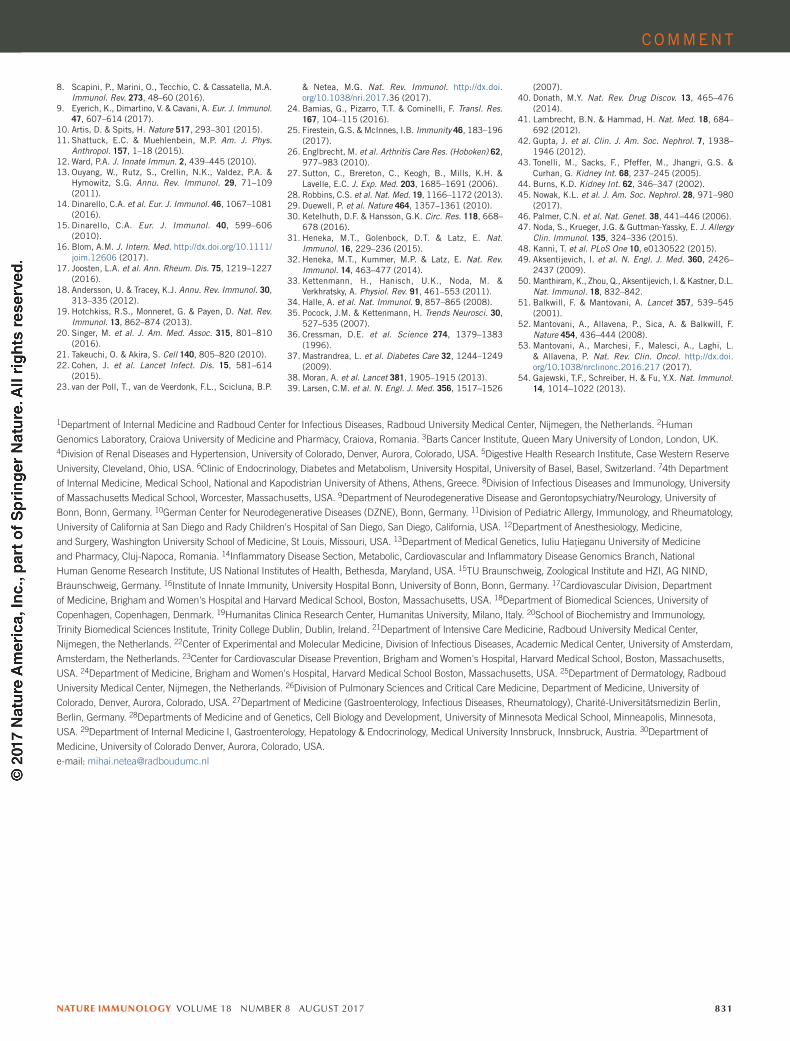
Basic elements of resolutionMechanisms that shut down the inflamma-tory response have paramount importance in the return to homeostasis (Fig. 2). Resolution is not simply the elimination of the stressing agent but instead is an active process involving functional reprogramming of cells through ad hoc production of mediators. Several mecha-nisms inhibit inflammation. The cytokine IL-10 suppresses the production of proin-flammatory cytokines13 and is mainly derived from regulatory T cells. IL-37, a member of the IL-1 family, broadly suppresses inflam-mation, as does the cytokine TGF-b, which is released from monocytes and platelets14. Cleaved extracellular domains of cytokine receptors, such as soluble TNFR and IL-1R, serve as decoy receptors and limit inflamma-tion by binding and neutralizing their respec-tive cytokines. Receptor antagonists, such as IL-1Ra, bind IL-1R without inducing an intracellular signal, thus inhibiting the bio-logical activity of the interleukins IL-1a and IL-1b (ref. 15). Complement inhibitors also modulate inflammation16, and prostaglan-dins and lipid mediators such as resolvins
patterns) or endogenous stress signals (dan-ger-associated molecular patterns) through germline-encoded pattern-recognition recep-tors (PRRs)3. PRRs are mainly expressed by myeloid cells, such as monocytes, macro-phages, neutrophils and dendritic cells, but are also expressed by lymphocytes, fibroblasts and epithelial cells4. Cellular stimulation triggers inflammatory processes through the release of proinflammatory cytokines and chemokines. The cytokines TNF and IL-1b have autocrine and paracrine effects leading to the local acti-vation of macrophages and neutrophils, but when these cytokines are released in large amounts, they can exert endocrine effects, such as induction of acute-phase proteins in the liver, platelet activation, fever, fatigue and anorexia. Cytokines activate endothelial cells, thus increasing vascular permeability and facil-itating entrance of immune cells into tissues at the site of infection, but they can also lead to capillary leakage, vasodilation and hypoten-sion5,6 (Fig. 1). The main function of chemo-kines is to recruit additional immune cells to the site of infection7; these cells include neutro-phils, which exert a crucial role in the phagocy-tosis and killing of pathogens8,9. The cytokine IFN-g, derived from type 1 helper T cells (TH1 cells), activates neutrophils, whereas the cyto-kine IL-22, derived from IL-17-producing helper T cells (TH17 cells) and innate lymphoid cells, acts on epithelial cells and subsequently stimulates the production and release of anti-microbial peptides, including defensins10.
In the bloodstream, activated monocytes and neutrophils release cytokines, which in
Inflammation is viewed as the driving factor in many diseases, including atherosclerosis,
cancer, autoimmunity and infections1, and it is a major contributor to age-related con-ditions2. The classical definition of inflam-mation—comprising rubor (redness), calor (warmth), dolor (pain) and tumor (swelling), as described by Celsus (30 bc–38 ad), and functio laesa (loss of function), as added by Galen (129–210 ad)—has persisted in modern times. Functionally, inflammation is broadly defined as a protective response of the organ-ism to stimulation by invading pathogens or endogenous signals such as damaged cells, thus resulting in the elimination of the initial cause of injury, the clearance of necrotic cells and tissue repair. However, owing to the complex and often simultaneous molecular, immuno-logical and physiological processes involved in the inflammatory response, clearly defin-ing inflammation presents a challenge. Here, we provide a guide to the sequence of events initiated during inflammation (Fig. 1) and the main mechanisms leading to the resolution of inflammation (Fig. 2), and we include an overview of the most important characteristics of the inflammatory process in various tissues and diseases (Table 1).
Basic elements of inflammationInflammation is induced when host cells sense evolutionarily conserved structures on pathogens (pathogen-associated molecular
A guiding map for inflammationMihai G Netea1,2, Frances Balkwill3, Michel Chonchol4, Fabio Cominelli5, Marc Y Donath6, Evangelos J Giamarellos-Bourboulis7, Douglas Golenbock8, Mark S Gresnigt1, Michael T Heneka9,10, Hal M Hoffman11, Richard Hotchkiss12, Leo A B Joosten1,13, Daniel L Kastner14, Martin Korte15, Eicke Latz8,10,16, Peter Libby17, Thomas Mandrup-Poulsen18, Alberto Mantovani19, Kingston H G Mills20, Kristen L Nowak4, Luke A O'Neill20, Peter Pickkers21, Tom van der Poll22, Paul M Ridker23,24, Joost Schalkwijk25, David A Schwartz26, Britta Siegmund27, Clifford J Steer28, Herbert Tilg29, Jos W M van der Meer1, Frank L van de Veerdonk1 & Charles A Dinarello1,30
Biologists, physicians and immunologists have contributed to the understanding of the cellular participants and biological pathways involved in inflammation. Here, we provide a general guide to the cellular and humoral contributors to inflammation as well as to the pathways that characterize inflammation in specific organs and tissues.
A full list of affiliations can be found at the end of
the paper.
826 VOLUME 18 NUMBER 8 AUGUST 2017 NATURE IMMUNOLOGY
INFLAMMAT IONCOMMENT

NATURE IMMUNOLOGY VOLUME 18 NUMBER 8 AUGUST 2017 827
pathogens to the primary site of infection, whereas systemic activation may result in dis-seminated intravascular coagulation, micro-vascular thrombosis and bleeding22 (Table 1). Enhanced adherence of leukocytes and plate-lets to the endothelial surface, and subsequent transmigration, results in vascular inflam-mation and disruption of the endothelial-cell barrier, thus causing leakage of intravascular proteins into the extravascular space, tissue edema and decreased microvascular perfusion. When severe, these abnormalities may lead to organ dysfunction and even death23.
Acute intestinal inflammationAcute intestinal inflammation is protective by eliminating infectious, toxic and other injuri-ous agents, while initiating the process of repair. Chronic inflammation in the gastrointestinal tract results from repeated acute injury and/or impaired resolution of inflammation, thus leading to conditions such as chronic gastritis and peptic ulcer disease, chronic pancreatitis,
vulnerable to secondary infections19.Here, we provide an up-to-date guide to
inflammation and summarize the main fea-tures of acute or chronic inflammation in spe-cific human diseases (Table 1).
SepsisIn sepsis, the release of danger-associated molecular patterns or alarmins from injured host cells activates PRRs, which also recog-nize pathogen-associated molecular patterns, thus giving rise to a vicious cycle of sustained hyperinflammation20. However, an ineffective antimicrobial host defense often accompanies this condition. Proinflammatory cytokines produced after recognition of the invading pathogens by PRRs21 can help protect the host but also can promote tissue injury during overwhelming sepsis. Unrestrained activation of complement contributes to organ failure, and C5a blockade improves the outcome of experimental sepsis. Local activation of the coagulation system in sepsis helps to confine
exert negative feedback loops by suppressing the transcription and release of cytokines. Acute-phase proteins induced during inflam-mation, such as a-1 antitrypsin, have broad anti-inflammatory properties17. Additional anti-inflammatory mechanisms involve stress hormones, particularly corticosteroids and catecholamines; negative regulators of Toll-like-receptor signaling, such as IRAK-M and A20; and microRNAs, such as miR-146 or miR-125. Neuroimmunoregulatory mecha-nisms (the so-called immunological reflex) provide anti-inflammatory negative feedback, which is triggered by peripheral sensory input transmitted through the afferent vagus nerve to the brainstem and is followed by activation of the efferent vagus and splenic nerve18, release of norepinephrine in the spleen and secretion of acetylcholine by a subset of CD4+ T cells, thereby inhibiting production of proinflam-matory cytokines by macrophages. However, an anti-inflammatory response that is too pronounced or persistent may render the host
Pathogens/PAMPs
Cellrecruitment
Mφ
Mφ
AMPs
CoagulationPlatelets
Endothelialactivation
Liver
TH1
TH17
ROS
C1 C3 C5 MAC
MBL
C3a C5a
Complement system
Tissue damageHost
defense
Epithelialhost defense
Acute-phaseproteins:• AAT• CRP• Ferritin
IL-6TNFIL-1
IL-1α(DAMPs)
• Fever• Somnolence• Anorexia• Pain
Hypothalamus
TNFIL-1IL-6
IL-22
IL-1IL-23
IL-12IL-18
Phagocytosisand killing
IFN-γIL-8
CCL2CCL3
TNFIL-1
IL-17
Figure 1 The immunological mechanisms leading to the induction of inflammation during the first stages of host defense against invading pathogens. AAT, a1-antitrypsin; PAMPs, pathogen-associated molecular patterns; AMPs, antimicrobial peptides; DAMPs, danger-associated molecular patterns; MAC, membrane attack complex; ROS, reactive oxygen species; CRP, C-reactive protein; MBL, mannose-binding lectin; Mf, macrophage or monocyte; CCL, C-C-motif chemokine ligand.
Deb
bie
Mai
zels
/Spr
inge
r Nat
ure
COMMENT
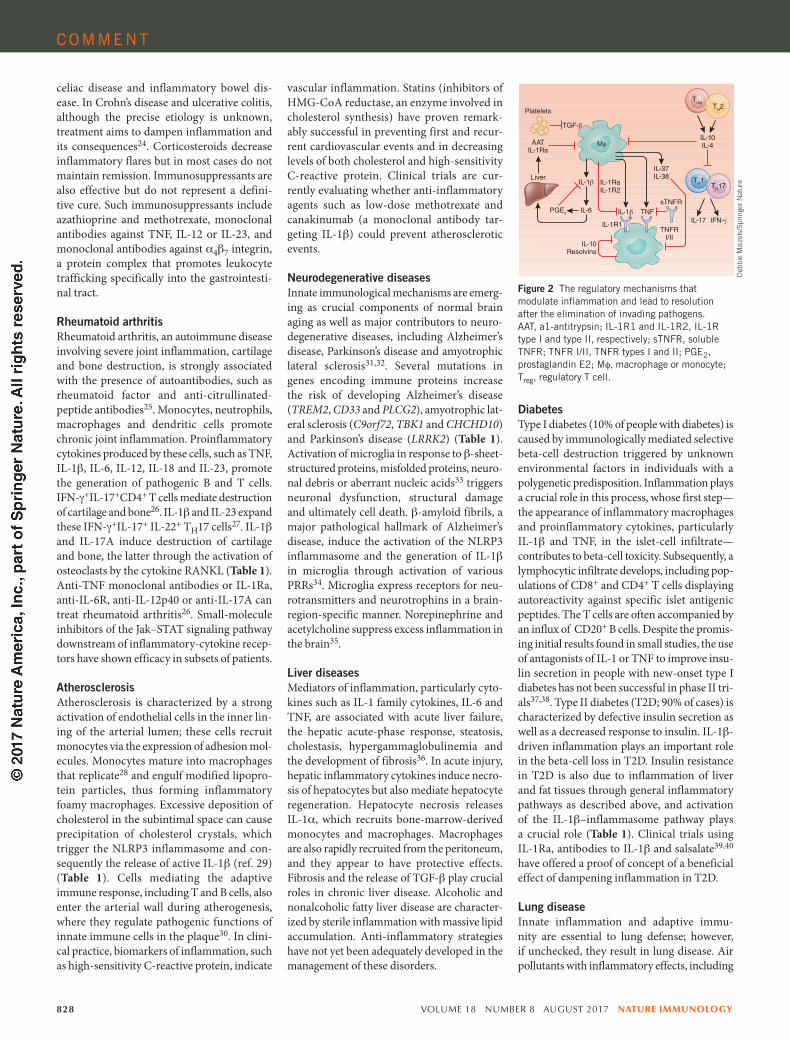
DiabetesType I diabetes (10% of people with diabetes) is caused by immunologically mediated selective beta-cell destruction triggered by unknown environmental factors in individuals with a polygenetic predisposition. Inflammation plays a crucial role in this process, whose first step—the appearance of inflammatory macrophages and proinflammatory cytokines, particularly IL-1b and TNF, in the islet-cell infiltrate—contributes to beta-cell toxicity. Subsequently, a lymphocytic infiltrate develops, including pop-ulations of CD8+ and CD4+ T cells displaying autoreactivity against specific islet antigenic peptides. The T cells are often accompanied by an influx of CD20+ B cells. Despite the promis-ing initial results found in small studies, the use of antagonists of IL-1 or TNF to improve insu-lin secretion in people with new-onset type I diabetes has not been successful in phase II tri-als37,38. Type II diabetes (T2D; 90% of cases) is characterized by defective insulin secretion as well as a decreased response to insulin. IL-1b-driven inflammation plays an important role in the beta-cell loss in T2D. Insulin resistance in T2D is also due to inflammation of liver and fat tissues through general inflammatory pathways as described above, and activation of the IL-1b–inflammasome pathway plays a crucial role (Table 1). Clinical trials using IL-1Ra, antibodies to IL-1b and salsalate39,40 have offered a proof of concept of a beneficial effect of dampening inflammation in T2D.
Lung diseaseInnate inflammation and adaptive immu-nity are essential to lung defense; however, if unchecked, they result in lung disease. Air pollutants with inflammatory effects, including
vascular inflammation. Statins (inhibitors of HMG-CoA reductase, an enzyme involved in cholesterol synthesis) have proven remark-ably successful in preventing first and recur-rent cardiovascular events and in decreasing levels of both cholesterol and high-sensitivity C-reactive protein. Clinical trials are cur-rently evaluating whether anti-inflammatory agents such as low-dose methotrexate and canakinumab (a monoclonal antibody tar-geting IL-1b) could prevent atherosclerotic events.
Neurodegenerative diseasesInnate immunological mechanisms are emerg-ing as crucial components of normal brain aging as well as major contributors to neuro-degenerative diseases, including Alzheimer’s disease, Parkinson’s disease and amyotrophic lateral sclerosis31,32. Several mutations in genes encoding immune proteins increase the risk of developing Alzheimer’s disease (TREM2, CD33 and PLCG2), amyotrophic lat-eral sclerosis (C9orf72, TBK1 and CHCHD10) and Parkinson’s disease (LRRK2) (Table 1). Activation of microglia in response to b-sheet-structured proteins, misfolded proteins, neuro-nal debris or aberrant nucleic acids33 triggers neuronal dysfunction, structural damage and ultimately cell death. b-amyloid fibrils, a major pathological hallmark of Alzheimer’s disease, induce the activation of the NLRP3 inflammasome and the generation of IL-1b in microglia through activation of various PRRs34. Microglia express receptors for neu-rotransmitters and neurotrophins in a brain-region-specific manner. Norepinephrine and acetylcholine suppress excess inflammation in the brain35.
Liver diseasesMediators of inflammation, particularly cyto-kines such as IL-1 family cytokines, IL-6 and TNF, are associated with acute liver failure, the hepatic acute-phase response, steatosis, cholestasis, hypergammaglobulinemia and the development of fibrosis36. In acute injury, hepatic inflammatory cytokines induce necro-sis of hepatocytes but also mediate hepatocyte regeneration. Hepatocyte necrosis releases IL-1a, which recruits bone-marrow-derived monocytes and macrophages. Macrophages are also rapidly recruited from the peritoneum, and they appear to have protective effects. Fibrosis and the release of TGF-b play crucial roles in chronic liver disease. Alcoholic and nonalcoholic fatty liver disease are character-ized by sterile inflammation with massive lipid accumulation. Anti-inflammatory strategies have not yet been adequately developed in the management of these disorders.
celiac disease and inflammatory bowel dis-ease. In Crohn’s disease and ulcerative colitis, although the precise etiology is unknown, treatment aims to dampen inflammation and its consequences24. Corticosteroids decrease inflammatory flares but in most cases do not maintain remission. Immunosuppressants are also effective but do not represent a defini-tive cure. Such immunosuppressants include azathioprine and methotrexate, monoclonal antibodies against TNF, IL-12 or IL-23, and monoclonal antibodies against a4b7 integrin, a protein complex that promotes leukocyte trafficking specifically into the gastrointesti-nal tract.
Rheumatoid arthritisRheumatoid arthritis, an autoimmune disease involving severe joint inflammation, cartilage and bone destruction, is strongly associated with the presence of autoantibodies, such as rheumatoid factor and anti-citrullinated-peptide antibodies25. Monocytes, neutrophils, macrophages and dendritic cells promote chronic joint inflammation. Proinflammatory cytokines produced by these cells, such as TNF, IL-1b, IL-6, IL-12, IL-18 and IL-23, promote the generation of pathogenic B and T cells. IFN-g+IL-17+CD4+ T cells mediate destruction of cartilage and bone26. IL-1b and IL-23 expand these IFN-g+IL-17+ IL-22+ TH17 cells27. IL-1b and IL-17A induce destruction of cartilage and bone, the latter through the activation of osteoclasts by the cytokine RANKL (Table 1). Anti-TNF monoclonal antibodies or IL-1Ra, anti-IL-6R, anti-IL-12p40 or anti-IL-17A can treat rheumatoid arthritis26. Small-molecule inhibitors of the Jak–STAT signaling pathway downstream of inflammatory-cytokine recep-tors have shown efficacy in subsets of patients.
AtherosclerosisAtherosclerosis is characterized by a strong activation of endothelial cells in the inner lin-ing of the arterial lumen; these cells recruit monocytes via the expression of adhesion mol-ecules. Monocytes mature into macrophages that replicate28 and engulf modified lipopro-tein particles, thus forming inflammatory foamy macrophages. Excessive deposition of cholesterol in the subintimal space can cause precipitation of cholesterol crystals, which trigger the NLRP3 inflammasome and con-sequently the release of active IL-1b (ref. 29) (Table 1). Cells mediating the adaptive immune response, including T and B cells, also enter the arterial wall during atherogenesis, where they regulate pathogenic functions of innate immune cells in the plaque30. In clini-cal practice, biomarkers of inflammation, such as high-sensitivity C-reactive protein, indicate
828 VOLUME 18 NUMBER 8 AUGUST 2017 NATURE IMMUNOLOGY
Liver
Treg
Platelets
sTNFRTNF
Mφ
TH2
TH1 TH17
TGF-β
IL-10IL-4
IL-37IL-38
IFN-γIL-17TNFRI/II
IL-1R1
IL-1β
IL-1β
IL-10Resolvins
PGE2
AATIL-1Ra
IL-1RaIL-1R2
IL-6
Figure 2 The regulatory mechanisms that modulate inflammation and lead to resolution after the elimination of invading pathogens. AAT, a1-antitrypsin; IL-1R1 and IL-1R2, IL-1R type I and type II, respectively; sTNFR, soluble TNFR; TNFR I/II, TNFR types I and II; PGE2, prostaglandin E2; Mf, macrophage or monocyte; Treg, regulatory T cell.
Deb
bie
Mai
zels
/Spr
inge
r Nat
ure
COMMENT

NATURE IMMUNOLOGY VOLUME 18 NUMBER 8 AUGUST 2017 829
Table 1 Specific characteristics of inflammation in various tissues and diseasesDisease/tissue Main characteristics of inflammation Main pathways/markers Specific complications Immunotherapy
Sepsis • Exaggerated inflammation and inappropriate endothelial activation combined with immunoparalysis
• Increased cytokines/acute-phase proteins
• Activation of complement, coagulation and endothelial cells
• Septic shock
• Multiple-organ failure
• Opportunistic infections
• Personalized immunotherapy: in hyperinflammation, IL-1Ra, anti-C5a; in immunoparalysis, rIFN-g, GM-CSF, anti-PD1, rIL7
Inflammation of the gastrointestinal tract
• Permanent structural and functional alterations
• Ulcers, strictures, fistulas
• Disturbed motility and barrier function
• Increased circulating cytokines and acute-phase proteins
• Decreased neutrophil function
• Peptic ulcer disease
• Chronic pancreatitis
• Celiac disease
• Crohn’s disease
• Ulcerative colitis
• Corticosteroids
• Antibodies to TNF, IL-12, IL-23 or integrin a4b7
Rheumatoid arthritis • Autoantibodies/immune complexes
• Proinflammatory cytokines
• Macrophage influx
• Pathogenic T and B cells
• TNF, IL-1b, IL-6, IL-12, IL-18 and IL-23
• IFN-g+IL-17+IL-22+ TH17 cells
• RANKL
• Anti–citrullinated peptides
• Joint inflammation
• Cartilage destruction
• Anti-TNF, IL-1Ra, Anti-IL-6R, Anti-IL-12p40, Anti-IL-17A
• JAK–STAT inhibitors
Atherosclerosis • Dyslipidemia and cholesterol deposition
• Monocyte and lymphocyte influx in intima
• Activation of inflammasomes and cytokines
• Inflammasome and cytokines
• hsCRP
• Angina pectoris
• Acute myocardial infarction
• Stroke
• Statins, including methotrexate and anti-IL-1b (canakinumab) currently in trials
Neurodegenerative diseases
• Peripheral infection/inflammation–induced activation of microglial cells
• b-amyloid fibrils
• TREM2, CD33, PLCG2• LRRK2, C9orf72, TBK1, CHCHD10
• Activation of inflammasomes and IL-1b
• Alzheimer’s disease
• Parkinson’s disease
• Amyotrophic lateral sclerosis
• Not yet available
Liver disease • Acute liver failure
• Hepatic acute-phase response
• Steatosis
• Cholestasis, hypergamma - globulinemia, fibrosis
• IL-1a and other proinflammatory cytokines
• TGF-b for fibrosis
• Acute and chronic hepatitis
• Nonalcoholic fatty liver disease
• Cirrhosis
• Not yet available
Diabetes • Infiltration of pancreatic islets with innate and adaptive immune cells and beta-cell apoptosis in T1D
• Low-grade innate inflammation in adipose tissue, liver and islets; insulin resistance and beta-cell apoptosis in T2D
• Proinflammatory cytokines IL-1b and TNF
• In T1D, also T cell–mediated beta-cell killing
•Macrovascular com-plications (myocardial infarction, stroke, clau-dication)
•Microvascular complications (kidney, ocular, neuronal)
• Anti-IL-1 (anakinra, canakinumab)
• Anti-TNF
Lung disease • Inflammation and hyper-reactivity
• Fibrosis
• TH2 and IL-4/IL-5/IL-13 allergic responses (asthma)
•Polymorphonuclear leukocyte and macrophage infiltrate, cytokines (COPD)
• TGF, integrin avb6, platelet-derived growth factor b (idiopathic pulmonary fibrosis)
• Asthma
• COPD
• Idiopathic pulmonary fibrosis
• Corticosteroids
• Anti-IL5
Chronic kidney disease
• Low-grade inflammation • NLRP3 inflammasome, IL1b, IL-6, PGE2, TGF-b
• Kidney insufficiency • IL-1Ra (anakinra)
• IL-1 soluble receptor (rilonacept)
Inflammatory skin diseases
• Inflammation with exaggerated TH2 (Alzheimer’s disease) or TH17 (psoriasis)
• Inflammation in apocrine glands (HS)
• TH17, TH2, antimicrobial peptides
• TH2, filaggrin
• IL-1b and TNF (HS)
• Psoriasis
• Atopic dermatitis
• HS
• Antibodies to TNF, IL-17, IL-17R, IL-23 (psoriasis)
• Anti-TNF and anti-IL-1 (HS)
Autoinflammatory syndromes (e.g., deficiency of IL-1Ra, FMF, HIDS, cryopyrin-associated periodic syndrome)
• Sterile inflammation in joints and peritoneum, fever, systemic inflammation
• Inflammasome/IL-1b pathway
IL-1/IL-1Ra balance
• NF-kB perturbations
• Type I IFN production
• Amyloid deposition (FMF)
• Anti-IL-1 therapies (anakinra, canakinumab, gevokizumab, rilonacept)
• TNF inhibitors
• JAK–STAT inhibitors
Cancer-related inflammation
• Infiltration of tumor-associated macrophages with strong immunosuppressive activity
• M2 macrophage phenotype
• Checkpoint proteins PD-1, PD-L1 and CTLA-4
• IL-1b, IL-6, TNF, IL-4, IL-10 and TGF-b
• Pentraxin-3
• T cell exhaustion and anergy
• Tumor progression
• Checkpoint blockade: antibodies to PD-1, PD-L1 and CTLA-4
• Immunostimulatory: BCG, muramyl dipeptide (mifamurtide), b-glucan
FMF, familial Mediterranean fever; HIDS, hyperimmunoglobulinemia D and periodic fever syndrome; HS, hidradenitis suppurativa; hsCRP, high-sensitivity C-reactive protein; T1D, type I diabetes; COPD, chronic obstructive pulmonary disease; BCG, Mycobacterium bovis bacillus Calmette–Guerin; GM-CSF, granulocyte-macrophage colony-stimulating factor; r, recombinant.
COMMENT
830 VOLUME 18 NUMBER 8 AUGUST 2017 NATURE IMMUNOLOGY
components and functions downstream of IL-1 in mouse models of carcinogenesis (Table 1). The type of inflammatory reaction determines the clinical severity of cancer. T cell–driven inflammation, characterized by an interferon signature, is associated with a better progno-sis54, whereas high macrophage infiltration is generally associated with poorer prognosis, especially when markers of type 2 polarization are considered53.
Conclusions and future perspectivesThe heterogeneous nature of the inflammatory response depends on the type of disease and the organ in which it occurs, and inflamma-tion can have both protective effects and col-lateral deleterious consequences for the host. The examples of successful therapies target-ing inflammation underscore the importance of understanding inflammatory pathways to enable further therapeutic advances.
ACKNOWLEDGMENTSWe thank all our colleagues in the field for their contributions to knowledge of inflammation. D.L.K. was supported by the Intramural Research Program of the National Human Genome Research Institute (NHGRI) at the US National Institutes of Health. M.G.N. was supported by an ERC Consolidator Grant (no. 310372), a Spinoza Grant from the Netherlands Organization for Scientific Research and a Competitiveness Operational Programme Grant from the Romanian Ministry of European Funds (FUSE). K.L.N. was supported by American Heart Association postdoctoral fellowship award 12POST11920023. F.C. was supported by NIH grants DK042191, DK055812, DK091222 and DK097948. F.B. was supported by an ERC Advanced Grant (ERC322566) and a Cancer Research UK Programme Grant (A16354). C.A.D. was supported by NIH grant AI15614. L.A.J. was supported by a Competitiveness Operational Programme grant from the Romanian Ministry of European Funds (HINT, ID P_37_762; MySMIS 103587) and a Dutch Arthritis Foundation grant (NR-12-2-303). K.H.G.M. was supported by grants from Science Foundation Ireland. P.L. was supported by the RRM Charitable Fund and The National Heart, Lung, and Blood Institute (R01 HL080472). B.S. was supported by the German Research Foundation SPP1656, 749/7-1, 749/10-1, the German Cancer Foundation, the German Israel Foundation and the Horizon 2020 program. D.A.S. was supported by NIH grant R01-HL097163. A.M. was supported by ERC, AIRC and Fondazione Cariplo.
COMPETING FINANCIAL INTERESTSThe authors declare no competing financial interests.
1. Eltzschig, H.K. & Carmeliet, P. N. Engl. J. Med. 364, 656–665 (2011).
2. Franceschi, C. & Campisi, J. J. Gerontol. A Biol. Sci. Med. Sci. 69 (Suppl. 1), S4–S9 (2014).
3. Akira, S., Uematsu, S. & Takeuchi, O. Cell 124, 783–801 (2006).
4. Iwasaki, A. & Medzhitov, R. Nat. Immunol. 5, 987–995 (2004).
5. Dinarello, C.A. Eur. J. Immunol. 37 (Suppl. 1), S34–S45 (2007).
6. Dinarello, C.A. Curr. Top. Microbiol. Immunol. 216, 133–165 (1996).
7. Bonecchi, R. et al. Front. Biosci. (Landmark Ed.) 14, 540–551 (2009).
involves systemic inflammatory responses, and frequent comorbidities are rheumatological or cardiovascular in nature47. In hidradenitis sup-purativa, also known as acne inversa, a devas-tating skin disorder bearing characteristics of both autoinflammatory and autoimmune dis-orders, histopathology reveals heavy lesional deposits of TNF, IL-1b, IL-23 and IL-17, as well as activation of both CD4+ and CD8+ T cells48.
Autoinflammatory syndromesAutoinflammatory syndromes can be defined as disorders with abnormally increased inflam-mation, which are mediated predominantly by the cells and molecules of the innate immune system, with a substantial host predisposition. The enhanced inflammatory state encom-passes the production of proinflammatory pyrogenic cytokines, particularly IL-1b, and hence fever and acute-phase responses are common prominent signs. From a theoreti-cal viewpoint, autoinflammatory syndromes result from excessive production and/or bio-logical activity of inflammatory mediators, or from a lack of endogenous inhibition. The prototypical condition associated with the lat-ter is deficiency of IL-1Ra, in which a lack of inhibition of IL-1 bioactivity leads to excessive inflammation49. Other important syndromes are familial Mediterranean fever, cryopyrinop-athies, and hyperimmunoglobulinemia D and periodic fever syndrome. An exaggerated IL-1b response is a hallmark of many autoinflamma-tory disorders, for which interference with IL-1 action is the preferred therapy50.
CancerAll the usual components of the inflammatory response reside in the tumor microenviron-ment but often exhibit ‘corrupted’ functions. Cancer-related inflammation is a key com-ponent of the tumor microenvironment51,52 and includes inflammatory cells, especially tumor-associated macrophages, which affect all aspects of cancer including growth, genetic instability, angiogenesis and metastasis53. Tumor-associated macrophages contribute to cancer immunosuppression by produc-ing prostaglandins, products of tryptophan metabolism, and by expressing checkpoint-blockade triggers (such as PD-L1). T and B cells, neutrophils, mast cells and eosinophils are also cellular components of cancer-related inflammation. Inflammatory cytokines such as TNF, IL-6 and IL-1 are important mediators of intercellular communication in cancer-related inflammation, along with many other mem-bers of the chemokine family51,52. The humoral arm of innate immunity also participates in cancer-related inflammation. Pentraxin-3, a fluid-phase PRR, interacts with complement
endotoxin, can exacerbate asthma but can also initiate the disease. Whereas IL-4 and dendritic cells promote inflammation mediated by type 2 helper T cells (TH2 cells), airway epithelia produce IL-1a, IL-1b, IL-25, IL-33 and thymic stromal lymphopoetin, each of which recruits and activates ILC2 cells, eosinophils and baso-phils, thereby enhancing inflammation and remodeling of the airway41. The cytokines IL-4 and IL-13 contribute to the interaction between innate and adaptive immunologi-cal mechanisms that promote inflammatory airway disease. IL-5 promotes differentiation and activation of eosinophils, and clinical tri-als targeting the IL-5 pathway by blocking the cytokine or its receptor in asthma have indi-cated benefits in people with high TH2 profiles.
Chronic kidney diseaseChronic kidney disease is a low-grade inflam-matory process. Inflammatory macrophages infiltrate the kidney and induce the release of proinflammatory cytokines and mediators such as IL-1b, TNF, IL-6, IL-23, reactive oxygen species, nitric oxide and inducible nitric oxide synthase. Cytokines such as TNF or TGF-b1 produced locally during kidney inflammation decrease kidney expression of the nephropro-tective proteins Klotho and PGC-1a and lead to suboptimal induction of these proteins. Circulating IL-1b, IL-1Ra, IL-6 and C-reactive protein are elevated in people with advanced stages of chronic kidney disease42, thus predict-ing a decline in kidney function43. IL-1b con-tributes to tubular interstitial fibrosis, promotes tubular epithelial–myofibroblast transdifferen-tiation, cytokine gene expression, production of prostaglandin E2 by mesangial cells and interstitial fibrosis mediated by TGF-b (ref. 44). Treatment with the IL-1 soluble receptor trap (rilonacept) decreases concentrations of C-reactive protein, improves dilation mediated by brachial artery flow and decreases vascular oxidative stress in people with chronic kidney disease45.
Inflammatory skin diseasesIn atopic dermatitis, penetration of external stimuli (for example, allergens) through an impaired skin barrier leads to an exaggerated TH2 response46. Local immunological imbal-ance causes further skin-barrier deterioration as IL-4 and IL-13 downregulate the expression of major skin-barrier genes such as filaggrins, thus leading to a vicious circle. In psoriasis, a T cell–driven disease with contributions of innate and adaptive immunity, inflammation is driven by signaling through NF-kB and TH17- and TH1-type cytokines, and therapies targeting TNF, IL-12/IL-23 and IL-17 have been found to be effective (Table 1). Psoriasis
COMMENT
NATURE IMMUNOLOGY VOLUME 18 NUMBER 8 AUGUST 2017 831
(2007).40. Donath, M.Y. Nat. Rev. Drug Discov. 13, 465–476
(2014).41. Lambrecht, B.N. & Hammad, H. Nat. Med. 18, 684–
692 (2012).42. Gupta, J. et al. Clin. J. Am. Soc. Nephrol. 7, 1938–
1946 (2012).43. Tonelli, M., Sacks, F., Pfeffer, M., Jhangri, G.S. &
Curhan, G. Kidney Int. 68, 237–245 (2005).44. Burns, K.D. Kidney Int. 62, 346–347 (2002).45. Nowak, K.L. et al. J. Am. Soc. Nephrol. 28, 971–980
(2017).46. Palmer, C.N. et al. Nat. Genet. 38, 441–446 (2006).47. Noda, S., Krueger, J.G. & Guttman-Yassky, E. J. Allergy
Clin. Immunol. 135, 324–336 (2015).48. Kanni, T. et al. PLoS One 10, e0130522 (2015).49. Aksentijevich, I. et al. N. Engl. J. Med. 360, 2426–
2437 (2009).50. Manthiram, K., Zhou, Q., Aksentijevich, I. & Kastner, D.L.
Nat. Immunol. 18, 832–842.51. Balkwill, F. & Mantovani, A. Lancet 357, 539–545
(2001).52. Mantovani, A., Allavena, P., Sica, A. & Balkwill, F.
Nature 454, 436–444 (2008).53. Mantovani, A., Marchesi, F., Malesci, A., Laghi, L.
& Allavena, P. Nat. Rev. Clin. Oncol. http://dx.doi.org/10.1038/nrclinonc.2016.217 (2017).
54. Gajewski, T.F., Schreiber, H. & Fu, Y.X. Nat. Immunol. 14, 1014–1022 (2013).
& Netea, M.G. Nat. Rev. Immunol. http://dx.doi.org/10.1038/nri.2017.36 (2017).
24. Bamias, G., Pizarro, T.T. & Cominelli, F. Transl. Res. 167, 104–115 (2016).
25. Firestein, G.S. & McInnes, I.B. Immunity 46, 183–196 (2017).
26. Englbrecht, M. et al. Arthritis Care Res. (Hoboken) 62, 977–983 (2010).
27. Sutton, C., Brereton, C., Keogh, B., Mills, K.H. & Lavelle, E.C. J. Exp. Med. 203, 1685–1691 (2006).
28. Robbins, C.S. et al. Nat. Med. 19, 1166–1172 (2013).29. Duewell, P. et al. Nature 464, 1357–1361 (2010).30. Ketelhuth, D.F. & Hansson, G.K. Circ. Res. 118, 668–
678 (2016).31. Heneka, M.T., Golenbock, D.T. & Latz, E. Nat.
Immunol. 16, 229–236 (2015).32. Heneka, M.T., Kummer, M.P. & Latz, E. Nat. Rev.
Immunol. 14, 463–477 (2014).33. Kettenmann, H., Hanisch, U.K., Noda, M. &
Verkhratsky, A. Physiol. Rev. 91, 461–553 (2011).34. Halle, A. et al. Nat. Immunol. 9, 857–865 (2008).35. Pocock, J.M. & Kettenmann, H. Trends Neurosci. 30,
527–535 (2007).36. Cressman, D.E. et al. Science 274, 1379–1383
(1996).37. Mastrandrea, L. et al. Diabetes Care 32, 1244–1249
(2009).38. Moran, A. et al. Lancet 381, 1905–1915 (2013).39. Larsen, C.M. et al. N. Engl. J. Med. 356, 1517–1526
8. Scapini, P., Marini, O., Tecchio, C. & Cassatella, M.A. Immunol. Rev. 273, 48–60 (2016).
9. Eyerich, K., Dimartino, V. & Cavani, A. Eur. J. Immunol. 47, 607–614 (2017).
10. Artis, D. & Spits, H. Nature 517, 293–301 (2015).11. Shattuck, E.C. & Muehlenbein, M.P. Am. J. Phys.
Anthropol. 157, 1–18 (2015).12. Ward, P.A. J. Innate Immun. 2, 439–445 (2010).13. Ouyang, W., Rutz, S., Crellin, N.K., Valdez, P.A. &
Hymowitz, S.G. Annu. Rev. Immunol. 29, 71–109 (2011).
14. Dinarello, C.A. et al. Eur. J. Immunol. 46, 1067–1081 (2016).
15. Dinarello, C.A. Eur. J. Immunol. 40, 599–606 (2010).
16. Blom, A.M. J. Intern. Med. http://dx.doi.org/10.1111/joim.12606 (2017).
17. Joosten, L.A. et al. Ann. Rheum. Dis. 75, 1219–1227 (2016).
18. Andersson, U. & Tracey, K.J. Annu. Rev. Immunol. 30, 313–335 (2012).
19. Hotchkiss, R.S., Monneret, G. & Payen, D. Nat. Rev. Immunol. 13, 862–874 (2013).
20. Singer, M. et al. J. Am. Med. Assoc. 315, 801–810 (2016).
21. Takeuchi, O. & Akira, S. Cell 140, 805–820 (2010).22. Cohen, J. et al. Lancet Infect. Dis. 15, 581–614
(2015).23. van der Poll, T., van de Veerdonk, F.L., Scicluna, B.P.
1Department of Internal Medicine and Radboud Center for Infectious Diseases, Radboud University Medical Center, Nijmegen, the Netherlands. 2Human
Genomics Laboratory, Craiova University of Medicine and Pharmacy, Craiova, Romania. 3Barts Cancer Institute, Queen Mary University of London, London, UK. 4Division of Renal Diseases and Hypertension, University of Colorado, Denver, Aurora, Colorado, USA. 5Digestive Health Research Institute, Case Western Reserve
University, Cleveland, Ohio, USA. 6Clinic of Endocrinology, Diabetes and Metabolism, University Hospital, University of Basel, Basel, Switzerland. 74th Department
of Internal Medicine, Medical School, National and Kapodistrian University of Athens, Athens, Greece. 8Division of Infectious Diseases and Immunology, University
of Massachusetts Medical School, Worcester, Massachusetts, USA. 9Department of Neurodegenerative Disease and Gerontopsychiatry/Neurology, University of
Bonn, Bonn, Germany. 10German Center for Neurodegenerative Diseases (DZNE), Bonn, Germany. 11Division of Pediatric Allergy, Immunology, and Rheumatology,
University of California at San Diego and Rady Children's Hospital of San Diego, San Diego, California, USA. 12Department of Anesthesiology, Medicine,
and Surgery, Washington University School of Medicine, St Louis, Missouri, USA. 13Department of Medical Genetics, Iuliu Hațieganu University of Medicine
and Pharmacy, Cluj-Napoca, Romania. 14Inflammatory Disease Section, Metabolic, Cardiovascular and Inflammatory Disease Genomics Branch, National
Human Genome Research Institute, US National Institutes of Health, Bethesda, Maryland, USA. 15TU Braunschweig, Zoological Institute and HZI, AG NIND,
Braunschweig, Germany. 16Institute of Innate Immunity, University Hospital Bonn, University of Bonn, Bonn, Germany. 17Cardiovascular Division, Department
of Medicine, Brigham and Women's Hospital and Harvard Medical School, Boston, Massachusetts, USA. 18Department of Biomedical Sciences, University of
Copenhagen, Copenhagen, Denmark. 19Humanitas Clinica Research Center, Humanitas University, Milano, Italy. 20School of Biochemistry and Immunology,
Trinity Biomedical Sciences Institute, Trinity College Dublin, Dublin, Ireland. 21Department of Intensive Care Medicine, Radboud University Medical Center,
Nijmegen, the Netherlands. 22Center of Experimental and Molecular Medicine, Division of Infectious Diseases, Academic Medical Center, University of Amsterdam,
Amsterdam, the Netherlands. 23Center for Cardiovascular Disease Prevention, Brigham and Women's Hospital, Harvard Medical School, Boston, Massachusetts,
USA. 24Department of Medicine, Brigham and Women's Hospital, Harvard Medical School Boston, Massachusetts, USA. 25Department of Dermatology, Radboud
University Medical Center, Nijmegen, the Netherlands. 26Division of Pulmonary Sciences and Critical Care Medicine, Department of Medicine, University of
Colorado, Denver, Aurora, Colorado, USA. 27Department of Medicine (Gastroenterology, Infectious Diseases, Rheumatology), Charité-Universitätsmedizin Berlin,
Berlin, Germany. 28Departments of Medicine and of Genetics, Cell Biology and Development, University of Minnesota Medical School, Minneapolis, Minnesota,
USA. 29Department of Internal Medicine I, Gastroenterology, Hepatology & Endocrinology, Medical University Innsbruck, Innsbruck, Austria. 30Department of
Medicine, University of Colorado Denver, Aurora, Colorado, USA.
e-mail: [email protected]
COMMENT