A Guide to the Sea Urchin Reproductive Cycle and … · By Philip James and Sten Siikavuopio A...
20
By Philip James and Sten Siikavuopio A Guide to the Sea Urchin Reproductive Cycle and Staging Sea Urchin Gonad Samples
Transcript of A Guide to the Sea Urchin Reproductive Cycle and … · By Philip James and Sten Siikavuopio A...
By Philip James and Sten Siikavuopio
A Guide to the Sea Urchin
Reproductive Cycle and Staging Sea Urchin
Gonad Samples
This publication is intended to be used as a guide for
anybody with a need to understand, or an interest, in the
reproductive cycle of sea urchins. It includes the follow-
ing: a description of the reproductive cycle of sea urchins;
the factors that cause variations in the size and quality
of the gonad; methods for sampling sea urchin gonads;
and a guide to reading the histology slides of sea urchin
gonads in order to be able to classify them into the dif-
ferent stages of the reproductive cycle. In order to further
assist with this process Appendix One shows examples of
histology slides taken from the two populations described
in Figure 2. These clearly show the general pattern associ-
ated with the reproductive cycle of sea urchins but also
the enormous variation that can occur between popula-
tions. The authors hope that this guide will enable those
working with sea urchins to follow the reproductive cycle
of selected populations and that it will contribute signifi-
cantly to the knowledge of the reproductive cycle of sea
urchins that occur in various populations around the coast
of Norway.
The authors are willing to assist with any queries that
may arise from the information contained in the guide
and their contact details are as follows:
Philip James ([email protected])
Sten Siikavuopio ([email protected])
Nofima AS
Muninbakken 9-13, Breivika
P.O.Box 6122
NO-9291 Tromsø
Tel. +47 77 62 90 00
Fax +47 77 62 91 00
E-mail: [email protected]
ISBN 978-82-7251-976-5
Use of this publication
The authors wish to sincerely thank FHF for funding the
production of this guide. Thanks to all those who contrib-
uted to the collection of the urchin samples used in the
guide, Tor Evensen for translation of text from English to
Norwegian, Atle Mortensen for editing, Vidar Mortensen
for use of urchin photographs and Oddvar Dahl for
artwork and typesetting. The authors would also like to
express their gratitude to the technical staff at the Veteri-
nærinstituttet in Harstad where all of the slides that were
used in the making of this guide were processed.
Acknowledgements
1
(see following section for details of how to take sea
urchin samples). The GI can then be calculated using the
following formulae:
GI (%) = Wet weight of gonad (g) /
Total wet weight of sea urchin (g) x 100
Factors that affect sea urchin gonads
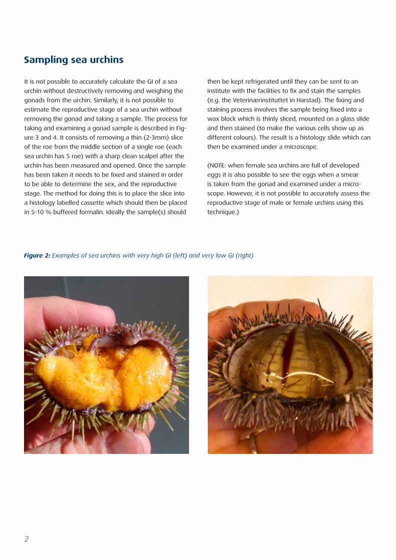
The GI of urchins in the wild can vary hugely and can be
less than 1%, or, as high as 20% (see Figure 2) whilst
for cultured sea urchins GI values can be as high as 35%.
Factors that affect GI are feed availability, environmental
conditions (e.g. daylight period, water temperature and
presence/absence of water currents) and the reproduc-
tive cycle of the urchin. The latter also has a significant
impact on the quality of the sea urchin gonad and this is
discussed in more detail in the ‘Reproductive cycle’ sec-
tion. Because of the natural variation in urchin GI, large
differences can occur both between individuals within a
population and between urchin populations that are very
close to one another. Therefore it is extremely difficult
to accurately predict the size and quality of sea urchins
gonads from any given population before the sea urchin
is opened and the GI is calculated.
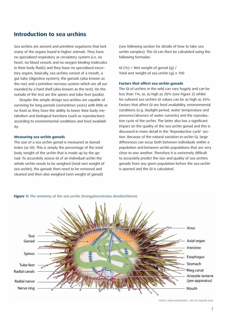
Sea urchins are ancient and primitive organisms that lack
many of the organs found in higher animals. They have
no specialized respiratory or circulatory system (i.e. no
heart, no blood vessels and no oxygen binding molecules
in their body fluids) and they have no specialized excre-
tory organs. Basically, sea urchins consist of a mouth, a
gut tube (digestive system), the gonads (also known as
the roe) and a primitive nervous system which are all sur-
rounded by a hard shell (also known as the test). On the
outside of the test are the spines and tube feet (podia).
Despite this simple design sea urchins are capable of
surviving for long periods (sometimes years) with little or
no food as they have the ability to lower their body me-
tabolism and biological functions (such as reproduction)
according to environmental conditions and feed availabil-
ity.
Measuring sea urchin gonads
The size of a sea urchin gonad is measured as Gonad
Index (or GI). This is simply the percentage of the total
body weight of the urchin that is made up by the go-
nad. To accurately assess GI of an individual urchin the
whole urchin needs to be weighed (total wet weight of
sea urchin), the gonads then need to be removed and
cleaned and then also weighed (wet weight of gonad)
Introduction to sea urchins
Figure 1: The anatomy of the sea urchin Strongylocentrotus droebachiensis.
Photo: Vidar Mortensen / art: by oddVar dahl
2
then be kept refrigerated until they can be sent to an
institute with the facilities to fix and stain the samples
(e.g. the Veterinærinstituttet in Harstad). The fixing and
staining process involves the sample being fixed into a
wax block which is thinly sliced, mounted on a glass slide
and then stained (to make the various cells show up as
different colours). The result is a histology slide which can
then be examined under a microscope.
(NOTE: when female sea urchins are full of developed
eggs it is also possible to see the eggs when a smear
is taken from the gonad and examined under a micro-
scope. However, it is not possible to accurately assess the
reproductive stage of male or female urchins using this
technique.)
It is not possible to accurately calculate the GI of a sea
urchin without destructively removing and weighing the
gonads from the urchin. Similarly, it is not possible to
estimate the reproductive stage of a sea urchin without
removing the gonad and taking a sample. The process for
taking and examining a gonad sample is described in Fig-
ure 3 and 4. It consists of removing a thin (2-3mm) slice
of the roe from the middle section of a single roe (each
sea urchin has 5 roe) with a sharp clean scalpel after the
urchin has been measured and opened. Once the sample
has been taken it needs to be fixed and stained in order
to be able to determine the sex, and the reproductive
stage. The method for doing this is to place the slice into
a histology labelled cassette which should then be placed
in 5-10 % buffered formalin. Ideally the sample(s) should
Sampling sea urchins
Figure 2: Examples of sea urchins with very high GI (left) and very low GI (right)
3
Figure 3: The process for sampling the roe of a sea urchin and preparing a histology sample: (A and B) the equipment
required includes a sea urchin opener, a spoon, a scalpel; a histology cassette and a plastic container with 5-10% formalin
solution; (C) weighing the whole live sea urchin; (D) measuring the test diameter of the urchin; (E) opening the urchin; (F)
removing all 5 gonads; (G) weighing the 5 gonads; (H) removing a small section from the middle of the gonad; (I) placing
sample into histology cassette; ( J) labelling the plastic container and histology cassette; and (K) placing the sample in
formalin solution.
A
E
H
B
F
I
C
G
J
D
K
4
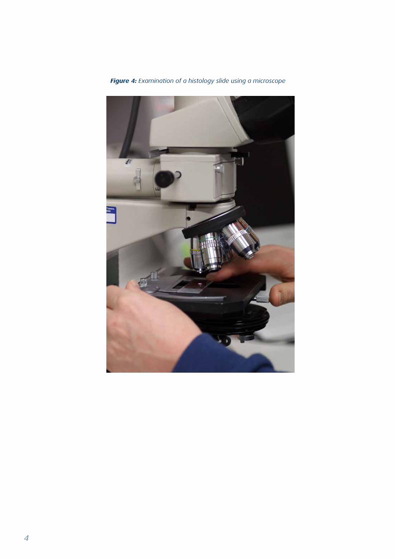
Figure 4: Examination of a histology slide using a microscope
5
Understanding the reproductive cycle of sea urchins is im-
portant to any potential sea urchin fishery in Norway as
the gonad is the only part of the urchin that has any com-
mercial value and the reproductive cycle affects both the
size and the quality of the gonad and subsequently its
value. Anybody involved in the aquaculture of sea urchins
also need to understand both the reproductive stage of
individuals and the reproductive cycles of populations in
any given area where they are operating.
Description of sea urchin reproductive cycle
Adult sea urchins are either male or female, with a nor-
mal sex ratio of 1:1, they both normally spawn once per
year and release their gametes (eggs or sperm) into the
water column (this is called broadcast spawning) where
mixing and fertilisation of the eggs occurs. Normally, in
Norway, spawning occurs around April when a sharp drop
in the size of the gonads occurs. Following spawning in
spring/early summer the urchins go through a dormant
stage when the gonad is generally small and in poor con-
dition. In late summer the gonads slowly increase in size
as it produces storage cells which increase in both size
The reproductive cycle of sea urchins
and number. In early to mid winter gametogenesis (the
formation of reproductive cells) occurs and the number of
storage cells in the gonad reduces and these are replaced
with reproductive cells. The cues that stimulate game-
togenesis are not fully understood but the primary cue
is believed to be changing photoperiod. The number of
reproductive cells within the gonad builds up over winter
until the urchin is once again in spawning condition in late
winter/Spring. The cues that trigger spawning are also
unclear but the primary cues are believed to be tempera-
ture and environmental factors such as algal blooms and
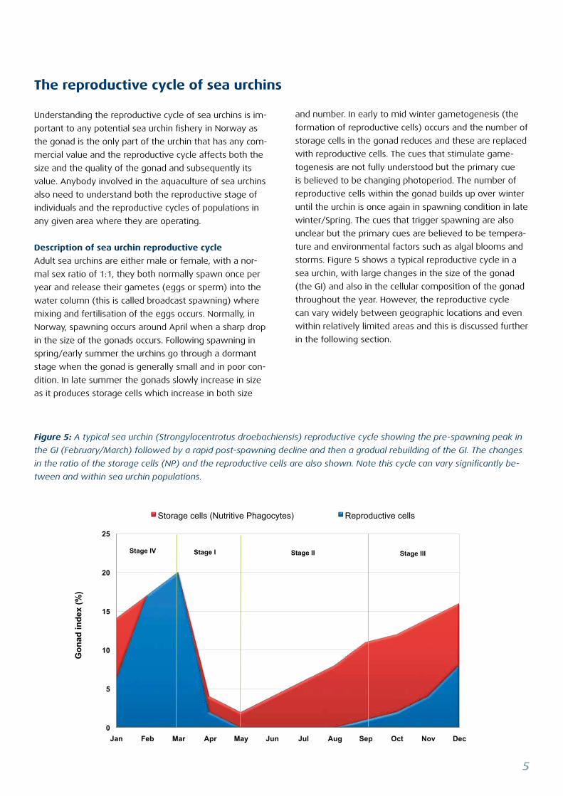
storms. Figure 5 shows a typical reproductive cycle in a
sea urchin, with large changes in the size of the gonad
(the GI) and also in the cellular composition of the gonad
throughout the year. However, the reproductive cycle
can vary widely between geographic locations and even
within relatively limited areas and this is discussed further
in the following section.
Figure 5: A typical sea urchin (Strongylocentrotus droebachiensis) reproductive cycle showing the pre-spawning peak in
the GI (February/March) followed by a rapid post-spawning decline and then a gradual rebuilding of the GI. The changes
in the ratio of the storage cells (NP) and the reproductive cells are also shown. Note this cycle can vary significantly be-
tween and within sea urchin populations.
0
5
10
15
20
25
Jan Feb Mar Apr May Jun Jul Aug Sep Oct Nov Dec
Gon
ad in
dex
(%)
Storage cells (Nutritive Phagocytes) Reproductive cells
Stage IV Stage I Stage II Stage III
6
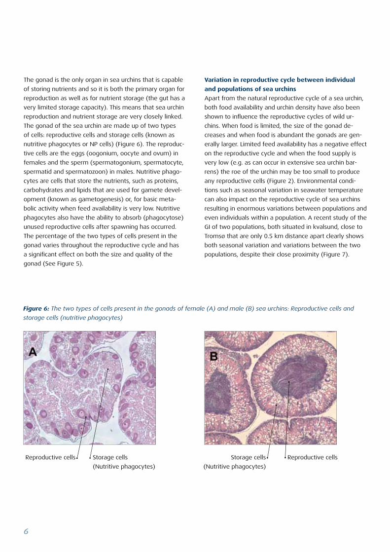
Figure 6: The two types of cells present in the gonads of female (A) and male (B) sea urchins: Reproductive cells and
storage cells (nutritive phagocytes)
The gonad is the only organ in sea urchins that is capable
of storing nutrients and so it is both the primary organ for
reproduction as well as for nutrient storage (the gut has a
very limited storage capacity). This means that sea urchin
reproduction and nutrient storage are very closely linked.
The gonad of the sea urchin are made up of two types
of cells: reproductive cells and storage cells (known as
nutritive phagocytes or NP cells) (Figure 6). The reproduc-
tive cells are the eggs (oogonium, oocyte and ovum) in
females and the sperm (spermatogonium, spermatocyte,
spermatid and spermatozoon) in males. Nutritive phago-
cytes are cells that store the nutrients, such as proteins,
carbohydrates and lipids that are used for gamete devel-
opment (known as gametogenesis) or, for basic meta-
bolic activity when feed availability is very low. Nutritive
phagocytes also have the ability to absorb (phagocytose)
unused reproductive cells after spawning has occurred.
The percentage of the two types of cells present in the
gonad varies throughout the reproductive cycle and has
a significant effect on both the size and quality of the
gonad (See Figure 5).
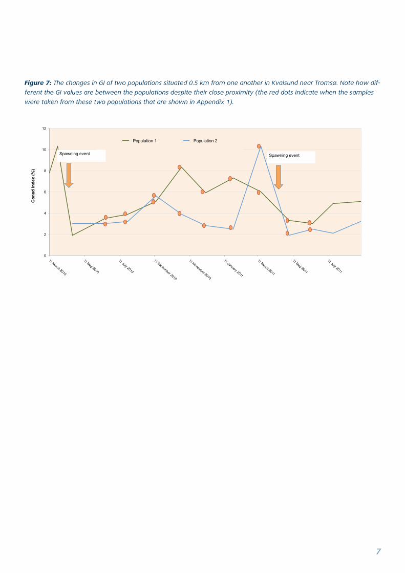
Variation in reproductive cycle between individual
and populations of sea urchins
Apart from the natural reproductive cycle of a sea urchin,
both food availability and urchin density have also been
shown to influence the reproductive cycles of wild ur-
chins. When food is limited, the size of the gonad de-
creases and when food is abundant the gonads are gen-
erally larger. Limited feed availability has a negative effect
on the reproductive cycle and when the food supply is
very low (e.g. as can occur in extensive sea urchin bar-
rens) the roe of the urchin may be too small to produce
any reproductive cells (Figure 2). Environmental condi-
tions such as seasonal variation in seawater temperature
can also impact on the reproductive cycle of sea urchins
resulting in enormous variations between populations and
even individuals within a population. A recent study of the
GI of two populations, both situated in kvalsund, close to
Tromsø that are only 0.5 km distance apart clearly shows
both seasonal variation and variations between the two
populations, despite their close proximity (Figure 7).
Reproductive cells Reproductive cellsStorage cells
(Nutritive phagocytes)
Storage cells
(Nutritive phagocytes)
7
Figure 7: The changes in GI of two populations situated 0.5 km from one another in Kvalsund near Tromsø. Note how dif-
ferent the GI values are between the populations despite their close proximity (the red dots indicate when the samples
were taken from these two populations that are shown in Appendix 1).
0
2
4
6
8
10
12
11 March 2010
11 May 2010
11 July 2010
11 September 2010
11 November 2010
11 January 2011
11 March 2011
11 May 2011
11 July 2011
Gon
ad In
dex
(%)
Population 1 Population 2
Spawning event Spawning event
8
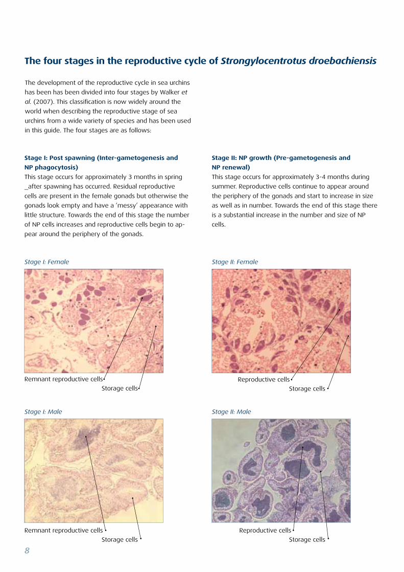
The development of the reproductive cycle in sea urchins
has been has been divided into four stages by Walker et
al. (2007). This classification is now widely around the
world when describing the reproductive stage of sea
urchins from a wide variety of species and has been used
in this guide. The four stages are as follows:
The four stages in the reproductive cycle of Strongylocentrotus droebachiensis
Stage II: NP growth (Pre-gametogenesis and
NP renewal)
This stage occurs for approximately 3-4 months during
summer. Reproductive cells continue to appear around
the periphery of the gonads and start to increase in size
as well as in number. Towards the end of this stage there
is a substantial increase in the number and size of NP
cells.
Stage I: Post spawning (Inter-gametogenesis and
NP phagocytosis)
This stage occurs for approximately 3 months in spring
_after spawning has occurred. Residual reproductive
cells are present in the female gonads but otherwise the
gonads look empty and have a ’messy’ appearance with
little structure. Towards the end of this stage the number
of NP cells increases and reproductive cells begin to ap-
pear around the periphery of the gonads.
Stage I: Male Stage II: Male
Stage I: Female Stage II: Female
Remnant reproductive cells
Storage cells
Reproductive cells
Storage cells
Remnant reproductive cells
Storage cells
Reproductive cells
Storage cells
9
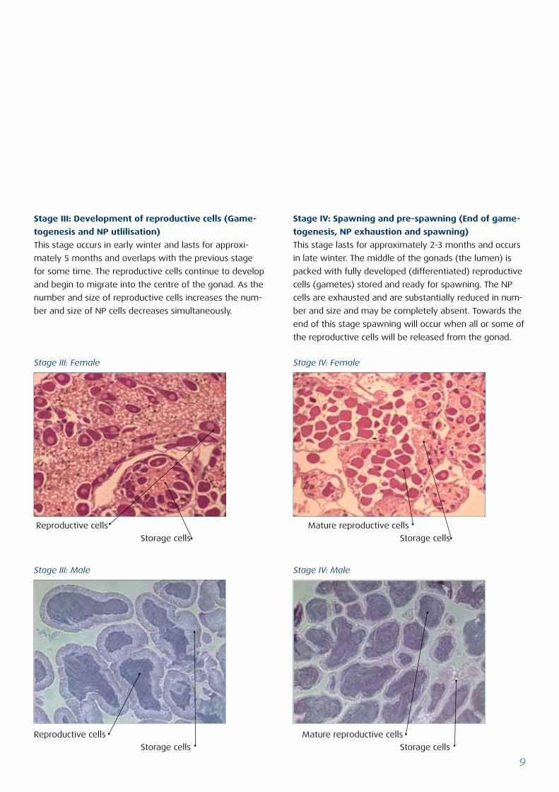
Stage IV: Spawning and pre-spawning (End of game-
togenesis, NP exhaustion and spawning)
This stage lasts for approximately 2-3 months and occurs
in late winter. The middle of the gonads (the lumen) is
packed with fully developed (differentiated) reproductive
cells (gametes) stored and ready for spawning. The NP
cells are exhausted and are substantially reduced in num-
ber and size and may be completely absent. Towards the
end of this stage spawning will occur when all or some of
the reproductive cells will be released from the gonad.
Stage III: Development of reproductive cells (Game-
togenesis and NP utlilisation)
This stage occurs in early winter and lasts for approxi-
mately 5 months and overlaps with the previous stage
for some time. The reproductive cells continue to develop
and begin to migrate into the centre of the gonad. As the
number and size of reproductive cells increases the num-
ber and size of NP cells decreases simultaneously.
Stage III: Male Stage IV: Male
Stage III: Female Stage IV: Female
Reproductive cells
Storage cells Storage cells
Mature reproductive cells
Reproductive cells
Storage cells
Mature reproductive cells
Storage cells
10
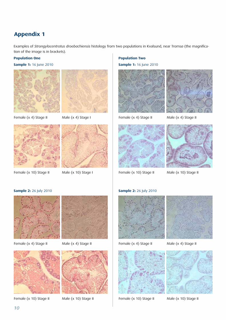
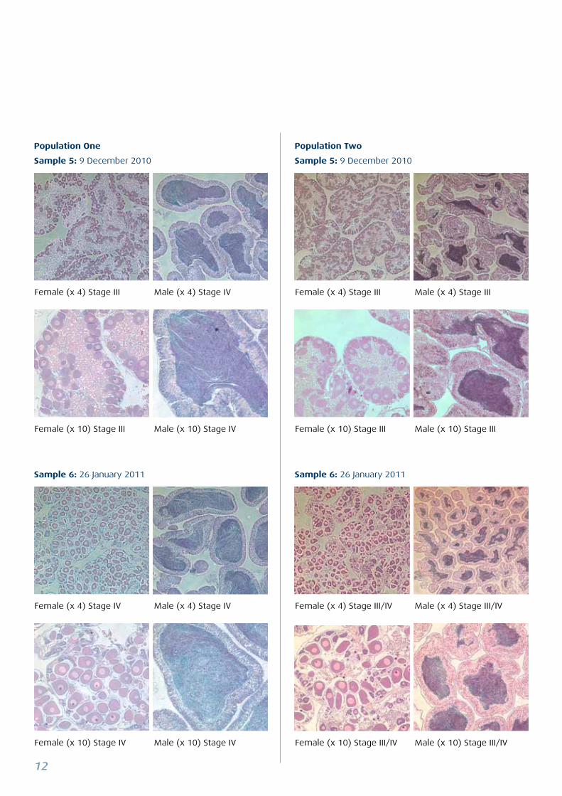
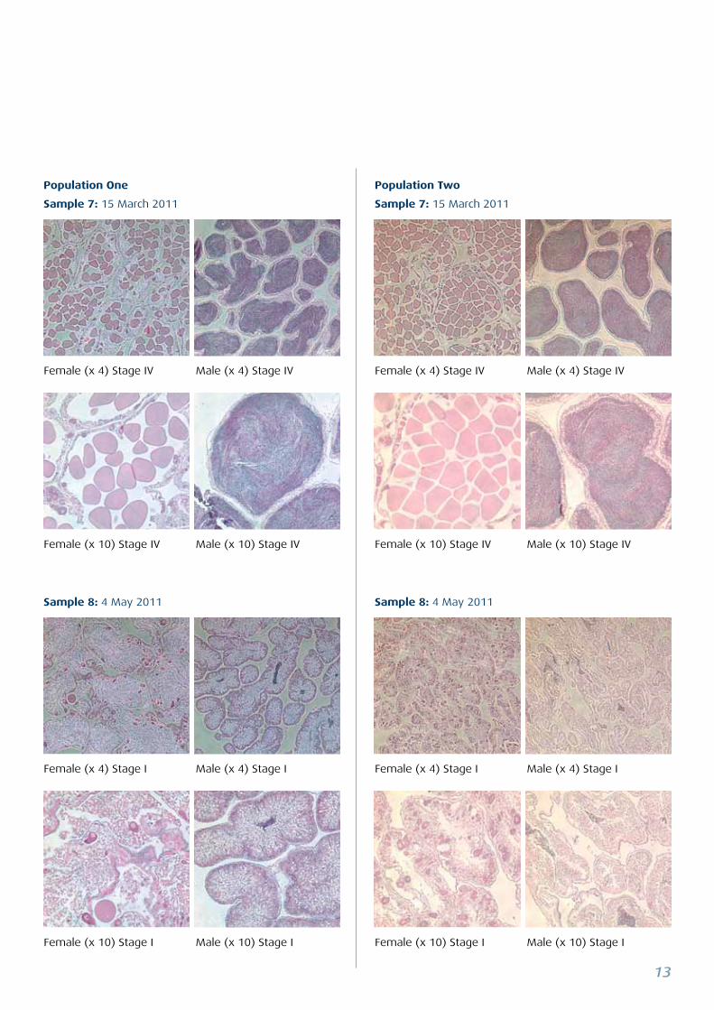
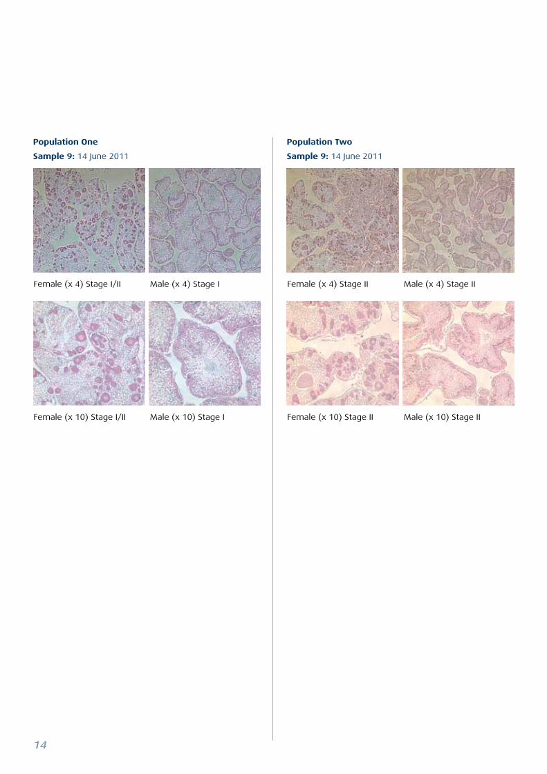
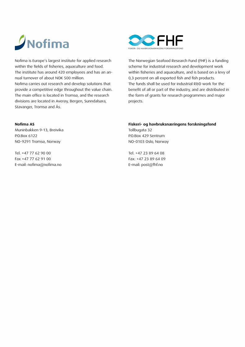
Examples of Strongylocentrotus droebachiensis histology from two populations in Kvalsund, near Tromsø (the magnifica-
tion of the image is in brackets).
Population One
Sample 1: 16 June 2010
Sample 2: 26 July 2010 Sample 2: 26 July 2010
Population Two
Sample 1: 16 June 2010
Appendix 1
Female (x 4) Stage II Male (x 4) Stage I Female (x 4) Stage II Male (x 4) Stage II
Female (x 10) Stage II Male (x 10) Stage I Female (x 10) Stage II Male (x 10) Stage II
Female (x 4) Stage II Male (x 4) Stage II Female (x 4) Stage II Male (x 4) Stage II
Female (x 10) Stage II Male (x 10) Stage II Female (x 10) Stage II Male (x 10) Stage II
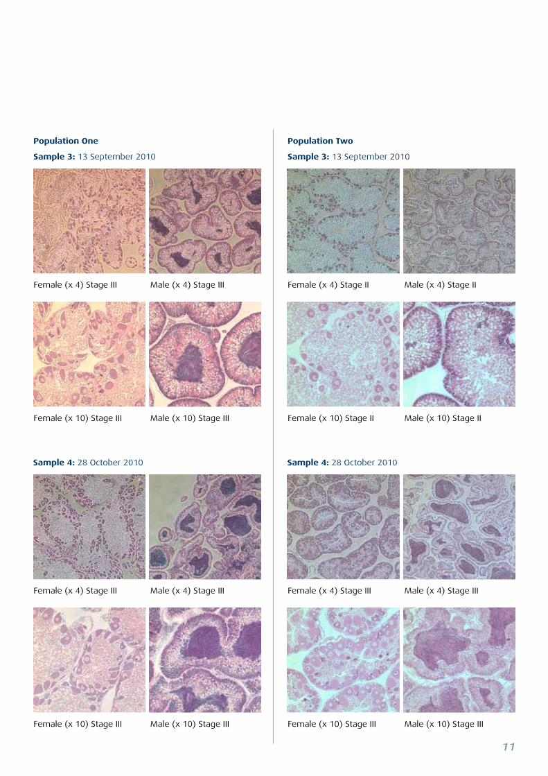
11
Sample 4: 28 October 2010 Sample 4: 28 October 2010
Female (x 4) Stage III Male (x 4) Stage III Female (x 4) Stage II Male (x 4) Stage II
Female (x 10) Stage III Male (x 10) Stage III Female (x 10) Stage II Male (x 10) Stage II
Female (x 4) Stage III Male (x 4) Stage III Female (x 4) Stage III Male (x 4) Stage III
Female (x 10) Stage III Male (x 10) Stage III Female (x 10) Stage III Male (x 10) Stage III
Population One
Sample 3: 13 September 2010
Population Two
Sample 3: 13 September 2010
12
Population One
Sample 5: 9 December 2010
Sample 6: 26 January 2011
Population Two
Sample 5: 9 December 2010
Sample 6: 26 January 2011
Female (x 4) Stage III Male (x 4) Stage IV Female (x 4) Stage III Male (x 4) Stage III
Female (x 10) Stage III Male (x 10) Stage IV Female (x 10) Stage III Male (x 10) Stage III
Female (x 4) Stage IV Male (x 4) Stage IV Female (x 4) Stage III/IV Male (x 4) Stage III/IV
Female (x 10) Stage IV Male (x 10) Stage IV Female (x 10) Stage III/IV Male (x 10) Stage III/IV
13
Population One
Sample 7: 15 March 2011
Sample 8: 4 May 2011
Population Two
Sample 7: 15 March 2011
Sample 8: 4 May 2011
Female (x 4) Stage IV Male (x 4) Stage IV Female (x 4) Stage IV Male (x 4) Stage IV
Female (x 10) Stage IV Male (x 10) Stage IV Female (x 10) Stage IV Male (x 10) Stage IV
Female (x 4) Stage I Male (x 4) Stage I Female (x 4) Stage I Male (x 4) Stage I
Female (x 10) Stage I Male (x 10) Stage I Female (x 10) Stage I Male (x 10) Stage I
14
Population One
Sample 9: 14 June 2011
Population Two
Sample 9: 14 June 2011
Female (x 4) Stage I/II Male (x 4) Stage I Female (x 4) Stage II Male (x 4) Stage II
Female (x 10) Stage I/II Male (x 10) Stage I Female (x 10) Stage II Male (x 10) Stage II

15
For your own notes:

16

Nofima is Europe’s largest institute for applied research
within the fields of fisheries, aquaculture and food.
The institute has around 420 employees and has an an-
nual turnover of about NOK 500 million.
Nofima carries out research and develop solutions that
provide a competitive edge throughout the value chain.
The main office is located in Tromsø, and the research
divisions are located in Averøy, Bergen, Sunndalsøra,
Stavanger, Tromsø and Ås.
Nofima AS
Muninbakken 9-13, Breivika
P.O.Box 6122
NO-9291 Tromsø, Norway
Tel. +47 77 62 90 00
Fax +47 77 62 91 00
E-mail: [email protected]
The Norwegian Seafood Research Fund (FHF) is a funding
scheme for industrial research and development work
within fisheries and aquaculture, and is based on a levy of
0,3 percent on all exported fish and fish products.
The funds shall be used for industrial R&D work for the
benefit of all or part of the industry, and are distributed in
the form of grants for research programmes and major
projects.
Fiskeri- og havbruksnæringens forskningsfond
Tollbugata 32
P.O.Box 429 Sentrum
NO-0103 Oslo, Norway
Tel. +47 23 89 64 08
Fax: +47 23 89 64 09
E-mail: [email protected]