A Generative Model for Brain Tumor Segmentation in...
8
A Generative Model for Brain Tumor Segmentation in Multi-Modal Images Bjoern H. Menze 1,2 , Koen Van Leemput 1,3,4 , Danial Lashkari 1 Marc-Andr´ e Weber 5 , Nicholas Ayache 2 , and Polina Golland 1 1 Computer Science and Artificial Intelligence Laboratory, Massachusetts Institute of Technology, USA 2 Asclepios Research Project, INRIA Sophia-Antipolis, France 3 Radiology, Massachusetts General Hospital, Harvard Medical School, USA 4 Information and Computer Science, Aalto University, Finland 5 Diagnostic Radiology, Heidelberg University Hospital, Germany Abstract. We introduce a generative probabilistic model for segmenta- tion of tumors in multi-dimensional images. The model allows for differ- ent tumor boundaries in each channel, reflecting difference in tumor ap- pearance across modalities. We augment a probabilistic atlas of healthy tissue priors with a latent atlas of the lesion and derive the estima- tion algorithm to extract tumor boundaries and the latent atlas from the image data. We present experiments on 25 glioma patient data sets, demonstrating significant improvement over the traditional multivariate tumor segmentation. 1 Introduction Limited therapy options require a careful diagnostic for patients with brain tu- mors. A multitude of available brain imaging sequences gives rise to patient data sets that include multi-parametric, multi-modal, and multi-temporal vol- umes even in standard clinical settings. Quantitative analysis of a lesion in these data poses a challenging computational problem. In this paper, we present a fully automated method for channel-specific tumor segmentation in such multi- dimensional images. Generative probabilistic models of spatial tissue distribution and appearance have enjoyed popularity for tissue classification as they exhibit good general- ization to unseen images [1–3]. Encoding spatial prior knowledge for a lesion, however, is difficult. Tumors may be modeled as outliers relative to the expected shape [4, 5] or image signal of healthy tissues [2, 6]. In [2], for example, a crite- rion for detecting outliers is used to generate a tumor prior in a subsequent EM segmentation which is treating tumor as an additional tissue class. Alternatively, the spatial prior for the tumor can be derived from the appearance of tumor- specific bio-markers [7, 8]. The tumor classification methods can be augmented with spatial regularization using a Markov Random Field prior [9] or a boundary finding step [2, 10] to ensure spatial contiguity of the segmentation results. Discriminative approaches directly learn the difference between the appear- ance of the lesion and other tissues and do not rely on spatial priors [11–16]. They do, however, often require substantial amounts of training data and typi- cally come at the cost of manual interaction for initialization and postprocessing.
-
Upload
truongthuy -
Category
Documents
-
view
221 -
download
1
Transcript of A Generative Model for Brain Tumor Segmentation in...
A Generative Model for Brain TumorSegmentation in Multi-Modal Images
Bjoern H. Menze1,2, Koen Van Leemput1,3,4, Danial Lashkari1
Marc-Andre Weber5, Nicholas Ayache2, and Polina Golland1
1 Computer Science and Artificial Intelligence Laboratory,Massachusetts Institute of Technology, USA
2 Asclepios Research Project, INRIA Sophia-Antipolis, France3 Radiology, Massachusetts General Hospital, Harvard Medical School, USA
4 Information and Computer Science, Aalto University, Finland5 Diagnostic Radiology, Heidelberg University Hospital, Germany
Abstract. We introduce a generative probabilistic model for segmenta-tion of tumors in multi-dimensional images. The model allows for differ-ent tumor boundaries in each channel, reflecting difference in tumor ap-pearance across modalities. We augment a probabilistic atlas of healthytissue priors with a latent atlas of the lesion and derive the estima-tion algorithm to extract tumor boundaries and the latent atlas fromthe image data. We present experiments on 25 glioma patient data sets,demonstrating significant improvement over the traditional multivariatetumor segmentation.
1 Introduction
Limited therapy options require a careful diagnostic for patients with brain tu-mors. A multitude of available brain imaging sequences gives rise to patientdata sets that include multi-parametric, multi-modal, and multi-temporal vol-umes even in standard clinical settings. Quantitative analysis of a lesion in thesedata poses a challenging computational problem. In this paper, we present afully automated method for channel-specific tumor segmentation in such multi-dimensional images.
Generative probabilistic models of spatial tissue distribution and appearancehave enjoyed popularity for tissue classification as they exhibit good general-ization to unseen images [1–3]. Encoding spatial prior knowledge for a lesion,however, is difficult. Tumors may be modeled as outliers relative to the expectedshape [4, 5] or image signal of healthy tissues [2, 6]. In [2], for example, a crite-rion for detecting outliers is used to generate a tumor prior in a subsequent EMsegmentation which is treating tumor as an additional tissue class. Alternatively,the spatial prior for the tumor can be derived from the appearance of tumor-specific bio-markers [7, 8]. The tumor classification methods can be augmentedwith spatial regularization using a Markov Random Field prior [9] or a boundaryfinding step [2, 10] to ensure spatial contiguity of the segmentation results.
Discriminative approaches directly learn the difference between the appear-ance of the lesion and other tissues and do not rely on spatial priors [11–16].They do, however, often require substantial amounts of training data and typi-cally come at the cost of manual interaction for initialization and postprocessing.
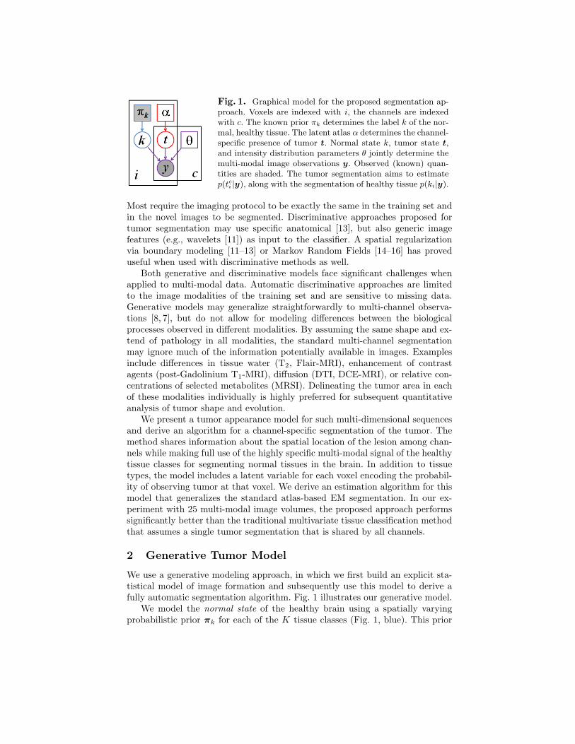
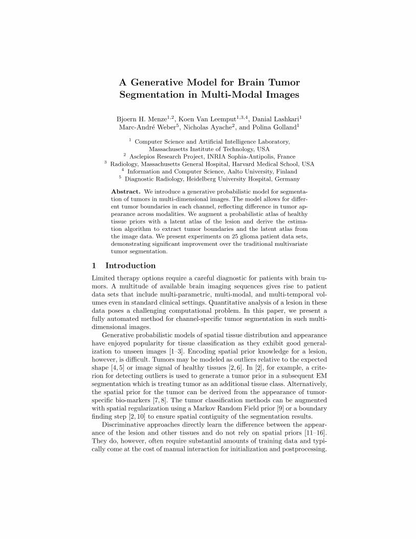
Fig. 1. Graphical model for the proposed segmentation ap-proach. Voxels are indexed with i, the channels are indexedwith c. The known prior πk determines the label k of the nor-mal, healthy tissue. The latent atlas α determines the channel-specific presence of tumor t. Normal state k, tumor state t,and intensity distribution parameters θ jointly determine themulti-modal image observations y. Observed (known) quan-tities are shaded. The tumor segmentation aims to estimatep(tci |y), along with the segmentation of healthy tissue p(ki|y).
Most require the imaging protocol to be exactly the same in the training set andin the novel images to be segmented. Discriminative approaches proposed fortumor segmentation may use specific anatomical [13], but also generic imagefeatures (e.g., wavelets [11]) as input to the classifier. A spatial regularizationvia boundary modeling [11–13] or Markov Random Fields [14–16] has proveduseful when used with discriminative methods as well.
Both generative and discriminative models face significant challenges whenapplied to multi-modal data. Automatic discriminative approaches are limitedto the image modalities of the training set and are sensitive to missing data.Generative models may generalize straightforwardly to multi-channel observa-tions [8, 7], but do not allow for modeling differences between the biologicalprocesses observed in different modalities. By assuming the same shape and ex-tend of pathology in all modalities, the standard multi-channel segmentationmay ignore much of the information potentially available in images. Examplesinclude differences in tissue water (T2, Flair-MRI), enhancement of contrastagents (post-Gadolinium T1-MRI), diffusion (DTI, DCE-MRI), or relative con-centrations of selected metabolites (MRSI). Delineating the tumor area in eachof these modalities individually is highly preferred for subsequent quantitativeanalysis of tumor shape and evolution.
We present a tumor appearance model for such multi-dimensional sequencesand derive an algorithm for a channel-specific segmentation of the tumor. Themethod shares information about the spatial location of the lesion among chan-nels while making full use of the highly specific multi-modal signal of the healthytissue classes for segmenting normal tissues in the brain. In addition to tissuetypes, the model includes a latent variable for each voxel encoding the probabil-ity of observing tumor at that voxel. We derive an estimation algorithm for thismodel that generalizes the standard atlas-based EM segmentation. In our ex-periment with 25 multi-modal image volumes, the proposed approach performssignificantly better than the traditional multivariate tissue classification methodthat assumes a single tumor segmentation that is shared by all channels.
2 Generative Tumor Model
We use a generative modeling approach, in which we first build an explicit sta-tistical model of image formation and subsequently use this model to derive afully automatic segmentation algorithm. Fig. 1 illustrates our generative model.
We model the normal state of the healthy brain using a spatially varyingprobabilistic prior πk for each of the K tissue classes (Fig. 1, blue). This prior

(atlas) is estimated from prior examples and is assumed to be known. At eachvoxel i, the atlas defines a multinomial distribution for the tissue label ki:
p(ki = k) = πki. (1)
The normal state ki is shared among all C channels at voxel i. In our experimentswe assume K = 3, representing gray matter, white matter and cerebrospinalfluid (CSF).
We model the tumor state using a spatially varying “latent” probabilisticatlas α, similar to [10] (Fig. 1, red). At each voxel i, this atlas provides a scalarparameter αi that defines the probability of observing tumor at that voxel.Parameter αi is unknown and is estimated as part of the segmentation process.We define a latent tumor state tci ∈ {0, 1} that indicates the presence of tumor inchannel c at voxel i and model it as a Bernoulli random variable with parameterαi. We form a binary tumor state vector ti = [t1i , . . . , t
Ci ]T indicating the tumor
presence for all c observations at voxel i, with probability
p(ti;αi) =∏c
p(tci ;αi) =∏c
αtci
i · (1− αi)1−tci . (2)
Image observations yci are generated by Gaussian intensity distributions for
each of the K tissue classes and the C channels, with mean µck and variance
vck, respectively (Fig. 1, purple). In tumor tissue (i.e., if tci = 1) the normal
observations are replaced by intensities from another set of channel-specificGaussian distributions with mean µc
K+1 and variance vcK+1, representing the
tumor class. Letting θ denote the set of all mean and variance parameters, andyi = [y1
i , . . . , yCi ]T denote the vector of the intensity observations at voxel i, we
define the data likelihood:
p(yi|ti, ki;θ) =∏c
p(yci |tci , ki;θ)
=∏c
[N (yc
i ; µcki, vc
ki)1−tc
i · N (yci ; µc
K+1, vcK+1)tc
i
], (3)
where N (· ; µ, v) is the Gaussian distribution with mean µ and variance v.Finally, the joint probability of the the latent atlas and the observed variables
p(yi, ti, ki;θ, αi) = p(yi|ti, ki;θ) · p(ti;αi) · p(ki) (4)
is the product of the components defined in Eqs. (1-3).
3 Maximum Likelihood Parameter Estimation
We seek Maximum Likelihood estimates of the model parameters {θ,α}:
〈θ, α〉 = arg max〈θ,α〉
p(y1, . . . ,yN ;θ,α) = arg max〈θ,α〉
N∏i=1
p(yi;θ,α),

where N is the number of voxels in the volume and
p(yi;θ,α) =∑ti
∑ki
p(yi, ti, ki;θ,α).
Observing that evaluating the objective function involves summing over valuesof ti and ki in Eq. (4), we use Jensen’s inequality to perform the optimizationusing an iterative, EM-style minorization technique [17]. Letting {θ, α} denotethe current parameter estimates, we can compute the posterior probability ofany of the 2C tumor state vectors ti, writing out the components of Eq. (4):
qi(ti) , p(ti|ki,yi; θ, α) ∝∑ki
p(yi|ti, ki; θ)p(ti; αi)p(ki), (5)
and∑
tiqi(ti) = 1. Based only on the intensity channels that do not show tumor
(tci = 0), we also compute the posterior probability of tissue k at voxel i:
wik(ti) , p(ki|ti,yi; θ, α) ∝ πki
∏c
N (yci ; µc
k, vck)1−tc
i ,
and∑
k wik(ti) = 1 for all ti. Using qi(·) and wik(·), we arrive at closed-formupdate expressions that guarantee increasingly better estimates of the modelparameters. The updates are intuitive: the latent tumor prior is an average ofthe corresponding posterior estimates
αi ←∑ti
qi(ti)
(1C
∑c
tci
)and the intensity parameters are updated with the weighted statistics of the datafor the healthy tissues (k = 1, . . . ,K)
µck ←
∑i
∑tiqi(ti)wik(ti)(1− tci ) yc
i∑i
∑tiqi(ti)wik(ti)(1− tci )
, vck ←
∑i
∑tiqi(ti)wik(ti)(1− tci ) (yc
i − µck)2∑
i
∑tiqi(ti)wik(ti)(1− tci )
and for the tumor class:
µcK+1 ←
∑i
∑tiqi(ti) tci y
ci∑
i
∑tiqi(ti) tci
, vcK+1 ←
∑i
∑tiqi(ti) tci (yc
i − µcK+1)2∑
i
∑tiqi(ti) tci
.
We iterate the estimation of the parameters {θ, α} and the computation of theposterior probabilities {qi(·), wik(·)} until convergence.
4 Tumor segmentation
Once we have an estimate of the model parameters {θ, α}, we can evaluate theprobability that tumor is visible in channel c of voxel i by summing over all theconfigurations ti for which tci = 1:
p(tci = 1|yi; θ, α) =∑ti
tci p(ti|yi; θ, α) =∑ti
tci qi(ti). (6)
We then assign channel c of voxel i to tumor if p(tci = 1|yi; θ, α) > 0.5.

5 Extensions
To augment the generative model outlined above with further physiologicalknowledge, we derive and implement extensions considering the expected shape,multivariate signal and structural appearance of the tumor.
Little spatial context is used in the basic model, as we assume the tumorstate ti in each voxel to be independent from the state of other voxels (Eq. 6and Eq. 3). It is only the atlas πk that encourages smooth classification forthe healthy tissue classes by imposing similar priors in neighboring voxels. Toencourage a similar smoothness of the tumor labels, we extend the latent atlasα to include a Markov Random Field (MRF) prior:
p(t1, . . . , tN ;β,α) ∝∏c
∏i
αtci
i (1− αi)1−tci exp
[− β
2
∑j∈Ni
tci (1− tcj) + tcj(1− tci )].
Here, Ni denotes the set of the six nearest neighbors of voxel i, and β is a pa-rameter governing how similar the tumor states tend to be in neighboring voxels.When β = 0, there is no interaction between voxels and the model reduces to theone described in Section 2. For β 6= 0, the posteriors qi(ti) are no longer givenby Eq. (5), and their exact computation becomes infeasible. However, relaxingthe MRF to a mean-field approximation [18] we derive an efficient approximatealgorithm. We let
nci =
∑j∈Ni
∑tj
tcj qj(tj)
denote the currently estimated “soft” count of neighbors that show tumor inchannel c. The mean-field approximation implies
p(ti|αi) '∏c
γtci
i (1−γi)(1−tci ), where γi =
αi
αi + (1− αi) exp[− β(2nc
i − 6)]
to replace the previously defined p(ti|αi) in Eq. (4), leading to smoothed esti-mates of the tumor segmentations.
Moreover, we want to account for the non-homogeneity in the appearanceof the tumor class, as gliomas show characteristic substructures such as activeand necrotic areas and edema. We model this via a straightforward extensionsof the tissue classes to include more than one class for tumor in a second mod-ification to our approach. Finally, to consider higher-order interactions in themultivariate biological signal yi of healthy tissue, we can relax the conditionalindependence of observations across channels by using multivariate Gaussians inthe data likelihood in Eq. (3). We report tests of these three extensions in thenext section.
6 Experiments
We evaluate our approach on a data set of 25 patients with glioma. The dataset comprises T1, T2, FLAIR-, and post-Gadolinium T1 MR images. Tumorswere outlined by a rater in three planes intersecting with the tumor center. We

Fig. 2. Examples of channel-specific segmentation results for four different modalities,in two patients. The outlines of regions with p(tci = 1|yi; θ, α) > 0.5 are shown in red.The proposed method localizes the tumor reliably in post-therapeutic images (below),where surgery has led to significant deviations from normalcy for healthy tissues.
register all images of a patient to the FLAIR volume by using affine registrationand segment the volume into the three healthy and an outlier class using afreely available implementation of the EM segmentation with bias correction [1].Outliers are defined as being more than three standard deviations away fromthe centroid of any of the three normal tissue classes.
We apply our algorithm to the bias field corrected volumes and initializeintensity parameters with values estimated in the initial segmentation. Whenusing multivariate distributions N (·;µ;V ),we initialize off-diagonal element in Vto zero. When modeling the tumor class with multiple Gaussian densities weinitialize the means of additional subclasses to random values. We use outliersin the initial segmentation to initialize the latent atlas α, setting αi for pixels ofthe outlier class to 0.7 and otherwise to 0.3. We typically observe convergenceafter 10 to 15 steps. For comparison, we also implement an EM segmentationtreating tumor as one of the tissue classes, with a weak MRF prior for spatialregularization, similar to [2]. We use the same data and initializations as above,but augment the atlas by a tumor prior obtained by smoothing the outlier classof the initial segmentation with a 3cm kernel. This alternative segmentation isapplied to every single image volume individually in a first experiment, and to themultivariate features of the whole multi-modal volume in a second experiment.To evaluate the classification maps we calculate Dice scores for both methods[19].
Fig. 2 illustrates results for two different subjects. We note that the tumorboundaries change across different modalities and the proposed method capturesthis variation well, even in post-therapeutic images. The method produces fewfalse positives which can be easily eliminated in a post-processing step. We eval-

●●
●
● ● ● ● ● ●
● ●
1e−02 1e−01 1e+00 1e+01 1e+02
0.0
0.2
0.4
0.6
0.8
1.0
Beta
Dic
e sc
ore
Fig. 3. Sensitivity to the MRF parameter β. Indicatedare the median (solid line) and the interquartile rangesof the average Dice scores of all 25 data set. Whilesome regularization is beneficial, the segmentation per-formance is relatively insensitive to the choice of theonly model parameter β.
uate the robustness and accuracy of our method in a series of experiments. First,we test the sensitivity of the performance to the MRF parameter β that governsthe smoothness of the resulting segmentations. It is the only parameter to beadjusted in our model. We find the performance of the algorithm to be relativelystable for a wide range of β values (Fig. 3), irrespective of size, location or shapeof the tumor (i.e., β ∈ [1; 50]). For simplicity, we set β = 1 in all further ex-periments. In the second experiment, we test different model options for normaltissue and tumor classes. We find little differences between approaches that usenon-zero off-diagonal elements in the covariance matrix of the intensity likelihoodand those that do not. Modeling tumor by three Gaussians improves the resultfor some cases, but leads to a somewhat lower performance on average. In a thirdexperiment, we compare our approach to the two alternative EM segmentationmethods (Fig. 4). Here, we find the proposed channel-specific segmentation toalways perform significantly better (p < 5 · 10−4, paired Cox-Wilcoxon test); itimproves the absolute value of the Dice score over all four modalities for nearlyall data sets by 0.1 to 0.2 (Fig. 4, right).
7 Conclusions
We present a generative model for tumor appearance in multi-modal image vol-umes of the brain that provides channel-specific segmentations. We derive anestimation algorithm and demonstrate superior performance over standard mul-tivariate EM segmentation. Unlike discriminative tumor segmentation methods,our model is applicable to any set of multi-modal image volumes, and is fullyautomatic. Further extensions of the model may consider structure of the tumor,or temporal evolution in longitudinal data sets.
Acknowledgements. This work was supported by the German Academy of SciencesLeopoldina (Fellowship Programme LPDS 2009-10), the Academy of Finland (133611),INRIA CompuTumor, NIH NIBIB NAMIC U54-EB005149, NIH NCRR NAC P41-RR13218, NIH NINDS R01-NS051826, NIH R01-NS052585, NIH R01-EB006758, NIHR01-EB009051, NIH P41-RR014075 and the NSF CAREER Award 0642971.
References
1. Van Leemput, K., Maes, F., Vandermeulen, D., Suetens, P.: Automated model-based tissue classification of MR images of the brain. IEEE TMI 18 (1999) 897–908
2. Prastawa, M., Bullitt, E., Ho, S., Gerig, G.: A brain tumor segmentation frameworkbased on outlier detection. MedIA 8 (2004) 275–83
3. Pohl, K.M., Fisher, J., Levitt, J.J., Shenton, M.E., Kikinis, R., Grimson, W.E.L.,Wells, W.M.: A unifying approach to registration, segmentation, and intensitycorrection. In: LNCS 3750, Proc MICCAI. (2005) 310–318
4. Zacharaki, E.I., Shen, D., Davatzikos, C.: Orbit: A multiresolution framework fordeformable registration of brain tumor images. IEEE TMI 27 (2008) 1003–17
●
u m c u m c u m c u m c
0.0
0.4
0.8
Dic
e sc
ore
T1 image T1 gad image T2 image Flair image
●
●
u m u m u m u m
−0.
20.
20.
6
Dic
e sc
ore
T1 image T1 gad image T2 image Flair image
Fig. 4. Benefits of the channel-specific segmentation. Boxplots show median, quartilesand outliers for the Dice scores of all 25 subjects, for all four modalities. Our channel-wise segmentation (c, green) improves over both multiple univariate (u, blue) andmultivariate (m, red) segmentation, both in the absolute terms (left) and with respectto patient-specific differences (right). The right figure shows c−u and c−m.
5. Bach Cuadra, B., Pollo, C., Bardera, A., Cuisenaire, O., Thiran, J.P.: Atlas-basedsegmentation of pathological brain MR images using a model of lesion growth.IEEE TMI 23 (2004) 1301–14
6. Gering, D.T., Grimson, W.E.L., Kikinis, R.: Recognizing deviations from normalcyfor brain tumor segmentation. In: LNCS 2488, Proc MICCAI. (2002) 388–95
7. Moon, N., Bullitt, E., van Leemput, K., Gerig, G.: Model-based brain and tumorsegmentation. In: Proc ICPR. (2002) 528–31
8. Prastawa, M., Bullitt, E., Moon, N., Leemput, K.V., Gerig, G.: Automatic braintumor segmentation by subject specific modification of atlas priors. Acad Radiol10 (2003) 1341–48
9. Van Leemput, K., Maes, F., Vandermeulen, D., Colchester, A., Suetens, P.: Auto-mated segmentation of multiple sclerosis lesions by model outlier detection. IEEETMI 20 (2001) 677–88
10. Raviv, T.R., Menze, B.H., Van Leemput, K., Stieltjes, B., Weber, M.A., Ayache, N.,Wells III, W., Golland, P.: Joint segmentation via patient-specific latent anatomymodel. In: Proc MICCAI-PMMIA. (2009)
11. Cobzas, D., Birkbeck, N., Schmidt, M., Jagersand, M., Murtha, A.: 3D variationalbrain tumor segmentation using a high dimensional feature set. In: Proc ICCV.(2007) 1–8
12. Lefohn, A., Cates, J., Whitaker, R.: Interactive, GPU-based level sets for 3D braintumor segmentation. In: LNCS 2878, Proc MICCAI. (2003) 564–72.
13. Ho, S., Bullitt, E., Gerig, G.: Level-set evolution with region competition: auto-matic 3D segmentation of brain tumors. In: Proc ICPR. (2002) 532–35
14. Gorlitz, L., Menze, B.H., Weber, M.A., Kelm, B.M., Hamprecht, F.A.: Semi-supervised tumor detection in magnetic resonance spectroscopic images using dis-criminative random fields. In: LNCS 4713, Proc DAGM. (2007) 224–233
15. Lee, C., Wang, S., Murtha, A., Greiner, R.: Segmenting brain tumors using pseu-doconditional random fields. In: LNCS 5242, Proc MICCAI. (2008) 359–66
16. Wels, M., Carneiro, G., Aplas, A., Huber, M., Hornegger, J., Comaniciu, D.: Adiscriminative model-constrained graph cuts approach to fully automated pediatricbrain tumor segmentation in 3D MRI. In: LNCS 5241, Proc MICCAI. (2008) 67–75
17. Hunter, D., Lange, K.: A tutorial on MM algorithms. American Statistician 58(2004) 30–37
18. Jordan, M.I., Gaharamani, Z., Jaakola, T.S., Saul, L.K.: An introduction to vari-ational methods for graphical models. Machine Learning 37 (1999) 183–233
19. Dice, L.: Measure of the amount of ecological association between species. Ecology26 (1945) 297–302