A Geminivirus lnduces Expression of a Host DNA Synthesis · PDF fileA Geminivirus lnduces...
16
The Plant Cell, Vol. 7, 705-719, June 1995 O 1995 American Society of Plant Physiologists A Geminivirus lnduces Expression of a Host DNA Synthesis Protein in Terminally Differentiated Plant Cells Steven Nagar,a Thomas J. Pedersen,bKevin M. Carrick,' Linda Hanley-Bowdoin,b and Dominique Robertsonai' a Department of Botany, North Carolina State University, Raleigh, North Carolina 27695-7612 Department of Biochemistry, North Carolina State University, Raleigh, North Carolina 27695-7622 Geminiviruses are plant DNA viruses that replicate through DNA intermediates in plant nuclei. l h e viral components required for replication are known, but no host factors have yet been identified. We used immunolocalizationto show that the replication proteins of the geminivirus tomato golden mosaic virus (TGMV) are located in nuclei of terminally differentiated cells that have left the cell cycle. In addition, TGMV infection resulted in a significant accumulation of the host DNA synthesis protein proliferating cell nuclear antigen (PCNA). PCNA, an accessory factor for DNA polymerase 6, was not present at detectable levels in healthy differentiated cells. The TGMV replication protein AL1 was sufficient to induce accumulation of PCNA in terminally differentiated cells of transgenic plants. Analysis of the mechanism(s) whereby AL1 induces the accumulation of host replication machinery in quiescent plant cells will provide a unique op- portunity to study plant DNA synthesis. INTRODUCTION Geminiviruses are a family of plant viruses characterized by a twin icosahedral morphology and single-stranded DNA ge- nomes (for review, see Lazarowitz, 1992; Timmermans et al., 1994). They replicate their small, circular genomes through double-stranded intermediates in plant nuclei using a rolling circle mechanism (Saunders et al., 1991; Stenger et al., 1991; Heyraud et al., 1993; Stanley, 1995). Geminiviruses encode only a few proteins for their replication (Elmer et al., 1988a; Hanley-Bowdoin et al., 1990; Sunter et al., 1990). There is no evidence that these viral proteins function as DNA polymer- ases, suggesting that geminiviruses must depend on plant DNA replication machinery. This reliance on host enzymes is similar to that seen during adenovirus, polyomavirus, and papillomavirus replication in animal cells. Analysis of these viruses and their replication proteins has contributed signifi- cantly to our knowledge of DNA replication and cell cycle regulation in animal cells (Sherr, 1994; Waga and Stillman, 1994). Geminiviruses have the Same potential for plant sys- tems and are unique in this capacity because most other known plant viruses replicate through RNA intermediates. The geminivirus tomato golden mosaic virus (TGMV) has a bipartite genome consistingof two -2.6-kb DNA components, designated A and B (Figure 1; Bisaro et al., 1982). The genome components are arranged similarly with divergent transcrip- tion units separated by a 5'intergenic region that includes the To whom correspondence should be addressed at Department of Botany, Box 7612, North Carolina State University, Raleigh, NC 27695-7612. -200-bp common region (Hamilton et al., 1984). The common region, which is highly conserved between the two genome components, contains the viral origin of replication (Revington et al., 1989; Lazarowitz et al., 1992) and transcriptional ele- ments (Hanley-Bowdoinet al., 1989; Sunter and Bisaro, 1989; Eagle et al., 1994). TGMV encodes seven open reading frames. Two of these specify the AL1 and AL3 proteins; these proteins are involved in viral replication. The AL1 protein is essential and sufficient for viral DNA replication in the presence of host factors (Elmer et al., 1988a; Hanley-Bowdoinet al., 1990). AL1 shows limited sequence identity to site-specific endonucleases that mediate initiation of rolling circle replication (Ilyina and Koonin, 1992). Recent experiments have shown that TGMV AL1 functions as an endonuclease to cleave specifically the replicationorigin within a conserved hairpin motif (B.M. Orozco and L. Hanley-Bowdoin,unpublished results). TGMV AL1 also functions as an origin recognition protein by specifically bind- ing to a 13-bp motif, 5'-GGTAGTAAGGTAG, on the left side of the common region (Fontes et al., 1992). lnteraction between AL1 and its recognition sequence is essential for replication and is virus specific (Fontes et al., 1994a, 1994b). The same DNA sequence also acts as a negative transcriptional element in the autoregulation of AL1 expression (Eagle et al., 1994). The AL3 protein is not required for TGMV replication but greatly enhances the levels of viral DNA accumulation by an unknown mechanism (Elmer et al., 1988a; Sunter et al., 1990). Geminiviruses rely on their plant hosts for all of the other enzymes and factors required for their replication. Plants contain multiple DNA polymerases that catalyze replication
Transcript of A Geminivirus lnduces Expression of a Host DNA Synthesis · PDF fileA Geminivirus lnduces...
The Plant Cell, Vol. 7, 705-719, June 1995 O 1995 American Society of Plant Physiologists
A Geminivirus lnduces Expression of a Host DNA Synthesis Protein in Terminally Differentiated Plant Cells
Steven Nagar,a Thomas J. Pedersen,b Kevin M. Carrick,' Linda Hanley-Bowdoin,b and Dominique Robertsonai' a Department of Botany, North Carolina State University, Raleigh, North Carolina 27695-7612
Department of Biochemistry, North Carolina State University, Raleigh, North Carolina 27695-7622
Geminiviruses are plant DNA viruses that replicate through DNA intermediates in plant nuclei. l h e viral components required for replication are known, but no host factors have yet been identified. We used immunolocalization to show that the replication proteins of the geminivirus tomato golden mosaic virus (TGMV) are located in nuclei of terminally differentiated cells that have left the cell cycle. In addition, TGMV infection resulted in a significant accumulation of the host DNA synthesis protein proliferating cell nuclear antigen (PCNA). PCNA, an accessory factor for DNA polymerase 6, was not present at detectable levels in healthy differentiated cells. The TGMV replication protein AL1 was sufficient to induce accumulation of PCNA in terminally differentiated cells of transgenic plants. Analysis of the mechanism(s) whereby AL1 induces the accumulation of host replication machinery in quiescent plant cells will provide a unique op- portunity to study plant DNA synthesis.
INTRODUCTION
Geminiviruses are a family of plant viruses characterized by a twin icosahedral morphology and single-stranded DNA ge- nomes (for review, see Lazarowitz, 1992; Timmermans et al., 1994). They replicate their small, circular genomes through double-stranded intermediates in plant nuclei using a rolling circle mechanism (Saunders et al., 1991; Stenger et al., 1991; Heyraud et al., 1993; Stanley, 1995). Geminiviruses encode only a few proteins for their replication (Elmer et al., 1988a; Hanley-Bowdoin et al., 1990; Sunter et al., 1990). There is no evidence that these viral proteins function as DNA polymer- ases, suggesting that geminiviruses must depend on plant DNA replication machinery. This reliance on host enzymes is similar to that seen during adenovirus, polyomavirus, and papillomavirus replication in animal cells. Analysis of these viruses and their replication proteins has contributed signifi- cantly to our knowledge of DNA replication and cell cycle regulation in animal cells (Sherr, 1994; Waga and Stillman, 1994). Geminiviruses have the Same potential for plant sys- tems and are unique in this capacity because most other known plant viruses replicate through RNA intermediates.
The geminivirus tomato golden mosaic virus (TGMV) has a bipartite genome consisting of two -2.6-kb DNA components, designated A and B (Figure 1; Bisaro et al., 1982). The genome components are arranged similarly with divergent transcrip- tion units separated by a 5'intergenic region that includes the
To whom correspondence should be addressed at Department of Botany, Box 7612, North Carolina State University, Raleigh, NC 27695-7612.
-200-bp common region (Hamilton et al., 1984). The common region, which is highly conserved between the two genome components, contains the viral origin of replication (Revington et al., 1989; Lazarowitz et al., 1992) and transcriptional ele- ments (Hanley-Bowdoin et al., 1989; Sunter and Bisaro, 1989; Eagle et al., 1994). TGMV encodes seven open reading frames. Two of these specify the AL1 and AL3 proteins; these proteins are involved in viral replication. The AL1 protein is essential and sufficient for viral DNA replication in the presence of host factors (Elmer et al., 1988a; Hanley-Bowdoin et al., 1990). AL1 shows limited sequence identity to site-specific endonucleases that mediate initiation of rolling circle replication (Ilyina and Koonin, 1992). Recent experiments have shown that TGMV AL1 functions as an endonuclease to cleave specifically the replication origin within a conserved hairpin motif (B.M. Orozco and L. Hanley-Bowdoin, unpublished results). TGMV AL1 also functions as an origin recognition protein by specifically bind- ing to a 13-bp motif, 5'-GGTAGTAAGGTAG, on the left side of the common region (Fontes et al., 1992). lnteraction between AL1 and its recognition sequence is essential for replication and is virus specific (Fontes et al., 1994a, 1994b). The same DNA sequence also acts as a negative transcriptional element in the autoregulation of AL1 expression (Eagle et al., 1994). The AL3 protein is not required for TGMV replication but greatly enhances the levels of viral DNA accumulation by an unknown mechanism (Elmer et al., 1988a; Sunter et al., 1990).
Geminiviruses rely on their plant hosts for all of the other enzymes and factors required for their replication. Plants contain multiple DNA polymerases that catalyze replication

706 The Plant Cell
CR AL1
encapsidation
expression
TGMV A
AL3 replication
B R 1 movement
Figure 1. Genome Organization of TGMV.
The ALI, AL2, AL3, AL4, AR1, BL1, and BR1 open reading frames and their orientations are indicated by arrows. The functions of the various proteins encoded by TGMV are also given. The ALI and AL3 open reading frames encode the only TGMV proteins involved in viral repli- cation. The open boxes (CR) represent the common region, which includes the origin of replication.
and repair of their nuclear, plastid, and mitochondrial genomes (for review, see Aves and Bryant, 1993). Activities related to animal DNA polymerases a (Pola) and p have been identified in plant nuclei and are thought to be involved in replication and repair, respectively. There is no direct evidence for the involvement of the plant Pola-like enzyme in nuclear DNA repli- cation, but its activity was found to increase 100-fold in germinating maize roots (Coelloet al., 1992). A DNA polymer- ase activity related to animal DNA polymerase 6 (POIS; Richard et al., 1991) and auxillary activities required for DNA replica- tion, including primase, helicase, topoisomerase, ligase, and single-stranded DNA binding activities, have also been found in plants (for review, see Aves and Bryant, 1993). Plant genomes contain DNA sequences similar to those of proliferating cell nuclear antigen (PCNA; Suzuka et al., 1989; Kodama et al., 1991). In animal and funga1 cells, PCNA associates with POIS to promote processivity of the enzyme (Bravo et al., 1987; Brown and Campbell, 1993). Rice PCNA can confer processivity to human POIS (Matsumotoet al., 1994) and, thus, is functionally equivalent to animal PCNA. Calf thymus PCNA increases the processivity of two DNA polymerases from wheat embryos (Laquel et al., 1993), providing further evidence for S- and E-like DNA polymerases in plants. PCNA is involved in both
replicative and repair DNA synthesis, but only actively divid- ing'plant cells contain high levels of the protein (Kosugi et al., 1991; Citterio et al., 1992; Daidoji et al., 1992).
The host factors required for geminivirus replication have not been elucidated, and the mechanism whereby gemini- viruses become established in plant nuclei is not known. During development, plant cells leave the cell division cycle and con- tain no detectable levels of DNA replication enzymes or PCNA upon differentiation (Coello et al., 1992; Daidoji et al., 1992). In mature dicotyledonous plants, DNA replication and cell divi- sion are restricted to undifferentiated, meristematic cells in the growing tips of shoots and roots or in the cambium of the stem (Martinez et al., 1992; Staiger and Doonan, 1993). These observations raise two important questions about how gemini- viruses recruit host replication factors. 1s replication restricted to meristematic cells with their complement of replication ma- chinery? Alternatively, are geminiviruses able to modify their plant hosts by inducing the synthesis of replication enzymes in terminally differentiated cells, as has been observed for mammalian DNA tumor viruses (Nevins, 1992)? Some tran- sient replication experiments have shown that cell division is a prerequisite for the appearance of replicative forms of geminivirus DNA (Townsend et al., 1986; Briddon et al., 1989; Matzeit et al., 1991; Boulton et al., 1993), whereas other ex- periments have detected viral replication in the absence of cell division (Brough et al., 1992; Timmermans et al., 1992). Flow cytometry analysis has suggested that there is a correlation between geminivirus replication and the S phase of the plant cell cycle (Accotto et al., 1993). However, these experiments included nuclei from both meristematic and differentiated cells, thereby precluding analysis of a requirement for meristematic cells for geminivirus replication. Ultrastructural studies have detected geminivirus particles in nuclei of terminally differen- tiated cells (Rushing et al., 1987), but it is not known whether the virus actually replicated in these cells or whether viral particles and/or DNA accumulated after movement from meri- stematic cells.
To reconcile these conflicting results, it is necessary to de- termine whether geminivirus replication occurs in meristematic or differentiated cells in intact plants. In this study, we used immunolocalization of the TGMV replication proteins AL1 and AL3 and of a host factor associated with DNAsynthesis, PCNA, to identify and characterize the cell types that support geminivi- rus replication in TGMV-infected and transgenic plants.
RESULTS
AL1 and AL3 Are Localized to Nuclei of lnfected Cells
Although geminiviruses are dependent on their host plants for much of their replication machinery, there is little informa- tion regarding the relationship between geminivirus and plant

A Geminivirus lnduces PCNA Accumulation 707
DNA replication. Ultrastructural studies have shown viral par- ticles in nuclei, chloroplasts, and differentiated sieve tube elements, which lack nuclei at maturity (Esau, 1969; Rushing et al., 1987; Groning et al., 1990). To begin to study the cellular basis of geminivirus replication, we asked whether the TGMV replication proteins AL1 and AL3 are located in a subcellular compartment where plant DNA replication occurs, that is, the nucleus, the plastid, and/or the mitochondrion. In Figure 2, antibodies specific to AL1 or AL3 and secondary antibodies conjugated to the fluorochrome Cascade Blue were incubated with sectioned tissue from TGMV- and mock-infected Nicotiana bentbamiana plants. The infected tissue was from symptom- atic plants 12 to 16 days postinoculation. The mock-infected leaf tissue was from expanded leaves of equivalent age as de- termined by their size and position on the plant.
AL1 and AL3 antigens were both detected in nuclei of in- fected leaf cells (Figures 28 and 2C). No material that cross-reacted with either antiAL1 (Figures 2A and 2D) or anti- AL3 antibodies (data not shown) was detected in mock-infected leaf tissue. Xylem, which is dead at maturity and lacks nuclei, autofluoresced light blue and was distinguishable from the medium blue Cascade Blue labeling. Similar patterns of labeling and nuclear morphology were seen in plant tissue bombarded or agroinoculated with TGMV, suggesting that the changes were not an artifact of the inoculation procedure. These results established that AL1 and AL3 are concentrated in the nucleus, one of the three subcellular compartments in plant cells competent for DNA replication. The data in Figure 2 do not exclude the possibility that the AL1 and AL3 proteins are also present in other subcellular compartments at levels below the limits of detection of the assay. In Figure 2L, AL3 labeling can also be seen in the cytoplasm adjacent to the cell wall of stem cells. This observation is consistent with the results of biochemical experiments that detected the AL3 protein in cytoplasmic and organelle fractions but not in cell wall frac- tions from TGMV-infected N. benthamiana plants (Pedersen and Hanley-Bowdoin, 1994).
Many of the TGMV-infected cells that cross-reacted with anti- AL1 or antiAL3 antibodies displayed abnormal nuclear mor- phology. Most of the positively labeled cells did not show visible signs of necrosis, but their nuclei were hypertrophic and lo- cated near the center of the cell (Figures 28 and 2C). In contrast, uninfected nuclei were elongated and typically found adjacent to the cell wall (Figures 2A and 2D). Frequently, the nucleoli of infected cells were not distinct structures. In those nuclei with discernible nucleoii, labeling of AL1 and AL3 was observed throughout the nucleus and the nucleolus. In some cases, punctate nuclear staining was observed with both anti- AL1 and anti-AL3 primary antibodies (Figures 2E to 2G). It is not clear whether the punctate staining represented viral ag- gregates or DNA replication complexes (Bravo and MacDonald- Bravo, 1987; Cardoso et al., 1993). Distorted nuclei with fibril- lar bodies and viral aggregates have also been observed in electron micrographs of TGMV-infected cells (Rushing et al., 1987).
AL1 and AL3 Are in Terminally Differentiated Cells of lnfected Plants
Geminivirus replication has been studied primarily in transient transfection systems that have used actively dividing suspen- sion cell cultures (Matzeit et al., 1991; Brough et al., 1992; Timmermans et al., 1992; Boulton et al., 1993; Fontes et al., 1994a) or in tissue explants that were cultured under callus- inducing conditions (Elmer et al., 1988b; Lazarowitz et al., 1992; Stenger et al., 1992). There is no information regarding viral DNA replication in intact plants, and it is not known whether viral replication is confined to actively dividing cell populations or occurs in terminally differentiated cells of geminivirus- infected plants. We addressed these possibilities by determin- ing which cell types in infected leaf and stem tissues show expression of the AL1 and AL3 open reading frames.
In tobacco leaves, cell divisions are scattered and occur throughout the blade until the leaf is 50% of its final length (Poethig and Sussex, 1985). In Figure 2, leaf sections were from fully expanded leaves and, thus, did not contain dividing cells. AL1 and AL3 antigens were present in nuclei of differen- tiated leaf cells including epidermal, mesophyll, and phloem- associated cells (Figure 2). The distribution of AL1 and AL3 in the nuclei of contiguous cells in infected tissue did not resem- ble the random pattern of dividing cells found in developing tobacco leaves 1 to 2 cm in length (Poethig and Sussex, 1985). These results suggest that TGMV replicates in terminally differentiated cells in leaf tissue.
Stem tissue is composed predominantly of differentiated cells that constitute the vascular cylinder and surrounding pa- renchyma cells. A single cell layer, the cambium, remains active for cell division. Cambial cells divide to produce terminally differentiated cells of the xylem or phloem on either side of the cambium. The AL1 antigen was seen in groups of fully differentiated cells throughout the outer cortex, xylem paren- chyma, phloem, and pith of N. bentbamiana stem tissue (Figures 2K and 2L). It is not possible to identify with certainty the cells constituting the cambium in the stem sections, and we have not seen patterns of viral protein labeling that cor- respond to the cambium. As shown in Figure 2K, AL1 labeling was more intense in the large cortical cells than in the smaller xylem parenchyma cells. lnfected nuclei, with their hyper- trophied morphology and central location in the cell, were readily visible (Figures 2K and 2L) in contiguous cells of the stem cortex. No cross-reacting material was detected in com- parable sections of mock-inoculated stem tissue; therefore, the nuclei were difficult to visualize (Figure 2J). The nuclei of small, undifferentiated cells immediately adjacent to the xylem dis- played green autofluorescence in both the control and infected stem tissues that was distinct from Cascade Blue fluorescence (Figures 2J and 2K). These results showed that TGMV repli- cation occurs in differentiated cells and not in dividing cells of stem tissue and were consistent with those obtained with leaf sections.
The symptoms of TGMV infection in N. bentbamiana are

708 The Plant Cell
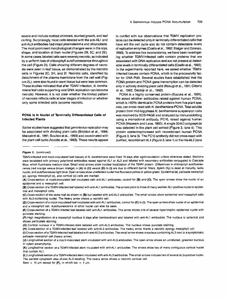
Figure 2. Localization of TGMV AL1 and AL3 Proteins in Nuclei of Differentiated Leaf and Stem Cells.
A Geminivirus lnduces PCNA Accumulation 709
severe and include mottled chlorosis, stunted growth, and leaf curling. Surprisingly, most cells labeled with the antiAL1 and antiAL3 antibodies had intact plasmalemma and chloroplasts. The most prominent morphological changes were in the size, shape, and location of their nuclei (Figures 28, 2C, and 2E). In some cases, labeled cells were severely necrotic, as indicated by a uniform loss of chlorophyll autofluorescence throughout the cell (Figure 21). Cells showing different degrees of necro- sis were seen in leaf tissue, as demonstrated by the necrotic cells in Figures 2C, 2H, and 21. Necrotic cells, identified by detachment of the plasma membrane from the cell wall (Fig- ure 2L), were also found in stem tissue but were less common. These studies indicated that after TGMV infection, N. bentha- miana leaf cells supporting vira1 DNA replication can become necrotic. However, it is not clear whether the limited pattern of necrosis reflects cells at later stages of infection or whether only some infected cells become necrotic.
PCNA 1s in Nuclei of Terminally Differentiated Cells of lnfected Plants
Earlier studies have suggested that geminivirus replication may be associated with dividing plant cells (Briddon et al., 1989; Matzeit et al., 1991; Boulton et al., 1993) and coordinated with the plant cell cycle (Accotto et al., 1993). These results appear
to conflict with our observations that TGMV replication pro- teins can be detected only in terminally differentiated cells that have left the cell cycle and do not contain detectable levels of replication enzymes (Coello et al., 1992; Staiger and Doonan, 1993). To address this inconsistency, we have been investigat- ing whether TGMV-infected cells contain proteins that are associated with DNA replication and are not present at detect- able levels in terminally differentiated cells (Coello et al., 1992). In the experiments reported here, we asked whether TGMV- infected tissues contain PCNA, which is the processivity fac- tor for DNA Po16. Severa1 studies have established that the PCNA protein and PCNA gene transcription can be detected only in actively dividing plant cells (Kosugi et al., 1991; Citterio et al., 1992; Daidoji et al., 1992).
PCNA is a highly conserved protein (Suzuka et al., 1991). We asked whether antibodies raised against human PCNA, which is >6O% identical to PCNA proteins from five plant spe- cies, can cross-react with N. benthamiana PCNA. Total soluble protein from mid-log phase N. benthamiana suspension cells was resolved by SDS-PAGE and analyzed by immunoblotting using a monoclonal antibody, PC10, raised against human ~
PCNA (Waseem and Lane, 1990). A single 33-kD polypeptide was detected in the plant cell extract (Figure 3, lane 4). This protein coelectrophoresed with recombinant human PCNA (Figure 3, lane 3). The PC10 antibody did not cross-react with purified, recombinant AL1 (Figure 3, lane 1) or the His-AL3 (lane
Figure 2. (continued).
TGMV-infected and mock-inoculated leaf tissues of N. benthamiana were fixed 16 days after agroinoculation unless otherwise stated. Sections were incubated with primary polyclonal antibodies raised against ALI or AL3 and labeled with secondary antibodies conjugated to Cascade Blue, which fluoresces medium blue. Small solid arrows show nuclear localization of the TGMV protein. Differences in chlorophyll autofluores- cence (red orange versus brown orange) in (A) to (C) versus (D) to (I) are due to different barrier filters. Xylem (Xy) is dead at maturity, lacks nuclei, and autofluoresces light blue. Open arrows show uninfected nuclei that fluoresce yellow or yellow green. Epidermal (e), palisade mesophyll (p), spongy mesophyll (s), and cortical (c) cells are marked. (A) Cross-section of mock-inoculated leaf incubated with anti-ALI antibodies; control for (B) and (C). The open arrows show the nuclei of an epidermal and a mesophyll cell. (B) Cross-section of a TGMV-infected leaf labeled with antiALl antibodies. The arrows point to three of many swollen ALI-positive nuclei in epider- mal and mesophyll cells. (C) Cross-section of the same leaf as shown in (B) but labeled with antiAL3 antibodies. The small arrows show epidermal and mesophyll cells with AL3-containing nuclei. The heavy arrow shows a necrotic cell. (D) Cross-section of a mock-inoculated leaf incubated with anti-ALI antibodies; control for (E) to (I). The open arrows show nuclei of an epidermal and a mesophyll cell. Autofluorescence of other nuclei can also be seen. (E) Cross-section of a TGMV-infected leaf labeled with anti-ALI antibodies. The arrow shows one of several hypertrophic epidermal nuclei with punctate staining. (F) High magnification of a mesophyll nucleus 6 days after bombardment and labeled with anti-ALI antibodies. The nucleus is spherical and shows particulate staining. (G) Cortical nucleus in a TGMV-infected stem labeled with anti-AL3 antibodies. The nucleus shows punctate staining. (H) Cross-section of a TGMV-infected leaf labeled with antiAL3 antibodies. The heavy arrow marks a necrotic spongy mesophyll cell. (I) Cross-section of a TGMV-infected leaf labeled with anti-AL3 antibodies. The small arrow shows a nucleus containing AL3 next to a symptomatic palisade mesophyll cell (heavy arrow). (J) Longitudinal section of a mock-inoculated stem incubated with antiAL3 antibodies. The open arrow shows an uninfected, greenish nucleus in xylem parenchyma. (K) Longitudinal section of a TGMV-infected stem incubated with anti-ALI antibodies. The arrows show two of many contiguous cortical nuclei that contain ALI. (L) Longitudinal section of aTGMV-infected stem incubated with antiAL3 antibodies. The small arrows indicate two of several AL3-positive nuclei. The parietal cytoplasm also shows AL3 labeling. The heavy arrow shows a necrotic cortical cell. Bars = 10 pm except for (F), in which bar = 1 pm.

710 The Plant Cell
AL1AL3
human PCNAplant cell extract
AL1-
PCNA-
HJS-AL3 -
-79
-50
-35-28
-20
1 2 3 4
Figure 3. Protein Gel Blot Analysis of Whole-Cell Extract Proteins Pre-pared from N. benthamiana Suspension Cells.Bacterially produced AL1 (lane 1), His-AL3 (lane 2), and PCNA (lane3) (10 ng each) and plant cell extract proteins (lane 4; 100 ug) wereresolved by SDS-PAGE and assayed by immunoblotting using anti-PCNA antibodies (PC10) and rabbit anti-mouse antibodies conjugatedto horseradish peroxidase as the primary and secondary antibodies,respectively. The positions of the molecular mass standards are indi-cated at right in kilodaltons. (+) indicates that a component was partof the extract separated on the protein gel; (-) indicates that a compo-nent was lacking.
2) protein. These results showed that the anti-PCNA antibodyspecifically recognizes N. benthamiana PCNA.
We then used the anti-PCNA antibody in immunolocaliza-tion experiments to examine TGMV- and mock-infected tissuesat identical stages of development for the presence of PCNA(Figure 4). The leaf tissue was from fully expanded leaves thatno longer contained actively dividing cells (Poethig and Sussex,1985). The PCNA antigen was found in nuclei of cells fromTGMV-infected leaf tissue (Figures 4B, 4F, and 4G). No PCNAlabeling was seen in mock-infected leaf tissue from fully ex-panded leaves (Figure 4A) or from leaves ~10 to 20% of theirfinal length. Analogous results were obtained for stem sec-tions of nondividing, differentiated cortical tissue. The anti-PCNA antibody labeled nuclei of infected stem tissue (Figures4D, 4E, and 4H) but did not label mock-inoculated stem tissue(Figure 4C). We were unable to detect PCNA in soluble pro-tein extracts from TGMV-infected tissue (data not shown) usinga variety of protein extraction and immunological techniques.This was probably due to problems with extraction of PCNAprotein from infected tissue or with dilution of the PCNA anti-gen in the extracts by proteins from uninfected cells.
One interpretation of our results is that the accumulationof PCNA in TGMV-infected tissue is related to a generalizedwound response resulting from viral infection. To address thispossibility, we asked whether N. benthamiana plants infectedwith the potyvirus potato virus X (PVX; Hemenway et al., 1988)also contain PCNA. PVX is an RNA virus that encodes its ownRNA-dependent RNA polymerase and replicates in the cyto-plasm. Sections from symptomatic PVX-infected leaves that
had been prepared and analyzed identically to TGMV-infectedplants showed no nuclear staining when incubated with theanti-PCNA antibody (data not shown). These results demon-strated that induction of PCNA accumulation is not due tovirus infection in general, but rather is specific to geminivirusinfection.
The distribution of PCNA in TGMV-infected tissue was simi-lar to the labeling patterns for AL1 andAL3. In leaf tissue, nucleiof a variety of differentiated cell types, including epidermal,palisade mesophyll, and spongy mesophyll cells, were labeledwith the anti-PCNA antibody (Figures 4B, 4F, and 4G). In stemtissue, PCNA was detected in fully expanded cells through-out the outer cortex and pith. Labeled cells in both tissues weregenerally clustered (Figures 4B and 4D), possibly reflectingsimultaneous invasion of viral DNA into contiguous cells dur-ing the infection process. The PCNA-positive cells in leaveswere highly vacuolated and had well-developed chloroplasts,both characteristic of differentiated cells. However, their nucleiwere hypertrophic and centrally located, similar to nuclei con-taining AL1 and AL3 antigens. PCNA labeling was uniformthroughout the nucleus and was not excluded from the nucleo-lus (Figures 4E, 4F, and 4H).
The similarities between PCNA, AL1, and AL3 labeling pat-terns may reflect expression of PCNA and the viral replicationproteins in the same cells. This hypothesis was addressed byusing anti-PCNA and anti-AL1 antibodies simultaneously toprobe leaf tissue (Figure 5). For these experiments, the anti-PCNA and anti-AL1 primary antibodies were detected usingsecondary antibodies conjugated to Cascade Blue and BODIPY-FL, respectively. This approach colocalized PCNA and AL1to nuclei of a number of differentiated cell types (data notshown). Colocalization of PCNA and AL1 in parenchyma cellsnear the leaf midvein is shown in Figures 5B and 5C, respec-tively. These results established that PCNA and AL1 can occurtogether in terminally differentiated cells.
PCNA Is in Terminally Differentiated Cells of PlantsTransgenic for AL1
DNA tumor viruses induce their hosts to produce the enzymesnecessary for viral DNA replication, usually through the ac-tion of a single or a few viral proteins (Chellappan et al., 1992;Fanning and Knippers, 1992; Nevins, 1992). Frequently, theseviral proteins also function in the initiation of viral replicationand/or control of viral gene expression (Fanning and Knippers,1992). Earlier studies have established that AL1 is involvedin both of these processes during the TGMV life cycle (Eagleet al., 1994; Fontes et al., 1994a, 1994b), making it a goodcandidate for the geminivirus protein responsible for PCNAaccumulation in TGMV-infected plants. To address this, weasked whether TGMV AL1 can induce the accumulation ofPCNA in transgenic N. benthamiana plants that contain theAL1 coding sequence under the control of the constitutive 35Spromoter from cauliflower mosaic virus and the 3' end of thenopaline synthase gene (Hanley-Bowdoin et al., 1990).

A Geminivirus Induces PCNA Accumulation 711
Figure 4. Localization of PCNA in Differentiated Stem and Leaf Cells of TGMV-lnfected Plants.
Sections were from plant material 10 days after agroinoculation unless otherwise stated. PCNA was labeled with PC10 antibodies and detectedusing an ABC horseradish peroxidase method.(A) Cross-section of a mock-inoculated leaf. There is no PCNA staining, and nuclei are difficult to see.(B) Cross-section of a TGMV-infected leaf. The arrows show PCNA-containing nuclei in mesophyll cells.(C) Longitudinal section of a mock-inoculated stem. The open arrows show uninfected nuclei.(D) Longitudinal section of a TGMV-infected stem. PCNA-containing nuclei are found in three cortical cells (arrows).(E) Longitudinal section of a TGMV-infected stem. The arrow shows a distorted nucleus containing PCNA.(F) Cross-section of a TGMV-infected leaf. The arrow shows a swollen, PCNA-containing nucleus in a palisade mesophyll cell.(G) Cross-section of a TGMV-infected leaf 18 days after agroinoculation. The arrow shows a spongy mesophyll nucleus with PCNA labeling.(H) Longitudinal section of a stem showing a PCNA-containing nucleus (arrow) in a cortical cell with well-developed chloroplasts.Bars = 10 um for (A) to (H).

712 The Plant Cell
Figure 5. Colocalization of PCNA and ALL
Cross-sections of TGMV-infected N. benthamiana leaf tissue were incubated with mouse anti-PCNA monoclonal and rabbit anti-AL1 polyclonalantibodies followed by incubation with anti-mouse biotinylated secondary antibodies and Cascade Blue-conjugated streptavidin and anti-rabbitBODIPY-FL-conjugated secondary antibodies. Two nuclei from midvein parenchyma cells show Colocalization.(A) Combined bright-field microscopy and UV fluorescence of a TGMV-infected leaf cross-section.(B) Same cross-section as shown in (A) but with PCNA localization (Cascade Blue fluorescence).(C) Same cross-section as shown in (A) but with AL1 localization (BODIPY-FL).The arrows show labeled nuclei. Xy, nonspecific xylem autofluorescence. Bar in (C) = 10 urn.
Tissue from transgenic plant line pMON455-9985, which wasshown previously to express functional AL1 protein (Hanley-Bowdoin et al., 1990), was excised from fully expanded leavesand probed with anti-PCNA and anti-AL1 antibodies. PCNAand AL1 were detected in nuclei of a variety of differentiatedcell types from transgenic tissue (Figure 6), albeit not asstrongly as in TGMV-infected nuclei. The pattern of AL1 andPCNA labeling in transgenic plant tissues was sporadic. Thiscontrasted with the pattern of labeling in virus-infected plants,which showed contiguous cells with AL1 or PCNA antigens.This most likely reflected variations in transgene expressionfrom the cauliflower mosaic virus 35S promoter. The propor-tion of cells labeled with AL1 antibodies was equivalent to theproportion of cells labeled with PCNA, suggesting that thosecells expressing AL1 also expressed PCNA. The morphologyof nuclei in cells expressing AL1 was similar to that of cellsexpressing PCNA (compare Figures 6B and 6C). Recent ex-periments have colocalized AL1 and PCNA to the same cells(data not shown).
PCNA was seen only in meristematic tissue of healthy, wild-type plants (Figure 7). We did not detect PCNA in healthy leavesthat were fully expanded, corresponding in development tothe TGMV-infected leaves, or in immature leaves that were ~2cm in length. PCNA-labeled cells of healthy floral meristemswere small, isodiametric, and uniform in size, lacked chlo-roplasts, and had round nuclei that, due to the small size ofthe cell, occupied a large proportion of the cell volume. Thismorphology was distinct from labeled cells of transgenic (Fig-ure 6) and virus-infected (Figure 4) plants, which were muchbigger, had large central vacuoles, and, in photosynthetictissues, contained chloroplasts. No PCNA antigen was detected
in differentiated leaf cells from N. benthamiana plants trans-formed with three tandem copies of TGMV B DNA (pMON337line 3427; Elmer et al., 1988a; data not shown), thereby estab-lishing that increased PCNA accumulation in the AL1 transgenicplants was not a general consequence of plant transforma-tion. The presence of AL1 and PCNA in terminally differentiatedleaf cells of transgenic plants expressing AL1 established thatAL1 can induce PCNA accumulation in intact plants indepen-dent of other viral proteins and the geminivirus infectionprocess.
DISCUSSION
Although DNA replication and cell division are central to thegrowth and development of all living organisms, little is knownabout the molecular mechanisms that mediate and coordinatethese processes in plants. This lack of information is strikingin view of the unique character of plant development. In ani-mal systems, studies of DNA tumor viruses have facilitatedthe identification and functional analysis of host replication andcell cycle regulatory factors. This is best demonstrated by ex-periments on the replication of simian virus 40 (SV40) and itsintegration into the mammalian cell cycle (Hamel et al., 1992;Wagaetal., 1994). It has been difficult to apply a similar strategyto the analysis of plant DNA replication and cell division be-cause the majority of plant viruses, including some DNAviruses, replicate through RNA intermediates. Geminivirusreplication has been studied extensively in plant tissue cul-ture, but there has been no information regarding viral

A Geminivirus Induces PCNA Accumulation 713
Figure 6. AL1 and PCNA Localization in Plants Transformed with the AL1 Gene.
Labeling was performed as given for Figure 4 using either PC10 or anti-AL1 primary antibodies as indicated.(A) Cross-section of a leaf midvein parenchyma cell from a control, nontransgenic plant incubated with anti-PCNA antibodies. The open arrowshows a wild-type unlabeled nucleus.(B) Cross-section of a leaf midvein parenchyma cell from a transgenic plant expressing AL1 incubated with anti-PCNA antibodies. The arrowshows a PCNA-containing nucleus.(C) Cross-section of a leaf midvein parenchyma cell from a transgenic plant expressing AL1 incubated with anti-AL1 antibodies. The arrow showsan AL1-containing nucleus.(D) Cross-section of a control, nontransgenic leaf incubated with anti-PCNA antibodies. The open arrows show elongated nuclei adjacent to thewalls in leaf palisade mesophyll and epidermal cells.(E) Cross-section of a leaf from a transgenic plant expressing AL1 incubated with anti-PCNA antibodies. The arrow shows a nucleus of a spongymesophyll cell with PCNA staining.(F) Cross-section of a leaf midvein parenchyma cell from a transgenic plant expressing AL1 incubated with anti-PCNA antibodies. The arrowshows a PCNA-containing nucleus.Bar in (F) = 10 urn for (A) to (F).

714 The Plant Cell
Figure 7. PCNA in N. benthamiana Floral Meristems from Healthy Plants.Tissue was stained as given for Figure 4.(A) Low magnification of meristematic cells. The arrow shows a PCNA-containing nucleus.(B) Same cells as shown in (A) but at a higher magnification. Arrow indicates the nucleus shown in (A).Bars in (A) and (B) = 10 \im.
replication in plants. In this research, we used immunolocali-zation to study geminivirus replication in plants. We showedthat geminivirus replication proteins are located in the nucleiof terminally differentiated cells in intact plants and thatgeminivirus infection induces the accumulation of the host DNAsynthesis protein, PCNA, in the same cells. We also showedthat the geminivirus replication protein, AL1, is sufficient toinduce the accumulation of PCNA in terminally differentiatedcells of transgenic plants. Together, these results strongly sug-gest that geminiviruses can induce the accumulation of DNAsynthesis machinery in quiescent cells of intact plants and thatAL1 plays a key role in the induction process, analogous tothe DNA tumor antigen proteins of mammalian viruses (Nevins,1992).
Immunolocalization experiments demonstrated that the AL1and AL3 replication proteins of the geminivirus TGMV are lo-cated in the nuclei of infected cells in intact plants (Figure 2).The nuclear localization of AL1 is consistent with its essentialroles as the origin recognition protein (Fontes et al., 1994a,1994b) and the nicking enzyme for initiation of rolling circlereplication (B.M. Orozco and L. Hanley-Bowdoin, unpublishedresults). AL3 is not required for replication but greatly enhancesthe level of viral DNA accumulation (Elmer et al., 1988a; Sunteret al., 1990). The nuclear location of AL3 suggests that it mayalso participate directly in replication or in stabilization of na-scent DNA. Immunolocalization experiments further establishedthat AL1 and AL3 are present in a variety of terminally differen-tiated cells of leaf and stem tissues (Figure 2). TGMV particleshave also been found in differentiated leaf cells (Rushing etal., 1987). TGMV infection of N. benthamiana may be unusualbecause of the large number of cell types that contain viralproteins and particles. In general, geminiviruses are thoughtto be confined to phloem parenchyma and companion cells
and to developing and mature sieve elements of vascular tis-sue (Hull, 1989; Horns and Jeske, 1991).
TGMV-infected cells and transgenic cells expressing AL1displayed an altered morphology, with their nuclei becomingspherical. Differentiated cells containing the PCNA antigenalso showed a change in nuclear structure. TGMV-infectednuclei occupied a much higher proportion of the total cell vol-ume than nuclei from transgenic plants expressing AL1(compare Figures 4F and 4G with 6E and 6F), indicating thatother factors in addition to AL1 are necessary to cause all ofthe alterations in nuclear morphology associated with geminivi-rus infection (Rushing et al., 1987). It is interesting that thenuclei of healthy, differentiated cells were elongated and foundadjacent to the cell wall (Figures 6A and 6D), whereas nucleiof meristematic cells were round, located toward the cell cen-ter (Figure 7B), and more closely resembled the TGMV-infectednuclei (Figures 4F and 4G). Nuclear rounding and migrationhave also been observed in alfalfa cortical cells after induc-tion of nodule formation by Rhizobium and preceding celldivision (Cooper and Long, 1994). Some infected cells showedpunctate labeling of AL1, AL3, and PCNA. The punctate label-ing may represent replication complexes (Hozak et al., 1994).PCNA has been associated with DNA replication foci in mam-malian cells (Bravo and MacDonald-Bravo, 1987; Cardoso etal., 1993). Alternatively, the punctate labeling could reflect fix-ation artifacts or the stage of viral infection. Different PCNAlabeling patterns have been seen depending on the methodof fixation (Waseem and Lane, 1990). Fibrillar spheres and in-clusion bodies characteristic of geminivirus infections (Rushinget al., 1987) might also distort labeling patterns of nuclearantigens.
Localization of TGMV AL1, TGMV AL3, and viral particlesto terminally differentiated cells of plants raises an important
A Geminivirus lnduces PCNA Accumulation 715
question: 1s geminivirus replication confined to cells competent for cell division or can geminiviruses induce the accumula- tion of host factors necessary for viral DNA replication? One possibility is that geminiviruses replicate in actively dividing plant cells, which provide the requisite host replication factors, and then move into differentiated cells. Cell-to-cell movement of DNA and the geminivirus movement protein BL1 in the ab- sence of viral replication was recently shown by microinjection of mature leaf cells (Noueiry et al., 1994). However, our results and those of Horns and Jeske (1991), who did not find gemini- virus DNA in actively dividing plant cells, cannot be explained by this hypothesis. Recent studies have shown that plasmodes- mata, the intercellular connections through which plant viruses move cell to cell, have distinct structural properties in different cell types (for review, see Lucas and Wolf, 1993). The plas- modesmata of meristematic cells may not be compatible with geminivirus movement proteins, thereby preventing gemini- viruses from moving into or out of meristematic cell populations in plants.
A second possibility is that geminiviruses replicate in termi- nally differentiated plant cells induced to accumulate host DNA replication factors. This hypothesis is supported by the local- ization of AL1 and AL3 to terminally differentiated cells and by the accumulation of the host DNA synthesis factor, PCNA, in the same cells. Unlike animals, most cells in mature plants have the capacity to dedifferentiate, resume cell division, and form new plants. Differentiated plant cells normally require wounding or hormone application to reenter the cell cycle (Gorst et al., 1991; Kosugi et al., 1991; Hemerly et al., 1993). The de- tection of PCNA in differentiated cells of transgenic plants expressing AL1 indicated that the AL1 protein can provide the stimulus necessary to induce the dedifferentiation process. Two lines of evidence suggest that AL1 is likely to act at the leve1 of cell cycle control. First, high levels of PCNA, which were the same as those detected in TGMV-infected plants, have been found only in actively dividing cells of plants (Kosugi et al., 1991; Citterio et al., 1992; Daidoji et al., 1992). Second, geminivirus-infected cells contain >104 viral genome copies per cell (Timmermans et al., 1992), indicating that they must be able to support efficient DNA replication, as occurs during S phase of the cell cycle. However, because PCNA is also as- sociated with DNA repair (Shivji et al., 1992), our data do not eliminate the possibility that the primary effect of AL1 is only on PCNA accumulation. Future experiments will determine whether AL1 also induces the expression of other cell cycle- related host factors.
Some of the morphological changes associated with dediffer- entiation of plant cells, that is, nuclear rounding and migration to the cell center, were observed in TGMV-infected and AL1- transformed plants. However, a decrease in cell size, a disap- pearance of the central vacuole, and an increase in cytoplasmic contents, which are also characteristic of undifferentiated plant cells (Gorst et al., 1991), were not seen (compare Figures 4G and 78). AL1 may only be able to trigger the early stages of
the dedifferentiation process leading to the accumulation of host replication factors, and other signals may be required for full dedifferentiation. Alternatively, AL1 may actively interfere with the progression through G2 phase and full dedifferenti- ation, thereby locking infected plant cells into S phase. This idea is consistent with the facts that cell division could not be detected in infected leaf cells and that transgenic tobacco cells expressing AL1 have significantly increased doubling times in suspension culture (T.J. Pedersen and L. Hanley-Bowdoin, unpublished results). Progression through G2 into M phase requires the activation of cell cycle-dependent protein kinases, such as cdc2. The SV40 large T antigen interferes with G2 transit by preventing the activation of cyclin B-cdc2 complexes during lytic infection (Scarano et al., 1994).
The ability of TGMV AL1 to induce the accumulation of a host protein associated with DNA replication in terminally differentiated plant cells suggested that it is a plant homolog of mammalian tumor antigens. Three very different mammalian DNA viruses encode proteins that modify cell cycle controls by interfering with the action of the host tumor suppressor pro- teins, pRb and p53 (Hamel et al., 1992; Nevins, 1992; Sherr, 1994). Binding of pRb by the SV40 large T antigen, adenovi- rus ElA, and the human papillomavirus E7 proteins results in the release of host transcription factors (Chellappan et al., 1992; Morris et al., 1994; Mudrak et al., 1994), which then in- duce expression of proteins required for DNA replication (Nevins, 1992). The adenovirus E1A protein can also activate transcription of the PCNA gene directly (Morris et al., 1994). TGMV AL1 may use similar mechanisms to induce synthesis of plant DNA replication proteins. The sequence-specific DNA binding activity of AL1 could be involved in direct transcrip- tional regulation of host genes. Alternatively, AL1 may interact with a plant tumor suppressor protein(s) and modify cell cycle regulation in terminally differentiated cells of infected plants. The feasibility of this idea is strengthened by the recent identifi- cation in Arabidopsis of cyclin D homologs showing conservation of the pRb interaction motif (Soni et al., 1995).
Some components of the plant cell cycle have been identi- fied by homology with fungal and animal proteins and by differential cDNA screening of synchronized suspension cells (for review, see Staiger and Doonan, 1993; Doerner, 1994). However, there is only limited functional information on plant cell cycle proteins, and this information is primarily from yeast complementation experiments and plant expression studies (Colasanti et al., 1993; Bergouniouxet al., 1994; Fobert et al., 1994). There is no direct evidence for the existence of tumor suppressor proteins in plants. Plant homologs to pRb or to the related p107 tumor suppressor protein have not been found. A protein related to the tumor suppressor protein p53 has been detected (Georgieva et al., 1994), but its function has not been demonstrated. The approaches based on homology and com- plementation of animal and fungal systems have provided valuable information regarding plant cell cycle components but are unlikely to uncover plant-specific features. Analysis of
716 The Plant Cell
the mechanism(s) whereby ALI induces the synthesis of a host DNA synthesis protein now provides a unique opportunity to study plant cell cycle regulation directly.
METHOOS
Plant Materlals
Nicotiana benthamiana plants were grown in a controlled environmental chamber at 25OC, 65% humidity, and a 14-hr-lightll0-hr-dark pho- toperiod. Plants were agroinoculated with tomato golden mosaic virus (TGMV) using mixed cultures of Agrobacterium fumefaciens contain- ing the T-DNA plasmids pMON337 (1.5 tandem copies of TGMV A) and pMON393 (1.5 tandem copies of TGMV B), as described by Elmer et al. (1988b). Tissues were harvested from infected plants exhibiting chlorosis and leaf curling at 12 to 18 days postinfection and from cor- responding plants mock-inoculated with water. Alternatively, plants at the 4tO 6 leaf stage were bombarded using a Bio-Rad Biolistic PDS- IOOOlHe system, according to the manufacturer's directions. Plasmid DNA containing partia1 tandem copies of the TGMV A (pTG1.3A) or TGMV B (pTG1.46) components (Fontes et al., 1994a) was purified using a Qiagen plasmid purification kit (Qiagen, Inc., Chatsworth, CA). DNA (5 wg of each plasmid) was coated onto I .O-pm microprojectiles and bombarded into plants at a pressure of 1300 psi. Aluminum foi1 was used to shield all but one expanded leaf on each plant. Tissues were harvested from systemically infected plants and control plants at 6 days postbombardment.
N. benthamiana plants transformed with the TGMV ALI protein (pMON455 line 9985; Hanley-Bowdoin et al., 1990) or TGMV B (pMON393 line 3427; Elmer et al., 1988a) were germinated on media containing 0.43% Murashige and Skoog salts (GIBCO BRL), 3% su- crose, 0.01% inositol, 0.0001 O/o thiamine-HCI, 0.02% KH2P04, 0.8% agar supplemented with 300 mglL kanamycin monosulfate and were transferred to soil at 3 weeks. Fully expanded leaves from 6-week-old plants were processed identically to TGMV-infected tissues. Symp- tomatic potato virus X (PVX)-infected N. benfhamiana plants were generated as described by Hemenway et al. (1988) and harvested at 5 days postinoculation. Vegetative and floral meristematic tissues were harvested from 6-week-old healthy plants (N. benfhamiana) and processed as described in the following sections.
Fixation and Sectioning of Plant Tissues
Leaves and stems were cut into 0.5- to 1 .O-cm pieces and fixed for 48 hr at room temperature in 10 mM PBS, pH 7.2, containing 4% form- aldehyde prepared from powdered paraformaldehyde (Fisher). Tissues were then stored in the fixative at 4°C until sectioned. Fixed tissues were embedded in 5% low-gelling-temperature agarose (Type XI; Sigma) in 10 mM PBS, pH 7.2, before sectioning. Thick tissue sec- tions (50 to 60 pm) were cut using a Vibratome 1000 (Technical Products International, Inc., St. Louis, MO). Tissue sections were then transferred to 10 mM PBS, pH 7.2, 0.1% BSA and stored at 4OC.
lmmunolocalization of Vira1 and Host Repllcatlon Proteins
All incubations were done at room temperature. Tissue sections were blocked in 0.5% goat IgG (Sigma) in 10 mM PBS, pH 7.4,0.1% BSA
(PBS-BSA) for 1 hr. After a 5-min rinse in PBS-BSA, sections were incubated for 1 hr with either an AL1-specific (Hanley-Bowdoin et al., 1990) or an AL3-specific (Pedersen and Hanley-Bowdoin, 1994) rab- bit polyclonal antiserum diluted 1:250 in PBS-BSA. After three 10-min rinses in PBS-BSA, sections were incubated for 1 hr with a Cascade Blue-conjugated goat anti-rabbit secondary antibody (Molecular Probes, Eugene, OR) diluted 1:250 in PBS-BSA. After three 10-min rinses in PBS-BSA, the sections were mounted on Fisher ProbeOn Plus slides in 10 mM PBS, pH 7.4, that contained 90Vo glycerol. Visu- alization of the ALI- and AL3-specific signals was achieved using a Nikon Optiphot-2 microscope with epifluorescence (Southern Micro Instruments, Atlanta, GA). Color photographs were produced using a Nikon UFX camera system and Kodak Gold ASA 400 film.
Proliferating cell nuclear antigen (PCNA) was localized in Vibratome sections of TGMV-infected, transgenic AL1 and healthy meristematic tissues using a Vectastain Elite ABC horseradish peroxidase kit (Vec- tor Laboratories, Burlingame, CA) according to the manufacturer's instructions with the following modifications. Sections were pretreated with 0.3% H202 in methanol for 30 min to quench endogenous per- oxidase activity and remove chlorophyll. After incubation for 1 hr with 1:lOO diluted mouse monoclonal antibodies raised against human PCNA (PC10; Santa Cruz Biotechnology, Inc., Santa Cruz, CA), sec- tions were incubated with biotinylated secondary antibodies and then with the avidin-biotinylated enzyme complex for 1 hr each. The 3-aminc- 9-ethylcarbazole substrate (Vector Laboratories) was used at a devel- opment time of 20 min. ldentical staining parameters were used for negative tissue controls. Sections were mounted as described previ- ously and imaged using a Nikon Optiphot microscope and Nomarski differential interference contrast optics. Photographs were produced using a Nikon UFX camera system and Kodak Royal Gold ASA 25 film.
For colocalization of TGMV ALl and host PCNA, fluorescent im- munolocalization was performed as described previously, but a biotinylated goat anti-mouse secondary antibody followed by Cascade Blue-conjugated streptavidin was used for PCNA, and a BODIPY-FL- conjugated goat anti-rabbit secondary antibody (Molecular Probes) was used for AL1.
Protein Analysis
Whole-cell extract proteins were prepared from exponentially grow- ing suspension cell cultures of N. benthamiana. Cells were collected on Whatman No. 1 filter paper, weighed, and transferred to a chilled mortar. Cells were ground under liquid N2 to a fine powder and sus- pended in whole-cell extract buffer (40 mM Tris-HCI, pH 7.5, 5 mM MgC12, O5 M sucrose, 10 mM P-mercaptoethanol, 0.8 mM phenylmeth- ylsulfonyl fluoride, 0.01 mM benzamidine, 0.05 mM E-aminocaproic acid, 5 p g h L leupeptin) at 2 mUg fresh weight. Cell debris was removed by filtration through Miracloth (Calbiochem, San Diego, CA). The fil- trate was adjusted to 500 mM NaCI, mixed for 1 hr at 4OC, and centrifuged at 125,0009 (1 hr at 4OC) to remove insoluble material. Solu- ble proteins were resolved by SDS-PAGE (12.5% polyacrylamide), transferred to nitrocellulose, and analyzed by immunoblotting (Pedersen and Hanley-Bowdoin, 1994). Protein blots were blocked in TBS (20 mM Tris-HCI, pH 7.4, 137 mM NaCI) and 5% nonfat dry milk (TBSM). Blots were incubated with the anti-PC10 antibody diluted 1:2000 in TBSM. Proteins were visualized after incubation with a sheep anti-mouse anti- body conjugated to horseradish peroxidase (diluted 1:5000 in TBS and 0.1% Tween 20) using an enhanced chemiluminescence detection sys- tem (Amersham).
The recombinant ALI protein was produced in Escherichia coli BL21(DE3) transformed with the bacterial expression cassette

A Geminivirus lnduces PCNA Accumulation 717
pMON1539, containing the AL1 open reading frame under the control of the T7 promoter (Hanley-Bowdoin et al., 1990). The ALI protein was purified from the refractile body fraction using SDS-PAGE as described previously (Hanley-Bowdoin et al., 1990). The recombinant His-AL3 protein was produced from E. coli transformed with the bacterial ex- pression cassette pNSB195, containing an AL3 open reading frame fusion with 10 histidine codons at its 5' end (Pedersen and Hanley- Bowdoin, 1994). The recombinant His-AW protein was purified from the refractile body fraction by Ni+-affinity chromatography in the pres- ente of 6 M guanidine-HCl (Pedersen and Hanley-Bowdoin, 1994). Recombinant human PCNA was expressed in E. coli and purified by Q-Sepharose and phenyl-Sepharose chromatography (Fien and Stillman, 1992).
ACKNOWLEDGMENTS
We thank Drs. Steven Bachenheimer, Flora Banuett, and Ross Whetten for critically reading the manuscript. We are grateful to Dr. Paul Fisher for providing purified, recombinant human PCNA protein and to Dr. Cynthia Hemenway for the WX-infected plants. We also acknowledge Dr. Tim Petty for providing the TGMV plasmids used for bombardment and Renee Werenko for help with the color prints. This work was sup- ported by National Science Foundation Grants Nos. MCB-9306608 (to D.R.) and DMB-9104150 (to L.H.-B.) and by United States Depart- ment of Agriculture Grant No. 93-37301-8786 (to L.H.-B). T.J.P. was supported by Public Health Service-National lnstitutes of Health post- doctoral fellowship No. A108511.
Received February 27, 1995; accepted April 26, 1995.
REFERENCES
Accotto, G.P., Mullineaux, P.M., Brown, S.C., and Marie, D. (1993). Digitaria streak geminivirus replicative forms are abundant in S-phase nuclei of infected cells. Virology 195, 257-259.
Aves, S.J., and Bryant, J.A. (1993). The molecular genetics and bio- chemistry of DNA replication. In Molecular and Cell Biology of the Plant Cell Cycle, J.C. Ormrod and D. Francis, eds (Dordrecht, The Netherlands: Kluwer Academic Publishers), pp. 45-56.
Bergounioux, C., Perennes, C., Hemerly, A.S., Qin, L.X., Sarda, C., Inzb, D., and Gadal, P. (1994). A cdc2 gene of &$unia hybrida is differentially expressed in leaves, protoplasts and during various cell cycle phases. Plant MOI. Biol. 20, 1121-1130.
Bisaro, D.M., Hamllton, W.D.O., Coutts, R.H.A., and Buck, K.W. (1982). Molecular cloning and characterization of the two DNA com- ponents of tomato golden mosaic virus. Nucleic Acids Res. 10,
Boulton, M.I., Pallaghy, C.K., Chatani, M., Macfarlane, S., and Davies, J.W. (1993). Replication of maize streak virus mutants in maize protoplasts-Evidence for a movement protein. Virology 192, 85-93.
Bravo, R., and MacDonald-Bravo, H. (1987). Existence of two popu- lations of cyclin/proliferating cell nuclear antigen during the cell cycle: Association with DNA replication sites. J. Cell Biol. 105, 1549-1554.
4913-4922.
Bravo, R., Frank, R., Blundell, P.A., and MacDonald-Bravo, H. (1987). CyclinlPCNA is lhe auxiliary protein of DNA polymerase-6. Nature
Briddon, R.W., Watts, J., Markham, P.G., and Stanley, J. (1989). The coat protein of beet curly top virus is essential for infectivity. Virol-
Brough, C., Sunter, G., Gardiner, W., and Bisaro, D. (1992). Kinetics of tomato golden mosaic virus replication and coat protein promoter activity in Nicotiana tabacum protoplasts. Virology 187, 1-9.
Brown, W.C., and Campbell, J.L. (1993). lnteraction of proliferating cell nuclear antigen with yeast DNA polymerase 6. J. Biol. Chem.
Cardoso, M.C., Leonhardt, H., and Nadal-Ginard, B. (1993). Rever- sal of terminal differentiation and control of DNA replication: Cyclin A and cdk2 specifically localize at subnuclear sites of DNA replica- tion. Cell 74, 979-992.
Chellappan, S., Kraus, V.B., Kroger, B., Munger, K., Howley, P.M., Phelps, P.C., and Nevlns, J.R. (1992). Adenovirus ElA, simian vi- rus 40 tumor antigen, and human papillomavirus E7 protein share the capacity to disrupt the interaction between transcription factor E2F and the retinoblastoma gene product. Proc. Natl. Acad. Sci.
Citterio, S., Sgorbati, S., Levi, M., Colombo, B.M., and Sparvoli, E. (1992). PCNA and total nuclear protein contentas markers of cell proliferation in pea tissue. J. Cell Sci. 102, 71-78.
Coello, P., Rodriguez, R., Garcia, E., and Vazquez-Ramos, J.M. (1992). A DNA polymerase from maize axes: Its purification and pos- sible role. Plant MOI. Biol. 20, 1159-1168.
Colasanti, J., Cho, S.-O., Wlck, S., and Sundaresan, V. (1993). Lo- calization of the functional ~ 3 4 ~ ~ ~ homolog of maize in root tip and stomatal complex cells: Association with predicted division sites. Plant Cell 5, 1101-1111.
Cooper, J.B., and Long, S.R. (1994). Morphogenetic rescue of Rhizo- bium meliloti nodulation mutants by trans-zeatin secretion. Plant Cell
Daidoji, H., Takasaki, Y., and Nakane, P.K. (1992). Proliferating cell nuclear antigen (PCNA/cyclin) in plant proliferating cells- Immuno- histochemical and quantitative analysis using autoantibody and murine monoclonal antibodies to PCNA. Cell Biochem. Funct. 10,
Doerner, P.W. (1994). Cell cycle regulation in plants. Plant Physiol.
Eagle, P.A., Orozco, B.M., and Hanley-Bowdoin, L. (1994). A DNA sequence required for geminivirus replication also mediates tran- scriptional regulation. Plant Cell 6, 1157-1170.
Elmer, J.S., Brand, L., Sunter, G., Gardlner, W.E., Bisaro, D.M., and Rogers, S.G. (1988a). Genetic analysis of tomato golden mosaic virus. II. Requirement for the product of the highly conserved ALI coding sequence for replication. Nucleic Acids Res. 16,7043-7060.
Elmer, J.S., Sunter, G., Gardlner, W.E., Brand, L., Browning, C.K., Bisaro, D.M., and Rogers, S.G. (1988b). Agrobacrerium-mediated inoculation of plants with tomato golden mosaic virus DNAs. Plant MOI. Biol. 10, 225-234.
326, 515-517.
Ogy 172, 628-633.
268, 21706-21710.
USA 89, 4549-4553.
6, 215-225.
123-132.
106, 823-827.
Esau, K. (1969). The Phloem. (Berlin: Gebruder Borntraeger).
Fanning, E., and Knippers, R. (1992). Structure and function of sim- ian virus-40 large tumor antigen. Annu. Rev. Biochem. 61, 55-85.
Fien, K., and Stillman, B. (1992). ldentification of replication factor C from Sacchammyces cerevisiae: A component of the leading strand DNA replication complex. MOI. Cell. Biol. 12, 155-163.

718 The Plant Cell
Fobert, P.R., Coen, E.S., Murphy, G.J.P., and Doonan, J.H. (1994). Patterns of cell division revealed by transcriptional regulation of genes during the cell cycle in plants. EM60 J. 13, 616-624.
Fontes, E.P.B., Luckow, V.A., and Hanley-Bowdoin, L. (1992). A geminivirus replication protein is a sequence-specific DNA binding protein. Plant Cell 4, 597-608.
Fontes, E.P.B., Gladfelter, H.J., Schaffer, R.L., Petty, I.T.D., and Hanley-Bowdoin, L. (1994a). Geminivirus replication origins have a modular organization. Plant Cell 6, 405-416.
Fontes, E.P.B., Eagle, P.A., Sipe, P.S., Luckow, V.A., and Hanley- Bowdoin, L. (1994b). lnteraction between a geminivirus replication protein and origin DNA& essential for viral replication. J. Biol. Chem.
Georgieva, E.I., Lopez-Rodas, G., and Loidl, P. (1994). Maize em- bryo germination. II. Proteins related to nuclear proto-oncogene- and tumor suppressor gene-products. Planta 192, 125-129.
Gorst, J., Sek, F.J., and John, P.C.L. (1991). Levels of p34cdc2-like protein in dividing, differentiating and dedifferentiating cells of car- rot. Planta 185, 304-310.
Groning, R.R., Frlschmuth, T., and Jeske, H. (1990). Replicative form DNA of abutilon mosaic virus is present in plastids. MOI. Gen. Ge- net. 220, 485-488.
Hamel, P.A., Gallie, B.I., and Phillips, R.A. (1992). The retinoblastoma protein and cell cycle regulation. Trends Genet. 8, 180-185.
Hamilton, W.D.O., Stein, V.E., Coutts, R.H.A., and Buck, K.W. (1984). Complete nucleotide sequence of the infectious cloned DNA com- ponents of tomato golden mosaic virus: Potential coding regions and regulatory sequences. EM60 J. 3, 2197-2205.
Hanley-Bowdoin, L., Elmer, J.S., and Rogers, S.G. (1989). Functional expression of the leftward open reading frames of the A component of tomato golden mosaic virus in transgenic tobacco plants. Plant Cell 1, 1057-1067.
Hanley-Bowdoin, L., Elmer, J.S., and Rogers, S.G. (1990). Expres- sion of functional replication protein from tomato golden mosaic virus in transgenic tobacco plants. Proc. Natl. Acad. Sci. USA 87,
Hemenway, C., Fang, R.X., Kaniewski, W., Chua, N.-H., and Tumer, N.E. (1988). Analysis of the mechanism of protection in transgenic plants expressing the potato virus X coat protein or its antisense
Hemerly, A.S., Ferreira, P., de Almeida Engler, J., Van Montagu, M., Engler, G., and InzB, D. (1993). cdc2A expression in Arabidop- sis is linked with competence for cell division. Plant Cell5,17I1-1723.
Heyraud, F., Matzeit, V., Kammann, M., Schaefer, S., Schell, J., and Gronenborn, B. (1993). ldentification of the initiation sequence for viral-strand DNA synthesis of wheat dwarf virus. EM60 J. 12,
Horns, T., and Jeske, H. (1991). Localization of abutilon mosaic virus (AbMV) DNAwithin leaf tissue byin situ hybridization. Virology 181,
Hozak, P., Jackson, D., and Cook, P. (1994). Replication factories and nuclear bodies: The ultrastructural characterization of replica- tion sites during the cell cycle. J. Cell Sci. 107, 2191-2202.
Hull, R. (1989). The movement of viruses in plants. Annu. Rev. Phytopathol. 27, 213-240.
Ilyina, T., and Koonin, E. (1992). Conserved sequence motifs in the initiator proteins for rolling circle DNA replication encoded by diverse
269, 8459-8465.
1446-1450.
RNA. EMBO J. 7, 1273-1280.
4445-4452.
580-588.
replicons from eubacteria, eucaryotes and archaebacteria. Nucleic Acids Res. 20, 3279-3285.
Kodama, H., Ito, M., Oshnishi, N., Suzaka, I., and Komamine, A. (1991). Molecular cloning of the gene for plant proliferating-cell nu- clear antigen and expression of this gene during the cell cycle of Catharanthus roseus cells. Eur. J. Biochem. 157, 459-503.
Kosugi, S., Suruka, I., Ohashi, Y., Murakami, T., and Arai, Y. (1991). Upstream sequences of rice proliferating cell nuclear antigen (PCNA) gene mediate expression of PCNA-GUS chimeric gene in meristems of transgenic tobacco plants. Nucleic Acids Res. 19, 1571-1576.
Laquel, P., Litvak, S., and Castroviejo, M. (1993). Mammalian prolifer- ating cell nuclear antigen stimulates the processivity of two wheat embryo DNA polymerases. Plant Physiol. 102, 107-1 14.
Lazarowitz, S. (1992). Geminiviruses: Genome structure and gene function. Crit. Rev. Plant Sci. 11, 327-349.
Lazarowitz, S.G., Wu, L.C., Rogers, S.G., and Elmer, J.S. (1992). Sequence-specific interaction with the viral AL1 protein identifies a geminivirus DNA replication origin. Plant Cell 4, 799-809.
Lucas, W., and Wolf, S. (1993). Plasmodesmata: The intercellular or. ganelles of green plants. Trends Cell Biol. 3, 308-315.
Martinez, M.C., Jorgensen, J.-E., Lawton, M.A., Lamb, C.J., and Doerner, P.J. (1992). Spatial pattern of cdc2 expression in relation to meristem activity and cell proliferation during plant development. Proc. Natl. Acad. Sci. USA 89, 7360-7364.
Matsumoto, T., Hata, S., Suzuka, I., and Hashimoto, J. (1994). Ex- pression of functional proliferating-cell nuclear antigen from rice (Oryza sativa) in fscherichia coli. Eur. J. Biochem. 223, 179-187.
Matzeit, V., Schaefer, S., Kammann, M., Schalk, H.J., Schell, J., and Gronenborn, B. (1991). Wheat dwarf virus vectors replicate and express foreign genes in cells of monocotyledonous plants. Plant Cell 3, 247-258.
Morris, G.F., Labrie, C., and Mathews, M.B. (1994). Modulation of transcriptional activation of the proliferating cell nuclear antigen pro- moter by the adenovirus-E1A 243-residue oncoprotein depends on proximal activators. MOI. Cell. Biol. 14, 543-553.
Mudrak, I., Ogris, E., Rotheneder, H., and Wintersberger, E. (1994). Coordinated trans activation of DNA synthesis- and precursor- producing enzymes by polyomavirus large T antigen through inter- action with the retinoblastoma protein. MOI. Cell. Biol. 14,1886-1892.
Nevins, J. (1992). E2F-A link between the Rb tumor suppressor pro- tein and viral oncoproteins. Science 258, 424-429.
Noueiry, A.O., Lucas, W.J., and Gilbertson, R.L. (1994). Two pro- teins of a plant DNA virus coordinate nuclear and plasmodesmal transport. Cell 76, 925-932.
Pedersen, T.J., and Hanley-Bowdoin, L. (1994). Molecular charac- terization of the AL3 protein encoded by a bipartite geminivirus. Virology 202, 1070-1075.
Poethig, R.S., and Sussex, I.M. (1985). The developmental morphol- ogy and growth dynamics of the tobacco leaf. Planta 165,158-169.
Revington, G.N., Sunter, G., and Bisam, D.M. (1989). DNAsequences essential for replication of the B genome component of tomato golden mosaic virus. Plant Cell 1, 985-992.
Richard, M.C., Litvak, S., and Castroviejo, M. (1991). DNA polymer- ase B from wheat embryos: A plant 6-like DNA polymerase. Arch. Biochem. Biophys. 287, 141-150.
A Geminivirus lnduces PCNA Accumulation 719
Rushing, A.E., Sunter, G., Gardiner, W.E., Dute, R.R., and Bisaro, D.M. (1987). Ultrastructural aspects of tomato golden mosaic virus infection in tobacco. Phytopathology 77, 1231-1236.
Saunders, K., Lucy, A., and Stanley, J. (1991). DNA forms of the geminivirus African cassava mosaic virus consistent with a rolling circle mechanism of replication. Nucleic Acids Res. 19,2325-2330.
Scarano, F.J., Laffin, J.A., Lehman, J.M., and Friedrich, T.D. (1994). Simian virus 40 prevents activation of M-phase-promoting factor dur- ing lytic infection. J. Virol. 68, 2355-2361.
Sherr, C.J. (1994). G1 phase progression: Cycling on cue. Cell 79,
Shivji, M., Kenny, M., and Wood, R. (1992). Proliferating cell nuclear antigen is required for DNA excision repair. Cell 69, 367-374.
Soni, R., Carmichael, J.P., Shah, Z.H., and Murray, J.A.H. (1995). A family of cyclin D homologs from plants differentially controlled by growth regulators and containing the conserved retinoblastoma protein interaction motif. Plant Cell 7, 85-103.
Staiger, C., and Doonan, J. (1993). Cell division in plants. Curr. Opin. Cell Biol. 5, 226-231.
Stanley, J. (1995). Analysis of African cassava mosaic virus recom- binants suggests strand nicking occurs within the conserved nonanucleotide motif during the initiation of rolling circle DNA repli- cation. Virology 206, 707-712.
Stenger, D.C., Revington, G.N., Stevenson, M.C., and Bisaro, D.M. (1991). Replicational release of geminivirus genomes from tandemly repeated copies: Evidence for rolling-circle replication of a plant vi- ral DNA. Proc. Natl. Acad. Sci. USA 88, 8029-8033.
Stenger, D.C., Davis, K.R., and Bisaro, D.M. (1992). Limited replica- tion of tomato golden mosaic virus DNA in explants of nonhost species. MOI. Plant-Microbe Interact. 5, 525-527.
Sunter, G., and Bisaro, D.M. (1989). Transcription map of the B ge- nome component of tomato golden mosaic virus and comparison with A component transcripts. Virology 173, 647-655.
551-555.
Sunter, G., Hertitz, M.D., Hormuzdi, S.G., Brough, C.L., and Bisaro, D.M. (1990). Genetic analysis of tomato golden mosaic virus: ORF AL2 is required for coat protein accumulation while ORFAL3 is neces- sary for efficient DNA replication. Virology 179, 69-77.
Suzuka, I., Daidoju, H., Matsuoka, M., Kadowaki, K., Takasaki, Y., Nakane, P.K., and Moriuchi, T. (1989). Gene for proliferating-cell nuclear antigen (DNA polymerase 6 auxiliary protein) is present in both mammalian and higher plant genomes. Proc. Natl. Acad. Sci.
Suzuka, I., Hata, S., Matsuoka, M., Kosugi, S., and Hashimoto, J. (1991). Highly conserved structure of proliferating cell nuclear anti- gen (DNA polymerase S auxiliary protein) gene in plants. Eur. J. Biochem. 197, 571-575.
Timmermans, M.C.P., Das, O.P., and Messing, J. (1992). trans Repli- cation and high copy numbers of wheat dwarf virus vectors in maize cells. Nucleic Acids Res. 20, 4047-4054.
Timmermans, M., Das, O., and Messing, J. (1994). Geminiviruses and their uses as extrachromosomal replicons. Annu. Rev. Plant Physiol. Plant MOI. Biol. 45, 79-112.
Townsend, R., Watts, J., and Stanley, J. (1986). Synthesis of vira1 DNA forms in Nicotiana plumbaginifolia protoplasts inoculated with cassava latent virus (CLV): Evidence for the independent replica- tion of one component of the CLV genome. Nucleic Acids Res. 14,
Waga, S., and Stillman, B. (1994). Anatomy of a DNA replication fork revealed by reconstitution of SV40 DNA replication in vitro. Nature
Waga, S., Bauer, G., and Stlllman, B. (1994). Reconstitution of com- plete SV40 DNA replication with purified replication factors. J. Biol. Chem. 269, 10923-10934.
Waseem, N.H., and Lane, D.P. (1990). Monoclonal antibody analysis of the proliferating cell nuclear antigen (PCNA). J. Cell Sci. 96,
USA 86, 3189-3193.
1253-1266.
369, 207-212.
'
121-129.

DOI 10.1105/tpc.7.6.705 1995;7;705-719Plant Cell
S Nagar, T J Pedersen, K M Carrick, L Hanley-Bowdoin and D Robertsonplant cells.
A geminivirus induces expression of a host DNA synthesis protein in terminally differentiated
This information is current as of May 23, 2018
Permissions X
https://www.copyright.com/ccc/openurl.do?sid=pd_hw1532298X&issn=1532298X&WT.mc_id=pd_hw1532298
eTOCs http://www.plantcell.org/cgi/alerts/ctmain
Sign up for eTOCs at:
CiteTrack Alerts http://www.plantcell.org/cgi/alerts/ctmain
Sign up for CiteTrack Alerts at:
Subscription Information http://www.aspb.org/publications/subscriptions.cfm
is available at:Plant Physiology and The Plant CellSubscription Information for
ADVANCING THE SCIENCE OF PLANT BIOLOGY © American Society of Plant Biologists