A Gastrosplenic Trunk in Association with a Replaced ...
4
312 © 2020 Journal of the Anatomical Society of India | Published by Wolters Kluwer - Medknow Address for correspondence: Prof. Petru Matusz, Department of Anatomy and Embryology, “Victor Babes” University of Medicine and Pharmacy Timisoara, Eftimie Murgu Square, No. 2, RO–300040 Timisoara, TM, România. E‑mail: [email protected] Access this article online Website: www.jasi.org.in DOI: 10.4103/JASI.JASI_78_19 Quick Response Code: Abstract The authors report the case of a 51‑year‑old male that shown to have, independently of the vascular lesions of the lower limbs, the presence of a gastrosplenic trunk (GST) in association with a replaced common hepatic artery (CHA), arising from the superior mesenteric artery by multidetector computed tomography angiography. The GST with an endoluminal diameter of 5.6 mm at its origin and a length of 18.8 mm arose from the anterior wall of the AA at the level of middle 1/3 rd of the L1 vertebral body. The replaced CHA with an endoluminal diameter at the origin of 4.8 mm has a lateral right ascendent path, initial in front of the superior mesenteric vein, in the middle portion penetrating the pancreatic parenchyma, in the terminal portion crossing the anterior aspect of hepatic portal vein, and finally fork in the gastroduodenal artery and hepatic artery proper. The knowledge of the variations of the GST and the replaced CHA is important for planning and performing procedures such as duodenopancreatectomy and liver transplantation. Keywords: Anatomic variants, gastrosplenic trunk, multidetector computed tomography angiography, replaced common hepatic artery, superior mesenteric artery A Gastrosplenic Trunk in Association with a Replaced Common Hepatic Artery Arising Independently from the Superior Mesenteric Artery: A Case Report using Multidetector Computed Tomography Angiography Laura‑Andreea Bolintineanu, Nicoleta Iacob 1 , Petru Matusz, Agneta Maria Pusztai Department of Anatomy and Embryology, “Victor Babes”University of Medicine and Pharmacy, Timisoara, România, 1 Department of Multidetector Computed Tomography and Magnetic Resonance Imaging, Neuromed Diagnostic Imaging Centre,Timisoara, România How to cite this article: Bolintineanu LA, Iacob N, Matusz P, Pusztai AM. A gastrosplenic trunk in association with a replaced common hepatic artery arising independently from the superior mesenteric artery: A case report using multidetector computed tomography angiography. J Anat Soc India 2020;68:312‑5. Introduction The trifurcation of the celiac trunk (CT) first described by Haller in 1756 and known in classical anatomy as “tripod of Haller” divides into the left gastric artery (LGA), common hepatic artery (CHA), and splenic artery (SA). [1] Reviewing the literature, Venieratos et al. [2] highlight the gastrosplenic trunk (GST) in classifications made by Lipsutz (1917), Adachi (1928), Morita (1935), and Michel (1955). Song et al., [3] analyzing CT variations on 5002 patients, highlighted GST in 2.86% of cases, out of which, in 2.64% of cases, the presence of GST is associated with a hepatomesenteric trunk (HMT), and in 0.22% of cases, GST is associated with the separate origin of CHA from the abdominal aorta (AA). Analyzing the CHA variants, the authors highlight in 3% of cases the origin of CHA from the superior mesenteric artery (SMA). The study documents, by multidetector computed tomography (MDCT) angiography, an extremely rare case with the presence of a GST in association with a replaced CHA arising from the SMA. Case Report We report the case of a 51‑year‑old male with a 22‑year history of smoking and peripheral vascular disease of the lower limbs for 6 years. Using MDCT angiography (64‑slice MDCT system; SOMATOM Sensation, Siemens Medical Solutions, Forchheim, Germany), the patient was shown to have, independently of the vascular lesions of the lower limbs, the presence of a GST in association with a replaced CHA arising from the SMA [Figure 1]. The GST with an endoluminal diameter of 5.6 mm at its origin and a length of 18.8 mm arose from the anterior wall of the AA at the level of middle 1/3 rd of the L1 vertebral body, 8 mm above the origin of the SMA. It forks into LGA with an endoluminal diameter at the origin of 2.9 mm and SA with an endoluminal diameter at the origin of 5.4 mm; the trunk of SMA with an endoluminal diameter at the origin of 9.6 mm had a length of 37.4 mm to the point of origin of the replaced CHA. The CHA with an endoluminal diameter at This is an open access journal, and arcles are distributed under the terms of the Creave Commons Aribuon‑NonCommercial‑ShareAlike 4.0 License, which allows others to remix, tweak, and build upon the work non‑commercially, as long as appropriate credit is given and the new creaons are licensed under the idencal terms. For reprints contact: [email protected] Received: 15 June 2019 Accepted: 16 January 2020 Available online: 28 February 2020 Article Info Case Report [Downloaded free from http://www.jasi.org.in on Monday, August 24, 2020, IP: 10.232.74.22]
Transcript of A Gastrosplenic Trunk in Association with a Replaced ...

312 © 2020 Journal of the Anatomical Society of India | Published by Wolters Kluwer - Medknow
Address for correspondence: Prof. Petru Matusz, Department of Anatomy and Embryology, “Victor Babes” University of Medicine and Pharmacy Timisoara, Eftimie Murgu Square, No. 2, RO–300040 Timisoara, TM, România. E‑mail: [email protected]
Access this article online
Website: www.jasi.org.inDOI: 10.4103/JASI.JASI_78_19
Quick Response Code:
AbstractThe authors report the case of a 51‑year‑old male that shown to have, independently of the vascular lesions of the lower limbs, the presence of a gastrosplenic trunk (GST) in association with a replaced common hepatic artery (CHA), arising from the superior mesenteric artery by multidetector computed tomography angiography. The GST with an endoluminal diameter of 5.6 mm at its origin and a length of 18.8 mm arose from the anterior wall of the AA at the level of middle 1/3rd of the L1 vertebral body. The replaced CHA with an endoluminal diameter at the origin of 4.8 mm has a lateral right ascendent path, initial in front of the superior mesenteric vein, in the middle portion penetrating the pancreatic parenchyma, in the terminal portion crossing the anterior aspect of hepatic portal vein, and finally fork in the gastroduodenal artery and hepatic artery proper. The knowledge of the variations of the GST and the replaced CHA is important for planning and performing procedures such as duodenopancreatectomy and liver transplantation.
Keywords: Anatomic variants, gastrosplenic trunk, multidetector computed tomography angiography, replaced common hepatic artery, superior mesenteric artery
A Gastrosplenic Trunk in Association with a Replaced Common Hepatic Artery Arising Independently from the Superior Mesenteric Artery: A Case Report using Multidetector Computed Tomography Angiography
Laura‑Andreea Bolintineanu, Nicoleta Iacob1, Petru Matusz, Agneta Maria PusztaiDepartment of Anatomy and Embryology, “Victor Babes”University of Medicine and Pharmacy, Timisoara, România, 1Department of Multidetector Computed Tomography and Magnetic Resonance Imaging, Neuromed Diagnostic Imaging Centre,Timisoara, România
How to cite this article: Bolintineanu LA, Iacob N, Matusz P, Pusztai AM. A gastrosplenic trunk in association with a replaced common hepatic artery arising independently from the superior mesenteric artery: A case report using multidetector computed tomography angiography. J Anat Soc India 2020;68:312‑5.
IntroductionThe trifurcation of the celiac trunk (CT) first described by Haller in 1756 and known in classical anatomy as “tripod of Haller” divides into the left gastric artery (LGA), common hepatic artery (CHA), and splenic artery (SA).[1] Reviewing the literature, Venieratos et al.[2] highlight the gastrosplenic trunk (GST) in classifications made by Lipsutz (1917), Adachi (1928), Morita (1935), and Michel (1955). Song et al.,[3] analyzing CT variations on 5002 patients, highlighted GST in 2.86% of cases, out of which, in 2.64% of cases, the presence of GST is associated with a hepatomesenteric trunk (HMT), and in 0.22% of cases, GST is associated with the separate origin of CHA from the abdominal aorta (AA). Analyzing the CHA variants, the authors highlight in 3% of cases the origin of CHA from the superior mesenteric artery (SMA). The study documents, by multidetector computed tomography (MDCT) angiography, an extremely rare case with the presence of a GST in association with a replaced CHA arising from the SMA.
Case ReportWe report the case of a 51‑year‑old male with a 22‑year history of smoking and peripheral vascular disease of the lower limbs for 6 years. Using MDCT angiography (64‑slice MDCT system; SOMATOM Sensation, Siemens Medical Solutions, Forchheim, Germany), the patient was shown to have, independently of the vascular lesions of the lower limbs, the presence of a GST in association with a replaced CHA arising from the SMA [Figure 1]. The GST with an endoluminal diameter of 5.6 mm at its origin and a length of 18.8 mm arose from the anterior wall of the AA at the level of middle 1/3rd of the L1 vertebral body, 8 mm above the origin of the SMA. It forks into LGA with an endoluminal diameter at the origin of 2.9 mm and SA with an endoluminal diameter at the origin of 5.4 mm; the trunk of SMA with an endoluminal diameter at the origin of 9.6 mm had a length of 37.4 mm to the point of origin of the replaced CHA. The CHA with an endoluminal diameter at
This is an open access journal, and articles are distributed under the terms of the Creative Commons Attribution‑NonCommercial‑ShareAlike 4.0 License, which allows others to remix, tweak, and build upon the work non‑commercially, as long as appropriate credit is given and the new creations are licensed under the identical terms.
For reprints contact: [email protected]
Received: 15 June 2019 Accepted: 16 January 2020 Available online: 28 February 2020
Article Info
Case Report
[Downloaded free from http://www.jasi.org.in on Monday, August 24, 2020, IP: 10.232.74.22]

Bolintineanu, et al.: Gastrosplenic trunk and a replaced common hepatic artery from the superior mesenteric artery
Journal of the Anatomical Society of India ¦ Volume 68 ¦ Issue 4 ¦ October-December 2019 313
the origin of 4.8 mm has a lateral right ascendant path, initial in front of the superior mesenteric vein (SMV), in the middle portion penetrating the pancreatic parenchyma, in the terminal portion crossing the anterior aspect of hepatic portal vein (HPV), and finally after 36.9 mm, fork in gastroduodenal artery (GDA) and hepatic artery proper (HAP) which are forking in the right and left branches.
Consent
For the X‑ray examination by using a 64‑slice MDCT system, and use of iodinated contrast agents, a written informed consent was obtained from the patient. Also, a written informed consent has been requested from the
patient for publication of this case report and accompanying images.
DiscussionEmbryology
The developmental changes in ventral segmental arteries (VSAs) result in CT and SMA anatomical variations.[1] From top to bottom, the four VSAs (from 10th to 13th) become the LGA, SA, CHA, and SMA, respectively. These four VSAs are united in the initial stages of embryo‑fetal development by “longitudinal anastomosis.” According to the extent of the resorption/retention of these structures, many anatomical variants of CT and SMA develop.[1] In our case,
Figure 1: Multidetector computed tomography angiography of the AA and liver. (a‑c) ‑ volume rendering technique (VRT) images of the AA (a) anterolateral left aspect; (b) anterior aspect; (c) anterior aspect after substraction of the AA. (d and e) ‑ maximum intensity projection (MIP) images of the liver; (d) coronal aspect; (e) transversal aspect. AA: Abdominal aorta, GST: Gastrosplenic trunk, LGA: Left gastric artery, SA: Splenic artery, SMA: Superior mesenteric artery, RCHA: Replaced common hepatic artery, GDA: Gastroduodenal artery, HAP: Hepatic artery proper, RBr: Right branch, LBr: Left branch, HPV: Hepatic portal vein, SMV: Superior mesenteric vein, L: Liver
d
cba
e
[Downloaded free from http://www.jasi.org.in on Monday, August 24, 2020, IP: 10.232.74.22]
Bolintineanu, et al.: Gastrosplenic trunk and a replaced common hepatic artery from the superior mesenteric artery
314 Journal of the Anatomical Society of India ¦ Volume 68 ¦ Issue 4 ¦ October-December 2019
the development of GST in association with the replaced CHA arising from SMA is embryologically similar to the development of GST in association with HMT (Morita Type IV). On the one hand, the longitudinal anastomosis persists between the 10th and the 11th VSA. The 11th root of the VSA regresses and its distal part connects through longitudinal anastomosis with the 10th VSA to form the GST. On the other hand, the 13th VSA will form SMA; the 12th root of the VSA regresses, and its distal part connects through longitudinal anastomosis with the 13th VSA to lead to the replaced CHA from SMA. The difference between HMT formation and replaced CHA arising from SMA is represented by more distal connection of 12th VSA to the 13th VSA (will form SMA) compared to the connection level in the case of HMT.
Anatomic variations and clinical implications
Lipsutz in 1917[2] described four types of the variational pattern of CT, the Type IV being represented by GST in association with CHA arising from the AA, with a prevalence of 3.62% of cases. Adachi[4] highlights the GST in association with ARHA from SMA in 1.98% of cases. The GST in association with HMT has been highlighted with a prevalence between 0.40%[4] and 2.64%.[3]
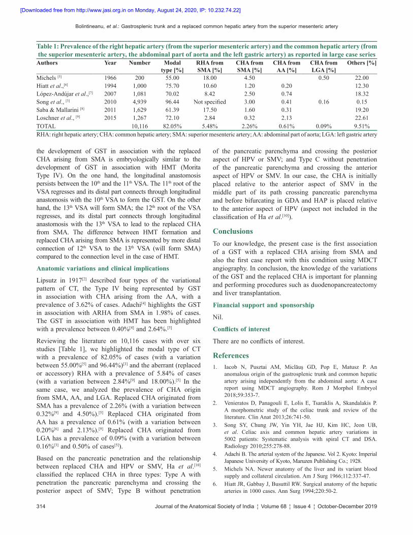
Reviewing the literature on 10,116 cases with over six studies [Table 1], we highlighted the modal type of CT with a prevalence of 82.05% of cases (with a variation between 55.00%[5] and 96.44%)[3] and the aberrant (replaced or accessory) RHA with a prevalence of 5.84% of cases (with a variation between 2.84%[9] and 18.00%).[5] In the same case, we analyzed the prevalence of CHA origin from SMA, AA, and LGA. Replaced CHA originated from SMA has a prevalence of 2.26% (with a variation between 0.32%[9] and 4.50%).[5] Replaced CHA originated from AA has a prevalence of 0.61% (with a variation between 0.20%[6] and 2.13%).[9] Replaced CHA originated from LGA has a prevalence of 0.09% (with a variation between 0.16%[3] and 0.50% of cases[5]).
Based on the pancreatic penetration and the relationship between replaced CHA and HPV or SMV, Ha et al.[10] classified the replaced CHA in three types: Type A with penetration the pancreatic parenchyma and crossing the posterior aspect of SMV; Type B without penetration
of the pancreatic parenchyma and crossing the posterior aspect of HPV or SMV; and Type C without penetration of the pancreatic parenchyma and crossing the anterior aspect of HPV or SMV. In our case, the CHA is initially placed relative to the anterior aspect of SMV in the middle part of its path crossing pancreatic parenchyma and before bifurcating in GDA and HAP is placed relative to the anterior aspect of HPV (aspect not included in the classification of Ha et al.[10]).
ConclusionsTo our knowledge, the present case is the first association of a GST with a replaced CHA arising from SMA and also the first case report with this condition using MDCT angiography. In conclusion, the knowledge of the variations of the GST and the replaced CHA is important for planning and performing procedures such as duodenopancreatectomy and liver transplantation.
Financial support and sponsorship
Nil.
Conflicts of interest
There are no conflicts of interest.
References1. Iacob N, Pusztai AM, Miclăuş GD, Pop E, Matusz P. An
anomalous origin of the gastrosplenic trunk and common hepatic artery arising independently from the abdominal aorta: A case report using MDCT angiography. Rom J Morphol Embryol 2018;59:353‑7.
2. Venieratos D, Panagouli E, Lolis E, Tsaraklis A, Skandalakis P. A morphometric study of the celiac trunk and review of the literature. Clin Anat 2013;26:741‑50.
3. Song SY, Chung JW, Yin YH, Jae HJ, Kim HC, Jeon UB, et al. Celiac axis and common hepatic artery variations in 5002 patients: Systematic analysis with spiral CT and DSA. Radiology 2010;255:278‑88.
4. Adachi B. The arterial system of the Japanese. Vol 2. Kyoto: Imperial Japanese University of Kyoto, Maruzen Publishing Co.; 1928.
5. Michels NA. Newer anatomy of the liver and its variant blood supply and collateral circulation. Am J Surg 1966;112:337‑47.
6. Hiatt JR, Gabbay J, Busuttil RW. Surgical anatomy of the hepatic arteries in 1000 cases. Ann Surg 1994;220:50‑2.
Table 1: Prevalence of the right hepatic artery (from the superior mesenteric artery) and the common hepatic artery (from the superior mesenteric artery, the abdominal part of aorta and the left gastric artery) as reported in large case series
Authors Year Number Modal type [%]
RHA from SMA [%]
CHA from SMA [%]
CHA from AA [%]
CHA from LGA [%]
Others [%]
Michels [5] 1966 200 55.00 18.00 4.50 0.50 22.00Hiatt et al.,[6] 1994 1,000 75.70 10.60 1.20 0.20 12.30López‑Andújar et al.,[7] 2007 1,081 70.02 8.42 2.50 0.74 18.32Song et al., [3] 2010 4,939 96.44 Not specified 3.00 0.41 0.16 0.15Saba & Mallarini [8] 2011 1,629 61.39 17.50 1.60 0.31 19.20Loschner et al., [9] 2015 1,267 72.10 2.84 0.32 2.13 22.61TOTAL 10,116 82.05% 5.48% 2.26% 0.61% 0.09% 9.51%RHA: right hepatic artery; CHA: common hepatic artery; SMA: superior mesenteric artery; AA: abdominal part of aorta; LGA: left gastric artery
[Downloaded free from http://www.jasi.org.in on Monday, August 24, 2020, IP: 10.232.74.22]

Bolintineanu, et al.: Gastrosplenic trunk and a replaced common hepatic artery from the superior mesenteric artery
Journal of the Anatomical Society of India ¦ Volume 68 ¦ Issue 4 ¦ October-December 2019 315
7. López‑Andújar R, Moya A, Montalvá E, Berenguer M, De Juan M, San Juan F, et al. Lessons learned from anatomic variants of the hepatic artery in 1,081 transplanted livers. Liver Transpl 2007;13:1401‑4.
8. Saba L, Mallarini G. Anatomic variations of arterial liver vascularization: An analysis by using MDCTA. Surg Radiol Anat 2011;33:559‑68.
9. Löschner C, Nagel SN, Kausche S, Teichgräber U. Hepatic arterial supply in 1297 CT‑angiographies. Rofo 2015;187:276‑82.
10. Ha HI, Kim MJ, Kim J, Park SY, Lee K, Jeon JY. Replaced common hepatic artery from the superior mesenteric artery: Multidetector computed tomography (MDCT) classification focused on pancreatic penetration and the course of travel. Surg Radiol Anat 2016;38:655‑62.
[Downloaded free from http://www.jasi.org.in on Monday, August 24, 2020, IP: 10.232.74.22]