Dubbed by Mike Harding as one of the hardest-working duos ...

INFORMATION TO USERS
This manuscript has been reproduced from the microfilm master. UMI
films the text directly from the original or copy submitted. Thus, some
thesis and dissertation copies are in typewriter face, while others may
be from any type of computer printer.
The quality of this reproduction is dependent upon the quality of the
copy submitted. Broken or indistinct print, colored or poor quality
illustrations and photographs, print bleedthrough, substandard margins,and improper alignment can adversely affectreproduction.
In the unlikely. event that the author did not send UMI a completemam1script and there are missing pages, these will be noted. Also, ifunauthorized copyrightmaterial had to be removed, a note will indicate
the deletion.
Oversize materials (e.g., maps, drawings, charts) are reproduced by
sectioning the original, beginning at the upper left-hand comer and
continning from left to right in equal sectionswith small overlaps. Each
original is also photographed in one exposure and is included inreduced form at the back of the book.
Photographs included in the original mamJscript have been reproduced
xerographically in this copy. Higher quality 6" x 9" black and white
photographic prints are available for any photographs or illustrations
appearing in this copy for an additional charge. Contact UMI directly
to order.
UMIA Bell & Howellmtormanon Company
300 North Zeeb Road. Ann Arbor. MI48106-1346 USA313/761-4700 800:521-0600


PART I:
SYNTHESIS OF 7-CIS CONTAINING 3-DEHYDRORETINAL ISOMERS
AND THEIR BINDING PROPERTIES WITH BOVINE OPSIN
PART II:
SYNTHESIS AND PROPERTIES OF A SERIES OF LOWER
HOMOLOGS OF B-CAROTENE
A DISSERTATION SUBMITTED TO THE GRADUATE DIVISION OF THEUNIVERSITY OF HAWAII IN PARTIAL FULFILLMENT OF THE
REQUIREMENTS FOR THE DEGREE OF
DOCTOR OF PHILOSOPHY
IN
CHEMISTRY
DECEMBER 1995
By
Rongliang Chen
Dissertation Committee:
Robert S. H. Liu, ChairmanEdgar F. KieferJohn D. Head
Marcus A. TiusHarry Y. Yffi!l~~?to

UNI Number: 9615509
UMI Microfonn 9615509Copyright 1996, by UMI Company. All rights reserved.
This microfonn edition is protected against unauthorizedcopying under Title 17, United States Code.
UMI300 North Zeeb RoadAnn Arbor, MI 48103

TO MY FAMILY
iii

Acknowledgments
I wish to express my gratitude to Professor R. S. H. Liu for his guidance in
research, patienceand encouragement.
I am also verygrateful to the following people for their assistance andsupport:
Dr. AIAsato, for helpful advice and suggestions on the synthesisof 3
dehydroretinal isomersandmini-carotenes.
Dr. LetticiaColmenares, for sharing with me valuable techniques in opsin
extraction and binding studies.
Wesley Yoshida, for recording the 500 MHz 1H-:NNlR spectra and theDynamic
NMR spectra of cis-mini-S
1. R. Thiel, for his helpwith HyperChem calculations and editingIntroduction and
Results sections of part I of this manuscript.
RajeevMuthyala, foreditingExperimentalsection of part I and thefirst three
sections of part II of this manuscript.
MeirongXu, for her understanding and loving support.
I also would like to thank the Department of Chemistry and ProfessorR. S. H. Liu
for the generous financial supportin the form of teaching assistantship and research
assistantship.
iv

Abstract
Part I:
A new synthetic route, starting from 3-hydroxy-B-ionone, was developed for the
synthesis of hindered 7-cis isomers of 3-dehydroretinal which led to six 7-cis isomers of 3
dehydroretinal and four 7-cis isomers of 3-hydroxyretinal. The structures of these isomers
were characterized with uv-Vis and 1H-NMR.
HyperChem program was used to calculate structural data such as dihedral angels,
charge density, total energy of the 3-dehydroretinal isomers and synthetic intermediates to
account for the observed UV properties and photochemistry. The 7-cis isomers were
found to have a C5-C6-C7-C8 dihedral angel in the range of 60 -700 while the 7-trans
isomers have the corresponding dihedral angles in the range of 40-500 .
The binding of these new isomers with bovine opsin was investigated. While the
7-cis and 7,9-dicis isomers of 3-dehydroretinal and 3-hydroxyretinal formed pigments with
opsin without isomerization, the 7,13-dicis, 7,9, 13-tricis, 7,1l-dicis and 7,9,1l-tricis
isomers of 3-dehydroretinal all showed instability in the binding media and isomerized
during the binding process. The Spectra Calc software package was employed to analyze
the pattern of isomerization and the likely configuration of the chromophore in the pigments
formed. 7,13-Dicis and 7,9,13-tricis 3-dehydroretinal have been shown to isomerize to 7
cis and 7,9-dicis during the binding process and the newly formed pigments are of 7-cis
and 7,9-dicis configuration. The analysis of binding curves of 7,ll-dicis 3-dehydroretinal
indicates that two pigments were likely formed during the binding process, one with 7,11
dicis configuration, the other with 7-cis configuration. 7,9,1l-Tricis 3-dehydroretinal was
shown to form a pigment of the 7,9-dicis configuration.
v

Part II.
A seriesof lower B-carotene homologs (dubbed mini-carotenes) were prepared and
their properties and photostationarystates were investigated.
The cis isomerof the mini-3, lowest member of this series, showed dynamic NMR
behavior at muchlower temperature than the 7-cis compounds in the retinal series. It also
showed thermall,7-H-shift reaction, which was rarely observed in the retinal series. The
unusual red-shifted UV absorptionof cis-mini-S has beenattributed to the secondary orbital
overlap in its spiralstructure.
Photochemistry of both cis-mini-S and trans-mini-S wasstudied. The cis-trans
isomerization was found to be accompanied by other irreversible sigmatropic 1,5-H-shift
and electrocyclization reactionsto give complex mixtures.
Photostationary state of mini-5 consists of about60% 9-cis isomerand 40% all
trans isomers. No 7-cis isomers were detected. Preliminary workwith mini-7 indicated
that it is relatively inactive toward light.
vi

Table of Contents
Acknowledgments .iv
Abstract v
List of Tables xiv
List of Figures xii
List of Schemes xvii
List of Abbreviations xviii
PART I: SYNTHESIS OF '·CIS CONTAINING 3·DEHYDRORETINAL
ISOMERS AND THEIR BINDING PROPERTIES WITH BOVINE
OPSIN
Introduction 1
A. The Chemistry of Vision 2
1. Visual pigments and visual cycles 2
2. The structure of rhodopsin 5
3. Protein-chromophore interaction in rhodopsin 9
4. The stereoselectivity of the binding cavity of rhodopsin 10
B. Synthetic strategies for retinal and 3-dehydroretinal 14
1. General methodology '" 14
a. C10+CI0 14
b. C14 + C6 15
c. C11 + C9 17
d. C13 + C7 19
e. C18 + C2 20
2. Synthesis of 7-cis isomers of retinal 25
C. The goal of this study , , 26
vii

Experimental 28
A. General Information , 28
1. Numbering of carbon skeleton 28
2. Geometrical configuration of double bonds , 28
3. Number, structure and name of compounds 28
B. Materials , 31
C. Measurement of physical properties 31
D. General procedures for sensitized photoisomerization 32
1. Sensitized irradiation in NMR tubes 32
2. Irradiation in vials 32
E. General procedure for protein binding studies 33
1. Preparation of Schiff base and protonated Schiff base , 33
2. Extraction of opsin 33
3. Binding of opsin with isomers of 3-dehydroretinal and 3-
hydroxyretinal , , , 34
F. Calculations 35
1. Molecular Modeling with HyperChem 35
2. "Curvefit" with Spectra Calc 36
G. Synthesis 37
Preparation of Diethyl-2-methy1-3-cyano-2-propeny1phosphonate .37
Preparation of tris-( trifluoroethy1)-2-methy1-3-cyano-2-
propeny1phosphonate 38
Synthesis of 3-dehydro.-B-ionone 38
Synthesis of 3-dehydro-B-iono1 39
Synthesis of 3-dehydro-B-ionone ethylene ketaL AO
Synthesis of 3-methoxy-B-ionone AO
viii

Synthesis of 3-methoxy-3-dehydro-B-ionone 41
Synthesis of 3-dehydro-B-ionylideneacetonitrile 42
Synthesis of 3-methoxy-B-ionylideneacetonitrile 42
Synthesis of 3-methoxy-3-dehydro-B-ionylideneacetonitrile 43
Synthesis of ethyl 3-dehydro-B-ionylidene acetate 44
Synthesis of 3-dehydro-B-ionylideneethanol. 44
Synthesis of 3-dehydro-B-ionylideneacetaldehyde 45
Synthesis of 3-hydroxy-B-ionone ethylene ketal 46
Synthesis of 3-hydroxy-B-ionone 47
Synthesis of 3-hydroxy-B-ionylideneacetonitrile 47
Synthesis of 7-cis 3-hydroxy-B-ionylideneacetonitrile 48
Synthesis of 7,9-dicis 3-hydroxy-B-ionylideneacetonitrile 49
Synthesis of 7-cis 3-hydroxy-B-ionylideneacetaldehyde 49
Synthesis of 7,9-dicis ~-hydroxy-B-ionylideneacetaldehyde 50
Synthesis of 3-methoxy-3-dehydro-B-ionylideneacetaldehyde 50
Synthesis of 7-cis 3-hydroxyretinonitrile 51
Synthesis of 7, l3-dicis 3-hydroxyretinonitrile " 51
Synthesis of7,9-dicis 3-hydroxyretinonitrile 52
Synthesis of 7,9, 13-tricis 3-hydroxyretinonitrile 53
Synthesis of 7, ll-dicis 3-hydroxyretinonitrile 53
Synthesis of7,9,1l-tricis retinonitrile 54
Synthesis of 7-cis 3-hydroxyretinal 54
Synthesis of7,13-dicis 3-hydroxyretinal 55
Synthesis of7,9-dicis 3-hydroxyretinal 55
Synthesis of 7,9, l3-tricis 3-hydroxyretinal , " 55
Synthesis of7-cis and 7,13-dicis 3-tosylretinonitrile 0 •••••••••• 56
ix

Synthesis of7,9-dicis and 7,9,I3-tricis 3-tosylretinonitrile 56
Synthesis of 7-cis and 7, I3-dicis 3-dehydroretinonitrile 56
Synthesis of 7,9-dicis and 7,9, I3-tricis 3-dehydroretinonitriles 57
Synthesis of 7,9, Ll-tricis 3-dehydroretinonitrile 57
Synthesis of 7,9, Ll-tricis 3-dehydroretinonitrile 57
Synthesis of 7-cis and 7, 13-dicis 3-dehydroretinal 57
Synthesis of 7,9-dicis and 7,9, I3-tricis 3-dehydroretinal 57
Synthesis of 7, l l-dicis 3-dehydroretinal 58
Synthesis of7,9,1l-tricis 3-dehydroretinal 58
Synthesis of 9-cis, 13-cis and all-trans 3-methoxy-3-
dehydroretinonitrile , " 58
Synthesis of 9-cis, 13-cis and all-trans 3-methoxy-3-dehydroretinal 58
Results 60
A. Photochemistry of 3-dehydro-B-ionone and other derivatives 62
1. Sensitized irradiation of 3-dehydro-B-ionone 62
2. Irradiation of 3-dehydro-B-ionylideneacetonitriIe 63
3. Photoisomerization of ethyI3-dehydro-B-ionylideneacetate 63
4. Photoisomerization of 3-dehydro-B-ionyIideneacetaidehyde 64
5. Photoisomerization of 3-dehydro-B-ionylideneethanol. 64
B. New approach to 3-dehydroretinal isomers '" . '" 65
1. Synthesis of 3-hydroxy-B-ionone 65
2. Photoisomerization of 3-hydroxy-B-ionylideneacetonitrile 66
3. Synthesis of 7-cis 3-bydroxyretinonitrile and other 7-cis isomers
(7, 13-dicis, 7,9-dicis and 7,9,13-tricis) 66
4. Synthesis of7,1l-dicis and 7,9,II-tricis isomers 67
x

5. Synthesis of 7-cis, 7,13-dicis, 7,9-dicis, 7,9, 13-tricis 3-
hydroxyretinals 68
6. Synthesis of 7-cis isomers of 3-dehydroretinonitrile 70
7. Synthesis of 3-dehydroretinal isomers 71
8. Synthesis of 3-methoxy-3-dehydroretinal 74
9. HPLC separations of isomers of 3-hydroxyretinal, 3-dehydro-3-
methoxyretinal and 3-dehydroretinal 76
C. Opsin binding results and Spectral analysis with Spectra Calc 78
1. Opsin binding of 7-cis 3-dehydroretinal 78
2. Spectra Calc analysis of the binding curves of7-cis 3-dehydroretinal 79
3. Opsin binding of7,9-dicis 3-dehydroretinal 80
4. Opsin binding of 7, 13-dicis 3-dehydroretinal 82
5. Spectra Calc analysis of the binding curves of 7, 13-dicis 3-
dehydroretinal 82
6. Opsin binding of 7,9, 13-tricis 3-dehydroretinal 84
7. Spectra Calc analysis of the binding curves of 7,9, 13-tricis 3-
dehydroretinal 85
8. Opsin binding of 7, 11-dicis 3-dehydroretinal 86
9. Spectra Calc analysis of the binding curves of7,11-dicis 3-
dehydroretinal 87
10. Opsin binding of7,9,1l-tricis 3-dehydroretinal.. 88
11. Spectra Calc analysis of the binding curves of 7,9, l l-tricis 3-
dehydroretinal 89
12. Opsin binding of9-cis 3-methoxy-3-dehydroretinal 89
13. Opsin binding of7-cis 3-hydroxyretinal 91
14. Opsin binding of7,9-dicis 3-hydroxyretinal. 92
xi
Discussion 93
A. The unusual 1,7-H-shift reaction of the 3-dehydro CIS intermediates 94
B. Comparison of UV absorptions of retinal and 3-dehydroretinal isomers. . . . .. 102
C. Pigment formation of 3-hydroxyretinal and 3-dehydroretinal isomers. . . . . . . .. 104
1. Binding of 3-hydroxyretinal isomers with opsin 105
2. Binding of 3-dehydroretinal isomers with opsin 105
3. Binding of 3-methoxy-3-dehydroretinal isomers with opsin 106
D. The analysis of the four unstable 3-dehydroretinal isomers 107
1. The isomerization of 7, 13-dicis 3-dehydroretinal during incubation 108
2. The isomerization of 7,9, 13-tricis retinal during incubation 110
3. The isomerization of7,11-dicis 3-dehydroretinal during incubation 112
4. The isomerization of 7,9, 11-tricis 3-dehydroretinal during incubation. . .. 114
E. The opsin shift of the synthetic visual pigments.. . . . . . . . . . . . . . . . . . . . . . . . . . . . . . . .. 116
F. Conclusions 117
G. Future A2 retinal studies 118
PART II: PREPARATION AND PROPERTIES OF A SERIES OF
LOWER HOMOLOGS OF 8·CAROTENE
Introduction 119
A. Some important carotenoids and their biological functions. . . . . . . . . . . . . . . . . . . .. 119
B. Photophysical studies of carotenoids and mini-carotenes , 120
C. The goal of this study...... . . .. . . .. . .. .. .. .. .. .. .. . . .. . .. .. . . .. .. .. . .. .. .. .. .. 123
Experimental 124
A. General Information. . . . . . . . . . . . . . . . . . . . . . . . . . . . . . . . . . . . . . . . . . . . . . . . . . . . . . . . . . . . . . . .. 124
1. Numbering of Carbon Skeleton 124
2. Direct irradiation procedure. . . . . . . . . . . . . . . . . . . . . . . . . . . . . . . . . . . . . . . . . . . . . . . . . . . .. 124
xii

B. Material , 125
C. Synthesis 125
1. Preparation of trans-mini-3 125
2. Preparation of cis-mini-3 " , 126
3. Thermal isomerization of cis-mini-3 127
4. Preparation of 3, 3'-didehydro-mini-3 l27
5. Preparation of mini-5 129
6. Preparation of mini-7 l30
7. Preparation ofmini-8 (symmetrical) l30
8. Preparation of unsymmetrical mini-8 133
9. Preparation of mini-9 134
Results 136
A. UV A.max andextinction coefficients of mini-carotenes , 136
B. Solventeffecton the absorption of cis and trans mini-3 , l37
C. Dynamic 1H-NMR properties of cis-mini-3 137
D. Photochemistry of trans mini-3 , l41
E. Photoirradiation of cis-mini-S 142
F. Photochemistry of mini-5 143
G. Photochemistry of mini-7 145
Discussion l46
A. Unusual properties of cis-mini-3 146
B. Photochemistry of mini-carotenes 150
1. Photochemistry of trans and cis mini-3 151
2. Photochemistry of cis-mini-3 153
3. Photochemistry of mini-S 154
4. Photochemistry of mini-7 and mini-9 157
xiii

List of Tables
Table 1. Isomeric Rhodopsin Analogs 12
Table 2. 1H-NMR chemical shift data for 3-hydroxyretinal isomers 69
Table 3. IH-NMR Coupling constants for 3-hydroxyretinal isomers 69
Table 4. IH-NMR chemical shift data for 3-dehydroretinal isomers 72
Table 5. 1H-NMR coupling constant data for 3-dehydroretinal isomers 73
Table 6. 1H-NMR chemical shift data for 3-methoxy-3-dehydroretinal isomers 75
Table 7. 1H-NMR coupling constant data for 3-methoxy-3-dehydroretinal isomers 75
Table 8. Curve-fitting of the opsin binding curves of 7-cis 3-dehydroretinal 80
Table 9. Curve fitting of the opsin binding curves of 7, 13-dicis 3-dehydroretinal , .. 82
Table 10. Curve fitting of the opsin binding curves of 7,9, 13-tricis 3-dehydroretinal 85
Table 11. Curve fitting of the opsin binding curves of 7, 11-dicis 3-dehydroretinal 87
Table 12. Curve fitting of opsin binding curves of7,9,11-tricis 3-dehydroretinal 89
Table 13. Semi-empirical calculation results of7-cis C15 nitriles and esters 97
Table 14. Semi-empirical calculation results of C15 radicals 99
Table 15. Semi-empirical calcualtion results of the transition states for 1,7-H-shift. 99
Table 16. UV-VIS absorption maxima (Amax) of retinal and 3-dehydroretinal
isomers 101
Table 17. Calculated Dihedral angles (<1>6-7) of retinal and 3-dehydroretinal isomers 104
Table 18. UV a (Amax in nm) and pigment formation data of 3-hydroxyretinal
isomers 105
Table 19. UV-VIS absorption and pigment formation data of 3-dehydroretinal
isomers 106
Table 20. UV (Amax in nm) and Pigment formation data of 3-methoxyretinal
isomers 107
XIV

Table 21. The opsin shift of the rhodopsin analogs prepared in this project 116
Table 22. Extinction coefficients (e) of mini-carotenes (in hexane) 136
Table 23. Solvent independence of UV Amax (nm) of trans mini-3 and cis mini-S 137
Table 24. Rate of exchangebetween the two methyl groups in cis-mini-S 138
Table 25. The activation parametersfor the dynamic process of cis-mini-S 141
Table 26. Direct irradiation results of trans-mini-3 142
Table 27. Direct irradiation of cis-mini-S 143
Table 28. Direct irradiation of mini-5 in hexane 144
Table 29. Direct irradiation of mini-S in isopropanol.. 145
Table 30. Comparisonof UV absorption of cis and trans isomers 146
xv
List of Figures
Figure 1.The visual cycle of rhodopsin 3
Figure2. Intermediates in the bleaching of rhodopsin (vertebrate) .4
Figure 3a. Secondary structure model of rhodopsin 6
Figure 3b. Outersegment of a rod cell 6
Figure 4. The three-dimensional bundlemodel of rhodopsin 7
Figure 5. The recentRhodopsin model 8
Figure 6. A two dimensional map of the binding pocketof opsin 13
Figure 7. Labeling of protons in 3-methoxy-B-ionone .41
Figure 8. HPLC separation of 3-dehydroretinalisomers 77
Figure 9. HPLC separation of3-methoxy-3-dehydroretinal isomers 77
Figure 10. HPLCseparation of 3-hydroxyretinalisomers 77
Figure 11.Binding of 7-cis 3-dehydroretinal with opsin 78
Figure 12. Overlay of difference spectra of7-cis 3-hydroxyretinal 79
Figure 13. Binding of 7,9-dicis 3-dehydroretinalwith opsin 81
Figure 14. Overlay of difference spectra between each of the successive binding
curves of7,9-dicis 3-dehydroretinal and the initialcurve 81
Figure 15. Binding of 7,13-dicis 3-dehydroretinalwith opsin 83
Figure 16. Overlay of difference spectra between each of the successive binding
curves of 7,13-dicis 3-dehydroretinal and the initialcurve 83
Figure 17. Binding of 7,9,13-tricis 3-dehydroretinalwith opsin 84
Figure 18. Overlay of difference spectra between each of the successive binding
curves of 7,9,13-tricis 3-dehydroretinaland the initial curve 84
Figure 19. Binding of7,1l-dicis 3-dehydroretinalwith opsin 86
Figure 20. Overlay of difference spectra between each of the successive binding
curves of7,II-dicis 3-dehydroretinal and the initialcurve 86
XVI

Figure 21. Bindingof 7,9,11-tricis3-dehydroretinal with opsin 88
Figure 22. Overlayof difference spectra betweeneach of the successive binding
curvesof 7,9,11-tricis 3-dehydroretinal and the initialcurve 88
Figure 23. Bindingof 9-cis 3-methoxy-3-dehydroretinal withopsin 90
Figure 24. Overlay of difference spectra betweeneach of the successive binding
curvesof 9-cis 3-methoxy-3-dehydroretinal and the initial curve 90
Figure 25. Bindingof 7-cis 3-hydroxyretinal with opsin 91
Figure 26. Overlay of difference spectra betweeneach of thesuccessive binding
curvesof 7-cis3-hydroxyretinal and the initialcurve 91
Figure 27. Bindingof 7,9-dicis 3-hydroxyretinal with opsin 92
Figure28. Overlay of difference spectra betweeneach of the successive binding
curvesof 7,9-dicis 3-hydroxyretinal and the initialcurve 92
Figure 29. The 8-cisoidand transoidconformations of7-cis CIS nitriles 97
Figure 30. Overlay of the fitted curves and the bindingcurve of 7,13-dicis 3-
dehydroretinal 109
Figure 31. The area change of the componentpeaks during the binding of 7,13-
dicis3-dehydroretinal with opsin 110
Figure 32. Overlayof thefitted curves and the bindingcurve of7,9,13-tricis3-
dehydroretinal 111
Figure 33. The area change of component peaks during the binding of 7,9,13-tricis
3-dehydroretinal with opsin , 111
Figure 34. Overlayof the fitted curves and the bindingcurve of 7,11-dicis 3-
dehydroretinal , 113
Figure 35. The area changes of peak componentsduring thebinding of 7,l l-dicis
with opsin 113
xvii

Figure 36. Overlay of the fitted curves and the binding curve of 7,9,11-tricis 3-
dehydroretinal , 114
Figure 37. The area change of the component peaks during the binding 7,9,11-tricis
3-dehydroretinal with opsin 115
Figure 38. Six commercially important carotenoids 120
Figure 39. Structuresof mini-carotenes 122
Figure 40. The 0-0 excitation energies of S1 and S2 states in mini-carotenes 123
Figure 41. Enantiomeric conformers of cis-mini-S 137
Figure 42. 1HNMRof the 1,1-gem-dimethy1 of cis-mini-3 in to1uene-d6 at various
T 139
Figure 43. The plot ofln (k/T) versus Iff for cis-mini-S 140
Figure 44. Compounds with red-shifted UV absorption for the cis isomers 147
Figure 45. The Bis-S-trans and bis-S-cis conformation of cis-mini-3 147
Figure 46. Energy of mini-3 conformers at different C5-C6-C7-CT dihedral angles 148
Figure 47. Secondary orbital interaction in the HOMO and LUMO of cis-mini-3 149
Figure 48. The equilibrationof the two enantiomeric bis-S-cisconformers 149
Figure 49. HPLC separation of mini-3 mixtures 150
Figure 50. HPLC separation of mini-S mixtures 151
Figure 51. cis-trans isomerization result of trans-mini-S 152
Figure 52. Cis-trans isomerization diagram of cis-mini-3 154
Figure 53. Cis-trans isomerization of mini-5 in hexane 155
Figure 54. Cis-trans isomerization of mini-S in isopropanol 156
Figure 55. Potentialcurves of the C7-C8-doub1ebond 156
Figure 56. Potential curves of the C9-C9'-doub1e bond 157
xviii

List of Schemes
Scheme 1.Synthetic route for 7-cis retinal isomers 26
Scheme 2. The Cl3 + C2 + C5 synthetic route for 3-dehydroretinal 60
Scheme 3. Synthetic route for 7-cis 3-dehydroretinal 62
Scheme 4. Synthetic route for 3-methoxy-3-dehydroretinal 74
Scheme 5. 1,7-H-shift of Cl5 ester 95
Scheme 6. 1,7-H-Shift of7,9-dicis-9-CF3-retinal 96
xix

List of Abbreviations
Protonnuclear magnetic resonance
High pressure liquid chromatography
Multiplet
Protonated Schiff base
Singlet
Schiff base
Triplet
Tetrahydrofuran
Thin LayerChromatography
Ultraviolet
Ultraviolet-visible
2,3,7,8,12,13,17,18-0ctaethyl-2l-H,23-H-porphine zinc (II)
9-Borabicyclo[3.3.l]nonane
EthylB-ionylideneacetate
3-[(3-Cholamidopropyl)dimethylammonio]-l-propanesulfonate
B-Ionylideneacetonitrile
Diethy12-methyl-3-cyano-2-propenylphosphonate
Doublet
1,5-Diazabicyc1o[4.3.0]non-5-ene
1,8-Dizazbicyc1o[5.4.0]undec-7-ene
Ethyl 3-dehydro-B-ionylideneacetate
3-Dehydro-B-ionylideneacetonitrile
Diisobutylaluminum hydride
4-Dimethylaminopyridine
4-(2-Hydroxyethyl)-1-piperazine ethanesulfuric acid
PSB
s
SB
t
THF
TLC
UV
UV-Vis
Zinc porphine
m
BBN
C15 ester
CHAPS
C15 nitrile
C5 phosphonate
d
DBN
DBU
DehydroC15 ester
DehydroC15 nitrile
DffiAL-H
DMAP
HEPES
lH-NMR
HPLC
xx

PART I
SYNTHESIS OF '·CIS CONTAINING 3·DEHYDRORETINAL ISOMERS
AND THEIR BINDING PROPERTIES WITH OPSIN

Introduction
3-Dehydroretinal (also called vitamin A2 aldehyde) was first isolated from fresh
water fish in 1937 by G. Wald. l Its structure was later confirmed by Morton and
coworkers.' Research conducted in the 1970's showed that 3-dehydroretinal is a very
common visual chromophore among fresh water species and some amphibians.' Some
fully terrestrial lizard species also utilize 3-dehydroretinal in their visual pigment, as has
been shown in a recent research report." It is now generally believed that there are four
types of visual chromophores: retinal, 3-dehydroretinal, 3-hydroxyretinal and 4
hydroxyretinal. All mammals, birds and deep-sea marine species use solely retinal in their
retinal
AEtOHmax 383 nm (e = 43000)
3-dehydroretinal
,EtOHII. max 401 nm (e = 43000)
HOOH
~ ~ ~CHO
3-hydroxyretinal
,EtOHII. max 379 nm (e = 44000)
4-hydroxyretinal
AEtOHmax 377 nm (e = ?)
visual pigment 3-Hydroxyretinal is a visual chromophore which was identified in the late
80's in the visual pigment of some fly species.' 4-Hydroxyretinal was discovered by Kito
et al. to be one of the three chromophores in a bioluminescent squid," These chromophores
combine with an opsin apoprotein to form visual pigments.
1

The extra double bond in 3-dehydroretinal shifts the UV absorption Amax slightly
(-20 nm) towards longer wavelengths (red) compared to regular retinal. The visual
pigments formed with 3-dehydroretinal also shift towards the red by as much as - 50 nrn
compared to the retinal based visual pigment.' The red shift of visual pigment explains
why many fresh water species employ 3-dehydroretinal based visual pigment, because the
ambient light in a fresh water environment is often reddish in color. For the terrestrial
lizard species with 3-dehydroretinal-hac;;ed pigment, a contrast theory' has heen suggested
to explain the advantage of using 3-dehydroretinal over retinal: green vegetation is highly
reflective in the spectral region above 700 nrn. Retinal based visual pigments have
negligible sensitivity in this region while 3-dehydroretinal based visual pigments still have
substantial sensitivity because of its red shifted UV absorption. Insect integument not
matching the far-red spectral reflectance of green vegetation (chlorophylls) would have
greater contrast and, therefore, would be easier to detect.
A. The Chemistry of Vision
1. Visual pigments and visual cycles
Visual pigments are located in light sensitive cells, the photoreceptors, in the retina.
Most vertebrate eyes possess two kinds of photoreceptors: rods, for vision in dim light and
cones, for vision in bnght light and color vision. The pigments formed by retinal and 3
dehydroretinal with rod opsin are called rhodopsin and porphyropsin respectively, and their
pigments with cone opsin are called iodopsin and cyanopsin respectively. Rhodopsin is the
most abundant visual pigment in nature, and thus the most widely studied visual pigment.
In the visual pigment of bovine rhodopsin, l l-cis retinal chromophore is linked to
the e--amino group of lysine 296 in the protein opsin via a protonated Schiff base linkage.
2

Upon light absorption, the retinal chromophore isomerizes to all-trans. The isomerization
leads to a series of conformational changes in the protein which triggers a cascade of
enzymatic reactions. Some aspects of this process leads to a neural excitation, which,
transmitted from one neuron to another along the optic pathways to the brain, ends in
exciting visual sensations.
The proposed visual cycle of rhodopsin is shown in Figure 1. Upon light
absorption. rhodopsin was bleached (shown by loss of the purple color of rhodopsin) to
opsin and all-trans retinal. There are quite a few transient intermediates during this
photobleaching process as shown separately in Figure 2. Among several possible
pathways to regenerate the l l-cis retinal as indicated in the cycle, Rando has suggested that
the most likely pathway is for all trans retinal from photobleaching to be reduced to retinol,
esterified, isomerized to l l-cis-retinol through the help of an isomerohydrolase, and finally
oxidized to l l-cis retinal which recombines with opsin to generate rhodopsin,"
Rhodopsin
+OP7 ~Sin
Ll-cis retinal
l l-cis retinol
l l-cis retinol ester
isomerase?~
isomerase?~
isomerase?~
..
all-trans retinal
all-trans retinol
all-trans retinol ester
Figure 1. The visual cycle of rhodopsin
The primary step (Figure 2) in the visual cycle is the only step in the vision process
3

Rhodopsin (498 nm)
<25 ps + In>
Photorhodopsin (560 nm)
40 ps +> -251°C
Bathorhodopsin (529 nm)
t+BSI-rhodopsin (477 nm)
36 ns + > -14<J'C
Lumirhodopsin (490 nm)
75 us +> -4<J'C
Metarhodopsin I (478 nm)
200 ms +> -lSOC
Metarhodopsin II (380 nrn)
I h+ > <J'C
All-trans retinal (380 nm)+
Opsin
Figure 2. Intermediates in the bleaching of rhodopsin (vertebrate). Approximate maxima
of absorption are given in parenthesis and the temperature above which an intermediate
transforms to the next one in the sequence is shown by the arrows. Approximate decay
constants near room temperature are shown on the left side of arrows (modified from
Becker's figure").
that involves light. A series of intermediates exist prior to chemical scissioning of
rhodopsin to opsin and trans retinal. The first intermediate photorhodopsin was discovered
by Shichida et al.;1l,12 it is formed in less than 13 ps (10-12 s) and is considered to have a
highly distorted l l-trans double bond." Photorhodopsin thermally decays to
4

bathorhodopsin which also has a distorted trans chromophore but this distortion is of a
lesser degree than in photorhodopsin. Until relatively recently, it was widely believed that
bathorhodopsin then decays directly to lumirhodopsin at room temperature. However
Randall et al.14has established that a new blue-shifted intermediate (BS!) is formed
subsquent to bathorhodopsin and approaches an equilibrium with bathorhodopsin before
decaying to the lumi intermediate. The authors also suggested that the photon energy,
initially stored in highly distorted trans chromophore, is transmitted to the protein during
the batho-to-BSI transition. BSI then decays to lumi and meta-I intermediates, and finally
to the key intermediate metarhodopsin-II, an unprotonated Schiff base. Under
physiological conditions the metarhodopsin-Il intermediate is assumed to be responsible for
eventual activation of an enzyme cascade, leading to closing of sodium channels in the
plasma membrane and neuronal transmission of the visual signal. 15,16
2. The structure of rhodopsin
Rhodopsin is an integral membrane protein with molecular weight of different
species ranging from 35,000 to 40,000. The most extensively studied bovine opsin is
about 39,000 and consists of 348 amino acids. 17,18 It has three distinct regions in rod cells,
the intradiscal, the disk membrane-embedded, and the cytoplasmic (Figure 3a and 3b). The
N (amino acid) and C (carboxylate) terminals of the polypeptide reside, respectively, in the
intradiscal and cytoplasmic sides. The seven hydrophobic helices embedded in the lipid
bilayer membrane, which account for about 50% of the 348 amino acids, comprise a
binding cavity for retinal. Unlike bacteriorhodopsin - the tertiary structure of which has
been clearly estabilished in the late. 1970's from electron diffraction data" - the tertiary
structure of rhodopsin is still not fully established due to the lack of crystal structure.
However efforts by numerous groups, which utilize a variety of techniques such as circular
dichroism spectrocopy," infrared linear dichroism," neutron
5

A B c 'p E F G
0\
J'OCytoplasmic ·OOC.APAVQSTETKSVT ..... TSAE A
MSNrR • SATTQKAE RNCMV 00. H K K L
Rp~ F a E K F T GQ G a Q
-TV ---T- V l__ E.I~AAa E K T pL
T LyV N,L P VVR
Y \40 H N K E A ",RTV;'iQN -1.1- LccGKN
NFL LV I Y E AliA", vFTV IIV 1M II
GFPI A,LN Viv VG
F A GaL AlyM pylY'1" L~o LO AV 5 LA.j\~T FC Y I LF )OOyyN
, F I A '\!9 \'!V MV F zxc At.\F LLv'" GG E 'C A L I V I pL~ ~T 5AAY FGGF FATL A A IIPL GAY FFA Membrane
• SML TTT 0LID G F P p vHF yFAV TIPA.... F qQ '5 T Y L L H G V L.r.;'\ Y 1/1 F V F1 1 F r.t-t-~__ L lIDC,,-yR5~ Fv l T PA looH G ..... -5 H __ G_
L • G T I..... E ::10 a Gs 0 FYyQpAEFp YfVFGP 'E ................ UNTE E
I SA V G ..... "1 H"'"''''NGT VGTK. ,.tacSCGloyyTP IntradiscaJ
~ EGp u~ IC~
. NFYVPFS
Figure 3a. Secondary structure model of rhodopsin showing the
three domains: cytoplasmic, membrane-embedded, and intradiscal.
The boundaries between the domains shown by the horizontal lines
are approximate. Single-letter abbreviations are used for the amino
acids. The 5 tryptophans responsible for the fluorescence are
highlighted by circles around the letters. The site of the protonated
retinyl Schiff base (Lys-296, boxed with plus sign outside) and the
Glu-II3, the counterion (boxed with the minus sign outside) are
shown (Khorana et al.22).
§~ .c::=::> Diac: - - 30CA R.pOl I Spocl"Q~
§ Plumo loIomb""oc::=::>C:=::J
c:::=::JJ •C:=::J _ Cyloplumlc: S;.. co - c 150.\c:::=::Jc:::=::Jc=:::::>~r ,,,..,,,,.0 S,...
Figure 3b. Outer segment of a rod cell.
The intradiscal space is greately exaggerated
(Becker 10)

1. Intr edl sc al End
2. - 25% 01 Ma~:s
3. Carbohydrale:sAttached andOo nt aln s N Tur rnlnu s
1. Cytopla:lmic 'End
2. -25% 01 Mass
3. Co nt ains CarbonTerminus
-4. Tr a n sducln BindinoSite
1. Hydrophobic Core
2. -50% 01 Mass
3. Uearly Alpha Helices
4. Retinal Site atLysine 206
........................ .....•. .
:' •.....: -',
:/ \~.' ~· .· .· .· .· .· .· .· .: '0: .. .
: ;. :
!t'
I ~ \ 1 l '\ I I. ~f~ - I
.............=: .
T
Io
-75Ar
v
o6A
1\o
6A
Io
28A
Disk Interlor
Cytopla"sm Side,
'o>.Q
coua....J
-...l
r---30A-~
Figure4. The three-dimensional bundle model of rhodopsin (Hargrave,', modified by Becker"),

scattering experiment," and more recently, Fourier transform infrared spectroscopy,"
electron cryo-microscopy." site specific mutagenesis and cross-linking.Y" solid state
NMR,29 isotopic labeling and photoaffinitystudies» have gained a reasonably clear picture
of the three dimensional structure of rhodopsin. Figure 4 shows the first three-dimensional
model proposed by Hargrave in 1983.23 Figure 5 the most recent model deduced by Han
et al.29 is based on NMR constraints and the helix arrangement proposed by Baldwin in
•Figure 5. The recent Rhodopsin model by Smith et al.29
1993.31 The main features of this model are: (1) the helicesare represented by shaded
circles in three levels; the darkest shading is toward the cytoplasmic surface of the protein
and the lightest shading toward the intradiscalsurface of the protein. (2) Retinal
chromophore is located around the middle level of the helices. (3) Glutamate 113, which
has been identified as the counterion for the protonatedSchiff base, is located near the
8
intradiscal end of helix III and apporaches C12 of retinal from beneath the retinal plane.
One of the oxygens from the Glu 113 carboxylate is facing C12 at a distance of -3.0 A.
while the other points away from the chromophore chain. (4) The B-ionone ring is
positioned between helices III and VI based on a cross-linking experiments by Nakanishi
and coworkers in which a photoactivatable group at C3 of the B-ionone ring has been
shown to react specifically with two adjacent residues on helix VI, Trp265 and Leu266. 30
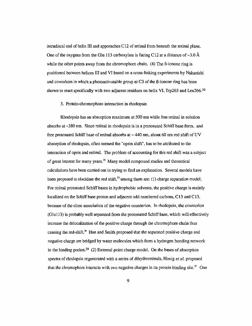
3. Protein-chromophore interaction in rhodopsin
Rhodopsin has an absorption maximum at 500 nm while free retinal in solution
absorbs at -380 nm. Since retinal in rhodopsin is in a protonated Schiff base form. and
free protonated Schiff base of retinal absorbs at - 440 nm, about 60 nm red shift of UV
absorption of rhodopsin, often termed the "opsin shift", has to be attributed to the
interaction of opsin and retinal. The problem of accounting for this red shift was a subject
of great interest for many years." Many model compound studies and theoretical
calculations have been carried out in trying to fmd an explanation. Several models have
been proposed to elucidate the red shift," among them are: (1) charge separation model.
For retinal protonated Schiff bases in hydrophobic solvents. the positive charge is mainly
localized on the Schiff base proton and adjacent odd numbered carbons, C 15 and C 13,
because of the close association of the negative counterion. In rhodopsin, the counterion
(Glu1l3) is probably well separated from the protonated Schiff base, which will effectively
increase the delocalization of the positive charge through the chromophore chain thus
causing the red-shift." Han and Smith proposed that the separated positive charge and
negative charge are bridged by water molecules which form a hydrogen bonding network
in the binding pocket-? (2) External point charge model. On the bases of absorption
spectra of rhodopsin regenerated with a series of dihydroretinals, Honig et at. proposed
that the chromophore interacts with two negative charges in its protein binding site." One
9

charge acts as a counterion to the PSB, while a second charge, situated near the middle of
the retinal chain, generates a red shift in the chromophore's absorption band. A more
recent data set obtained of dihydroretinal pigments in native membranes has modified the
position of the second point charge to near C 13.36 However, which amino acid residue
plays the role of this second point charge has not been clearly established. (3) The ring
chain conformation model. This newly proposed model focuses on the C5-C6-C7-C8
dihedral angle which affects the conjugation between the ring and the chain parts of the
chromophore. Ab initio calculations have demonstrated that the retinal chromophore in
bacteriorhodopsin, unlike the free retinal, which has a C5-C6-C7-C8 dihedral angle of
about 400, maintains a planar conformation between the ring and the chain." This
conformation change from a distorted conjugation to full conjugation certainly will
contribute to the opsin shift The same calculation on the other hand, concluded that in
rhodopsin the C6-C7 bond is still considerably twisted.
4. The stereoselectivity of the binding cavity of rhodopsin
There are basically two ways to probe the binding site of the apoprotein opsin in
rhodopsin. One is a technique called site specific mutagenesis, which modifies the protein
by replacing the concerned amino acid in the vicinity of chromophore. Another way is to
modify the chromophore, i.e. synthesizing retinal analogs and study the binding of retinal
analog with opsin. The former technique has been used by Khorana and coworkers" and
other groups39,4O in their studies of rhodopsin and bacteriorhodopsin, while the latter
approach has been the most often used method by organic chemists in probing the opsin
chromophore interaction and the shape of chromophore binding site."
The early observation that in addition to l l-cis retinal only the structurally similar 9
cis isomer formed a pigment analog, while the other four known isomers at that time (all
trans, 13-cis, 9,13-dicis, and 11,13-dicis) either failed to give pigment or did not yield
10
conclusive results, led Wald and coworkers to conclude that the binding site of opsin is
highly specific." However, two independent observations in the late 1970's and early
1980's prompted a reevaluation of this postulate. First, Nakanishi and coworkers showed
by extraction of chromophore from the pigment analog derived from 9,13-dicis retinal that
the pigment analog retained the original geometrical integrity," though later, more careful
and detailed work by Trehan et al. indicated there was a considerable amount of
isomerization in the [mal pigment," Second, seven new isomers of retinal, six containing
the hindered 7-cis geometry (Table 1), were shown to form new pigment analogs with
absorption properties substantially blue shifted ("-max450 to 462 nm) from those of other
known isomeric rhodopsins (480 to 500 nm)." These results indicated that opsin displays
little selectivity in being able to accept nearly all the cis, dicis, even tricis isomers, the only
exception being the all-trans and the 13-cis isomers.w
Even though opsin has been shown to form pigment with a variety of retinal
analogs, its selectivity is still reflected in pigment yield and the rate of pigment formation:
higher yield and faster rate of pigment formation are observed for the native chromophore
l l-cis retinal and the structurally similar 9-cis retinal than other analogs.
Numerous structurally modified retinal analogs have been synthesized and their
binding properties with opsin investigated. Among them are isotopically labeled retinal
derivatives.f" fluorinated retinals,48,49 alkylated retinals,50 retinals with aryl and naphthyl
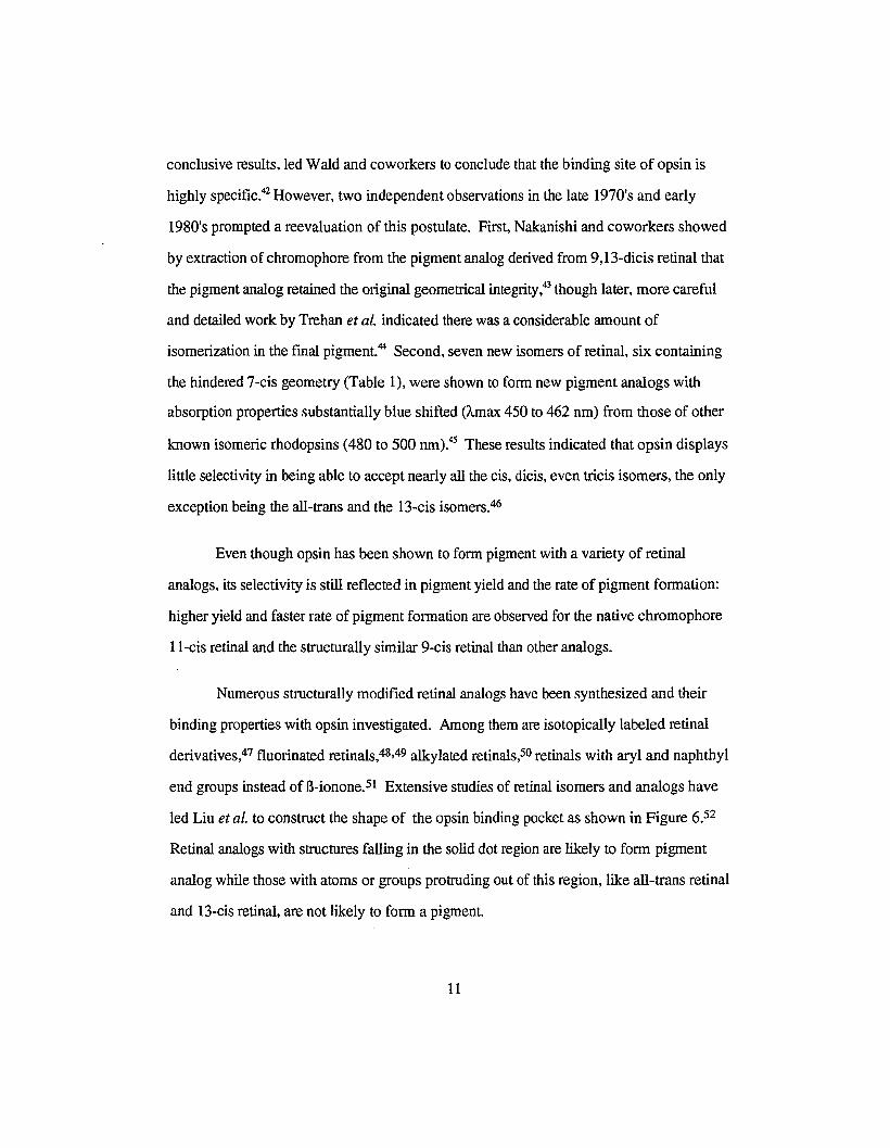
end groups instead of B-ionone.51 Extensive studies of retinal isomers and analogs have
led Liu et al. to construct the shape of the opsin binding pocket as shown in Figure 6.52
Retinal analogs with structures falling in the solid dot region are likely to form pigment
analog while those with atoms or groups protruding out of this region, like all-trans retinal
and 13-cis retinal, are not likely to form a pigment.
11

Table 1. Isomeric Rhodopsin Analogs
Retinal Isomers Pigment Analog Amax (om) Reference
all-trans none 42
13-cis none 42
7-cis 450 45
9-cis 483 42
l l-cis 498 42
7,9-dicis 460 45
7,13-dicis 455 45
7,II-dicis 455 45
9,II-dicis 480 42
9,13-dicis 481 42
11,13-dicis 498 (?) 42
7,11,13-tricis ?* 53
7,9, 13-tricis 455 45
7,9,1l-tricis 462 45
9,11,13-tricis ?* 54
all-cis ?* 53
* Unstable isomers.
12

Q
'...
..
..•o
I
r
\
o ••I
I
."
f
.r1' • Y :'1I I ,V.., • I : •
~, ~k - '. .t .' .......,:-, 12~. -:(J .. " • • .' ""J"\ .' ""••• n.", • • • ' ..v, '" I"',. .' v·, ..·... v '.:.... ':.t().' ••,.... ~ -r • 'iJ I
)1-' ,tv u 'iJ '. ':' '1'9 '. • V n.I ~ I' .. 0 "
0_~
• : Ii, lJ, M ••••'::'"
......... ~. 0 • n. ' ..', I"'" .-.' ", '. 0 •.r • "1'... ...... ". \ . . .' . .().1., •.;' • :\ '. • • 0....... 'iJ. • '. 0 :
D'o '. L,."
• "~~"O •• -.... N.. ,
.~ .: ...• " 04'
:'iJ..:-'---'~ 'lo ..or .. ,
I
: , 0
?-··.1fVtI ,,,,,
~
Ga
~
w
Figure 6. A two dimensional map of thebinding pocket of opsin. The solid dots are projected positions ofcarbon atoms of all
isomeric rhodopsin chrornophores, Theperimeter is the summation of vander Waals radii of outmost carbon atoms. The
triangles and circles arethose of thenonbinding 13-cis and all-trans isomers.

B. Synthetic strategies for retinaland 3-dehydroretinal
Thesynthetic routes leading to retinal and retinal analogs in most cases follow
methodology developed for the synthesisof vitamin A and carotenoids.P The most
commonsynthetic methods were Wittig and Homer condensations of carbonyl compounds
with triphenylphosphonium halides or dialkyl phosphonates, Grignard or Nef reactions of
carbonylcompounds with metal acetylides, aldol condensations, Refonnatsky reactionof
carbonylcompounds with (J.- or y-haloesters and nitrilesand Knovenagel-Doebner
condensations of aldehydeswith compounds possessing activated hydrogens. The
syntheticroutefor retinoids is often named according to how the C20 skeletonof retinal is
assembled from smallercarbon units. Among the many combinations used in the assembly
of the retinalskeleton are ClO + ClO, Cll + C9, Cl3 + C7, Cl4 + C6, C15 + C5, C16 +
C4, C18+ C2. For each assembling method, there are also a variety of reactions to carry it
out. Somerepresentatives of thesesynthetic strategiesare briefly reviewed in the following
section.
1. General methodology
a. ClO + ClO
Oneexample of this methodology is the condensation of B-cyclogeranyltriphenyl
phosphonium bromide (1) with the ClO aldehydic trieneacid (2) or ester (3) in the
presenceof sodium methoxide.t"
(1) (2, R= H, 3, R= C2H5)
14

COOR
(4, R= H, 5, R= C2HS)
Although the Wittig reaction usually gives cis-trans mixtures, the newly formed
7,8-double bond was completely in the trans configuration.
b. C14+C6
The technical synthesis of vitamin A developed by Isler et al. followed this route.F
Condensation of B-C14-aldehyde (6) with the Grignard derivative of cis-3-methyl-2
penten-4-yn-l-ol (pentol, 7) afforded the C20 diol8. Partial hydrogenation of the
condensation product over Lindlar catalyst afforded product 9, which after acetylation of
the primary hydroxyl group, was dehydrated and rearranged to all-trans vitamin A acetate.
Subsequent saponification then gave the corresponding vitamin A (10).
~CHO
(6)
+ •c"l ----.BrMgC" 't
OMgBr
(cis 7)
~~~
~ c~L. 'tI OH OH
•CHzOH
(I3-cis,8) (13-cis,9)
(all-trans, 10)
15

By a variation of the procedure, sterically hindered vitamin A isomers have been
prepared.f The Grignard reaction of the B-C-14 aldehyde with trans pento1 (7) led to the
acetylenic diol (l3-trans, 8). Acetylation followed by dehydration furnished 11,12
dehydroretinol (11), which on partial hydrogenation was transformed into l l-cis retinol.
Manganese dioxide oxidation of the latter gave the corresponding Ll-cis retinal (12).
11,13-Dicis retinal was obtained by a similar sequence of reactions starting from the
acetylenic intermediatevia 13-cis-11, 12-dehydroretinol.
~CHO
(6)
+ BrMgC,~OMgBr
(trans, 7)
~" C~OH
~ C"
I OH
(13-trans, 8)
IE .. ~"'C~OH
~ C'I~
(11)
• •
(12)
Starting from 3-dehydro-B-C14-aldehyde, all-trans, 11,13-dicis, and l l-cis vitamin
A2 as well as the corresponding aldehyde have been synthesized in an analogous way.59.60
This scheme however cannot be applied to the synthesis 7-cis containing isomers
because the dehydration step only afforded the 7,8-trans double bond.
16

In yet another variation of the versatile C14 + C6 route, starting from the isomeric
B-C-14 aldehyde, Eiter et al. effected the synthesis of 9-cis vitamin A acetate by the
following sequence of reactions.s! This synthetic scheme featured a surprisingly high
degree of selectivity (85%-89%) in the fmal base-induced (OBN) dehydrobromination of
the bromide 14 prepared by reaction of 13 with PBf3, wherein the 9-cis configuration was
introduced.
~CHO
(6)
+ ~ 0... C OM.,BrBrMgC'
(trans, 7)
OAe --- OAe
(13) (14)
DBN
OAe
(15)
c. Cll + C9
This combination scheme was introduced by Nakanishi et al.62 The condensation
of ethyl 3,7-dimethyl-2,4,6-nonatriene-8-ynoate (16) with 2,2,6-trimethylcyclohexanone
(17) afforded 18 in high yield (>80%). However, subsequent conversion to vitamin A
ester 5 was only accomplished in less than 20% yield.
17

(17)
,c~C02Et HC~
(16)
..c~C02Et~C~
~OH
(18)
., .
(5)
A different approach was reported by Attenburrow for the synthesis of 7,8
dehydrovitamin A by the following sequence of reactions.P
(:(~CH
C'"I +
(19)
o~
(20)
•
(21) (22)
It seems that partial hydrogenation of (22) may lead to the 7-cis isomer of retinal.
However Fagle et al.64 and Kini65 reported that 7,8-dehydrovitamin A would not undergo
partial hydrogenation to give 7-cis retinol although compound 23 has been partially
hydrogenated to the 7-cis product 24. 66
18

~eXC~ OH OMe ~OMe~ --_.~HO"" OMe
(23)
d. C13 +C7
(24)
Jacobs et at. reported a novel approach using this combinarion.s? Condensation of
B-ionone (25) with the lithium or sodium derivativesof the acetylenicether (26) yielded
the acetylenic C20 alcohol (27). Lithium aluminium hydridereduction or partial
hydrogenation produced the alcohol (28), which on acid hydrolysiswas converted to
retinal.
~o(25) (26)
•
(27) (28)
•
(all-trans, 12)
CHO
C13 + C7 strategycan also be accomplished by Wittig reactions. Pommer reported
an approachin which B-ionyltriphenylphosphonium bromide(29) was used to react with
19

..
the aldehydic acid or the aldehydic ester (30) to yield a mixture of all-trans and 9-cis
retinoic acid and the corresponding ethyl esters (5) respectively/"
~ P'(CoHshB, + OHC~C02R
(29)
(all-trans, 5)
+
(30)
(9-cis,5)
To apply this scheme to the synthesis of 7-cis isomers would require that the B
ionyltriphenylphosphonium bromide be isomerized to the 7-cis configuration. However
few studies have been done regarding the photoisomerization of Wittig salts.
e. Cl8 +C2
A large number of vitamin A derivatives were synthesized from the B-CI8-ketone
(31) by condensation with various C2 reagents listed in the following scheme.
(31)
C2 reagent
o C2
20
•
y
CH20Ac
y

Br- (C6HshP+CH2CH20H CH20H
Br-(C6HshP+CH2CN CN
(C2HsO)2P(O)CH2CN CN
Br" (C6HshP+CH2COOEt COOEt
Br-(C6HshP+CH2CONH2 CONH2
Br" (C6HshP+CH2CH(OEt) 2 CH(OEt)2
The Reformatsky reaction with bromoacetic acid esters has also been used in the
synthesis of vitamin A from the B-C18-ketone (31).68,69 Dehydrationof the resulting
hydroxy ester (32) gave mainly retro-vitaminA acid ethyl ester. Hydrolysis to the
corresponding acidfollowed by treatmentwith PC13 furnished a mixture of isomeric
vitamin A acidchlorides which could be reduceddirectlywith lithiumaluminum hydrideto
vitamin A or hydrolyzed to vitaminA acid.
~o(31) (32)
OHCOOR
" ..
(10)
f. CIS + CS
Matsui et al.70 reported a very interestingstereoselective synthesisof unhindered
21

retinal isomer whereby the C20 carbon skeleton was generated by the condensation of a
metal dienolate derivative of ethyl senecioate (33) with 13-C 15-aldehyde (34). The
stereochemical outcome of the Matsui condensation was governed by the nature of
counterion in the generation of the requisite dienolate. If potassium amide was used to
generate the dienolate of senecioate, all-trans retinoic acid (4) was produced; if sodium
amide or lithium amide was used, only l3-cis isomer was produced.
~CHO I~'- + ~C02Et
(all-trans, 32) (33)
~,~COOH
(all-trans, 4)
~, ~
COOH
(l3-cis, 4)
Starting from 9-cis B-CI5-aldehyde (34), 9-cis and 9,13-dicis isomers were
obtained:
22

~HO +
(9-cis,34) (33)
~ COOH
(9-cis,4) (9,13-dicis,4)
Isler et al. successfully applied the Matsui condensation to the synthesis of four
isomers (all-trans, 9-cis, 13-cis, 9,13-dicis) of vitamin A2 acid (36), alcohol and aldehyde
by starting from all-trans and 9-cis 3-dehydro-B-CI5 aldehyde (35):59
~CHO~y -
(all-trans, 35)
+
(33)
Matsui condensation•
~ COOH+
~ ~ ~
COOH
(all-trans, 36)
23
(13-cis, 36)

(X-)HO(9-cis, 35)
+ ~C02Et
(33)
Matsui condensationII
+
~ COOH
(9-cis, 36) (9,13-dicis, 36)
Wittig and Homer reactions have also been widely used for the synthesis of vitamin
A and derivatives (nitrile, acetate, aldehyde, acid) with the Cl5 + C5 strategy. The most
common procedure is to react C 15 aldehyde by reacting with a variety of phosphonium
salts (Wittig reaction) or phosphonate esters (Horner reaction).
Vitamin A acetate and vitamin A nitrile were made by the condensation of B-C15
aldehyde (34) with the phosphonium salt (37) by Pommer et al.56
~CHO
(34)
+ Br'(C~5)3P+CH2~C02R
(37)
(5)
Pommer et at. also reported the synthesisvia B-CI5 aldehyde and C5 phosphonate'".
24

Some of the most commonly used C5 phosphonium salts and phosphonates are
listed below:
(<;H,oJ,.P(OJCHz~Co,.R
(<;H,O),P(O)CH2~CNMead et al.72,73 have successfully adapted the Still and Gennari/" method, which
enhances the cis selectivity in the Wittig-Herner reaction, to the synthesis of l l-cis isomer
of 19,19,19-trifluorinated retinal by using a fluorinated C5 phospho nate. This method has
been widely used now to synthesize l l-cls isomers of retinal analogs.
(38) (39)
" "
CHO
(40)
+ isomers
2. Synthesis of 7-cis isomers of retinal
In the 1970's, Liu and Ramarnurthy reported that the photosensitized isomerization
of a series of l3-ionyl and l3-ionyliqene derivatives resulted in the stereoselective formation
of their stable 7-cis forms.75 This discovery eventually led to the successful synthesis of
all the missing isomers with 7-cis geometry. The most practical and convenient route has
25

been found to be Cl5 +C5 (Scheme 1). The 7-cis and 7,9-dicis building blocks. the Cl5
aldehydes, were prepared by DIBAL-H reduction of the corresponding nitriles. The nitriles
~CN
~CHO
hu•
sensitizer
C5 extension• • 7-cis isomers of retinal
•
Scheme 1. Synthetic route for 7-cis retinal isomers
was converted to aldehydes, and subsequent Homer-Emmons reactions (C 15 + C5)
yielded 7-cis, 7,9-dicis, 7,13-dicis and 7,9, 13-tricis isomers." Using the modified Still
and Gennari approach by D. Mead, 7,1l-dicis, 7,1l,13-tricis and 7,9,1l-tricis and all-cis
were prepared by A. Trehan et ai.53 7, l l-Dicis and 7,11, 13-tricis were also synthesized
using a different approach by Kini.65
C. The goal of this study
Isler et al. first reported the synthesis of six isomers of vitamin A2 and derivatives
in 1962.59 Since then only one new isomer (7-cis) was added by Liu et aU7 The lack of
progress in this area is attributed in part to the less stable nature of vitamin A2 compounds.
The opsin binding studies of 3-dehydroretinal were also limited due to the unavailability of
other isomers. So far, only three isomers (l l-cis, 9-cis and 7-cis) have heen used for
opsin binding studies. Pigment formation of l l-cis and 9-cis isomers with a variety of
opsins (crayfish'", bovine/? and octopus-s) have been reported. 3-Dehydroretinal is
different from retinal in that the ring portion is considerably more planar than retinal. How
this planarity affects the opsin binding and whether the other 7-cis isomers form pigments
26

with opsin or not are yet to be determined. The reported synthesis of 7-cis 3
dehydroretinal utilized the Cl8 + C2 approach, a method not applicable to the synthesis of
other dicis and tricis isomers containing 7-cis geometry. Whether the synthetic strategies of
7-cis retinal isomers can be applied to 3-dehydroretinal or not also needs to be established.
The goal of these studies is to develop a general synthetic scheme which can be
applied to the synthesis of all the 7-cis isomers, to isolate and identify the hitherto unknown
isomers 3-dehydroretinals, and investigate the opsin binding properties of these new
isomers.
27

Experimental
A. General Information
1. Numbering of carbon skeleton
The IUPAC system of numbering for carotenoids and retinoids will be used
throughout the work. Retinal numbered as such is shown below. All other lower synthetic
intermediates will be numbered in a similar way.
19 20
14
15CHO
all-trans 3-dehydroretinal
2. Geometrical configuration of double bonds
Geometrical configuration is indicated by citing the double bond or bonds with cis
configuration. It is assumed that other bonds have trans configuration. For example:
~ ~ 1~
CHO
13-cis 3-dehydroretinal
3. Number, structure and name of compounds
41
Diethyl 2-methyl-3-cyano-2-propenylphosphonate
28

42
Bis(2',2',2'-trifluoroethyl)-2-methyl-3-cyano-2-propenyl phosphonate
~Y
Y'~
Number
43
44
Y
COCH3 (Y' = H)
CH(OH)CH3 (Y' = H)
Name
3-dehydro-B-ionone
3-dehydro-B-ionol
45
46
C(CH3)(-OCH2CH20-)
(Y' =H)
COCH3
(Y'=MeO)
ketal
ionone
3-dehydro-B-ionone ethylene
3-methoxy-3-dehydro-B-
~YY'~ ~- ~
47
48
CN (Y' =H)
CN
(Y'=MeO)
3-dehydro-B-ionylideneacetonitrile
3-methoxy-3-dehydro-B-
ionylideneacetonitrile
29

49
50
35
COOEt (Y' = H)
CH20H (Y' = H)
CHO (Y' =H)
ethyl 3-dehydro-B-ionylideneacetate
3-dehydro-B-ionylideneethanol
3-dehydro-B-ionylideneacetaldehyde
51 (OCH2CH20) (Y' = OH) 3-hydroxy-B-iononeethylene ketal
52 (0) (Y' = OH) 3-hydroxy-B-ionone
53 (0) (Y'= CH30) 3-methoxy-B-ionone
54 CHCN (Y' = OH) 3-hydroxy-B-ionylideneacetonitrile
55 CHCN (Y' = CH30) 3-methoxy-B-ionylideneacetonitrile
56 CHCHO (Y' = OH) 3-hydroxy-B-ionylideneacetaldehyde
57 CHCHO (Y'= CH30) 3-methoxy-B-ionylideneacetaldehye
Y'
Y
58
59
60
CN (Y' = H)
CHO (Y' ="H)
CN (Y' = Tosyl)
3-hydroxyretinonitrile
3-hydroxyretinal
3-tosylretinonitrile
30

y
61 Y=CN (Y' =H) 3-dehydroretinonitrile
62 Y = CHO (Y' = H) 3-dehydroretinal
63 Y= CN (Y' = MeO) 3-methoxy-3-dehydroretinonitrile
64 Y= CHO (Y' = MeO) 3-methoxy-3-dehydroretinal
B. Materials
All the Horner-Emmons reactions. DffiAL-H reduction and hydroboration
reactions were carried out under inert atmosphere. the reagents and anhydrous solvents
were transferred via syringe or cannula. Anhydrous THF was obtained by distilling from a
benzophenone ketyl (from sodium and benzophenone) solution. Triplet sensitizer
benzanthrone was purified by recrystallization in ethanol/chloroform. p-Toluenesulfonyl
chloride was purified by recrystallization in chloroform. Diethylcyanomethylphosphonate
was prepared by Dr. A. Asato from triethylphosphite and bromoacetonitrile. Silica gel for
column chromatography (silica gel 60. 300-600 mesh) was from ICN Biomedicals. All
other chemicals used were purchased from Aldrich and used as received.
C. Measurement of physical properties
uv-VIS spectra were obtained on a Perkin-Elmer A,19 spectrophotometer
lH-NMR spectra were obtained on a General Electric QE-300. Deuterated
chloroform was used as solvent unless otherwise indicated.
31

HPLCwas used to monitor the progress of some reactions and separate isomers in
reactionmixtures. A complete HPLC system from Rainin Instrument Company, Inc. was
employedfor all the HPLC operations. Two types of columnswere used: Microsorb1M
Si-80-199-C5 (semi-preparative) and Microsorb'IN Si-80-125-C5 (analytical).
D. Generalprocedures for sensitized photoisomerization
The sensitized photoisomerization of B-ionyl and B-ionylidene derivatives were
conducted in eitherNMR tubes or Pyrex vials. The NMR tubemethod was used for the
purposeof quick analysis and monitoring the progressof reaction. The Pyrex vial method
was used mainly for preparative purpose.
1. Sensitized irradiation in NMR tubes
A solution of about 10 - 50 mg of a substrate, a triplet sensitizer (1 - 4% the weight
of substrate), and 0.5 ml of CDCl3 or C6D6 was takenin a cleanPyrex NMR tube and the
solutionwas degassed by three freeze-pump-thaw cycles. The tubeswere thensealed and
irradiated at constanttemperature (H20 bath) using a 200WHanovia medium pressure
mercury lamp. The absorbance of light by the substrate was prevented by using
appropriate Corningglass filters.
2. Irradiation in vials:
About0.2 - 2 g of a substrate and a sensitizer (1 % weight of substrate) were
dissolvedin a Pyrexvial with 20 - 30 ml benzene. The solution was deoxygenated by
passing through a stream of nitrogen for ten minutes. The vial was then sealed and
irradiatedusinga 450W Hanovia medium pressure mercury lamp. The progressof the
reactionwas followed either by TLC or NMR. After the reaction was complete, the
32

solution was transferred to a roundbottom flask and the solvent was removed by
evaporationon a rotaryevaporator. The crude product was purified by column
chromatography.
E. General procedure for protein bindingstudies
1. Preparation of Schiffbase and protonated Schiff base
The purified 3-dehydro or 3-hydroxyretinal isomers were dissolved inethanol to
make a solution withabsorbance between0.5 and 1. A few drops of n-butylamine (lM)
solution was added to thecuvette with the retinal solution. The UV-VIS spectrum of the
Schiff base was taken when no morechanges in UV-VIS absorbance wereobserved with
the addition ofn-butylamine. The UV-VIS spectrum ofprotonated Schiffbase was taken
by adding trifluoroacetic acid to the Schiff base solution.
2. Extraction of opsin
The isolation of opsinfrom cattle retina was carried out following theprotocol
originallydeveloped by Papermaster and Dreyer" and modifiedby Dennyand
Colmenares" of this laboratory.
Unlessotherwise specified, all following operations were doneat 4 0Cin a cold
room and under dim light.
The thawed retinas (200 pieces) were homogenized in 200 ml chilled 34% sucrose
(containing 10mM HEPES withp~=7, 65 mM NaCl and 2 mM MgCl2). The
homogenatewas centrifuged at 2000rpm for 10 min. The supernatantfraction wasdiluted
with 3 volumesof the HEPES bufferand centrifuged under vacuum at7,000rpm using
Beckmanrotor type27 for 15min to yield a pellet of crude rod outer segment.
33

The pellets were homogenized with 90 ml HEPES buffer using a homogenizer
(speed 60 rpm) for 1 min. Six 15-ml fractions of this suspension were floated by syringe
on six 15-ml chilled 34% sucrose solutions, and were centrifuged using rotor with swing
buckets under vacuum at 25,000 rpm for 45 min. The orange band was collected, and
bleached in the presence of 1 ml of 1M NH20H by illumination with a 550W projector
lamp using 3-73 cut-off filter for about 45 min. Excess hydroxylamine was removed by
repeated washings with HEPES buffer (5 times) and with distilled water (once), each time
spinning down at 15,000 rpm under vacuum for 15 min. The white pellet, lyophilized for
at least 12 h, was subsequently washed several times (8-10 times) with cold hexanes until
the retinal oxime was completely removed (monitored by UV-VIS spectroscopy). The
purified ROS was solubilized in 10 m1 of 2% CHAPS. The clear solution after
centrifugation at 25,000 rpm for 25 min was stored at -85 oc.
The concentration of opsin in the above solution was determined by taking an
aliquot (dilute with 2% CHAPS) to bind with ll-cis retinal in the dark. An ethanol
solution of l l-cis retinal with a concentration of about 0.5 mg/m1 (<15 ul) was transferred
by syringe to a 0.5 ml cuvette containing the opsin solution. Formation of the pigment was
manifested by the increase of absorbance at around 500 nm. The progress of binding was
monitored by UV-VIS spectroscopy until completion, when the absorbance remained
constant The concentration of the opsin was calculated based on the absorbance of the
pigment at Amax,500 nm (determined from the difference between the spectra after
completion of binding and after bleaching) and the extinction coefficient of rhodopsin,
which is 4 x 10⁴ molel-crrr l." Typical concentrations of 1-2 x 10⁻⁴ M were obtained.
3. Binding of opsin with isomers of 3-dehydroretinal and 3-hydroxyretinal
The above binding procedure was repeated using the synthetic isomers instead of
Ll-cis. CHAPS solubilized opsin was placed in a 0.5 ml cuvette and 2% CHAPS solution
34

was used as reference. After background correction, an aliquot «15 ul) of ethanol solution
with a concentration of about 0.5 - I mg/ml was added to the opsin. Pigment formation
was monitored with UV-VIS spectroscopy.
F. Calculations
1. Molecular Modeling with HyperChem
HyperChem, a commercial desktop molecular-design package from Autodesk Inc.•
features sophisticated modeling and visualization. molecular dynamics. classical and semi
empirical quantum mechanical calculations. built-in scripting language. and open
architecture. It was used in the modeling of retinal and 3-dehydroretinal isomers to obtain
dihedral angles. and also in the molecular orbital calculation for some of the CIS
intermediates to investigate why the 3-dehydro CIS intermediates are so apt to undergo
1.7-H-shift reaction. The general procedure and parameters used in the molecular
modeling are provided below:
Software version: HyperChemTM Release 3 for Windows
a. 20 sketch of an isomer was drawn first and a 3D representation of the molecule
was then generated using the HyperChem Model Builder.
b. The conformation of the molecule was first optimized through a quick molecular
mechanical calculation. MM+ force field which was developed for organic molecules was
chosen for this optimization. Options for the MM+ force filed are: NONE for cutoffs (all
nonbonded interactions were calculated), BOND DIPOLES for Electrostatic (bond dipoles
were used to calculate nonbonded electrostatic interactions),
c. The optimized molecule was then subjected to semiempirical quantum mechanical
calculation. The semiempirical method was AM I which is an improved MNDO method
35

and the most accurate method at this stage. SCF options are: Convergence limit =0.01
kcallmol. An SCF calculation ends when the difference in energy after two consecutive
iterations is less than this amount; Iteration limit = 100. This is the maximum number of
iterations for an SCF calculation. The calculation ends after this iteration even if it has not
reached the convergence limit (for the molecules calculated in this dissertation, all satisfied
the convergence limit, so this limit was never invoked to end the calculation); accelerated
convergence and RHF (restricted Hartree-Fock method) were chosen for the calculation of
all the closed shell molecules. For radicals, UHF (unrestricted Hartree-Fock method) was
chosen for the calculation.
d. After geometry optimization with the semiempirical quantum mechanical
calculation, dihedral angle and other structural data can be directly obtained using the
selection tool in HyperChem.
2. "Curvefit" with Spectra Calc.
Spectra Calc (Galactic Industries Corporation) is a software package for processing
or acquiring scientific data. It can be used for a variety of data analysis applications.
"Curvefit" is just one of the many application programs in this software package. It can be
used to deconvolute complex spectra into components by calculating the best fit of
Gaussian, Lorentzian, or Log normal bands (useful for modeling tailing peaks or peaks that
are "skewed" to one side). This program works by first asking for input of the number of
component bands present, their peak positions, their peak widths, and the peak types
(Gaussian, Lorentzian, Log normal, or mixture). The program takes these starting guesses
and iterates in order to find the combination of band heights, positions, and widths which
best fit the data file. This curve-fit program was used to analyze the opsin binding curves
of the A2 isomers, and identify the likely isomerization pattern of unstable multiple cis
isomers during the binding process.
36

The general procedure for the calculation includes the following steps:
a. The UV-VIS binding curves obtained from Perkin-ElmerA,19 instrument were
first converted to Spectra Calc file format by the me import program in Spectra Calc.
b. "Curvefit" calculation is selected from the Arithmetic menu.
c. Numbers of peaks, peak types, peak widths, peak heights and centers were
entered. Peak positions are fixed for all the calculations.
d. The maximum number of fitting passes is specified at 1,000. If the calculation
shows further improvement in fitting, more passes are given.
e. At theend of calculation (no further improvement in fitting by introducing
additionalpasses), the calculated curves (peaks), statistical error (X2) and other parameters
generated in the calculationare saved in a file.
f. Different initial peak parameters (position, height,width, number) were used and
steps c - e were repeated to obtain the best set of calculatedcurvecomponents for the
experimentalcurve. The "goodness" of fitting is judged by the X2 square (the smaller, the
better, acceptable value being less than 0.01) and visual inspection of how well the
experimental curve coincides with the calculated curve from the sum of component curves.
G. Synthesis
Preparation of Diethyl-2-methyl-3-cyano-2-propenylphosphonate (4 C2.Phosphonate)
A mixture oftriethylphosphite (23 g, 0.138 mol) and 4-chloro-3-methyl
acrylonitrile (15.9 g, 0.138 mol) was heated at l300C for 5 h, the crude product was
37

distilled (1350-1400C/0.8 mm Hg) to give 27 g of the C5 phosphate (90% yield, trans:cis
=55:45).
1H-NMR (CDCI3, 300MHz, 0 in ppm): cis form, 5.27 (s, broad, IH), 4.13 (m,
4H), 2.98 (d, J =23.9 Hz, 2H), 2.11 (d, J =3.0 Hz, 3H), 1.36 (t, J =6.0 Hz, 6H); trans
form, 5.29 (s, broad, 1H), 4.13 (m, 4H), 2.72 (d, J =23.9 Hz, 2H), 2.20 (d, J =3 Hz.
3H), 1.34 (t, J = 6.0 Hz, 6H).
Preparation of tris-(trifluoroethyn-2-methyl-3-cyano-2-propenylphosphonate (42
fluorinated C5_phosphonate)
A mixture oftris-(trifluoroethyl)phosphite (10.76 g, 32.8 mmol) and 4-bromo-3
methyl-acrylonitrile (3.5 g, 21.9 mmol) was refluxed at 1700C (bath temperature) for 48 h.
The dark mixture was distilled (llOoC/0.8 mm Hg) to give 5.6 g of pure product (79%
yield, trans:cis =55:45)
IH-NMR (CDCl3, 300MHz, 0 in ppm): cis form, 5.38 (d, J =5 Hz, 1H), 4.43
(rn, 4H), 3.14 (d, J =24.9 Hz, 2H), 2.10 (m, 3H); trans form, 5.34 (d, J =5.8 Hz, IH),
4.43 (m, 4H), 2.91 (d, 2H, J = 24.5 Hz), 2.22 (m, 3H).
Synthesis of 3-dehydro-B-ionone (43)83
B-Ionone (198 g, 1 mol) in carbon tetrachloride (1.21) was placed in a 2.5 I, round
bottom flask fitted with an efficient coil condenser, a mechanical stirrer, and a thermometer.
Sodium bicarbonate (100 g), calcium oxide (80 g), and N-bromosuccinimide (214 g, 1.2
mol) were added. The mixture was heated until boiling began, and heating was continued
for about 10 min. When the exothermic reaction subsided, the temperature was lowered to
400C, N, N-dimethylaniline (270 ml) was added, and the succinimide was filtered through
38

a fritted-glass funnel and washed with carbon tetrachloride. The solvent was distilled until
the pot temperature reached 900C, and the residue was heated under the atmosphere of
nitrogen for 2 h in a water bath. Pyridine (90 ml) was added, and the heating was
continued for another hour. The cooled reaction mixture was poured into cold water and
extracted with petroleum ether (bp 30-600C). The combined extracts were washed with
cold 2% sulfuric acid, water, and sodium bicarbonate solution. Vacuum distillation yielded
155 g (43% yield) of 3-dehydro-B-ionone. (bp lOO-llOoC/6 mmHg)
IH-NMR (CDCI3, 300MHz, 0 in ppm): 7.27 (lH, d, J7,8= 16.4 Hz, H-7), 6.21
(lH, d, J= 16.4 Hz, H-8), 5.88 (2H, s, H-3 and H-4), 2.31 (3H, s, 9-CH3), 2.11 (2H,
m, CH2-2), 1.91 (3H, s, 5-CH3), 1.08 (6H, s, l-CH3, l' -CH3).
Synthesis of 3-dehydro-B-ionol (44)
Trans 3-dehydro-B-ionol was prepared by NaBH4 reduction of 3-dehydro-B-ionone
following a procedure similar to that of Lugtenburg."
3-Dehydro-B-ionone (1.9 g, 10 mmol) was taken in 15 ml of methanol in an
Erlenmeyer flask, and the flask was placed in ice-water bath. To this about 0.5 g NaBH4
was added in three equal portions. The mixture was stirred at room temperature for 30
minutes and then quenched by adding a saturated solution of ammonium chloride. The
mixture was extracted with ether. The combined ether layer was dried over MgS04.
Evaporation of ether gave 1.8 g of crude 3-dehydro-B-ionol (95% yield). Pure 3-dehydro
B-ionolwas obtained after column chromatographic purification.
IH-NMR (CDCI3, 300MHz, 0 in ppm): all-trans 6.02 (lH, d, J7,8 = 16.0 Hz, H
7),5.56 (lH, dd, J7,8 = 16.0 Hz, J8,9 = 6.1 Hz, H-8), 6.84 (lH, d, J3,4 = 9.5 Hz, H-
39

4), 5.65 (IH, dt, J3,4 = 9.5 Hz, J3,2 = 4.7 Hz, H-3), 4.15 (lH, b, H-9), 2.00 (2H, m,
CH2-2), 1.80 (3H, s, 5-Me), 1.17 (3H, s, 9-Me), 1.02 (6H, s, I-Me, I-Me').
Synthesis of 3-dehydro-B-ionone ethylene ketal (4 S)
3-Dehydro-B-ionone(48 g, 0.25 mol), ethylene glycol (35 g), triethylorthoforrnate
(78 g) and p-toluenesulfonic acid (0.2 g) were dissolved in benzene (600 ml) and refluxed
for 2 h. After cooling down to room temperature, the mixture was washed with 5%
NaHC03 solution; then, the benzene layer was dried over MgS04. Benzene solvent was
removed on a rotary evaporator and the crude product mixture was distilled to give 45 g
(120-1250C/3 mm Hg) of pure 3-dehydro-B-ionone ethylene ketal.
1H-NMR (CDCI3, 300MHz, 8 in ppm): 6.22 (lH, d, J7,8 =16.0 Hz, H-7), 5.48
(lH, d, J=16.0 Hz, H-8), 5.81 (lH, d, J= 9.6 Hz, H-4), 5.73 (1H, dt, J3,4 = 9.6 Hz,
J2,3 = 4.2 Hz, H-3), 4.00 (2H, m, -OCH2-), 3.93 (2H, m, -CH20-), 2.06 (2H, m, CH2
2),1.80 (3H, s, 9-CH3), 1.54 (3H, s, 5-CH3), 0.99 (6H, s, 1-CH3, 1'-CH3).
Synthesis of 3-methoxy-B-ionone (S 3)83_
Concentrated sulfuric acid (7.2 ml) in methanol (180 ml) was cooled to OOC in a
500 ml flask fitted with a stirrer and a thermometer. Dehydro-B-ionone (18.0 g) was added
to the cold solution and stirred under an atmosphere of nitrogen at 0 - 50C for 24 h. The
reaction mixture was poured onto ice water (-200 ml), and a 50% NaOH solution was
added to the cold solution while stirring vigorously. The product was extracted with
petroleum ether three times, and the combined extracts were washed with water and dried
over anhydrous MgS04. Vacuum distillation yielded 11.5 g (55%) of 3-methoxy-B-
ionone, bp 11O-1150C (-2 mm Hg).
40

IH-NMR (CDCI3. 300MHz. 8 in ppm): 7.21 (lH. d. J7.8 =16.3 Hz. H-7). 6.10
(lH. d. J7,8 =16.4 Hz, H-8), 3.50 (lR, m, H-3), 3.40 (3H. s, CH30). 2.30 (3H. s. 9
Me), 1.75(3H. s, 5-Me), 1.11 (6H, broad singlet. I-Me, I-Me'), 2.45 (lH, dd, J4a,4b =
17.4 Hz, J3,4b =5 Hz, H-4b), 2.05 (lH, dd, J4a, 4b =17.8 Hz, J3,4a =10 Hz. H-4a),
1.85 (lH, dddd, H-2b), 1.42 (lH, t, J2a,3 =12.0 Hz, J2b.2a =12.0 Hz. H-2a). The
following structure illustrates the labeling of hydrogen a and b.
o
Figure 7. Labeling of protons in 3-methoxy-B-ionone
Synthesis of 3-methoxy-3-dehydro-B-ionone (46)
This was prepared by NBS bromination of 3-methoxy-B-ionone and a subsequent
HBr elimination.
3-Methoxy-B-ionone (1 g, 4.5 mmol), carbon tetrachloride (60 ml), NBS (1.2 g,
6.74 mmol), NaHC03 (0.5 g) and CaD (0.4 g) were added to a 100 ml round bottom
flask and refluxed for three hours. N, N-dimethylaniline (2 g) was added to the cooled
mixture and insoluble materials were filtered out. The filtrate was evaporated and the
residue was heated at 900C for I h. Anhydrous pyridine (2 ml) was then added and the
mixture was heated for an additional hour. Crude mixture was separated on silica gel
chromatography. The reaction gave very low yield of 3-methoxy-B-ionone, only 0.105 g
product (10%) was obtained.
41
lH-NMR (CDC13, 300MHz, 0 in ppm): 7.36 (lH, d, J7,8 =16.3 Hz, H-7), 6.17
(lH, d, J7,8 =16.3 Hz, H-8), 4.95 (lH, s, H-4), 3.63 (3H, s, CH30), 2.28 (3H, s, 9
Me), 2.17 (3H, s, CH2 at C-2), 1.95 (3H, s, 5-Me), 1.15 (6H, s I-Me, I-Me').
Synthesis of 3-dehydro-B-iQnylideneacetQnitrile (47)
This was prepared by Horner reaction of 3-dehydro-B-ionone and
diethylcyanomethylphosphonate in the presence of sodium hydride.
NaH (1.1 g, 60% in mineral oil) was suspended in 40 ml THE The mixture was
cooled to OOC and 4.8 g (0.027 mol) of diethylcyanomethylphosphonate in 30 ml THF was
added slowly. After stirring for 30 minutes at room temperature, 3-dehydro-B-ionQne (4.0
g, 0.021 mol) in 20 m111IF was introduced. The reaction was quenched with saturated
ammonium chloride after 3 h. 20% Ethyl etherlhexanes was used to extract the reaction
mixture. After column purification, 4.1 g of 3-dehydro-B-ionylideneacetonitrile (9-cis and
all-trans) was obtained (92% yield).
lH-NMR (CDC13, 300MHz, 0 in ppm): 9-cis form, 6.83 (lH, d, J = 16.1 Hz, H
8),6.60 (lH, d, J = 16.1 Hz, H-7), 5.85 (2H, m, H-3 and H-4), 5.11 (lH, s, H-lO),
2.22 (3H, s, 9-Me), 1.92 (3H, s, 5-Me), 2.07 (2H, d, CH2), 1.06 (6H, s, I-Me, I-Me');
all-trans form, 6.56 (lH, d, H-7), 6.26 (lH, d, H-8), 5.85 (2H, m, H-3 and H-4), 5.19
(tH, s, H-lO), 2.21 (3H, s, 9-Me), 1.86 (3H, s, 5-Me), 2.10 (2H, d, CH2), 1.04 (6H, s.
I-Me, I-Me').
Synthesis of 3-methQxy-B-iQnylideneacetQnitrile (55)
This was prepared by Horner reaction of 3-methoxy-B-iQnone and
diethylcyanomethylphosphonate in the presence of sodium hydride by following the same
procedure for 3-dehydrQ-B-iQnylideneacetQnitrile (47).
42

1H-NMR (COCl3, 300MHz, 8 in ppm): 9-cis form, 6.70 (lH, d, J =16.1 Hz, H
8),6.55 (lH, d, J =16.1 Hz, H-7), 5.12 ua, s, H-lO). 3.5 un, m, H-3), 3.39 (3H, s,
CH30), 2.05 (3H, s, 9-Me), 1.77 (3H, s, 5-Me), 1.09 (6H, s, I-Me, I-Me'); all-trans
form, 6.50 (lH, d, J = 16.0 Hz, H-7), 6.13 (lH, d. J =16.0 Hz, H-8), 5,11 (lH. s. H
10), 3.5 (lH, m, H-3), 3.38 (3H, s, CH30), 2.20 (3H, s, 9-Me), 1.72 (3H, s, 5-Me),
1.06 (6H, s, I-Me, I-Me'). Protons on the six membered ring have similar chemical shifts
and almost identical coupling patterns to that of 3-methoxy-B-ionone. they will not be listed
here and later for compounds derived from 3-methoxy-B-ionone.
Synthesis of 3-methoxy-3-dehydro-B-ionylideneacetonitrile (48)
This was prepared by NBS bromination of 3-methoxy-B-ionylideneacetonitrile and
a subsequent E2 elimination.
3-Methoxy-B-ionylideneacetonitrile (2.45 g. 0.01 mol), carbon tetrachloride (100
m1), NBS (2.67 g, 0.015 mol), NaHC03 (1.5 g) and CaO (1.1 g) were added to a 250 ml
round bottom flask and refluxed for three hours. The mixture was then filtered to remove
the insoluble salts and succinimide. The filtrate was evaporated and the residue was
transferred to another 100 ml round bottom flask, 50 ml methanol and 1 g of KOH pellets
were added to this flask and the mixture was refluxed for 2 h. The cooled reaction mixture
was poured onto 50 ml cold water and extracted with 20% ether/hexanes. The combined
extracts were dried over anhydrous MgS04. Evaporation of the extracts yielded about 2.3
g crude product. Pure product was obtained from column chromatography (1.2 g, 49%
yield).
1H-NMR (COC13, 300MHz, 8 in ppm): 9-cis form, 6.82 (lH, d, J =16.1 Hz, H
8), 6.67 (IH, d, J =16.1 Hz, H-7), 5.07 (lH, s, H-lO), 4.99 (lH, s, H-4), 3.68 (3H, s.
CH30), 2.19 (3H, s, 9-Me), 1.99 (3H, s, 5-Me), 2.08 (2H, s, CH2), 1.14 (6H, s, I-Me,
43

I-Me') ; all-trans form, 6.62 (lH, d, H-7), 6.26 (lH, d, H-8), 5.17 (lH, s, H-1O), 4.97
(lH, s, H-4), 3.67 (3H, s, CH30), 2.23 (3H, s, 9-Me), 1.94 (3H, s, 5-Me), 2.17 (2H,
s, CH2), 1.11 (6H, s, I-Me, I-Me').
Synthesis of ethyl 3-dehydro-B-ionylidene acetate (49)
This was prepared by the Homer reaction of 3-dehydro-B-ionone with
diethylcarbethoxyphosphonate in the presence of sodium hydride.
NaH (1.2 g, 60% in mineral oil) was suspended in 40 ml THF. The mixture was
cooled to OOC and 6.5 g (0.029 mol) of diethylcarbethoxyphosphonate in 30 ml THF was
added slowly. After stirring for 30 min at room temperature, 3-dehydro-B-ionone (3.6 g,
0.019 mol) in 20 m1 THF was introduced. The reaction was quenched with saturated
ammonium chloride after 3 h. 20% ethyl ether/hexanes was used to extract the reaction
mixture. After purification by chromatography, 4.4 g of 3-dehydro-ethyl-B
ionylideneacetate (9-cis : all-trans 10:90) was obtained.
1H-NMR (CDC13, 300MHz, () in ppm): 9-cis form, 8.31 (IH, d, 1 =16.6 Hz, H
8),6.54 (lH, d, 1 = 16.6 Hz, H-7), 5.78 (lH, d, J = 9 Hz, H-4), 5.71 (lH, s, H-1O),
5.65 (lH, dt, 13,4 = 9 Hz, J3,2 =4.7 Hz), 4.05 (2H, q, J = 7 Hz, -OCH2-CH3), 2.39
(3H, s, 9-Me), 1.74 (3H, s, 5-Me), 1.95 (2H, d, J =4.7 Hz, CH2-2), 0.98 (3H, t, 1 =7
Hz, -OCH2-CH3), 1.09 (6H, s, I-Me, I-Me'); all-trans form, 6.44 (lH, d, 1 = 16.2 Hz,
H-7), 6.15 (lH, d, J = 16.2 Hz, H-8), 5.78 (IH, d, J = 9 Hz, H-4), 5.86 (lH, s, H-1O),
5.65 (lH, dt, J3,4 =9 Hz, J3,2 =4.7 Hz), 4.05 (2H, q, J =7 Hz, -OCH2-CH3), 2.39
(3H, s, 9-Me), 1.72 (3H, s, 5-Me), 1.95 (2H, d, J = 4.7 Hz, CH2-2), 1.03 (3H, t, 1 = 7
Hz, -OCH2-CH3), 0.96 (6H, s, I-Me, I-Me').
Synthesis of 3-dehydro-B-ionylideneethano1 (50)
This was prepared by DIBAL-H reduction of ethyl 3-dehydro-B-ionylideneacetate.
44

EthyI3-dehydro-B-ionylideneacetate (2.0 g, 7.6 mmol) was dissolved in 20 ml
anhydrous THF. The solution was cooled to -78oC and 10 ml DIBAL-H (1.0 M hexanes
solution) was added via syringe. The reaction was allowed to warm up and stirred at room
temperature for 1 h. Excess DIBAL-H was destroyed by adding NaP solution. The
mixture was filtered to remove solid residues and the filtrate was extracted with ether.
After drying over MgS04 and solvent evaporation, the crude product was purified by
column chromatography. A total of 1.6 g (91% yield) of all-trans and 9-cis mixture
(90: 10) was obtained.
1H-NMR (CDC13, 300MHz, 0 in ppm): 9-cis form, 6.55 (lH, d, J =16.0 Hz, H
8), 6.20 (IH, d, J = 16.0 Hz, H-7), 5.85 (lH, d, J = 9.5 Hz, H-4), 5.74 (lH, dt, J3,4 =
9.5 Hz, J3,2 = 4.6 Hz, H-3), 5.60 (lH, t, JlO, 11 = 7.0 Hz, H-I0), 4.33 (2H, d, J = 7.0
Hz, -CH20H), 2.08 (2H, m, CH2-2), 1.94 (3H, s, 5-Me), 1.61 (3H, s, 9-Me), 1.03
(6H, s, I-Me, I-Me'); all-trans form, 6.13 (lH, d, J =16.0 Hz, H-7), 6.20 (lH, d, J =16.0 Hz, H-8), 5.85 (lH, d, J = 9.5 Hz, H-4), 5.74 (lH, dt, J3,4 = 9.5 Hz, J3,2 = 4.6
Hz, H-3), 5.67 (lH, t, JlO, 11 =7.0 Hz, H-IO), 4.33 (2H, d, J =7.0 Hz, -CH20H),
2.08 (2H, m, CH2-2), 1.87 (3H, s, 5-Me), 1.85 (3H, s, 9-Me), 1.02 (6H, s, I-Me, 1-
Me').
Synthesis of 3-dehydro-B-ionylideneacetaldehyde (35)
This was prepared by DIBAL-H reduction of 3-dehydro-B-ionylideneacetonitrile.
To a solution of3-dehydro-B-ionylideneacetonitrile (3 g, 0.014 mol) in dry THF
was added 28 ml DIBAL-H (1.0 Min hexane). The completion of the reaction was
checked by TLC analysis (1.5 h). The reaction was quenched with a mixture of silica gel,
water and ether. After stirring for three hours, the mixture was filtered and the filtrate was
dried over MgS04. The solvent was evaporated and the residue was purified by column
45
chromatography. Pure 3-dehydro-B-ionylideneacetaldehyde (2.25 g, 74% yield) was
obtained.
IH-NMR (CDCl3, 300MHz, 8 in ppm): all-trans, 10.14 (lH, d, J = 8 Hz, H-II),
6.74 (lH, d, J = 16.2 Hz, H-7), 6.32 (l H, d, J = 16.2 Hz, H-8), 5.97 (lH, d, J = 8.1
Hz, H-4), 5.86 (lH, s, H-IO), 5.80 (lH, m, H-3), 2.33 (3H, s, 9-Me), 2.11 (2H, m,
CH2-2), 1.95 (3H, s, 5-Me), l.05 (6H, s, I-Me, I-Me'); 9-cis, 10.18 (lH, d, J =8.0
Hz, H-Il), 7.23 (IH, d, J = 15.4 Hz, H-8), 6.63 (IH, d, J = 15.9 Hz, H-7), 5.88 (lH,
d, J = 8.0 Hz, IO-H), 5.86 (2H, m, overlapping H-3 and H-4), 2.14 (3H, s, 9-CH3),
2.12 (2H, m, 2-CH2), l.91(3H, S, 5-CH3), l.07 (6H, s, I-CH3, 1'-CH3).
Synthesis of 3-hydroxy-B-ionone ethylene ketal (5 1)
A 250-ml three-necked flask, equipped with a magnetic stirring bar, a nitrogen gas
inlet adapter and a reflux condenser fitted with a hose adapter leading to a mineral oil
bubbler, was charged with 100 ml BBN (0.5 M in THF) and 8 g of 3-dehydro-B-ionone
ethylene ketal in 40 ml anhydrous THF under a slow stream of nitrogen. The reaction
mixture was refluxed for 6 h. The excess BBN was destroyed at the end of the reaction by
dropwise addition of 10 ml methanol, followed by the addition in one portion of 10 ml
NaOH (3 M). The boric acid intermediate was then oxidized by dropwise addition of 8 ml
aqueous hydrogen peroxide (30%) to the well-stirred reaction mixture. After the addition
was complete, the mixture was stirred for an hour at 50-550C and then cooled to room
temperature. The aqueous layer is saturated with NaCI and 100 ml ether was added. The
upper organic layer was removed and the aqueous layer was extracted with ether twice.
The organic layer and the ether extracts were combined and the solvent was removed on a
rotary evaporator. The crude product was purified by flash column chromatography with
40% ethyl acetate/hexanes to give 3.8 g (54% yield) 3-hydroxy-B-ionone ethylene ketal.
46

IH-NMR (CDCl3, 300MHz, 0 in ppm): 6.17 (1H, d, J7,8 =16.0 Hz, H-7), 5.37
(lH, d, J7,8 =16.0 Hz, H-8), 3.96 (5H, m, H-3 and 2 x CH20), 1.68 (3H, s, 5-Me),
1.51 (3H, s, 9-Me), 1.04 (3H, s, I-Me), 1.02 (3H, s, I-Me'), 2.34 (1H, dd, J4a,4b =16.4 Hz, J3,4b =5.5 Hz, H-4b), 2.01 (lH, dd, J4a, 4b =16.0 Hz, J3,4a =9.6 Hz, H
4a), 1.76 (1H, dddd, J2b,2a =12.0 Hz, J2b,3 =3.2 Hz, J2b,4b =2 Hz, H-2b), 1.45
(1H, t, J2a,3 =12.0 Hz, J2a,2b =12.0 Hz, H-2a). Refer to Figure 7 for the labeling of
hydrogen a and b.
Synthesis of 3-hydroxy-B-ionone (52)
3-Hydroxy-B-ionone ethylene ketal 1.2 g (5 mmol), oxalic acid (- 20 mg), CH2Cl2
(15 ml) and water (15 ml) were mixed and refluxed for 16 h. The CH2C12 layer was then
separated from the water layer and dried over MgS04. After evaporating the methylene
chloride on a rotary evaporator, the residue was purified by flash chromatography and 0.9
g of 3-hydroxy-B-ionone was obtained (89% yield).
IH-NMR (CDCI3, 300MHz, 0 in ppm): 7.22 (JH, d, J7,8 =16.4 Hz, H-7), 6.12
(lH, d, J7,8 = 16.4 Hz, H-8), 4.00 (lH, m, H-3), 2.32 (3H, s, 9-Me), 1.78 (3H, s, 5
Me), 1.13 (3H, s, I-Me), 1.11 (3H, s, I-Me'), 2.44 (lH, dd, J4a,4b = 17.4 Hz, J3,4b =
5 Hz, H-4b), 2.12 (1H, dd, J4a, 4b = 17.8 Hz, J3,4a = 10 Hz, H-4a), 1.82 (1H, dddd,
H-2b, overlapped with 5-Me), 1.51 (lH, t, J2a,3 = 12.0 Hz, J2b,2a =12.0 Hz, H-2a).
Refer to Figure 7 for the labeling of hydrogen a and b.
Synthesis of 3-hydroxy-B-ionylideneacetonitrile (54)
Sodium hydride (0.76 g, 60% in mineral oil) was suspended in 20 ml dry THF in a
100 ml round bottom flask. The flask was cooled in an ice-water bath and 3.4 g (0.019
mol) of diethylcyanomethylphosphonate in 30 m1 THF was added slowly. The mixture
47

was stirred at room temperature for 30 min. To this, 2 g (0.010 mol) of 3-hydroxy-B
ionone in 15 ml THF was added. The mixture was stirred for 2 h. at room temperature and
then quenched by addition of saturated ammonium chloride. Solvent mixture of ethyl
acetate and hexanes (40:60) was used to extract the reaction products. After drying and
solvent evaporation, the product mixture was purified by column chromatography. A
mixture of all-trans and 9-cis of 54 totaling 1.78 g was obtained (77% yield).
IH-NMR (CDC13, 300MHz, 0 in ppm): 9-cis form, 6.69 (l H, d, J =16.1 Hz, H
8),6.53 (lH, d, J = 16.1 Hz, H-7), 5.12 (lH, s, H-lO), 4.0 (lH, m, H-3), 2.05 (3H, s,
9-Me), 1.76 (3H, s, 5-Me), 1.08 (6H, s, I-Me, I-Me'); all-trans form, 6.49 (lH, d, J =
16.0 Hz, H-7), 6.13 (lH, d, J = 16.0 Hz, H-8), 5,17 (lH, s, H-lO), 4.0 (lH, m, H-3),
2.19 (3H, s, 9-Me), 1.71 (3H, s, 5-Me), 1.06 (6H, s, I-Me, I-Me'). The hydrogens (H
2a, H-2b, H-4a, H-4b) on the six membered ring have similar chemical shifts and coupling
patterns to those of 3-hydroxy-B-ionone and they will not be listed here and later for the
compounds derived from 3-hydroxy-B-ionone or 3-methoxy-B-ionone.
Synthesis of 7-cis 3-hydroxy-B-ionylideneacetonitrile C7-cis54)
7-Cis 3-hydroxy-B-ionylideneacetonitrile was prepared by sensitized irradiation of
all trans 3-hydroxy-B-ionylideneacetonitri1e following the general procedure described.
Benzanthrone was used as the sensitizer and 0-52 Coming filter was used to cut off the
short wavelength light which can be absorbed by the substrate. The photostationary
composition was made up of 50 : 50 of 7-cis and 7,9-dicis 3-hydroxy-B
ionylideneacetonitrile. The two isomers were separated by silica gel chromatography.
1H-NMR (CDC13, 300MHz, 0 in ppm): 6.19 (lH, d, J =12.7 Hz, H-7), 6.09
(lH, d, J = 12.4 Hz, H-8), 5.31 (lH, s, H-lO), 4.01 (lH, m, H-3), 2.10 (3H, s, 9-
48

CH3), 1.55 (3H, s, 5-CH3), 1.081 and 1.077(6H, two overlapping singlets, l-CH3, 1'
CH3)·
Synthesis of 7,9-dicis 3-hydroxy-B-ionylideneacetonitrile C7,9-dicis 54)
The same procedure described above for 7-cis 3-hydroxy-B-ionylideneacetonitrile
also produced 7,9-dicis 3-hydroxy-B-ionylideneacetonitrile.
1H-NMR (CDCI3, 300MHz, 0 in ppm): 6.66 (IH, d, J =12,5 Hz, H-8), 6.29
(lH, d, J = 12.5 Hz, H-7), 5.14 (lH, s, H-IO), 4.02 (lH, m, H-3), 1.94 (3H, s, 9
CH3), 1.59 (3H, s, 5-CH3), 1.102 and 1.106 (6H, two overlapping singlets, l-CH3, 1'-
CH3)·
Synthesis of 7-cis 3-hydroxy-B-ionylideneacetaldehyde C7-cis 56)
DIBAL-H reduction of the 7-cis 3-hydroxy-B-ionylideneacetonitrile gave 7-cis 3
hydroxy-B-ionylideneacetaldehyde.
To a solution of7-cis 3-hydroxy-B-ionylideneacetonitrile (1.0 g, 4.3 mmol) in 20
ml dry THF at -78 0C was added 11 ml DIBAL-H (1.0 M in hexane). The completion of
the reaction was checked by TLC (1 h). The reaction was quenched with a mixture of silica
gel, water and ether, After stirring for 3 hours, the mixture was filtered and the filtrate was
dried over MgS04. The solvent was evaporated and the residue was purified by column
chromatography. Pure 3-hydroxy-B-ionylideneacetaldehyde (0.6 g, 60% yield) was
obtained.
IH-NMR (CDCI3, 300MHz, 0 in ppm): 10.07 (lH, d, J =8.1 Hz, H-ll), 6.25
(lH, d, J = 12.1 Hz, H-7), 6.16 (IH, d, J = 12.1 Hz, H-8), 5.99 (lH, d, J = 8.1 Hz, H-
49

10),4.01 (lH, m, H-3), 2.20 (3H, s, 9-CH3), 1.55 (3H, s, 5-CH3), 1.102 and 1.106
(6H, two overlapping singlets, 1-CH3, l'-CH3).
Synthesis of 7.9-dicis 3-hydroxy-B-ionylideneacetaldehyde (7,9-dicis 56)
7,9-Dicis 3-hydroxy-B-ionylideneacetaldehyde was prepared by DIBAL-H
reduction of 7,9-dicis 3-hydroxy-B-ionylideneacetonitrile following the same procedure
described for 7-cis 3-hydroxy-B-ionylideneacetaldehyde.
1H-NMR (CDC13, 300MHz, 8 in ppm): 10.10 (lH, d, J = 8.1 Hz, H-11), 6.95
(lH, d, J = 12.7 Hz, H-8), 6.32 (lH, d, J = 12.7 Hz, H-7), 5.82 (lH, d, J = 8.1 Hz, H
10),4.01 un, m, H-3), 1.99 (3H, s, 9-CH3), 1.56 (3H, s, 5-CH3), 1.131 and 1.127
(6H, two overlapping singlets, 1-CH3, l'-CH3).
Synthesis of 3-methoxy-3-dehydro-B-ionylideneacetaldehyde (57)
DIBAL-H reduction of 3-methoxy-3-dehydro-B-ionylideneacetonitrile afforded the
corresponding aldehyde.
1H-NMR (CDC13, 300MHz, 8 in ppm): 9-cis form, 10.16 (lH, d, JlO,l1 =8,0
Hz, H-11), 7.22 (lH, d, J = 16.0 Hz, H-8), 6.70 (lH, d, J = 16,0 Hz, H-7), 5.85 (lH,
d, JlO,l1 = 8.0 Hz, H-lO), 4.98 (lH, s, H-4), 3.70 (3H, s, CH30), 2.16 (3H, s, 9-Me),
1.96 (3H, s, 5-Me), 2.13 (2H, s, CH2-2), 1.11 (6H, s, I-Me, I-Me') ; all-trans form,
10.11 (lH, d, rio.u = 8.0 Hz, H-l1), 6.80 (lH, d, H-7), 6.30 (lH, d, H-8), 5.95 (lH,
d, JI0,11 =8.0 Hz, H-lO), 4.97 (lH, s, H-4), 3.67 (3H, s, CH30), 2.31 (3H, s, 9-Me),
1.94 (3H, s, 5-Me), 2.14 (2H, s, CH2-2), 1.10 (6H, s, I-Me, I-Me').
50

Synthesis of 7-cis 3-hydroxyretinonitrile O-cis 58)
NaH (0.3 g, 60% in mineral oil) was washed with dry THF to remove the mineral
oil and then suspended in 15 ml anhydrous THF at 00C. To this a 15 ml THF solution of
C5 phosphonate (1.03 g, 5 mmol) was added slowly. The mixture was warmed up to
room temperature, the excess NaH was allowed to settle. The supernatant was then
transferred to a dry round bottom flask and cooled to -780C. 7-Cis 3-hydroxy-B
ionylideneacetaldehyde (0.56 g, 2.5 mmol) in 15 ml THF was added to the cooled
solution. After stirring at -78°C for one hour, the reaction mixture was warmed up to
room temperature and stirred for an additional hour. The reaction was quenched with
saturated NH4Cl solution. After extraction with ether three times, the organic layer was
combined and dried over MgS04. Upon evaporation of solvent, the residue was separated
on column chromatography. Two fractions of product were obtained: 7, 13-dicis 3
hydroxyretinonitrile (30 mg) and 7-cis 3-hydroxyretinonitrile (575 mg, 85% yield).
lH-NMR (CD3CN, 300MHz, 0 in ppm): 6.86 (lH, dd, JIO,l1 =11.5 Hz, Jll,12
= 15.0 Hz, H-ll), 6.54 (lH, d, J11,12 = 15.0 Hz, H-12), 6.18 (IH, d, JIO,l1 = 11.5
Hz, H-IO), 6.13 (lH, d, J7,8 = 12.6 Hz, H-8), 5.92 (lH, d, J7,8 = 12.6 Hz, H-7), 5.18
(lH, s, H-14), 4.02 (lH, m, H-3), 2.20 (3H, s, 13-CH3), 1.89 (3H, s, 9-CH3), 1.54
(3H, s, 5-CH3), 1.081 and 1.077 (6H, two overlapping singlets, l-CH3, l' -CH3).
Synthesis of 7, 13-dicis 3-hydroxyretinonitrile (7, 13-dicis 58)
7,13-Dicis 3-hydroxyretinonitrile was isolated as a minor fraction in the synthesis
of 7-cis 3-hydroxyretinonitrile.
IH-NMR (CD3CN, 300MHz, 0 in ppm): 6.78 (lH, d, Jll,12 =15.0 Hz, H-12),
6.90 (lH, dd, JIO,l1 = 11.0 Hz, Jl1,12 = 15.2 Hz, H-ll), 6.29 (lH, d, JIO,l1 = 11.0
51

Hz, H-lO), 6.18 (lH, d, J7,8 = 12.6 Hz, H-8), 5.92 (lH, d, J = 12.6 Hz, H-7), 5.10
(lH, s, H-14), 4.02 (lH, m, H-3), 2.20 (3H, s, 13-CH3), 1.90 (3H, s, 9-CH3), 1.54
(3H, s, 5-CH3), 1.081 and 1.077 (6H, two overlapping singlets, l-CH3, 1'-CH3).
Synthesis of 7.9-dicis 3-hydroxyretinonitrile C7 .9-dicis 58)
7,9-Dicis 3-hydroxyretinonitrile was prepared by a procedure similar to that of7-cis
3-hydroxyretinonitrile starting from 7,9-dicis 3-hydroxy-B-ionylideneacetaldehyde.
NaH (0.1 g, 60% in mineral oil) was washed with dry THF to remove the mineral
oil and then suspended in 10 ml dry THF. To this a 10 ml THF solution of C5
phosphonate (0.093 g, 0.42 mmol) was added. After stirring for 10 minutes, the excess
NaH was allowed to settle. The supernatant was then transferred to a dry round bottom
flask and cooled to -78oC. 7,9-Dicis 3-hydroxy-B-ionylideneacetaldehyde (0.050 g, 0.21
mmol) in 5 ml THF was added to the cooled solution. After stirring at -78oC for 30
minutes, the reaction mixture was warmed up to room temperature and stirred for an
additional hour. The reaction was quenched with saturated NH4CI solution. After
extraction with ether, the organic layer was combined and dried over MgS04. Upon
evaporation of solvent, the residue was separated on silica gel column. 7,9-Dicis and a
minor fraction 7,9,13-tricis 3-hydroxyretinonitrile (total 59.1 mg, 93% yield) was
obtained.
IH-NMR (CD3CN, 300MHz, 0 in ppm): 6.96 (lH, dd, JIO,ll = 11.3 Hz, Jll,12
= 15.1 Hz, H-ll), 6.60 (lH, d, J7,8 = 12.5 Hz, H-8), 6.21 (lH, d, Jll,12 = 15.2 Hz,
H-12), 6.06 (lH, d, J7,8 = 12.4 Hz, H-7), 5.98 (lH, d, J 10,11 = 11.5 Hz, H-IO), 5.19
(lH, s, H-14), 4.00 (lH, m, H-3), 2.20 (3H, s, 13-CH3), 1.85 (3H, s, 9-CH3), 1.50
(3H, s, 5-CH3), 1.101 and 1.107 (6H, two overlapping singlets, I-CH3, l' -CH3).
52

Synthesis of 7,9, 13-tricis 3-hydroxyretinonitrile (7,9, 13-tricis 58)
7,9,13-Tricis 3-hydroxyretinonitrile was isolated as a minor product in the
synthesis of 7,9-dicis 3-hydroxyretinonitrile.
IH-NMR (CD3CN, 300MHz, 0 in ppm): 6.96 (lH, dd, JlO,11 = 11.3 Hz, Jll,12
= 15.1 Hz, H-ll), 6.72 (lH, d, Jl1,12 =15.1 Hz, H-12), 6.62 (lH, d, J7,8 = 12.5 Hz,
H-8), 6.08 (lH, d, J =12.4 Hz, H-7), 5.98 (lH, d, JlO,11 = 11.5 Hz, H-lO), 5.10 (lH,
s, H-14), 4.00 (lH, m, H-3), 2.20 (3H, s, 13-CH3), 1.85 (3H, s, 9-CH3), 1.50 (3H, s,
5-CH3), 1.101 and 1.107 (6H, two overlapping singlets, l-CH3, 1'-CH3).
Synthesis of 7, I1-dicis 3-hydroxyretinonitrile (7, I1-dicis 58)
A modified Horner reaction reported by StilI and Gennari was used for the
synthesis of 7, l l-dicis and 7,9, l l-tricis isomers because of its selectivity in the formation
of disubstituted double bonds (the double bond at C-l1).
A solution of the fluorinated C5 phosphonate (533 mg), 18-crown-6 (1.2 g,
purified by recrystallization in CH3CN) in 30 ml of anhydrous THF was cooled to -78oC
under argon and treated with 3.3 ml KN(TMS)2 (0.5 M in toluene). 7-Cis 3-hydroxy-B
ionylideneacetaldehyde (191.2 mg, 0.82 mmol) in 10 rnl anhydrous THF was then added
and the resulting mixture was stirred for I h at -78oC and 30 min at room temperature. The
reaction was quenched with saturated NH4CI, and the mixture extracted with ether. The
ether extracts were dried over MgS04 and evaporated. The residue was purified on column
chromatography (40% ethyl acetatelhexanes). A mixture of four isomers (7-cis, 7,13-dicis,
7, l l-dicis and minor 7,11, 13-tricis) were obtained with 7, l l-dicis as the major (>60%)
isomer.
53

IH-NMR (CD3CN, 300MHz, 0 in ppm): 6.64 (lH, t, JIO,ll = 12.4 Hz, Jll,12 =
12.8 Hz, H-ll), 6.61 (lH, d, 110,11 =12.4 Hz, H-IO), 6.16 (l H, d, 17,8 =12.5 Hz, H
8),5.95 (lH, d, 111,12 = 12.0 Hz, H-12), 5.95 (lH, d, 1 = 12.4 Hz, H-7), 5.34 (lH, s,
H-14), 3.90 (lH, m, H-3), 2.21 (3H, s, 13-CH3), 1.87 (3H, s, 9-CH3), 1.51 (3H, s, 5
CH3), 1.06 (6H, ss, l-CH3, l' -CH3).
Synthesis of 7,9, I1-tricis retinonitrile (7,9, I1-tricis 58)
A procedure similar to that of 7, l l-dicis 3-hydroxyretinonitrile was used for the
synthesis of 7,9, l l-tricis retinonitrile starting from 7,9-dicis 3-hydroxy-B
ionylideneacetaldehyde. Thus, 400 mg of 7,9-dicis-3-hydroxy-B-ionylideneacetaldehyde
was reacted with a mixture of 1.1 g of the fluorinated C5 phosphonate, 2.9 g of 18-C-6,
5.9 m1 of KN(TMS)2. After column chromatography, a mixture of four isomers (450
mg) 7,9-dicis, 7,9,13-tricis, 7,9,11-tricis (major, > 60% selectivity) and possibly all-cis
(unable to identify) were obtained.
IH-NMR (CD3CN, 300MHz, 0 in ppm): 6.68 (l H, t, 110,11 =11.4 Hz, 111,12 =
11.5 Hz, H-11), 6.56 (lH, d, 17,8 = 12.6 Hz, H-8), 6.42 (lH, d, 110,11 = 11.8 Hz, H
10),6.09 (lH, d, 17,8 =12.6 Hz, H-7), 5.88 (lH, d, 111,12 =11.8 Hz, H-12), 5.37
(lH, s, H-14), 3.87 (lH, m, H-3), 2.21 (3H, s, 13-CH3), 1.85 (3H, s, 9-CH3), 1.45
(3H, s, 5-CH3), 1.07 (6H, ss, l-CH3, 1'-CH3).
Synthesis of 7-cis 3-hydroxyretinal (7-cis 59)
7-Cis 3-hydroxyretinal was prepared by DmAL-H reduction of 7-cis 3
hydroxyretinonitrile by following asimilar procedure to that of 3-hydroxy-B
ionylideneacetaldehyde.
54

The crude product was purified on column chromatography and pure 7-cis isomer
was obtained from HPLC separation. HPLC condition: mobile phase,
CH30HffHFIHexane 0.7/17/82.3; Flow rate, 2 mllmin; Monitoring wavelength, 360
nm.
The IH-NMR data are shown in Tables 2 and 3 in the Results section.
Synthesis of 7,13-dicis 3-hydroxyretinal (7, 13-dicis 59)
OffiAL-H reduction of a mixture of7-cis and 7,13-dicis 3-hydroxyretinonitrile
afforded the 7-cis and 7,13-dicis 3-hydroxyretinal mixture. Pure 7,13-isomer was
obtained from HPLC separation. HPLC condition same as that of7-cis 3-hydroxyretinal.
The 1H-NMR data are shown in Tables 2 and 3 in the Results section.
Synthesis of 7,9-dicis 3-hydroxyretinal (7,9-dicis 59)
DffiAL-H reduction of 7,9-dicis and 7,9, 13-tricis 3-hydroxyretinonitriie mixture
afforded the 7,9-dicis and 7,9, 13-tricis 3-hydroxyretinals. Pure 7,9-dicis and 7,9,13-tricis
3-hydroxyretinal were obtained from HPLC separation. HPLC condition: mobile phase,
CH30Hlethyi acetate/hexane 0,7/20/79,3, flow rate, 2 ml/min: monitoring wavelength,
360 nrn.
The IH-NMR data are shown in Tables 2 and 3 in the Results section.
Synthesis of 7,9, 13-tricis 3-hydroxyretinal (7,9,13-59)
The same procedure for 7,9-dicis 3-hydroxyretinal was used.
The 1H-NMR data are shown in Tables 2 and 3 in the Results section.
55

Synthesis of 7-cis and 7,13-dicis 3-tosylretinonitrile (7 -cis and 7. 13-dicis 6 m
The conversion of the 3-hydroxyl group to 3-p-toluenesulfonyl (e.g, 3-tosyl) group
was accomplished by reacting the 3-hydroxyretinonitrile with p-toluenesulfonyl chloride
(tosyl chloride) in the presence of 4-N,N-dimethyaminopyridine (DMAP).
In a dry round bottom flask, 7-cis and 7,13-dicis retinonitrile mixture (51.6 mg,
0.174 mmol), tosyl chloride (66 mg, 0.348 mmol, recrystallized in chloroform) and DMAP
(64 mg, 0.552 mmol) were mixed in 10 rnl anhydrous methylene chloride, The mixture
was stirred overnight. Upon evaporation of solvent, the residue was purified on column
chromatography (20% ethyl acetatelhexane), 3-tosyl retinonitrile (7-cis and 7,13-dicis
mixture, 70.1 mg, 89% yield) was obtained.
Synthesis of 7.9-dicis and 7.9. 13-tricis 3-tosylretinonitrile {7,9-dicis and 7.9. 13-tricis 6 m
Procedure similar to that of 7-cis and 7,13-dicis isomers was employed using a
mixture of 7,9-dicis and 7,9, 13-tricis 3-hydroxyretinonitrile instead,
Synthesis of 7-cis and 7. 13-dicis 3-dehydroretinonitrile (7-cis and 7. 13-dicis 6 1)
7-Cis and 7,13-dicis 3-dehydroretinonitrile were prepared by carrying out an E2
elimination on the corresponding 3-tosylretinonitriles.
7-Cis and 7,13-dicis 3-tosylretinonitriles (224 mg, 0.5 mmol), KOH (83 mg,
excess), 18-crown-6 (264 mg, excess) were dissolved in chilled CH30H. Using chilled
methanol is necessary because the heat released when KOH is dissolved in methanol can
make the solution quite warm which will isomerize the 7-cis isomers to all-trans isomer.
The solution was stirred for 24 h. The reaction was worked up by adding cold water and
extracting with ether. The ether extracts were dried over MgS04 and evaporated. The
56

crude residue was purified on column chromatography (20% ethyl etherlhexanes). 7-Cis
and 7, 13-dicis 3-dehydroretinonitrile (122 mg, 61% yield) were obtained.
Synthesis of7,9-dicis and 7,9, 13-tricis 3-dehydroretinonitriles (7,9-dicis and 7,9, 13-tricis
61)
The same E2 reaction was carried out on 7,9-dicis and 7,9, 13-tricis 3
tosylretinonitrile to produce the corresponding 3-dehydroretinonitrile,
Synthesis of 7. I1-dicis 3-dehydroretinonitrile (7, ll-dicis 6 1)
Again the same E2 elimination reaction was applied to 7, l l-dicis 3-tosylretinonitrile
to obtain 7, l l-dicis 3-dehydroretinonitrile.
Synthesis of 7,9, ll-tricis 3-dehydroretinonitrile (7,9, Il-tricis 61)
E2 elimination of 7,9,11-tricis 3-tosylretinonitrile gave 7,9,11-tricis 3
dehydroretinonitrile.
Synthesis of 7-cis and 7, 13-dicis 3-dehydroretinal (7-cis and 7, 13-dicis 62)
DIBAL-H reduction of a mixture of 7-cis and 7,13-dicis 3-dehydroretinonitrile
afforded the corresponding 3-dehydroretinal. The isomers were separated on HPLC.
Condition: mobile phase, 2% ethyl ether/hexane; flow rate 2 ml/min; monitor wavelength,
360 nm. The 1H-NMR data are shown in Tables 4 and 5 in the Results section.
Synthesis of 7,9-dicis and 7,9, 13-tricis 3-dehydroretinal (7,9-dicis and 7,9, 13-tricis 62)
DIBAL-H reduction of 7,9-dicis and 7,9, 13-tricis 3-dehydroretinonitrile afforded
7,9-dicis and 7,9,13-tricis 3-dehydroretinals. HPLC was used to separate the mixtures
57
(condition same as that used to separate the 7-cis and 7,13-dicis isomers). The IH-NMR
data are shown in Tables 4 and 5 in the Results section.
Synthesis of 7. ll-dicis 3-dehydroretinal C7. ll-dicis 62)
DIBAL-H reduction of 7, l l-dicis 3-dehydroretinonitrile (mixture with other
isomers) afforded 7,11-dicis 3-dehydroretinal. Pure isomer was obtained by HPLC
separation. HPLC condition: mobile phase, 4% ethyl ether/hexane; flow rate 3 ml/min;
monitor wavelength, 360 nm. The IH-NMR data are shown in Tables 4 and 5 in the
Results section.
Synthesis of 7.9. ll-tricis 3-dehydroretinal C7.9. ll-tricis 6 2)
DIBAL-H reduction of 7,9, l l-tricis 3-dehydroretinonitrile (mixture with other
isomers) afforded 7,9,II-tricis 3-dehydroretinal. Pure isomer was obtained by HPLC
separation. HPLC condition: mobile phase, 4% ethyl ether/hexane; flow rate 2 ml/min;
monitor wavelength, 360 nm. The IH-NMR data are shown in Tables 4 and 5 in the
Results section.
Synthesis of 9-cis. 13-cis and all-trans 3-methoxy-3-dehydroretinonitrile (63)
The above three isomers were prepared by a procedure similar to that of 7-cis 3
hydroxyretinonitrile starting from a mixture of 9-cis and all-trans 3-methoxy-3-dehydro-B
ionylideneacetaldehyde.
Synthesis of 9-cis. 13-cis and all-trans 3-methoxy-3-dehydroretinal (64)
DIBAL-H reduction of the isomers of 3-methoxy-3-dehydroretinonitrile afforded
the isomers of 3-methoxy-3-dehydroretinal.
58

Pure isomers were obtained by HPLC separation. HPLC condition: mobile phase,
3% ethyl acetate/hexane saturated with acetonitrile; flow rate 2 ml/min; monitor wavelength,
410 nm. The IH-NMR data are shown in Tables 6 and 7 in the Results section.
59

Results
As has been discussed in the introduction, there are a variety of approaches to the
synthesis of retinal and retinal analogs. The approach we initially adopted for the synthesis
of novel 3-dehydroretinal isomers was similar to that used for the synthesis of 7-cis retinal
isomers already well established in this lab, i.e., the C13 + C2 + C5 route where the retinal
skeleton (C20) is built up from the readily available C13 starting material B-ionone (in the
case of retinal) or 3-dehyciro-B-ionone (in the case of vitamin A2). The synthetic scheme is
shown in Scheme 2 using all-trans isomer as an example:
~o C2 extension C5 extension
(43) (C15 intermediates, Y= CN, COOEt, CHO)
::::::,.~ ~y----~ • all-trans 3-dehydroretinal
Scheme 2. The C 13 + C2 + C5 synthetic route for 3-dehydroretinal
It is reasonable to assume that all the 7-cis isomers can be made by starting from
either 7-cis 3-dehydro-B-ionone or the 7-cis 3-dehydro C15 intermediate according to
scheme 2. Cis geometry at the other three positions (e.g. 9, 11, 13) can be generated by
manipulating the reaction conditions or the Wittig reagents used. Thus, photoisomerization
of 3-dehydro-B-ionone and the C15 intermediates to generate the 7-cis building blocks
becomes essential for a successful application of scheme 2 to the synthesis of all 7-cis
containing isomers of 3-dehydroretinal. Investigation of the photochemistry of 3-dehydro
B-ionone and 3-dehydro C15 intermediates however gave very unexpected results: unlike
60

B-ionol and the regular C15 intermediates in the retinal series which underwent one-way
isomerization to 7-cis isomer upon sensitized photoirradiation, none of their 3-dehydro
counterparts produced 7-cis in any significant amounts upon sensitized photoirradiation. A
1,7-hydrogen shift was found to be the dominating reaction for these dehydro compounds.
Since photoirradiation of 3-dehydro intermediates did not produce 7-cis isomers or
only produced 7-cis isomer as a minor component in the reaction mixture, synthesis of 7
cis A2 isomers cannot be accomplished by simply following the route for the synthesis of
retinal. A new approach was adopted: the C20 skeleton of the 7-cis isomers of 3
substituted retinal was assembled first; and then the double bond at C3-C4 position was
generated for the dehydro series through an elimination reaction. As demonstrated in
Scheme 3, by starting from a 3-substituted B-ionone, the 3-substituted C15 intermediates
can be prepared. Photochemistry of these C15 intermediates can be expected to be very
similar to that of regular C15 intermediates because they have the same chromophore. By
following the established procedure of 7-cis retinal isomers, we can obtain the 7-cis
isomers of 3-substituted retinal. 7-Cis isomers of 3-dehydroretinal can be obtained at last
through an E2 elimination involving a relatively acidic allylic proton. The alternate route of
using 4-substituted derivatives was shown to be unpromising" because of the subsequent
difficulty with the elimination reaction.
C2 Extension..y
~ eN hu
(52) (53, all-trans & 9-cis)
61

l50=-CNy
Cs Extension.. .. y
(53, 7-cis & 7,9-dicis) (58, 7-cis isomers)
Elimination. .. .
(61, 7-cis isomers)
Scheme 3. Synthetic route for 7-cis 3-dehydroretinal
The hydroxy group was chosen as the substituent at 3-position because it can be
readily modified to other good leaving groups to facilitate the elimination reaction. 3
Hydroxyretinal is also a naturally occurring visual chromophore. On our way to make the
7-cis 3-dehydroretinal isomers, the 7-cis isomers of the 3-hydroxyretinal visual
chromophore can also be synthesized with minor modifications.
A. Photochemistry of 3-dehydro-B-ionone and other derivatives
1. Sensitized irradiation of 3-dehydro-B-ionone
Irradiation of 3-dehydro-B-ionone in the presence of benzanthrone, zinc porphine
(2,3,7,8, 12,13,17, l8-octaethyl-21-H,23-H-porphine zinc (II» or Rose Bengal failed to
produce any cis isomers. Actually the starting trans isomer was literally unchanged even
after 10 h irradiation.
62
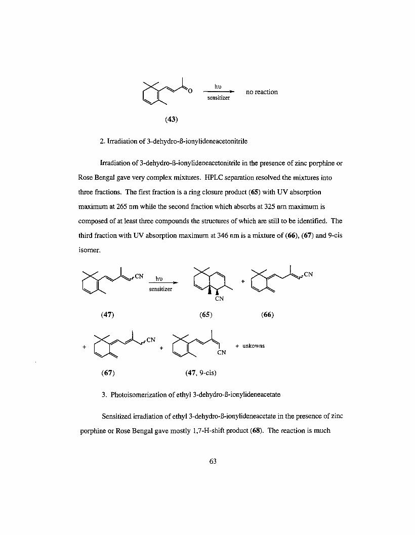
~o(43)
hu ..sensitizer
no reaction
2. Irradiation of 3-dehydro-B-ionylideneacetonitrile
Irradiation of 3-dehydro-B-ionylideneacetonitrile in the presence of zinc porphine or
Rose Bengal gave very complex mixtures. HPLC separation resolved the mixtures into
three fractions. The first fraction is a ring closure product (65) with UV absorption
maximum at 265 nm while the second fraction which absorbs at 325 nm maximum is
composed of at least three compounds the structures of which are still to be identified. The
third fraction with UV absorption maximum at 346 nm is a mixture of (66), (67) and 9-cis
isomer.
~CN~y -
hu
sensitizer.. +~CN
(47)
~# CN
+ +~
(67)
CN
(65)
~~~- 1N
(47,9-cis)
(66)
+ unkowns
3. Photoisomerization of ethy13-dehydro-B-ionylideneacetate
Sensitized irradiation of ethyl 3-dehydro-B-ionylideneacetate in the presence of zinc
porphine or Rose Bengal gave mostly 1,7-H-shift product (68). The reaction is much
63

cleaner than that of 3-dehydro-B-ionylideneacetonitrile. The photoirradiation was carried
out in three different solvents (acetone, chloroform and benzene). In acetone, there are
some minor reaction products which were suspected to be 7-cis and 7,9-dicis besides the
major 1,7-H-shift product, while in benzene and chloroform the reactions gave clean 1,7
H-shift product. Temperature also seems to have a significant effect on the reaction rate.
While 1H-NMR shows that irradiation at room temperature for 10 h can completely convert
the starting material, less than 20% of the starting material is converted to the 1,7-H-shift
product even after 15 h irradiation at DoC.
~,.rCOOEt~ y -
(49, all-trans & 9-cis)
hu
sensitizer•~COOEtU~' ~
(68)
4. Photoisomerization of 3-dehydro-B-ionylideneacetaldehyde
Sensitized photoisomerization of 3-dehydro-B-ionylideneacetaldehyde in the
presence of Rose Bengal or zinc porphine gave the 1,7-H-shift product (69) plus other
unidentified minor products.
~,.rCHO~y -
(35, all-trans & 9-cis)
hu
sensitizer•
(69)
+ other unidentifiedproducts
5. Photoisomerization of 3-dehydro-B-ionylideneethanol
Sensitized photoisomerization of 3-dehydro-B-ionylideneethanol in the presence of
Rose Bengal or zinc porphine again gave mostly 1,7-H-shift product (70). Small amounts
of 7,9-dicis and 7-cis products were also produced.
64

hu
sensitizer•
(50, all-trans & 9-cis)
~CH20H(50,7-cis)
(70)
+ csc=rCH20H
(50, 7,9-dicis)
B. New approach to 3-dehydroretinal isomers
1. Synthesis of 3-hydroxy-B-ionone
The synthesis of 3-hydroxy-B-ionone was carried out by hydroboration of 3
dehydro-B-ionone ethylene ketal followed by hydrolysis. Two borane reagents were tried
for this reaction: borane methyl sulfide complex (2.0 Min THF) and 9-BBN (9
borabicyclo[3.3.1]nonane, 0.5 M in THF). Both reagents required hours of reflux to
complete the reaction. In the case of 9-BBN, 3-hydroxy-B-ionone ethylene ketal was the
only hydroboration product isolated, while for the borane methyl sulfide complex, 4
hydroxy-B-ionone ethylene ketal was also isolated as a minor component.
9-BBN
THF, relux•
(45)
oxalicacid.CH2Cl2
H20, relux•
(51)
~OHO (52)
65

2. Photoisomerization of 3-hydroxy-B-ionylideneacetonitrile
3-Hydroxy-B-ionylideneacetonitrile was prepared by a Homer reaction of 3-
(53)
NaH
(EtO)2P(O)CH 2CN•~o
HOU'
(52)
hydroxy-B-ionone with triethylcyanomethylphosphonate.
~CNHOU Y -
Photoirradiation of 3-hydroxy-B-ionylideneacetonitrile in the presence of
benzanthrone or Rose Bengal resulted in one-way isomerization of the double bond at the
7-position. At the end of irradiation, a mixture of 7-cis and 7,9-dicis isomers in a ratio of
about 50: 50 was obtained. The two isomers were readily separated by silica gel
chromatography and both were low-melting white solids « 250C).
~CNHO
Rose Bengal
Corning filter3-73
• ~CNHO~ / ~
(53, all-trans & 9-cis) (53, 7-cis & 7,9-dicis)
3. Synthesis of7-cis 3-hydroxyretinonitrile and other 7-cis isomers (7, 13-dicis,
7,9-dicis and 7,9, 13-tricis)
7-Cis 3-hydroxyretinonitrile was synthesized by DIBAL-H reduction of 7-cis 3
hydroxy-B-ionylideneacetonitrile followed by C5 extension.
~CNHO~ / \
DIBAI:..-H• ~CHO
HO
(53, 7-cis) (56, 7-cis)
66

(EtO) zP(O)CH zC(CH 3)=CHCN..NaH
><0=HO~ / "=-~
CN
(58, 7-cis & 7, 13-dicis)
7,9-Dicis and 7,9, 13-tricis 3-hydroxyretinonitrile were synthesized starting from
(56, 7,9-dicis)
~CHO
HO~ /..DffiAL-H
7,9-dicis 3-hydroxy-B-ionylideneacetonitri1e.DereNHO
(53, 7,9-dicis)
(EtO) 2P(O)CH 2C(CH 3)=CHCN~
NaHHO
(58, 7,9-dicis & 7,9, 13-tricis)
There is no Ll-cis isomer formed from this Homer reaction.
4. Synthesis of7,1l-dicis and 7,9,1l-tricis isomers
Construction of the l l-cis double bond was accomplished by the method reported
by Still and Gennari.H This method used fluorine substituted C5 phosphonate, and a
strong base to enhance the formation of l l-cis double bond. The reaction was selective
( l l-cis accounted for more than 60% of all the possible isomers) but not specific, so the
syntheses of 7, Ll-dicis and 7,9, l l-tricis 3-hydroxyretinonitriles both resulted in a mixture
of several isomers. For 7,11-dicis, four isomers: 7-cis, 7, Ll-dicis, 7, 13-dicis and
7,11,13-tricis were isolated.
67

~CNHO~ I ~
(53, 7-cis)
DIBAL-H• ~CHOHO~ I ~
(56, 7-cis)
(CF3CH20hP(O)CH 2C(CH3)=CHCN•
KN(TMS) 2, 18-Crown-6 HO
(58, 7,1l-dicis & 7,1l,13-tricis)
The synthesis of the 7,9,11-tricis isomer was accomplished by starting from 7,9
dicis 3-hydroxy-B-ionylideneacetonitrile. Four isomers were isolated: 7,9-dicis, 7,9,13
tricis, 7,11,13-tricis and a possible all-cis (identified with UV).
~CN
HO~ I
(53, 7,9-dicis)
DIBAL-H•~CHO
HO~ I
(56, 7,9-dicis)
KN(TMS) 2, 18-Crown-6
..HO
(58, 7,9,l1-tricis & all-cis)
5. Synthesis of 7-cis, 7, 13-dicis, 7,9-dicis, 7,9, 13-tricis 3-hydroxyretinals (59)
The four isomers were prepared by DIBAL-H reduction of the corresponding
nitriles. Pure isomers were obtained from HPLC purification.
HO
(58, 7-cis isomers)
DIBAL-H ..HO
(59. 7-cis isomers)
1H-NMR data of these four isomers are shown in Tables 2 and 3.
68

0\\0
Table 2. IHNMR chemical shift (ppm) data of 3-hydroxyretinal isomers
isomers 7-H 8-H IO-H ll-H 12-H 14-H 15-H 5-Me 9-Me 13-Me
7-cis 5.94 6.16 6.25 7.05 6.34 5.97 10.11 1.55 1.90 2.30
7,9-dicis 6.07 6.64 6.05 7.14 6.28 5.97 10.11 1.51 1.86 2.31
7,13-dicis 5.94 6.17 6.25 6.94 7.26 5.85 10.18 1.50 1.90 2.15
7,9, 13-tricis 6.10 6.77 6.18 7.20 7.42 5.79 10.24 1.50 1.85 2.15
Table 3. IHNMR coupling constant (Hz) data for 3-hydroxyretinal isomers
isomers J7,8 JIO,l1 Jll,12 J14,15
7-cis 12.5 11.5 15.1 8.3
7,9-dicis 12.5 11.6 15.3 8.1
7,13-dicis 12.4 10.6 14.7 8.5
7,9, 13-tricis 12.3 11.4 14.9 7.8

6. Synthesisof 7-cis isomers of 3-dehydroretinonitrile
The doublebondat C3-C4can be generatedby E2 elimination. A mild reaction
condition has to be workedout becauseof the thermal sensitivity of the conjugated cis
double bonds of the chromophore chain. All-trans3-hydroxy-B-ionylideneacetonitrile was
used as a model compound to findoptimaleliminationconditions. The 3-hydroxy group
was first convertedto a mesylate groupby reactingwith methanesulfonyl chloride in the
presence of triethylamine. Elimination of the mesylate in the presenceofDBU (1,8
diazabicyclo[5.4.0]undec-7-ene) at room temperature proceeded very slowly; the reaction
was acceleratedgreatly by raising the temperature to 550 C.
~CNHO
(53, all-trans & 9-cis)
25°C. 15min.
(71, all-trans & 9-cis)
DBU.55°C•
<30 min.
~CN~~. -
(47, all-trans & 9-cis)
The same scheme was appliedto the 7-cis isomer of 3-hydroxyretinonitrile. While
the eliminationreaction workedout just like the model compound, about40% of the
elimination productwas trans3-dehydroretinonitrile. The elevated temperature apparently
had caused substantial isomerization. Afterseveral attempts in search of a milder
elimination condition, we discovered that the best method is to convert the 3-hydroxy
group to a tosylate group. The elimination was carried out at room temperature in the
presence of KOH and 18-crown-6.
70

HO
TsCI. DMAP ..CH2C1Z TsO
(58, 7-cis)
KOH. 18-Crown-6•
(61, 7-cis)
(60, 7-cis)
Other isomers (7,9-dicis, 7, 13-dicis, 7, l l-dicis, 7,9, Ll-tricis, 7,9, 13-tricis) were
synthesized in a similar way.
7. Synthesis of 3-dehydroretinal isomers (7-cis, 7, 13-dicis, 7,9-dicis, 7,9,13
tricis, 7, ll-dicis and 7,9, ll-tricis)
The above six isomers were prepared by DIBAL-H reduction of the corresponding
nitriles. Pure isomers were obtained from HPLC separation. 1H-NMR data are shown in
Table 4 and 5.
(61,7-cis) .
DIBAL-H
71
(62, 7-cis)

-Jtv
Table 4. 1HNMR chemical shift (in ppm) data of 3-hdehydroretinal isomers
isomers 7-H 8-H IO-H ll-H 12-H 14-H 15-H 5-Me 9-Me 13-Me
7-cis 5.95 6.18 6.27 7.06 6.34 5.97 10.11 1.59 1.97 2.31
7,9-dicis . 6.05 6.62 6.08 7.15 6.28 5.97 10.11 1.56 1.92 2.31
7, 13-dicis 5.98 6.23 6.34 7.04 7.37 5.78 10.17 1.57 1.96 2.12
7,9, 13-tricis 6.10 6.71 6.14 7.11 7.31 5.79 10.17 1.54 1.91 2.08
7,II-dicis 5.97 6.17 6.00 6.71 6.62 5.88 10.05 1.56 1.91 2.30
7,9,II-tricis 6.12 6.54 5.94 6.67 6.43 5.91 10.05 1.53 1.89 2.32
9-cis 6.34 6.82 6.12 7.23 6.32 5.98 10.11 1.93 2.04 2.31
9,13-dicis 6.35 6.80 6.15 7.12 7.25 5.85 10.20 1.60 1.95 2.15
13-cis 6.34 6.34 6.27 7.05 6.36 5.86 10.21 1.89 2.04 2.15
l l-cis 6.35 6.28 5.94 6.70 6.57 6.09 10.09 1.87 2.00 2.37
all-trans 6.34 6.30 6.22 7.12 6.37 5.97 10.09 1.86 2.02 2.31

-Jt.J
Table 5. 1HNMR coupling constant (in Hz) data for 3-dehydroretinal isomers
isomer J7,8 JIO,l1 J11,12 J14,15
7-cis 12.3 11.5 15.0 8.1
7,9-dicis 12.2 11.5 15.2 8.2
7,13-dicis 12.6 11.4 15.0 8.0
7,9, 13-tricis 12.6 11.3 15.0 8.1
7,II-dicis 12.3 11.1 12.3 8.0
7,9,II-tricis 12.5 11.9 12.2 8.0
9-cis 15.9 11.4 15.0 8.2
9,13-dicis 16.0 10.9 15.4 8.0
13-cis 16.1 11.5 15.0 8.0
l l-cis 16.0 11.7 12.2 8.1
all-trans 15.9 11.4 15.1 8.2

8. Synthesis of 3-methoxy-3-dehydroretinal
Three isomers (all-trans, 9-cis and 13-cis) of 3-methoxy-3-dehydroretinal were
synthesized using the following scheme:
~o(43)
~ 0 0Meo~ ~~
(53)
b---
~CNMeO
(56)
C, dII ~CN
MeoM ~~ -(48)
e, f, e CHO
(63, 9-cis, 13-cis, all-trans)
Scheme 4. Synthetic route for 3-methoxy-3-dehydroretinal
a) Concentrated H2S04, CH30H, SoC; b) (EtO)2P(O)CH2CN, NaH, THF; c) NBS,
CCI4, reflux; d) KOH, CH30H; e) DmAL-H, THF, -78oC; f) NaH,
(EtO)2P(O)CH2CH(CH3)=CHCN.
The 1H-NMR data of the three prepared isomers all-trans, 9-cis and 13-cis are
presented in Table 6 and 7.
74

--JUl
Table 6. i HNMR chemical shift (in ppm) data of 3-methoxy-3-dehydroretinal isomers
isomer 7-H 8-H IO-H ll-H 12-H 14-H 15-H 5-Me 9-Me 13-Me
all trans 6.40 6.28 6.23 7.22 6.39 5.87 10.05 1.86 2.01 2.30
9-cis . 6.42 6.30 6.29 7.14 7.38 5.78 10.18 1.89 2.03 2.13
13-cis 6.40 6.86 6.14 7.31 6.37 5.84 10.06 1.93 2.01 2.30
Table 7. IHNMR coupling constant (in Hz) data for 3-methoxy-3-dehydroretinal isomers
isomer 17,8 110,11 111,12 114,15
all-trans 16.1 11.6 15.0 8.1
9-cis 16.5 11.3 14.9 7.9
13-cis 16.0 11.6 15.0 8.1

9. HPLC separations of isomers of 3-hydroxyretinal, 3-dehydro-3-methoxyretinal and 3
dehydroretinal
Retinoids are easily isomerized or even destroyed in the presence of light and
oxidants. Considerable attention was given to the collection, handling and storage of the
synthesized isomers of.retinal and analogs. Since most of the synthetic reactions employed
often resulted in a mixture of isomers, separation of them had to be worked out to identify
the individual isomers. Regular column chromatography was not successful for the
separation of most of the isomeric mixtures. Therefore HPLC separation conditions had to
be worked out for separating the isomeric mixtures. Some of the representative HPLC
chromatograms are shown in Figures 8, 9 and 10.
76

Mobile Phase: 4% Ether/Hexane
Flow rate: 2 mllmin
7.9.1:3-lricu
oj 0)
7. I 3-&cis
t
61.
Figure 8. HPLC separation of 3-dchydrorctinaI Isomers,
all-trans
/'Mobile Phase: 3% ethyl acetatc!
Hexane saturated with acetonitrile
Flow rate: 2 ml/min
0.0
Figure 9. HPLC separation of 3-mcLhoxy-3·dehydroretinaI isomers.
IS.
•Mobile I'has<;: 0.7% CI1JOHI
17% THF/82.3%Hexanc
Flow rate: 2 ml/min
7.13-dkis
i "o.o
Figure 10. HPLC separation of 3-hydroxyreLinaI isomers.
77
.L55.
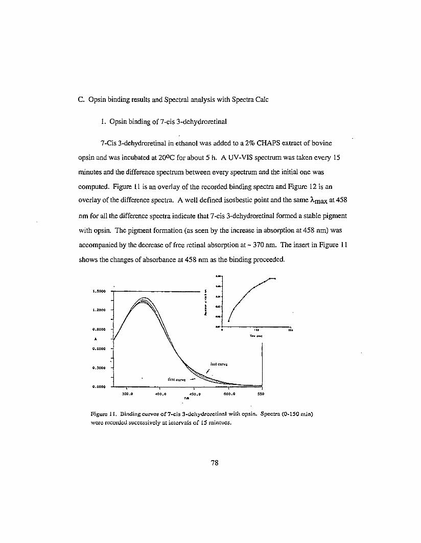
C. Opsin binding results and Spectral analysis with Spectra Calc
1. Opsin binding of 7-cis 3-dehydroretinal
7-Cis 3-dehydroretinal in ethanol was added to a 2% CHAPS extract of bovine
opsin and was incubated at 200C for about 5 h. A UV-VIS spectrum was taken every 15
minutes and the difference spectrum between every spectrum and the initial one was
computed. Figure 11 is an overlay of the recorded binding spectra and Figure 12 is an
overlay of the difference spectra. A well defined isosbestic point and the same Amax at 458
nm for all the difference spectra indicate that 7-cis 3-dehydroretinal formed a stable pigment
with opsin. The pigment formation (as seen by the increase in absorption at 458 nm) was
accompanied by the decrease of free retinal absorption at - 370 nm. The insert in Figure 11
shows the changes of absorbance at 458 nm as the binding proceeded.
1.5000
1.2000
0.9000
0.6000
0.3000
0.0000
3:10.0 ~oo.o 000.0
'01
Figure 11. Dindins curves of 7-cis 3-dchydrorctinnl with opsin. Spectra (0-150 min)
were recorded successively at intervals of 15 mintucs.
78

0.0000
o.o~oo
-0.0000
-0.0~~0
-0.0769
-0.1100
JJO.O ~OO.O ~O.O
na:100.0 GOO.O
Figure 12. Overlay of difference spcvtra between each of the successi~e opsin bindingcurves of 7-cis 3-dehYUroretinal and the initial curve.
2. Spectra Calc analysis of the binding curves of 7-cis 3-dehydroretinal
To confirm the assumption that formation of the 7-cis pigment from 7-cis 3
dehydroretinal was not complicated by isomerization process, Spectra Calc analysis of the
binding curves was carried out. The combination of three components, a 7-cis 3
dehydroretinal peak at 377 nm, a pigment peak at 458 nm, and a blue shifted random Schiff
base peak at 346 nm was found to fit the binding curves very well. Table 8 lists the
changes of area in percentages of all the individual components. X2 is the statistical error
of curvefitting.
79

Table 8. Curve-fitting of the opsin binding curves of 7-cis 3-dehydroretinal
Time Schiff base% 7-cis retinal% 7-cis pigment% 7-cis retinal and X2
(min) (346 nm) (377 nm) (458 nm) Schiff base
30 44.3 54.5 1.3 98.7 0.035
45 43.9 54.6 1.5 98.5 0.0095
60 43.1 54.1 2.7 97.3 0.0097
75 42.7 53.6 3.7 96.3 0.0093
90 42.4 53.1 4.5 95.5 0.0089
105 42.1 52.8 5.2 94.8 0.0088
120 41.8 52.5 5.7 94.3 0.0080
135 41.7 52.1 6.1 93.9 0.0076
150 41.8 51.8 6.4 93.6 0.0072
165 43.1 50.3 6.6 93.4 0.0069
180 43.3 50.1 6.6 93.4 0.0070
195 44.3 49.1 6.6 93.4 0.0066
210 44.3 49.1 6.6 93.4 0.0068
3. Opsin binding of 7,9-dicis 3-dehydroretinal
Opsin binding results of 7,9-dicis 3-dehydroretinal are shown in Figures 13 and
14. It is clear from the figures that 7,9-dicis 3-dehydroretinal also formed stable pigment
with opsin with a Amax at 466.8 nm.
80

0.3000
0.2000
e, :000
0.0000
~tI
~"
!
i~l'~
i ~"
~..uo
l:sl eurve • II "T_C.....
350.0 ~~o.o
noo.c:;o.o :'00.0
Figure 13. Binding curves of7.9-dicis 3-dehydroretinal with opsin. Spectra (O-63 min)
were recorded successively at intervals of 6 rnlntucs, Insert: the absorption change at 466nrn (Amaxof the difference spectra) over time.
0.20CO
I:1Slcurve
O. :200
o.o~oo
Ar~~l CIIl\'C
-e.eeco
-0. :200
-0.2000
3:;0.0 ~oo.o ~:;O.O :;00.0noo
Figure 14. Overlay of difference spcvtra between each of the successive opsin binding
curves of7.9-dicis 3-dehyclrorctinal and the initial curve.
81

4. Opsin binding of 7, 13-dicis 3-dehydroretinal
Opsin binding spectra and difference spectra of 7, 13-dicis 3-dehydroretinal are
shown in Figures 15and 16 respectively. There is no isosbestic point in either the binding
curves or the difference spectra. Unlike 7-cis and 7,9-dicis, the formation of pigment is
accompanied by anactual increase in the region of free dehydroretinal absorption. The
results suggested isomerization of the dehydroretinal chromophore.
5. SpectraCalc analysis of the binding curves of 7, 13-dicis 3-dehydroretinal
Since the likely isomerization product from 7, 13-dicis 3-dehydroretinal is 7-cis (see
discussion), the combination of four components 7, 13-dicis (367 nm) and 7-cis 3
dehydroretinal (377nm),7-cis pigment (458 nm) and a Schiff base peak (350 nm) was
applied to the curvefitting of the binding curves. Table 9 lists the changes of area in
percentages of all the individualcomponents. Excellent fitting is achieved for all the
binding curves withverysmall X2 values.
Table 9. Curve fitting of the opsin binding curves of 7, 13-dicis 3-dehydroretinal
Time Schiffbase 7,13-dicis 7-cis 7-cis pigment X2
area %(350) area % (367) area % (377) area % (458)
30 4.3 80.4 11.7 3.5 0.0035
60 4.4 79.4 12.3 3.9 0.0031
90 4.2 78.6 13.1 4.2 0.0029
120 4.1 77.8 13.8 4.3 0.0028
150 3.9 77.5 14.2 4.5 0.0026
180 3.6 77.1 14.6 4.6 0.0024
210 3.5 76.9 14.9 4.7 0.0022
240 3.3 76.7 15.1 4.9 0.0022
82

1.1000
O.Beoo
0.C600
0.2200
0.0000
~~0.0 ~00.0
nil000.0
Figure 15. Binding curves of7,13-dicis 3-dehydroretinal with opsin. Spectra (0-330min) were recorded successively at intervals of 30 mintucs.
0.1000
O.OBOO
0.0600
0.0200
0.0000
nil
Figure 16. Overlay of difference spcvtra between each of the successive opsin binding
curves of7,13-dicis 3-dehydrorctinal and the initial curve,
83

6. Opsin binding of 7,9, 13-tricis 3-dehydroretinal
Figures 17 and 18 show the binding results of 7,9, 13-tricis 3-dehydroretinal with
opsin. Similar to the result of 7,13-dicis 3-dehydroretinal, the difference curves suggested
isomerization during incubation.
300.0 J~O.O ~OO.O
O."':;0.0 SOO.O :;:;0.0
Figure 17. Binding curves of7,9,13-tricis 3-dehydroretinal with opsin. Spectra (0-420
min) were recorded successively at intervals of30 rnintucs.
o.oaso
0.0410
0.0270
0.0130
-0.00.10
-0.01:10
0'"
Figure 18. Overlay of difference spcvtra between each of the successive opsin binding
curves of 7,9 ,13-tricis 3-dehydroretinal and the initial curve.
84

7. Spectra Calc analysis of the binding curves of 7,9,13-tricis 3-dehydroretinal
Since 7,9-dicis 3-dehydroretinal formed stable pigment with opsin, and the Cl3
Cl4 double bond is a readily isomerizable double bond, we carried out the curve-fitting of
the 7,9, 13-tricis 3-dehydroretinal binding curves with four components: a Schiff base at
348.8 nm, 7,9,13-tricis 3-dehydroretinal at 362 nm, 7,9-dicis 3-dehdyroretinal at 369 nm
and a 7,9-dicis pigment peak at 466 nm. The curve-fitting results are shown in Table 10.
Table 10. Curve fitting of the opsin binding curves of 7,9, 13-tricis 3-dehydroretinal
Time (min) Schiff base 7,9,13-tricis 7,9-dicis 7,9-dicis pig- X2
(348.8 nm) (362 nm) (369 nm) ment (466 nm)
30 5.18 43.00 10.65 1.48 0.00168
60 4.78 42.92 12.26 1.74 0.00189
90 4.46 42.68 13.53 1.95 0.00168
120 4.14 42.54 14.51 2.29 0.00169
150 3.89 42.45 15.12 2.60 0.00155
180 3.74 42.44 15.53 2.98 0.00154
210 3.67 42.38 15.79 3.22 0.00149
240 3.61 42.35 15.79 3.43 0.00156
270 3.42 42.38 16.00 3.65 0.00163
300 3.32 42.43 16.02 3.84 0.00168
330 3.32 42.36 16.00 3.92 0.00167
360 3.27 42.34 15.97 4.04 0.00189
390 3.08 42.37 15.97 4.09 0.00197
420 3.09 42.26 15.91 4.17 0.00210
85

8. Opsin binding of 7, l l-dicis 3-dehydroretinal
In Figure 19 and 20 arc the opsin binding results of 7, ll-dicis 3-dehydroretinal.
The increase at 370 nm is possibly the result of isomerization and Schiff base formation.
The pigment formed initially has a Amax at 475 nm and it gradually shifted to shorter
wavelength as is shown in the insert in Figure 19.
0.7500
0.5920
0.4340
0.2760
0.1190
-0.0400
I ."
~j
1..
• I•• ... ,.. ...TI't.I_»
lUICWVC
350.0 400.0na
4:50.0 500.0 5:10.0
Figure 19. Binding curves of7,1l-dicis 3-dehydroretinal with opsin. Spectra (0-330
min) were recorded successively at intervals of 15 mintucs. Insert: the change of theAmaxof difference spectra over time.
0.3500
~0.2660
10.1820 .J
t-
0.0990
0.OJ40
-0.0700
350.0 400.0 450.0 500.0 ~.O
Figure 20. Overlay of difference spcvtra between each of the successive opsin binding
curves of7,11-dicis 3-dehydroretinal and the initial curve.
86

9. Spectra Calc analysis of the binding curves of 7, ll-dicis 3-dehydroretinal
The combination of four components: a Schiff base, 7,11-dicis 3-dehydroretinal, 7
cis 3-dehydroretinal, 7, 11-dicis pigment, 7-cis pigment was found to fit the binding curves
of 7, l l-dicis 3-dehydroretinal. Table 11 lists the results of curvefitting with Spectra Calc.
Table 11. Curve fitting of the opsin binding curves of 7, 11-dicis 3-dehydroretinal
Time Schiff base 7,11-dicis 7-cis (377 7-cis pigment 7,11-dicis pigment
(min) (358 nm) (372 nm) nm) (458 nm) (475 nm)
15 6.3 73.7 17.1 0 2.9
30 18.6 57.8 18.1 0 5.4
45 16.0 57.4 19.0 0 7.6
60 13.4 58.3 17.5 0 10.9
75 14.9 53.6 19.0 0 12.4
90 12.1 53.4 20.2 0 14.3
105 9.7 53.0 20.6 1.5 15.2
120 8.5 51.2 21.8 3.9 14.7
135 8.1 47.0 23.4 7.9 137
150 7.3 44.7 23.9 11.9 12.2
165 6.8 40.9 25.3 16.6 10.3
180 7.2 40.3 23.9 20.1 8.5
195 7.1 40.7 24.9 19.5 7.8
300 6.9 39.1 25.8 20.3 7.9
87

10. Opsin binding of 7,9, l l-tricis 3-dehydroretinal
In Figure 21 and 22 are the opsin binding results of 7,9, 11-tricis 3-dehydroretinal.
Schiff base formation caused the increase near the free aldehyde absorption maximum.
Unlike 7, l l-dicis, the pigment formed seemed to be stable and has a constant Amax of 454
nm throughout the binding process. ..I
(i u
i u
J ..,U .1 ," IIII
r_~.
1.Z~00
0.7158
A
0.Z336
-o.ooeo
:;50.0 400.0 "50.0n..
1100.0
"r-ISIS".O 600.0
Figure 21. Binding curves of7.9.11-tricis 3-dehydroretinal with opsin. Spectra (0-225
min) were recorded successively at intervals of 15 mintues. Insert: the absorption changeat 454 nm (A.max of the difference spectra) over time.
0.4!i00
0.35711
0.Z652
A
o.l7Ze
0.oe04
-O.OIZO
350.0 400.0 4S0.0nil
ISOO.O li5Q .0 600.1
Figure 22. Overlay of difference spcvtra between each of the successive opsin bindingcurves of7.9,ll-tricis 3-dehydroretinal and the initial curve.
88

11. Spectra Calc analysis of the binding curves of 7,9, 11-tricis 3-dehydroretina1
The binding curves of 7,9, 11-tricis 3-dehydroretinal were fitted with three
components: 7,9,11-tricis 3-dehydroretinal at 357 nm, 7,9-dicis 3-dehydroretinal at 363
nm and 7,9-dicis pigment at 466 nm. Table 12lists the curve-fitting results with Spectra
Calc.
Table 12. Curve fitting of opsin binding curves of 7,9, l l-tricis 3-dehydroretinal
Time (min) 7,9,11-tricis 7,9-dicis 7,9-dicis pigment X2
(357 nm) (363 nm) (466 nm)
15 0.494 0.073 0.011 0.2231
30 0.489 0.105 0.021 0.3224
45 0.484 0.116 0.033 0.2211
60 0.478 0.131 0.044 0.1202
75 0.472 0.142 0.056 0.1189
90 0.466 0.151 0.067 0.0140
105 0.463 0.158 0.075 0.0117
120 0.456 0.168 0.088 0.0087
135 0.452 0.171 0.097 0.0068
150 0.451 0.172 0.097 0.0087
165 0.452 0.174 0.097 0.0088
12. Opsin binding of9-cis 3-methoxy-3-dehydroretinal
9-Cis 3-methoxy-3-dehydroretinal formed a red shifted pigment with opsin readily
upon incubation. Figures 23 and 24 are the binding results of 9-cis 3-methoxy-3-
89

dehydroretinal with opsin. The pigment formed has a constant Amaxat 545 nm throughout
the binding process.
Figure 23. Binding curves of 9-cis 3-methoxy-3-dehydroretinal with opsin. Spectra (0
35 min) were recorded successively at intervals of 5 mintues. Insert: the absorptionchange at 545 nm (Amax of the difference spectra) over time.
0.7000
0.'600
0.2BOO
0.1400'
0.0000
0400.0 1100.0n.
5.50.0 600.0 6110.0
Figure 24. Overlay of difference spevtra between each of the successive opsin binding
curves of 9-cis 3-methoxy-3-dehydrorctinal and the initial curve.
90

13. Opsin binding of 7-cis 3-hydroxyretinal
In Figures 25 and 26 are the opsin binding results of 7-cis 3-hydroxyretinal. A
stable pigment with a "'max of 454 nm was formed.
i.eooo
r, :860
0.8720
-0.0700
.. II 'N UI "I-....
I
350.0 3BO.O ~oo.o ~20.0 4<0.0 ~60.0 ~o.o :100.0 :120.0nil
Figure 25. Binding curves of7-cis 3-hyclroxyretinal with opsin. Spectra (0-120 min)
were recorded successively at intervals of 15 mintucs, Insert: the absorption change at454 nm (Amaxof the difference spectra) over time.
0.0700
0.0160
-0.0380
• -0.0920
-0.1460
-0.2000
360.0 380.0 .00.0 .;0.0 .~o.o .80.0 .80.0 600.0 820.0nil
Figure 26. Overlay of difference spevtra between each of the successive opsin binding
curves of7-cis 3-hydroxyretinal and the initial curve.
91

14. Opsin binding of7,9-dicis 3-hydroxyretinal
Figure 27 and 28 are the opsin binding results of 7,9-dicis 3-hydroxyretinal. A
stab!e pigment with a Amaxof 448 nm was formed.
1. 4000
1.1200
0.8400
0.5600
0.2800
0.0000
340.0 360.0 300.0
.. .t ,.. u. ..."","1-
I~SI curve
460.0 480.0 500.0
Figure 27. Binding curves of7,9-dicis 3-hydroxyretinal with opsin. Spectra (0-135 min)were recorded successively at intervals of 15 mintucs, Insert: the absorption change at448 11111 (Ama" of the difference spectra) over time.
0.0600
0.0180
-0.0240
-0.0660
-0.1080 •
-0.1500
340.0 360.0 380.0 400.0 420.0 440.0 460.0 480.0 500.0
".
Figure 28. Overlay of difference spcvtra between each of the successive opsin binding
curves of 7.9-dicis 3-hydroxyrctinal and the initial curve.
92

Discussion
Six 7-cis 3-dehydroretinal isomers were prepared in this project Their bindings
with bovineopsin were also investigated. While the 3-dehydroretinal isomers
demonstrated a lot of similarities to their retinalcounterparts such as HPLC separation
profile, IH-NMRcoupling pattern, UV absorptionchangingpattern from all-trans to the
strained7-cis and other dicis and tricis isomers, sensitivity to light. heat and acidic residues
in solvent There are still considerable differences betweenthese two series. The most
unexpected difference is in their preparation: the different photochemistryof the 3-dehydro
CIS intermediates from their retinal counterparts compelled us to abandon a very
convenientapplication of the synthetic route of 7-cis retinal isomers to the synthesis of 3
dehydroretinal isomers. Another difference is the stability of the isomers during the opsin
binding process: the binding of the 7-cis and 7,9-dicis 3-dehydroretinals with opsin are
quite similar to that of their retinal counterparts; however7,13-dicis, 7,9,13-tricis, 7,11
dicis and 7,9,1l-tricis isomers seemed to isomerizeconsiderably faster than the
corresponding retinal isomers.
While it is well known that in the 7-cis isomers the ring and chain are highly twisted
out of plane,the exact degree of twisting has never beenmeasuredor calculated. By
comparingthe UVabsorptions of the different isomersbetween the retinal series and the 3
dehydroretinal series,we were able to reach a conclusion that the 7-cis isomers are twisted
to such an extent that the ring and the chain double bonds are substantially out of
conjugation. Molecularmodeling of the retinal and 3-dehydroretinal isomers were carried
out to-calculate the dihedral angles between the C5-C6 and C7-C8 double bonds which can
be used to characterize the degree of twisting betweenthe ring and the chain.
93

Efforts were also made to analyze the isomerization process of the unstable isomers
during the opsin binding process. Since there are considerable experimental difficulties
involved in identifying the isomerization product. a spectral analysis program called Spectra
Calc was used to deconvolute the complex UV-VIS spectra obtained during the binding
process, giving information related to possible pathways for the isomerization of these
isomers during the binding process.
A. The unusuall,7-H-shift reaction of the 3-dehydro Cl5 intermediates
Our sensitized photoisomerization results of a series of 3-dehydro Cl5
intermediates showed that the dominant product of the reaction is 1,7-H-shift product The
orbital symmetry rules of the frontier orbital theory predicts that for 1,7-H-shift reaction the
stereochemistry must be antarafacial for thermal conditions while suprafacial for
photochemical conditions. The two different pathways are illustrated below with a
substituted triene molecule:
rGu v
~v
hu•
suprafacial
hu ..suprafacial f)"z y "
HV
Both the photochemical and thermall,7-H-shift reactions have been observed."
For the sensitized photoirradiation of the 3-dehydro Cl5 intermediates carried out in this
project. only the thermal pathway was possible, because there were no singlet excited states
produced in the sensitized photoirradiation. The irradiation of the 3-dehydro Cl5
94

intermediates resulted in a two-step reaction: the triplet excited sensitization isomerization
to the 7-cis isomer intermediate, and then an immediate thermall,7-H-shift reaction. The
1,7-H-shift reaction from the 7-cis intermediate must be of relatively low activation energy
because the 7-cis intermediate was not observed even though IH-NMR and HPLC have
been used in an effort to detect them. While the 1,7-H-shift is so prevalent in the 3
dehydro CIS intermediates, it is rarely observed in the CIS intermediates in the synthesis
of isomers of parent retinal. The only example found in literature is the thermal 1,7-H-shift
of CIS ester (Scheme 5).'01 The reaction was used to convert the 9-cis geometry to 9-trans
ester.
mE H
9-eis
[1,7] H Shift
•~~E~H
H
!l
mH E
9-trans
[1,7] H Shift- ~~H~E
H
Scheme 5. 1,7-H-shift of CIS ester
The temperature for this I,7-H-shift reaction is considerably higher than the room
temperature in the 3-dehydro series.
Mead et al.88 also reported a facile 1,7-H-shift reaction of7,9-dicis-9-CF3-retinal. It
is believed that the CF3 group directed the molecule to the necessary 8-cisoid conformation
for the 1,7-H-shift reaction (Scheme 6).
95

• •
8-transoid
1,7-H-shift•
8-cisoid
Scheme 6. 1,7-H-Shift of 7,9-dicis-9-CF3-retinal
The ease with which the 3-dehydro CI5 intermediates underwent I,7-H-shift is
surprising. To rationalize the result, we tried a series of semi-empirical calculations to look
for the differences in structural properties between the 3-dehydro and the regular C15
intermediates which may be responsible for their different photochemical behavior. The
first step was the calculation of the ground state of these two types of C 15 intermediates.
The molecules with the 8-cisoid and 8-transoid conformations were optimized and their
electron charge distribution and distance between relevant atoms were compared. The
results of C15 ester, 3-dehydro C15 ester, C15 nitrile, 3-dehydro C15 nitrile are listed in
Table 13.
8-cisoid C15 nitrile
(8-c-C15-CN)
96
~10
I - 1f? eN5 5'
8-transoid C15 nitrile
(8-c-C l5-CN)

8-cisoid dehydro C15 nitrile
(8-c-DC l5-CN)
~10
I - f? eN~ 5 5'
8-transoid dehydro C 15 nitrile
(8-c- DC 15-CN)
Figure 29. The 8-cisoid and transoid conformations of 7-cis C15 nitriles
Table 13. Semi-empirical calculation results of 7-cis C 15 nitriles and esters
Molecules d (A)a e (C5)b e (C10)C Energy (kcal/molej'I
8-c-C15-CN 3.595 -0.078 -0.120 -3737
8-t-C15-CN 3.980 -0.078 -0.0135 -3736
8-c-DC15-CN 3.576 -0.054 -0.121 -3605
8-t-DC15-CN 3.977 -0.047 -0.0135 -3604
8-c-DC l5-CODEt 3.727 -0.048 -0.209 -4333
8-c-C 15-COOEt 3.645 -0.075 -0.214 -4464
a. The distance between carbon 5' and carbon 10. b. The electron charge on carbon 5. c.
The electron charge on carbon 10. d. The optimized binding energy (the total energy
relative to the same atoms that are not interacting).
The result that the 8-transoid and cisoid conformations (Figure 29) for both C15
nitrile and 3-dehydro C15 nitrile were of approximately equal energy indicates that the
required 8-cisoid conformation for 1,7-H-shift was readily accessible for the ground state
molecules of both compounds. There is not much difference between the 8-cisoid C15
nitrile and 8-cisoid 3-dehydro C 15 nitrile with regard to the distance the H has to migrate
(from C5' to CIO). Actually the only difference between the 8-cisoid C15 nitrile and the 8-
97

cisoid 3-dehydro C15 nitrile was that in the C15 nitrile, the electron charge at C5 was
44.4% percent higher than that in 3-dehydro C 15 nitrile; for the ester, the difference is
56%. However how this difference is related to the activity of 1,7-H-shift has not been
determined.
Though the 1,7-H-shift is a concerted process, its transition state may be imagined
to be composed of a H atom and the conjugated polyene radical of C 15 intermediates. The
H atom migrates from C5t to ClO.
8-cisoid C15 nitrile transition state
C5(:(CNHz
8-cisoid 3-dehydro C15 nitrile transition state
Semi-empirical calculations of the C15 ester and nitrile radicals were also carried
out to look for any significant differences between the regular and the 3-dehydro C15
intermediates. The radicals were first optimized using unrestricted Hartree-Fock (UHF)
method because of the open-shell nature of these radicals. The structure data were obtained
from the optimized conformation. Table 14 listed the results of the calculation.
As shown in Table 14, there is again not much difference between the dehydro C15
intermediates and the regular C15 intermediates. The only significant difference is still the
charge on carbon 5. The charge on carbon 5 in regular C15 is about 90% higher than that
in the 3-dehydro C15 intermediate.
98

Table 14. Semi-empirical calculation results of CIS radicals
Radicals d(A)a e (C5)b e(C5')C e (ClO)d Energy (kcal/rnolej''
8-c-C15-CN radical 3.123 -0.077 -0.203 -0.092 -3665
8-c-DC15-CN radical 3.189 -0.043 -0.199 -0.091 -3535
8-c-C15-COOEt radical 3.224 -0.074 -0.203 -0.180 -4391
8-c-DC15-COOEt radical 3.220 -0.039 -0.201 -0.179 -4262
a. The distance between carbon 5' and carbon 10. b. The electron charge on carbon 5. c.
The electron charge on carbon 5'. d. Theelectroncharge on carbon 10. e. The optimized
binding energy (the total energy relative tothesame atoms that are not interacting).
While the previous calculation focused on the optimized individual molecules, we
also tried to mimic the transition state byputting a hydrogen at equal distance between the
carbon 5' and Carbon 10 in the C15 nitrile radicals and then optimized the system (carried
out by J. R. Thiel). Table 15 listed the results of the calculation results for this imagined
transition state.
Table 15. Semi-empirical calcualtion results of the transition states for 1,7-H-shift
Molecules d(A)a e (5)b e(5')C e (lO)d Energy (kcal/mole'P
8-c-C15-CN transition state 3.837 -0.077 -0.209 -0.045 -3732
8-c-DC15-CN transition 3.155 -0.061 -0.185 -0.102 -3603
state
a. The distance between carbon 5' and carbon 10. b. The electron charge on carbon 5. c.
The electron charge on carbon 5'. d. Theelectroncharge on carbon 10. e. The optimized
binding energy (the total energy relative tothesame atoms that are not interacting).
99

A dramatic change in the electron charge on carbon lOis observed in this
calculation. In the dehydro C15 transition state, the electron charge on carbon 10 is more
than twice as high as that in the regular CI5 transition state. Since carbon 10 is the
destination of the H migration, with a high electron density, it will definitely attract the H
atom to move over. The distance between C5' and ClO is also significantly shorter (20%)
in the dehydro CI5 transition state than in the regular CI5 transition state. These two
factors, the distance between C5' and ClO and the electron charge on ClO, may be
responsible for the dominating 1,7-H-shift reactions in the 3-dehydro series.
100

.....o.-
Table 16. UV-Vis absorption maxima (Amax)of retinal and 3-dehydroretinal isomers
Hexanes Ethanol
retinal 3-dehydroretinal ~Amax retinal 3-dehydroretinal ~Amax
all-trans 368 a 385 17 383 a 401 b 18
13-cis 363 a 380 c 17 375 a 395 b 20
9-cis 363 a 380 17 373 a 391 b 18
Ll-cis 365 a 377 c 12 380 a 393 b 13
9, 13-dicis 359 a 375 16 368
7-cis 359 a 365 6 377 a 377 0
7,9-dicis 351 a 354 3 366 a 363 -3
7,13-dicis 357 a 356 -I 367
7,11-dicis 355 a 361 6 374 372 -2
7,9,II-tricis 345 a 348 3 357
7,9, 13-tricis 346 a 348 2 362 a
a. Y. Zhu & A. Trehan.89 b. Isler in reference." c. X. Li.91

B. Comparison of UV absorptions of retinal and 3-dehydroretinal isomers
The data in Table 16 (p97) show that the Amax. of UV-Vis absorption of 7-cis
isomers of 3-dehydroretinal is not significantly red-shifted and in some cases even blue
shifted when compared to the corresponding retinal isomers. This is in sharp contrast with
the 7-trans isomers which show a red-shift in the range of 12-17 nm in hexane and 13-20
nm in ethanol. According to the empirical method developed by Woodward and Fieser
for predicting the UV-VIS Amax of conjugated polyene system, an extra double bond
contributes about 30 nm to the Amax. of the parent molecule. The same amount of shift
would be expected for 3-dehydroretinal from retinal. The reason that the 7-trans shifted
only 12-17 nm and 7-cis did not show any significant shift lies in the fact that the ring and
the chain are not fully conjugated. In the 7-trans isomer, the van der Waals repulsion
between the 5-methyl and 8-hydrogen prevents a fully planar conformation, thus full
conjugation between the ring and the chain is not possible. In the 7-cis isomer, the van der
Waals repulsion is much more severe because now it involves two methyl groups.
~ CHO
7-trans 3-dehydroretinal
~
CHO
7-cis 3-dehydroretinal
The C(5)-C(6)-C(7)-C(8) dihedral angle (henceforth denoted <\>6-7) can be used to
characterize the degree of twisting between the ring and chain. There are a limited number
of X-ray reports about the dihedral angles of retinal isomers: all-trans retinal" is reported to
have a <\>6-7 dihedral angle of 58.30; ll-cis retinal", 41.4°; 13-cis retinal", 65.40. Other
methods such as long range coupling constant in NMR were also used to determine the
102

dihedral angles in retinal and some related lower homologs like B-ionone and CI5 synthetic
intermediates. Honig et al.96 reported that$6-7 in all-trans retinal and l l-cis retinal (in
solution) should be in the range of 300 to70°. Liu et al." studied the NMR properties of a
series of substituted 7-cis Cl5 intermediates, and their <1>6-7 was found to be in the range of
300 to 500. In another study, Liu et al.98 also reported <1>6-7 of all-trans and 7-cis 3
dehydro-B-ionone: all-trans, 35 ± 5°; 7-cis, 46±70. These numbers vary greatly and
were derived from different methods and for different physical states; so it is very difficult
to compare or apply the data to the isomers inTable 16. We have carried out the semi
empirical calcualtions of the isomers of retinal and 3-dehydroretinal. The dihedral angles
were obtained from the optimized conformations and are shown in Table 17. The 7-cis
isomers of both retinal and 3-dehydroretinal have<1>6-7 dihedral angles in the range of 58 to
700, significantly larger than those of7-transisomers which are in the range of 40-50°.
103

Table 17. Calculated Dihedral angles (<1>6-7) of retinal and 3-dehydroretinal isomers
Retinal 3-Dehydroretinal
<l>6-7a Energyb <l>6-7a Energyl'
all-trans 46.7 -4945 46.1 -4841
9-cis 46.8 -4945 45.8 -4814
13-cis 48.7 -4945 45.9 -4814
l l-cis 49.4 -4812 49.5 -4812
9,13-dicis 47.1 -4945 45.8 -4841
7-cis 60.7 -4942 60.3 -4811
7,9-dicis 66.4 -4942 58.4 -4809
7,II-dicis 60.2 -4939 58.8 -4814
7,13-dicis 61.6 -4941 61.2 -4810
7,9,II-tricis 63.4 -4940 65.8 -4808
7,9,13-tricis 63.8 -4942 59.0 -4810
a.In degree. b. Optimized binding energy (kcalfmole)
C. Pigmentformation of 3-hydroxyretinal and 3-dehydroretinal isomers
The newly prepared isomers of 3-hydroxyretinal, 3-dehydroretinal and 3
methoxyretinal wereincubated with opsin. The isomers were purified from HPLC before
the incubation. Thepigment formation was monitoredthrough UV-VIS absorption. The
pigmentformation and yield data are listed in Tables 17, 18, 19.
104

1. Binding of 3-hydroxyretinal isomers with opsin
As expected, the 7-cis and 7,9-dicis isomers of 3-hydroxyretinal formed stable
pigments with bovine opsin. The Amaxof the pigments are similar to that of 7-cis retinal
(450 nm) and 7,9-dicis retinal (460 nm). The pigment yields are lower than the 40%
reported for 7-cis and 7,9-dicis retinal. Neither 7, 13-dicis nor 7,9,13-tricis 3
hydroxyretinals formed significant amounts of pigment when incubated with bovine opsin
in preliminary experiments. No further effort was pursued due partly to the limited
availability of these two isomers.
Table 18. UV a ( Amax in nm) and pigment formation data of 3-hydroxyretinal isomers
isomers free aldehyde SBb PSBc Pigmentd yielde k1(xl04)f
7-cis 372.8 353.6 432.0 459.2 25% 8.92
7,9-dicis 357.6 342.4 414.4 452.4 20% 8.16
7, 13-dicis 364.0 348.0 422.4 g
7,9,13-tricis 357.6 341.6 416.8 g
a. in ethano1. b. n-butyl Schiff base in ethano1. c. protonated Schiff base in ethano1. d. in
detergent (CHAPS). e. Yield is relative to that of l l-cis retinal which is assumed to be
100%. It is calculated according to the method described in reference 99. f. The pseudo first
order rate constant (retinal isomers were used in excess) is calculated by plotting the
absorbance at the Amax of the pigment peak against time. The value is amplified by 104
times. From the tangents of the initial portion of the plot, the rate constants of the binding
were determined. g. Pigment formation was minimal.
2. Binding of 3-dehydroretinal isomers with opsin
Like 3-hydroxyretinal, the 7-cis and 7,9-dicis isomers readily formed pigment with
bovine opsin (Table 19). The other multiple cis isomers all showed instability in the
105

binding media (opsin detergent solution). The binding curves shown in the Results section
clearly demonstrated that isomerization occurred. A curve-fitting analysis has been used to
identify the possible isomerization products. The analysis results are discussed in the
following sections.
Table 19. UV-VIS absorption and pigment formation data of 3-dehydroretinal isomers-
isomer free aldehyde SB PSB Pigment vie1d k1(x104)
7-cis 377.2 356.4 437.2 458.8c 35% 7.42
7,9-dicis 363.2 352.0 426.4 466.8 40% 4.16
7, 13-dicis 367.2 350.4 426.4 b
7,9,13-tricis 362.4 348.0 420.8 b
7,11-dicis 372.4 358.0 435.6 b
7,9,11-tricis 357.2 345.2 416.4 b
a. Solvent and other conditions same as those described in Table 18. b. Isomerization of
the chromophore occurred with the pigment formation. c. Reported Amax at 464 nm."?
3. Binding of 3-methoxy-3-dehydroretinal isomers with opsin
3-Methoxyretinal is a very red-shifted retinal analog. Its UV absorption maximum
is about 50 nm longer than that of the corresponding all-trans retinal. The trans isomer
formed a pigment readily with bacterioopsin however with Amax (560 nm) blue shifted
relative to that of bacterioopsin (568 nm). The 9-cis isomer formed a stable pigment with
bovine opsin with UV Amax at 54~ nm. It is the most red-shifted bovine rhodopsin analog
prepared so far. The rate of the pigment formation was also faster than the 7-cis and 7,9
dicis isomers of 3-hydroxyretinal and 3-dehydroretinal. That 9-cis isomer binds with opsin
much faster than other isomers except l l-cis has been true for most of the retinal analogs
106

ever studied. The opsin binding pocket did not need to do a lot of adjustmentfor the 9-cis
isomer becauseof its spatial similarity to the native l l-cis isomers. Howeverfor 7-cis and
7,9-dicis isomers,considerable adjustment in the opsin conformation was needed, thus
slowing down the rate for binding.100
Table 20. UV ( Amax in nm) and Pigment formation data of 3-methoxyretinal isomers-
isomers free aldehyde SB PSB Pigment yield kI(x103)
all-transe 430.0 401.2 520.8 560b b b
9-cis 419.6 394.2 510.2 545c 45% 5.82
a. Solventand otherconditions same as those described in Table 18. b. Bindingwith
bacterioopsin (carried out by L. Colmenares), yield and rate constants were not determined.
c. Bindingwith bovine opsin.
D. The analysis of the four unstable 3-dehydroretinalisomers
To establishthe configuration of isomers in the pigmentdemandschromophore
extraction whichis a tediousprocess and precautionshave to be taken to minimizeany
isomerization during the extraction process. A common procedure is first to denature the
pigmentwith an organicsolvent so the chromophorecan be released from the protein. The
extractedfree chromophore can then be analyzed by HPLC and 1H-NMR. The
shortcomings of thischromophore extraction procedureare low recoveryyields, the need
of substantial material and unavoidabledark isomerization of theunstablegeometric
isomers. An improvedmethod is to add excess hydroxyamine which will convert the
retinalchromophore to its corresponding oxime resultingin retention of configuration.'?'
The difficulties involvedwith this method is the analysisand identification of the oximes,
for each isomerof retinal there is a pair of isomeric oximes (synand anti forms). To
identifythe configuration of the retinal chromophorewould require a completeseparation
107

and identification of the oxime isomers first Though the hydroxyamine method minimizes
the dark isomerization to a certain extent, it doesnotcompletely prevented the dark
isomerization in all cases.
In lightof the above difficulties associated withthe experimental procedure,we
resorted to thecurve resolution technique and useda software package called SpectraCalc
for the analysis of the binding curves recordedsuccessively during the incubation. Since
the UV-VISdata of the 3-dehydroretinal isomers areknown, byfitting in the UV-VIS
curvesof certainisomersand those of the pigments likely formed duringthe binding
processand examining the degree of fitting, reflecting how the fitted curvesoverlap with
the real binding curves, we can "identify" all apecies involved in the courseof binding
experiment and the nature of isomerization of the retinal chromophore.
1.The isomerization of 7,13-dicis 3-dehydroretinal during incubation
Sincethe C13-C14double bond in retinal is very susceptible to isomerizatonas
established in the case of 7,13-dicis retinal.!" the likely isomerization of 7,13-dicis3
dehydroretinal is to 7-cis-3-dehydroretinal. So in an initial attempt, we tried to fit the
bindingcurveswith three components:7-cis 3-dehydroretinal (withAmax at 377 nm),
7,13-dicis 3-dehydroretinal (with Amax at 367 nm), and7-cis pigment(with Amax at 458
nm). Howeverthe best calculationcurves still showed some discrepancy with the
experimental curvenear the blue edge of the curve (at about 350nm). This discrepancy
was removed whena peak at 350 nm was incorporated. Thiscan be rationalized as the
presence of a random Schiff base peak derived from themany freeamine groups on the
side of the protein bindingcavity in reactionwiththefree aldehyde. All twelve binding
curves, recorded successivelyat intervalsof 15minutes, were resolved by the Spectra Calc
curve fitting routine.
108

As shown in Figure 30, the sum (fitted curve) of the predicted component curves
(7,13-dicis 3-dehydroretinal, a random Schiff base from 7,13-dicis 3-dehydroretinal, 7-cis
3-dehydroretinal from 7,13-dicis 3-dehydrorctinal, and a pigment with 7-cis configuration)
matches the binding curve very well. This result supported our assumption that the 7,13
dicis 3-dehydroretinal isomerized to 7-cis, which then bound with opsin to form 7-cis
pigment While the pigment peak at 458 nm could also be interpreted as a hitherto
unknown 7,13-dicis pigment which coincidentally has identical A.max as that of the 7-cis
pigment, experimental observation that the pigment formed after the formation of7-cis 3
dehydroretinal parallels with 7,13-dicis retinal binding studies which showed by
chrornophore extraction that only the 7-cis retinal pigment was formed.l"
.,og .5-eon
~
Schilt bcee
nttod C:UlVO
blndlng curvo
300 350 -1-00 ""SO 500 550
Wavolont;lh (nm)
Figure 30. Overlay of the fitted curves and the binding curve of 7, 13-dicis 3
dchydrorctinal
109

81 6.0
80...."0"0;..,.cOJ 79
:::!<'S
77
...5.0 c
OJ
E.:'0Co
~ur:.
4.0
806020 40o76 -J--r--r-.....--r--r--,---.--.......,--;r-.-r-..-r-...-r-r--........--t- 3.0
100 120 140 160 180200
time (min)•o
7.13·dicis aldohydda
7·cis pigment
Figure 31. The area change of the component peaks during the binding of 7, 13-dicis 3
dehydroretinal with opsin.
2. The isomerization of7,9,13-tricis retinal during incubation
The easily isomerizable C13-CI4 double bond makes 7,9-dicis the most likely
isomerization product of 7,9, 13-tricis 3-dehydroretinal. Our attempt to fit the binding
curves with four components: a Schiff base peak at 348.8 nm, 7,9,13-tricis 3
dehydroretinal at 362 nm, 7,9-dicis 3-dehydroretinal at 369 nm and pigment with 7,9-dicis
configuration was quite successful. Figure 32 is an overlay of the fitted curves and the
experimental binding curve. The calculated and the experimental curves are virtully
indistinguishable which means that 7,9,13-dicis 3-dehydroretinal very likely isomerized to
7,9-dicis 3-dehydroretinal yielding the eventual stable 7,9-dicis pigment.
110

Joo J50 400
Waveleng:h <nm)
450 500 550
Figure 32. Overlay of the fitted curves and the binding curve of7,9,13-tricis 3dehydrorctinal
7.0
6.0
c..~ 5.0C.
J.O
40 80 120 160 200 240 280
72.0
70.0
68.0
66.0
tline (min)--0- 7,94ieis pigment
--e- 7,9.1J·lrieis aldehyde
Figure 33. The area change of component peaks during the binding of 7,9,13-tricis 3dchydrorctinal with opsin
111

3. The isomerization of 7, l I-dicis 3-dehydroretinal during incubation
Since the l l-cis double bond is another easily isomerizable double bond. we
initially tried to curve fit the 7,11-dicis 3-dehydroretinal binding curves with four
components: a Schiff base at 358 nm, 7,II-dicis 3-dehydroretinal at 372 nm, 7-cis 3
dehydroretinal at 377 nm, and a pigment with 7-cis configuration at 458 nm. However the
calculated curve did not match the experimental curve well. Careful analysis of the
difference curves (shown in the Results section) indicated that there might be two pigments
formed at different stages because the initial difference spectrum has a pigment Amax at 475
nm and in the end, the Amax changed to 458 nm which happened to be the Amax of
pigment of7-cis isomer. So the likely possibility is that the 7,1l-dicis also formed a
pigment with Amax at 475 nm. Initial curves agree very well with those calculated curves
from a Schiff base peak, 7, l l-dicis 3-dehydroretinal peak, 7-cis 3-dehydroretinal peak,
and only one pigment with Amax at 475 nm. Adding the 7-cis pigment component to the
initial curves caused considerable discrepancy. However the curve fitting of the later
binding curves requires the inclusion of the 7-cis pigment. Assigning the 475 nm pigment
to that of7,11-dicis is a little arbitrary. The most obvious concern is why the 7,lI-dicis
would form a pigment with a longer Amax than the 7-cis isomer pigment considering that
7,II-dicis isomer is more twisted than the 7-cis isomer. It may not be true to assume that
there is a direct correlation between the Amax of the pigments and the chromophore because
the protein chromophore interaction also contributes a lot to the pigment absorption. There
is a possibility that the protein perturbed the 7, 11-dicis chromophore more than the 7-cis
chromophore. In fact, in the retinal series, the 7, l l-dicis isomer formed a pigment (455
nm) which had a longer Amax than" that of7-cis (450 nm). Figure 34 shows excellent
agreement between the calcualted and the [mal experimental binding curves and Figure 35
is the change of the area of pigment peaks during the binding process.
112

·6
.4
.uCoofo
~.2
bIndIng curve
I
JSO 400 SOO SSO
Wavelength (nmj
Figure 34. Overlay of the fitted curves and the binding curve of7.11-di~is 3
dehydroretinal
CQI
E.2.0Clo
20.0
15.0
10.0
5.0
300200100~~~~~-"""'---r-----.----.--r---f- 0.0
400
29.0
24.0
-;;19.0QI
E~Clo
VI 14.0U~
9.0
4.0
·1.0a
time (min)o
•7·cis pigmont
7,t t-dicis pigment
Figure 35. The area changes of peak components during the binding of 7, l l-dlcis withopsin
113

4. The isomerization of 7,9, ll-tricis 3-dehydroretinal during incubation
It is also apparent from the binding curves of7,9,11-tricis 3-dehydroretinal that
isomerization OCCUlTed. Since the likely product of the 7,9,1l-tricis 3-dehydroretinal is
7,9-dicis, three components 7,9,ll-tricis 3-dehydroretinal (357 nm), 7,9-dicis 3
dehydroretinal (363 nm), and 7,9-dicis 3-dehydoretinal opsin pigment (466 nm) were tried
to fit in the binding curves. This assumption fits the later binding curves much better than
the initial five binding curves (see the X2 values in Table 12). There is a possibility that
there was more than one isomerized product involved, however attempts to fit the binding
curves with other components such as 7-cis and 7, l l-dicis isomer were not quite
successful. Figure 36 is an overlay of the final binding curve and the fitted curves. Figure
37 shows the changes of the area percentage of the components during the binding process.
DUee~ .5
~
o+-------~
.:ISO 400 450
Wavelength (nm)
500 550
Figure 36. Overlay of the fitted curves and the binding curve of7,9,11-t~icis3
dehydroretinal
114

90.0
...~;..,
oJ: 80.0...~
'"III
U0;:
--0\- 70.0r--
o 40 80
Time (mIn)
120
o
•
Figure 37. The area change of the component peaks during the binding 7 ,9,ll-tricis 3dehydroretinal with opsin
115

E. The opsin shift of the synthetic visual pigments
The opsin shift is defined as the difference of the reciprocal of the Amax of the
pigment and the protonated Schiff base. It reflects the contribution of protein perturbation
to the UV-VIS absorption of the pigment. Table 21 lists the opsin shift values of the
synthetic visual pigments prepared in this project. The opsin shift values of7-cis and 7,9
dicis isomers of retinal and l l-cis isomer of 3-dehydroretinal are also listed for the purpose
of comparison.
Table 21 The opsin shift of the rhodopsin analogs prepared in this projects
Isomers Protonated Schiff Pigment Opsin Shift
base (nm) (nm) (crrr l)
3-hydroxyretinal (7-cis) 432.0 459.2 1400
3-hydroxvretinal (7,9-dicis) 414.4 452.4 2200
3-Dehydroretinal (7-cis) 437.2 458.8 1100
3-Dehydroretinal (7,9-dicis) 426.4 466.8 1992
retinal (7-cis)b 425 450 1307
retinal (7,9-dicis)C 410 460 2070
ll-cis 3-dehydroretinald 471 520 2000
3-Methoxy-3-dehydroretinal (9-cis) 510.2 545 1400
l l-cis retinal 440 498 2650
9-cis retinal 440 483 2000
a. Solvent and other conditions same as those described in Table 18. b. reference 56.
c. reference 104. d. reference 37. e. reference 14.
116

F. Conclusions
1. A synthetic scheme for the preparation of 7-cis isomers of 3-dehydroretinal was
developed. Six 7-cis isomers (five are new) have been prepared through this scheme. The
geometry of the isomers have been properly identified through IH-NMR and UV-VIS
absorption spectra.
2. The binding interaction of these new isomers with bovine opsin were
investigated. While 7-cis and 7,9-dicis isomers formed stable pigments with opsin similar
to their retinal counterparts, the other four isomers 7, 13-dicis, 7,9, 13-tricis, 7,1l-dicis and
7,9, l l-tricis showed considerable isomerization during the opsin binding process. The
UV-VIS binding curves obtained during incubation were analyzed with a software package
called Spectra Calc. 7,13-Dicis and 7,9,13-tricis isomers were shown to have isomerized
to 7-cis and 7,9-dicis before pigment formation. The curves for 7,ll-dicis isomer are
consistent with possible formation of two isomeric pigments during the binding process.
7,9,11-Tricis isomer was shown to isomerize to 7,9-dicis, however the actual process,
especially at the early stage could be more complicated because of the apparent discrepancy
between part of the calculated curves and the observed curves.
(3) A new retinal analog, 3-methoxy-3-dehydroretinal was synthesized. Its UV
VIS absorption Amax is about 50 nm more to the red than that of retinal. Its 9-cis isomer
readily formed a pigment with opsin with a Amax at 545 nm.
(4) The photochemistry of the 3-dehydro C15 intermediates has been shown to be
different from that of retinal series.. Molecular modeling of these compounds revealed the
possible cause for the dominant 1,7-H-shift reaction in the 3-dehydro series.
117

G, Future A2 retinal studies
Photoisomerization plays an important role in a lot of physiological processes, To
understand the isomerization and be able to predict the outcome of the isomerization, recent
researches have been focusing on studying the nature of the excited states of the molecules
using time-resolved laser technique and resonance Raman spectroscopy. Koyama et al,
have studed the triplet states of7-cis, 9-cis, l l-cis and 13-cis retinal isomers with
picosecond absorption spectroscopy. lOS The isomerization process of these isomers can be
literally "seen" by taking the transient absorption of the isomers at a series of decay times
after excitation. Considering the fact that 3-dehydroretinal is the second most common
visual chromophore in nature, it is of interest and importance to study the photochemistry
of the 3-dehydroretinal series. Now that the availability of 3-dehydroretinal isomers is no
longer a problem, the photochemistry of this series should be thoroughly studied. The
solvent and concentration effect on the photoisomerization should also be investigated
because it has been demonstrated that a quantum chain process'" in the photoisomerization
of retinal can occur when reaching a certain concentration. The excited states of the 3
dehydroretinal series also need to be studied just like their retinal counterparts. While
excited state studies can lead to a better understanding of the photoisomerization process,
molecular modeling of the ground state molecules probably can help us understand the
thermal isomerization process. Since it is clear that the isomers of 3-dehydroretinal are not
as stable as their retinal counterparts, it will be interesting to compare the bond orders and
other structural data from molecular modeling to see how different these two series are.
118

PART II
PREPARATION AND PROPERTIES OF A SERIES OF LOWER
HOMOLOGS OF 8-CAROTENE

Introduction
A. Some important carotenoids and theirbiological functions
Carotenoids are a class of isoprenoids (and their oxygenated derivatives) with a
long chain of conjugated double bonds. They constitute one of the most widespread and
important groups of natural pigments and are responsible for the beautiful colors of many
fruits (pineapple, citrus fruits, tomatoes, paprika, rose hips and etc.) and flowers
(eschscholtzia, narcissus), as well as thecolors of many birds (flamingo, cock of rock,
ibis, canary), insects (lady bird), and marine animals (crustaceans, salmon).' 07 The
structures of six commercially important carotenoids are shown in Figure 38.
B-apo-8'-carotenal (C30)
o~
ethyl 8'-apo-B-carotene.8'-oate
o
citranaxanthin
B-carotene
119

o
ocanthaxanthin
Q
tD
o
Figure 38. Six commercially important carotenoids
The important biological functions of carotenoids include: (1) the long established
role as provitamin A;108 (2) the essential role as accessory light-harvesting pigments in
photosynthetic organisms and tissues, 109 e.g., algae and the chloroplasts of green plants;
(3) protection against damage by photosensitized oxidation in the reaction center of
photosynthesis. 110 This role are currently being investigated as antioxidants that may exert
an important protective action against many diseases, including cancer. f 11
B. Photophysical studies of carotenoids and mini-carotenes
Photophysical studies of excited states of carotenoids have been examined
extensively by many researchers in search of information such as those related to energy
transfer process between carotenoids and chlorophyll in photosynthesis. Emission
spectroscopy is one of the most often used techniques in this respect. 112 However there
are difficulties involved in dealing with carotenoids: first, most carotenoids are not stable
because of the long conjugated double bonds being very sensitive to oxidation. I 13 Second,
120

high resolution spectroscopic data are difficult to obtain from these molecules, making it
difficult to pinpoint the energy locations of excited states. Third, quantum yields of
emission for long conjugated polyenes are often extremely low (10-4 to 10-5) . 1 14 One
approach to avoid these problems is to study polyene series with shorter chains (fewer
double bond) and then extrapolate the results to gain information about the longer
carotenoids. Actually investigation of simple unsubstituted polyene and the more stable
a.,ro-diphenypolyenes have led to a much better picture about their excited state properties
including photochemical properties such as isomerization, cyclization and the effects of
substituents115. One of the most notable results is Kohler's discovery of a low-lying
excited singlet state of polyenes with Ag symmetry (thus a forbidden transition state).' 16
This discovery corrected the long-held misconception that the lowest lying excited singlet
state was the Bu state as predicted by simple molecular orbital theory and observed as a
strong absorption in the visible region of the spectrum. This discovery also raised the
important question about the location of this ACT state in carotenoids and its role in thel:>
energy transfer between the carotenoids and chlorophylls. I 17
For a better understanding of the excited state properties of polyenes, Asato and Liu
synthesized a new series of polyenes dubbed mini-carotenes (lower B-carotene homologs,
Figure 39). In collaboration with Gillbro, the fluorescence properties of this polyene series
were fully investigated. I I 8
mini-3
121

mini-S
mini-7
mini-9
Figure 39. Structures of mini-carotenes
Emission fromboth S I (2Ag state) and S2 (lBu state) were observed for mini-5,
mini-7 and mini-9, though the emission intensities of the two states were quite different.
For mini-5 and mini-7, the emissions from S I were dominating (the quantum yield ratios
of fs l/fs2 were 7000and 80 respectively) while in mini-9, S2 emission was dominating
(fs ltfs2 = 0.1). The energy of the S I state of B-carotene was estimated at 14,500 ± 1000
cm- I by extrapolationof the plot of the SI state energy versus the number of double bonds
(Figure 40). This result is in good agreement with the weak fluorescence result reported
later by Gillbro et al which put the S I state at 14,200 crrr l. I 19 Such a low-lying SI state
(corresponding to emission band at. 650 -750 nm) suggests the energy transfer from
carotenoids to chlorophyll in photosynthetic membranes is likely from S I of carotenoids to
the lowest Qy (650-700 nm) singlet excited state of Chlorophyll a instead of the higher Qx
(because SI to Qx wouldbe energetically unfavorable).
122

•30000.,.---------------,
•
em" 20000- • 51 energy• 52 energy
12I
10I
8I
610000 4--"'T-"-.,---r---,~__r-_r_-_r_-i
4
Number 01 conjuqatlon
Figure 40. The 0-0 excitation energies of 5 1 and 52 states in mini-carotenes
C. The goal of this study
The photophysical studies of this mini-carotene series have shown that they are a
very interesting and useful series of molecules. The photochemical properties of this series
are yet to be investigated. The emission study of this series also predicted that mini-8 will
likely exhibit dual emission from both the 2Ag and lBu states with equal intensities. In
this study, we would like to synthesize the mini-8 molecule so its dual emission properties
can be identified, and also investigate the photochemical properties of compounds in this
series especially the photoisomerization reaction.
123

Experimental
A. General Information
1. Numbering of Carbon Skeleton:
The carbon atom of mini-carotenes are numbered according to the IUPAC rules
for carotenoids. The numbering of mini-9 is shown below.
19 2016 17 3'
II13' 12' 10' 8'
9~ 9' 2'2
~ I~ ~ ~10 12 II'
3 17' 16'20' 19'
4 18
2. Direct irradiation procedure:
Mini-3: Hexane solution (approximately 5 x 10⁻³ M) was prepared in a quartz
test tube and bubbled with nitrogen to remove oxygen. The irradiation was carried out in
a Rayonet photochemical reactor. The light source was the unfiltered light from a low
pressure Hg lamp. The course of the reaction was followed by HPLC analysis with
aliquots taken at various intervals.
Mini-S: Hexane and isopropanol solutions (approximately 5 x 10⁻³ M) were
prepared in a Pyrex vial and bubbled with nitrogen. The sample was irradiated with a
200 W Hanovia medium pressure mercury lamp and the Corning 0-54 filter was used to
filter out the light with A< 300 nm. HPLC was used to follow the progress of the
reaction.
124

Mini-7: About 2 mg of the mini-7 was dissolved in CDCl3 in an NMR tube, and
the solution was degassed. The sample was irradiated with a Hanovia medium pressure
mercury lamp. The light was filtered with a Coming 0-51 filter (cut off < 360 nrn). The
reaction was followed by IH-NMR spectroscopy.
B. Material
All the mini-carotene samples from synthesis are purified by recrystallization.
The vinyl B-ionyl alcohol was prepared by Dr. Asato. Anhydrous THF was obtained by
distilling from a benzophenone ketyl solution. The PPh3·HBr was prepared by
introducing HBr gas to the methylene chloride solution of triphenylphosphine and
recrystallized in ether/methylene chloride. Citral was from IFF Inc.
C. Synthesis
1. Preparation of trans-mini-S
The mini-3 sample was prepared by a McMurry coupling of B-cyclocitral. The B
cyclocitral was prepared from citral anW20.
a. Preparation of B-cyclocitral: Citral ani! (from 152 g citral) in 150 m1 ether was
added dropwise to 95% sulfuric acid (500 g) at -20 °C during 30 min under a nitrogen
atmosphere, and stirred vigorously for another 45 min at -15 0C. The product was
poured onto ice (3 Kg) and extracted four times with ether. The ether extracts were then
washed with water to remove acid residue and dried over Na2S04. Vacuum distillation
of the extracts gave a colorless liquid (87 g, mixture of a.and ~-cyclocitral).
The above mixture of a. and ~-cyclocitralwas treated with potassium hydroxide
(20 g) in methanol (400 ml) at 0 0C for 5 h. The mixture was diluted with water (400
125

ml), saturated with NaCI, and extracted with ether. Distillation of the extracts yielded
pure B-cyclocitral (78 g).
b. McMurry coupling of B-cyclocitral: TiCl4 (30 ml, 1M in methylene chloride)
was added dropwise with a syringe to cooled (about 0 0c) anhydrous THF under argon.
Activated zinc dust (3.6 g, 56 mmol) was added in portions to the yellow suspension as
the reaction warmed to room temperature. The reaction mixture was heated to reflux for
about 30 min to obtain a fine, black suspension of "Titanium Reagent". After cooling
down to room temperature, a solution of B-cyclocitral (2.28 g, 15 rnrnol in 20 ml THF)
was added. The reaction mixture was stirred for a further 2 h at room temperature. The
reaction was quenched with 15% ammonia solution. The mixture was extracted with
20% etherlhexanes three times. After drying and evaporation of the solvent, the crude
solid was recrystallized in ether/methanol. Trans mini-3 (1.9 g, 93% yield) was obtained
as a white solid.
1H-NMR (CDCI3, 300MHz): 5.80 (2H, s, H-7, H-T); 2.00 (4H, t, J3,4 = 6.1 Hz,
CH2 (4 and 4')); 1.78 (6H, s, CH3 (5 and 5')); 1.61 (4H, m, J3,4 = 6.1 Hz, J3,2 =6.0 Hz,
CH2 (3 and 3')); 1.43 (4H, t, J3,4 = 6.0 Hz, CH2 (2 and 2')); 1.03 (l2H, s, Ll-gem-
dimethyl and l',l'-gem-dimethyl).
2. Preparation of cis-mini-3
The cis-mini-3 was prepared by sensitized photoisomerization of the trans isomer.
Benzanthrone was used as the sensitizer. The final product was purified by HPLC.
Condition: column, Dynarnax-60A C18 column; flow rate, 2 ml/min; mobile phase: 95%
ethanol; detector wavelength, 254 nm.
126

IH-NMR (CDC13, 300MHz): 5.95 (2H, s, H-7, H-8); 2.01 (2H, t, J3 4 = 6.1 Hz,,
CH2 (4)); 1.60 (2H, m, J3,4 =6.1 Hz, J2,3 =6.1 Hz, CH2 (3)); 1.44 (2H, t, J2,3 =6.1 Hz,
CH2 (2)); 1.38 (3H, s, CH3 (5»; 1.06 (6H, s, 1,1-gem-dimethyl).
3. Thermal isomerization of cis-mini-3
About 5 mg of cis-mini-3 was dissolved in deuterated toluene in an NMR tube.
The tube was then sealed, and the sample was heated in water bath at 85 0C for about 5 h.
The cis-mini-S underwent 1,7-H-shift to give the following product:
4 18
3'
17' 16'
IH-NMR (CDCI3): 6.30 (1H, d, J7,7' = 11.3 Hz, H-7); 6.12 (1H, d, J7,7' =11.3
Hz, H-7'); 5.00 (2H, t, J4,18 = 1.5 Hz, vinyl methylene protons (18»; 2.15 (2H, tt, J4,18
=1.5 Hz, J3,4 =6.1 Hz, CH2(4»; 1.80-1.20 (m, CH2(3), CH2(2), CH(5'), CH2(4'),
CH2(3'), CH2(2')); 1.12 (3H, d, 15',18' = 7.5 Hz, CH3(18')); 1.08 (12H, s, Ll-gem-
dimethyl and l',l'-gem-dimethyl).
4. Preparation of 3, 3'-didehydro-mini-3
3,3'-Didehydro-mini-3 was prepared by McMurry coupling of safranal.
~CHO
~
Safranal
,TiC14 + Zn
127
3,3'-didehydro-mini-3

a. Preparation of safranal. Safranal was prepared according to the reported
procedure. 121 a-Cyclocitral from citral was brominated at -78 0C to give 3,4-dibromo
product which readily underwent HBr elimination to produce 3-bromo-B-cyclocitral.
Reflux in 2,4,6-collidine of 3-bromo-B-cyclocitral yielded safranal.
Br
3-bromo-B-cyclocitralcc-cyclocitral
2,4,6-collidine
..
~CHO
~
gCHO
CH3
Brr
-HBr .. CXHO
1H-NMR (CDCI3): 10.13 (lH, s, CHG); 6.15 (IH, m, J3,4 = 9.5 Hz, J2,3 = 4.5
Hz, H-3); 5.86 (lH, dt, J3,4 =9.5 Hz, J2,4 =1.6 Hz, H-4); 2.15 (3H, s, CH3 (5»; 2.13
(2H, dd, J2,3 =4.5 Hz, J2,4 =1.6 Hz, CH2 (2)); 1.18 (6H, s, Ll-gem-dimethyl).
b. McMurry coupling of safranal. The procedure was similar to that for the
preparation of rnini-3. TiC14 (53 ml, 1M solution in methylene chloride), Zn (6.8 g, 106
mmol), and safranal (2.0 g, 13.3 mmol) were used for the reaction to give 1.6 g (90%
yield) pure product.
IH-NMR (CDC13): 5.96 (2H, s, H-7, H-T); 5.84 (2H, dt, J3,4 = 9.6 Hz, J2,4 =
1.5 Hz, H-4, H-4'); 5.72 (2H, m, J3,4 = 9.5 Hz, J2,3 = 4.5 Hz, H-3, H-3'); 2.08 (4H, dd,
J2,3 = 4.4 Hz, J2,4 = 1.6 Hz, CH2(2) and CH2(2'); 1.90 (6H, s, CH3(5) and CH3(5');
1.03 (l2H, s, 1,1-gem-dimethyl and 1',I'-gem-dimethyl).
128

5. Preparation of mini-5
Mini-5 was prepared by McMurry coupling of B-ionone.
TiCl4 (50 ml, 1M in methylene chloride) was added to cooled anhydrous THF (20
ml) under argon. Activated zinc dust (6.4 g, 0.1 mol) was then added in portions. The
reaction mixture was heated to reflux for about 30 min to obtain the so-called "Titanium
Regent". After cooling down to room temperature, a solution of B-ionone (2.4 g in 20 ml
THF) was added. The reaction mixture was stirred for a further 2 h at room temperature.
The workup procedure was similar to that used in the preparation of mini-3.
The white solid isolated from the reaction was a mixture of 9-cis and all-trans
mini-5 (cis/trans « 115,2.0 g, 91%). Enriched trans isomer can be obtained from
recrystallization. If methylene chloride and methanol mixture was used for
recrystallization, the cis to trans ratio was about 1 to 3. If ether and methanol or benzene
and methanol mixture was used, the ratio was less than 1 to 10. Pure cis and trans
isomers were obtained from HPLC separation. Semipreparative condition: column,
Dynamax-60A C18 column; flow rate, 2 ml/min; mobile phase, 30% methylene
chloride/acetonitrile; detector wavelength 312 nm. Analytical condition: column, C30
S5-200A (YMC, incorporated); flow rate, 2 ml/min; mobile phase, 10% water/methanol;
detector wavelength 312 nm.
IH-NMR (CDCI3): trans-mini-5, 6.65 (2H, d, J7,8 =16.0 Hz, H-8 and H-8');
6.20 (2H, d, J7,8 = 15.98 Hz, H-7 and H-T); 2.02 (4H, t, J3,4 = 6.0 Hz, CH2(4) and
CH2(4')); 1.97 (6H, s, CH3(9) andCH3(9')); 1.75 (6H, s, CH3(5) and CH3(5'); 1.62 (4H,
m, J3,4 =6.0 Hz, J2,3 =5.5 Hz, CH2(3) and CH2(3')); 1.46 (4H, t, J2,3 =5.5 Hz, CH2(2)
and CH2(2')); 1.03 (l2H, s, l.I-gem-dimethyl and CH3(l',I'-gem-dimethyl))
129

1H-NMR (CDC13): 9-cis-mini-5, 6.70 (2H, d, J7,8 = 16.0 Hz, H-8 and H-8');
6.10 (2H, d, J7,8 = 16.0 Hz, H-7 and H-T); 2.01 (4H, t, J3,4 = 6.0 Hz, CH2(4) and
CH2(4'»; 1.97 (6H, s, CH3(9) and CH3(9'»; 1.70 (6H, s, CH3(5) and CH3(5'); 1.60 (4H,
m, J3,4 = 6.0 Hz, J2,3 = 5.5 Hz, CH2(3) and CH2(3'»; 1.45 (4H, t, J2,3 = 5.5 Hz, CH2(2)
and CH2(2'»; 1.01 (l2H, s, CH3(l,1-gem-dimethyl) and CH3(l',l'-gem-dimethyl».
6. Preparation of mini-7
Mini-7 was prepared by McMurry coupling of B-ionylideneacetaldehyde.
TiC14 (30 ml, 1M solution in methylene chloride), Zn (3.92g, 61.3 rnmol) and
C15 aldehyde (1.64 g, 7.66 rnmol) were used. Pure rnini-7 (1.3 g, 86%) was obtained
after recrystallization in methylene/methanol.
IH-NMR (CDCI3) 6.62 (2H, m, J11,11' = 16.0 Hz, JlO,11 = 11.0 Hz, H-lland
H-ll'); 6.16 (2H, d, JlO,11 = 11.0 Hz, H-lO and H-lO'); 6.15 (2H, J7, 8 =16.0 Hz, H-7
and H-T); 6.12 (2H, J7, 8 =16.0 Hz, H-8 and H-8'); 2.02 (4H, t, J3,4 = 6.3 Hz, CH2(4)
and CH2(4'»; 1.94 (6H, s, CH3(9) and CH3(9'»; 1.71 (6H, s, CH3(5) and CH3(5'); 1.61
(4H, m, J3,4 = 6.3 Hz, J2,3 = 5.5 Hz, CH2(3) and CH2(3'»; 1.46 (4H, t, J2,3 =5.5 Hz,
CH2(2) and CH2(2'»; 1.02 (l2H, s, CH3(l,I-gem-dimethyl) and CH3(l',I'-gem-
dimethyl».
7. Preparation of mini-8 (symmetrical)
Unlike the synthesis of odd-numbered mini carotenes which can be prepared by
the McMurry coupling procedure of the requisite carbonyl precursors, mini-8 was
synthesized by the conventional Wittig reaction as shown in the following scheme:
130

C 17 aldehyde C15 phosphonium bromide
n-BuLi Jr
THF
Mini-8
a. Preparation of C17 aldehyde. C 17 aldehyde was prepared by C2 extension of
the C15 aldehyde. The procedure was similar to that used in the preparation of 3-dehydro
C15 aldehyde from 3-dehydro-B-ionone in Part 1.
b. Preparation of CIS phosphonium bromide.!" In a 100 rnl round bottom flask
equipped with Ar inlet, dropping funnel, magnetic stirrer and CaS04 drying tube was
suspended 4.0 g (11 mmol) of PPh3,HBr in 40 ml dry methanol. A solution of 2.5 g of
vinyl-B-ionol in 40 rnl of dry methanol was added dropwise over 30 min, stirring was
continued at room temperature for 75 h and the solution was concentrated under reduced
pressure. After keeping at reduced pressure (0.5 rom Hg) for a further 2.5 h, a glassy
material was obtained. Crystallization from Et20rrHF (10/1) afforded 5.2 g pure
product.
b. Wittig reaction of C17 aldehyde and CIS phosphonium bromide. CIS
phosphonium salt (1.56 g, 2.86 mmol) was dissolved in 20 rnl anhydrous THF and cooled
to -78 0C. n-BuLi (2.9 ml, 1M in THF) was then added and stirred for 20 min. A dark
131

red solution resulted, to which C17 aldehyde (0.35 g, 1.43 mmol) in 10 rnl THF was
added. The mixture was allowed to warm up to room temperature, and stirred for a
further 2 h. The reaction mixture was quenched with 0.5 M citric acid and extracted with
20% etherlhexanes. After evaporation of the solvent, the crude product was purified by
aluminum oxide column chromatography. Pure l l-cis isomer (first fraction) was
obtained directly from the column chromatography collection. Pure trans mini-8 was
obtained by recrystallization of the second (major) fraction from column chromatography
in ether/methanol. A total of31O mg product (cis + trans) was obtained.
1H-NMR (CDC13, 500 MHz): all-trans mini-8, 6.67 (2H, m, J11,ll' = 15.0 Hz,
J1O,ll = 11.0 Hz, H-11 and H-11'), 6.42 (2H, m, Jl1,12 = 15.0 Hz, H-12 and H-12'),
6.22 (2H, d, J 7,8 =16.0 Hz, H-7 and H-7'), 6.17 (2H, d, rio.u = 11.0 Hz, H-lO and H
10'),6.14 (2H, d, J7, 8 =16.0 Hz, H-8 and H-8'), 2.02 (4H, t, J3,4 = 6.3 Hz, CH2(4) and
CH2(4'», 1.95 (6H, s, CH3(9) and CH3(9'», 1.68 (6H, s, CH3(5) and CH3(5'), 1.61 (4H,
m, J3,4 = 6.3 Hz, J2,3 = 5.5 Hz, CH2(3) and CH2(3'», 1.46 (4H, t, J2,3 = 5.5 Hz, CH2(2)
and CH2(2'», 1.02 (l2H, s, CH3(l,1-gem-dimethyl) and CH3(1',1'-gem-dimethyl».
l l-cis-mini-B, 6.92 (lH, dd, J11,12 = 12.3 Hz, J12,12' = 11.9 Hz, H-12); 6.72
(2H, dd, J11',12' = 14.5 Hz, J12,12' = 11.9 Hz, H-12'); 6.68 (lH, d, JlO,ll = 11.7 Hz, H
10); 6.38 (lH, t, H-11); 6.23 (2H, d, J 7, 8 =16.0 Hz, H-7, H-7'); 6.20 (2H, m, H-10', H
II'); 6.15 (2H, d, J7, 8 =16.0 Hz, H-8, H-8'); 2.04 (4H, t, J3,4 = 6.3 Hz, CH2(4) and
CH2(4'»; 1.96 (3H, s, CH3(9»; 1.94 (3H, s, CH3(9'»; 1.72 (3H, s, CH3(5»; 1.70 (3H, s,
CH3(5'); 1.62 (4H, m, J3,4 = 6.3 Hz, J2,3 = 5.5 Hz, CH2(3) and CH2(3'»; 1.46 (4H, t,
J2,3 = 5.5 Hz, CH2(2) and CH2(2'~); 1.03 (6H, s, CH3(l,1-gem-dimethyl»; 1.02 (6H, s,
CH3(l ',1-gem-dimethylj).
132

8. Preparation of unsymmetrical mini-8
The symmetrical mini-8 turned out to be very unstable. The sample sent to our
collaborator for photophysical studies decomposed after a short period of time. The
following scheme was used to synthesize an unsymmetrical mini-8 which was much
more stable than the symmetrical one.
~ CHO+
all-trans retinal B-ionyltriphenylphosphonium bromide
n-BuLi.
THF
unsymmetrical mini-8
a. Preparation of B-ionyltriphenylphosphonium bromide. B
ionyltriphenylphosphonium bromide was prepared by the reaction of B-ionol and
triphenylphosphonium hydrobromide according to the reported procedure. 123
b. Wittig reaction of retinal with B-ionyltriphenylphosphonium bromide. To a
stirred solution of B-ionyltriphenylphosphonium bromide (0.5 g, 1.5 mmo1) in 20 ml of
anhydrous THF were added at -78 0C 0.6 ml ofBuLi (2.5 M solution in hexane) and all
trans retinal (284 mg, 1.5 mmole) in 10 ml THF. The mixture was stirred for 1 h at -78
0C, then warmed up to room temperature. After one hour, a saturated solution of NH4CI
was added. The mixture was extracted with hexanes three times. After drying and
evaporation of the combined hexane extracts, the residue was purified by aluminum
133

oxide chromatography. The fractions containing the mini-8 have a total yield of 76% of
the all-trans and 9'-cis isomers. Pure all-trans isomer was obtained by recrystallization
from a mixture of ether and methanol.
1H-NMR (CDCI3, 500 MHz): 6.64 (IH, dd, JIO,l1=11.7 Hz, lt1,l2=15 Hz,
11-H); 6.45 (lH, d, JIO',ll'=11.3, Il'-H); 6.41 (lH, d, J11,12=15 Hz, H-12); 6.37 (lH, d,
J10',11'=11.3 Hz, H-lO'); 6.18 (2H, s, H-7, H-8); 6.16 (lH, d, J7',8'=16.1, H-T); 6.15 (lH,
d, J 10.11 =11.7 Hz, H-lO); 6.12 (lH, d, J7'.8'=16.1, H-8'); 2.04 (4H, t, J3,4 = 6.3 Hz,
CH2(4) and CH2(4')); 1.971 (3H, s, 13-CH3); 1.969 (6H, s, 9-CH3, 9'-CH3); 1.63 (4H,
m, J3,4 = 6.3 Hz, J2,3 = 5.5 Hz, CH2(3) and CH2(3')); 1.727 (3H, s, 5-CH3); 1.719 (3H,
s,5'-CH3); 1.47 (4H, t, J2,3 = 5.5 Hz, CH2(2) and CH2(2')); 1.031(6H, s, 1,1-gem
dimethyl); 1.028 (6H, s, l',l'-gem-dimethyl).
9. Preparation of mini-9
Mini-9 was prepared by McMurry coupling of C 18 ketone.
a. Preparation ofCI8 ketone. CIS aldehyde (1.25 g, 5.84 mmol), acetone (1.02
g, excess), and 2 mllO% NaOH solution were mixed and stirred overnight. The solution
was then neutralized with 10% Sulfuric acid. The reaction mixture was extracted with
20% ether/hexanes. After drying and evaporation of the solvent, the crude product was
purified by recrystallization in ether/methanol, yielding 1.13 g pure product.
eRO+
1H-NMR (CDCl3): 7.60 (lH, dd, JlO, 11 = 11.8 Hz, J11, 12 = 15.0 Hz, H-Il);
6.42 (lH, d, J7, 8 = 16.1 Hz, H-7); 6.19 (lH, d, J7, 8 = 16.1 Hz, H-8); 6.16 (lH, d, J1O,
134

11 = 11.8 Hz, H-lO); 6.18 (IH, d, Jl1, 12 = 15.0 Hz, H-12); 2.30 (3H, s, CH3 (13)); 2.05
(3H, s, CH3 (9)); 2.02 (2H, t, J3,4 = 6.3 Hz, CH2 (4)); 1.72 (3H, s, CH3 (5)); 1.62 (2H,
m, J2,3 = 5.5 Hz, CH2 (3)); 1.47 (2H, t, J2,3 = 5.5 Hz, CH2 (2)); 1.03 (6H, s, Ll-gem
dimethyl).
b. McMurry coupling of the C18 ketone. TiCl4 (18 ml, 1M solution in
methylene chloride), Zn (2.38 g, 61.3 mmol) and C18 ketone (1.13 g, 7.66 mmol) were
used. Pure mini-9 (800 mg, 79%) was obtained after recrystallization in methylene
chloride/methanol.
IH-NMR (CDCl3) 6.78 (2H, d, Jll,12 = 14.8 Hz, H-12, H-12'); 6.64 (2H, dd,
JlO,l1 = 11.1 Hz, J11,12 = 14.8 Hz, H-11, H-11'); 6.13 (2H, d, JlO, 11 =11.1 Hz, H-lO,
H-lO'); 6.40 (4H, s, H-7, H-7', H-8, H-8'); 1.93 (6H, s, CH3(l3), CH3(l3'), 1.90 (4H, t,
J3,4 = 6.3 Hz, CH2(4), CH2(4')); 1.89 (6H, s, CH3(9) and CH3(9')); 1.62 (6H, s, CH3(5)
and CH3(5'); 1.51 (4H, m, J3,4 = 6.3 Hz, J2,3 = 5.5 Hz, CH2(3) and CH2(3')); 1.47 (4H,
t, J2,3 = 5.5 Hz, CH2(2) and CH2(2')); 0.94 (l2H, s, CH3(l,I-gem-dimethyl) and
CH3( I', 1'-gem-dimethyl).
135

Results
Aseries of six mini-carotenes have been prepared, of which 3,3-didehydro-mini-3
andmini-8 are newcompounds. Photoisomerization of mini-3, mini-5 and mini-7 have
been investigated and theirphotostationary compositions were determined. The interesting
properties of cis-mini-3 werealso studied in detail. Results of these studies are presented
in his section.
A. UV Amax and extinction coefficients of mini-carotenes
The extinctioncoefficients of mini-3, mini-5, mini-7, mini-9 in hexane were
determined. The Fieser-Kuhn empirical rules" are used to calculate the Amax and the
extinction coefficient of the mini-carotenes. Table 22 listed the results. As expected, the
UV-VIS absorption shiftsto longerwavelength and the extinction coefficient increases as
theconjugated doublebondchain gets longer. However the calculated extinction
coefficients are considerably higher than the measured values.
Table 22. Extinction coefficients (£) of mini-carotenes (in hexane)
compound UV Amax (nm) E (molel-crrr l ) Calculated CalculatedE
Amax(nm) (rnolerl-crrr'! )
trans-mini-3 244.3 7,800 239
cis-mini-S 254.4 6,800
mini-5 312.0 25,000 318.5 87,000
mini-? 370.7 46,000 373.7 120,000
mini-9 416.4 70,000 425.3 160,000
B-carotene92* 452.0 152,000 453.3 190,000
* "mini-l l"
136

B. Solvent effect on the absorption of cis and trans mini-3
The cis-mini-S was found to have UV absorption more to the red than the trans
mini-3. Possible factors causing this unusual property will be discussed in the next
section. The UV absorption spectra of trans and cis mini-3 in solvents of different
polarities were measured. It was found that solvent has very limited effect on the UV
absorption ofmini-3. The largest solvent induced shift (relative to hexane) was only 1.6
nm for the trans mini-3, and 3.6 nm for the cis-mini-S, Both of the largest shifts are found
in acetonitrile. The results are shown in Table 23.
Table 23. Solvent independence of UV Amax (nm) of trans mini-3 and cis mini-3
Solvent Hexane Ether Acetonitrile 95% Ethanol
Trans-mini-S 246.8 247.6 248.4 246.8
Cis-mini-S 254.4 254.8 258.0 255.6
C. Dynamic 1H-NMR properties of cis-mini-S
The nonplanar conformation of cis-mini-S makes the 1,1 (and 1',1') gem-dimethyl
protons not equivalent. However at room temperature, because of the rapid rotation of the
C6-C7 single bond (Figure 41), the two methyl groups exchange places so rapidly that they
>=X. J'~R
Figure 41. Enantiomeric conformers of cis-mini-S, R represents the other six
membered ring.
137

cannot be distinguished by NMR spectroscopy (only an averaged singlet signal can be
observed). With decreasing temperature, the rotation around the C6-C7 single bond
becomes slower and the two methyl protons can eventually be observed as separate signals.
This Dynamic NMR property of cis-mini-3 was studied in a temperature range of -20 to -90
oc. The averaged methyl group signal started to broaden when the temperature was
lowered to -57 0C (Figure 42). Below -69 0C (coalescence temperature), the broad signal
started to split into two signals, becoming two distinct singlets at
temperatures below -80 oc. The rate of exchange of the methyl groups can be calculated
from a complete line-shape analysis (CLA) using the modified Bloch equations, 124.125 or
using computer software such as DNMR5 126 which provides for automatic iterative
matching of the calculated and experimental spectra. Table 24 lists the rate of exchange
between the two methyl groups at various temperatures.
Table 24. Rate of exchange between the two methyl groups in cis-mini-3
Temperature (OC) Rate (k, s-l) life time (Ilk, s)
-90 64.72 0.01545
-87 77.28 0.01294
-84 99.27 0.01007
-81 127.5 0.007840
-78 167.87 0.005957
-75 185.9 0.005379
-69 240.37 0.004160
-66 320.48 0.003120
-63 449.50 0.002225
138

1, i-eli methyl T °C
-72 .
-69
- Gv
-63
-GO
·57
t
_------- ~~l....._.__
_--- ------.J"'_J"'-_~-___-__-.J.J ~__----~-----~
1\___--- ---.....1 '---~ 1..--_",...-..---"" _
-75
-78
.~-~~'I. , , ii' i i • , i·l I • ii' i • I • , . I •• , I '
2.2 2.0 i.o i.s 1.4 1.2 1.0 . 0.0ppm
-81 .
-34
-37
·90
Figure 42. ll-INMR of the l,l-gem-dimethy1 of cis-mini-S in tolucnc-dg at various T.
139

The freeenergyof activation for the exchangeprocess of cis mini-3 at coalescence
temperature werecalculated accordingto the standard equation127 (eq 1).
llG:1:C =RTdln(k/h) + In(2112Tcl7tllO)] =4.575Td9.972 + log(Tc/llO)] (1)
The enthalpy and entropyof activationfor theexchange process werecalculated
using equation2. The In (kIT) values were plottedagainst lIT (Figure 43). The enthalpy
was obtainedfromthe slope and the entropy was obtained from the intercept. In Table 25
are listedthe calculated values of these activation parameters.
In (kIT) =In (Kbfh) + IlS:1:1R - (&FIR) lIT (2)
OJ
-0.2
-0.7
y=11.498 - 2295.5x R"2 =0.986
0.00560.00540.00520.0050-1.2-I-----,,-----r----r--~lo.......,
0.0048
Iff
Figure43. The plot of In (k/T) versus lIT for cis-mini-S.
140

Table 25. The activation parameters for the dynamic process of cis-mini-S
Coalescence T 204 K
Chemical Shift difference (6.0) 68.82 Hz
6.G*c 10.6 Kcal/mole
6.S* -24.36 Callmole·K
6.H* 4.56 Kcal/mole
D. Photochemistry of trans mini-3
Direct photoirradiation (254 nm) of the trans mini-3 in hexane was carried out
according to the procedure described in the experimental section. The amount of trans and
cis isomers were monitored with HPLC. The results are listed in Table 26. Direct
irradiation caused the trans mini-3 to isomerize to cis mini-3, accompanied by other
photoreactions to give products not detected by HPLC at 254 nm. This was indicated by
decreasing total amount (cis + trans) of the mini-3 over time while the aliquot size for
HPLC analysis remained.
141

Table 26. Direct irradiation results of trans-mini-3
Time (min) t-mini-3 (x10-4 M) c-mini-3 (x10-4 M) total (x10-4 M) cit Ratio
0 13.89 0.00 13.89 0.00
20 12.93 0.47 13.40 0.037
45 11.23 0.95 12.18 0.084
75 10.55 1.38 11.93 0.131
105 8.45 1.57 10.02 0.185
135 7.05 1.70 8.75 0.241
165 5.89 1.81 7.70 0.307
195 5.16 1.77 6.93 0.343
255 3.72 1.64 5.36 0.442
285 2.94 1.59 4.53 0.541
345 1.97 1.31 3.28 0.662
405 1.41 1.10 2.51 0.782
465 0.91 0.74 1.65 0.812
E. Photoirradiation of cis-mini-S
Irradiation (254 nm) of an enriched sample of cis-mini-3 was carried out in the
same way as the trans-mini-3. Table 27 showed the results of the irradiation. Cis-rnini-3
isomerized to trans upon irradiation. Like trans-mini-S, the total amount of rnini-3 isomers
decreased over time, which indicated that cis-trans isomerization was not the only reaction
142

which occurred upon irradiation. Apparently there were other reactions which gave
products not detectable by HPLC using UV detection (254 nm).
Table 27. Direct irradiation of cis-mini-3
Time (min) c-mini-3 (xlO-4 M) t-mini-3 (xlO-4 M) total (xlO-4 M) cItRatio
0 7.924 1.218 9.142 6.506
15 7.371 1.50 8.871 3.984
30 6.867 2.384 9.251 2.881
45 6.308 2.682 8.990 2.352
60 5.826 2.959 8.785 1.969
75 5.178 3.031 8.209 1.708
90 4.743 3.114 7.857 1.523
105 4.319 3.237 7.556 1.334
120 3.859 3.239 7.098 1.191
150 3.066 3.046 6.112 1.007
180 2.446 2.775 5.221 0.881
F. Photochemistry of mini-5
The direct irradiation of mini-5 was carried out in hexane or isopropanol. In
hexane, the photostationary state was reached after about 3.5 h of irradiation. The
photostationary composition, determined by HPLC with UV detection at 312 nm consists
of 60% 9-cis and 40% all-trans isomer (area ratio without extinction coefficient correction,
Table 28).
143

Table 28. Direct irradiation of mini-5 in hexane.
Time (min) all-t-rnini-S % 9-c-mini-5 % cit Ratio
0 92.1 7.9 11.67
15 86.1 13.9 6.22
30 80.5 19.5 4.14
45 77.7 22.3 3.48
60 71.8 28.2 2.54
75 66.2 33.8 1.96
105 56.1 43.9 1.28
135 44.9 55.1 0.82
195 40.6 59.4 0.68
225 38.9 61.1 0.64
255 37.4 62.6 0.60
295 35.2 64.8 0.54
335 38.0 62.0 0.61
375 34.8 65.2 0.53
In isopropanol, the photostationary state was reached after 3.8 h of irradiation.
Similar to the result in hexane, the photostationary composition consists of about 60% 9-cis
and 40% all-trans isomer (area ratio without extinction coefficient correction, Table 29).
The disappearance (caused by reactions other than photoisomerization) of mini-5 was not
144

significant during the early stage of irradiation. However prolonged irradiation did cause
the mini-5 to change to other unidentified products.
Table 29. Direct irradiation ofmini-5 in isopropanol
Time (min) all-t-mini-5 % 9-c-mini-5 % cItRatio
0 92.1 7.9 11.67
15 86.1 13.9 6.22
30 80.5 19.5 4.14
45 77.7 22.3 3.48
60 71.8 28.2 2.54
75 66.2 33.8 1.96
105 56.1 43.9 1.28
135 44.9 55.1 0.82
195 40.6 59.4 0.68
225 38.9 61.1 0.64
255 37.4 62.6 0.60
G. Photochemistry of mini-7
Direct irradiation of mini-7 was carried out in a NMR tube. The irradiation reaction
was followed by 1HNMR spectroscopy, The 1HNMR spectrum of the rnini-7 remained
practically unchanged even after 4 h of irradiation. The trans isomer of mini-7 was
apparently the favored product from the relaxation of the excited state.
145

Discussion
A. Unusual properties of cis-mini-3
The UV absorption of cis-mini-3 has a maximum at 254 nm which is about 10 nm
to the red relative to the trans isomer. This result is certainly not commonly observed for
other conjugated cis and trans isomers. As can be seen from the compounds in Table 30, a
cis isomer usually has a blue shifted Amax when compared to their trans isomer because
steric crowding of the cis isomers usually results in various degrees of distortion in
conjugation of the double bonds when compared with their corresponding trans isomers.
All the cis isomers of retinal and 3-dehydroretinal have blue-shifted Amax relative to the all-
trans isomer. The only examples we know of having a red-shifted cis isomer are the
compounds shown in Figure 44. Compound I was made by Saltiel et al . and the cis
isomer is said to have a red-shifted UV absorption relative to the trans isomer.P This we
believe is a unique case in which the added ring does not allow twisting of single bonds.
The alternative is to twist double bond to relieve steric strain which causes red-shift.f"
Compound II was synthesized by Colmenares, the cis has a UV Amax at 253 nm while the
trans has a UV Amax at 249 nm in 95% ethanol. This rarely observed result we believe can
be attributed to secondary orbital interaction as discussed below.
Table 30. Comparison of UV absorption of cis and trans isomers
compound cis (nm) trans (nm)
stilbenea 280 320.5
B-ionolb 210 235
3-dehydro-B-iononec 312 (7-cis) 342
mini-5d 316.8 (9-cis) 322.8
a. in hexane'"; b. in ethanol!"; c. in hexane" ; d. in methylene chloride/acetonitrile
146

s=<oCis-I Trans-I
CF3
F3C CF3 F3CCF3
Cis-II Trans-II
Figure 44. Compounds with red-shifted UV absorption for the cis isomers.
To explain this abnormal property ofcis-mini-S, we have to examine the
conformation of cis-mini-3 in solution. The 6, 6'-bis-S-cis conformation (Figure 45)
should be the preferred conformation in solution because of the steric bulk of the
tetrasubstituted C-I (C-l') unit. This is similar to 2-t-butyl-1,3-butadiene or 2,5-di-t-butyl
1,3,5-hexatriene in which the s-cis conformations have been known to be dominant.F'
Because of the 5-methyl and 5'-methyl interaction, the bis-Scis conformation is twisted
with a C5-C6-C7-CT (and C5'-C6'-C7'-C7) dihedral angle estimated to be near 50°.
6,6'-Bis-S-cis conformer 6,6'-Bis-S-trans conformer
Figure 45. The Bis-S-trans and bis-S-cis conformation of cis-mini-3
147

Results from molecular mechanics calculations (MM2, MM+ in HyperChem,
canied out by Thiel, J. R.) arc in agreement; the energy minimized bis-S-cis conformer
(Figure 46, lower minimum on the right) is - 11 kcallmole more stable than the S-trans
(upper minimum on the left). The calculated results also show that bis-Scis conformer
should adopt a spiral conformation which we believe, accounts for theunusual red-shift of
UV absorption and other properties of cis-mini-S.
o- 90
Dihedral Angle
40 -t--~---r---r--...,
:" 180
50
60~
oS--
Figure 46. Energy of mini-3 conformers at different C5-C6-C7-C7' dihedral angles!"
In the spiral conformation of cis-mini-S, the 1,6-p,p orbitals of the triene unit are
oriented in a direction suitable for overlap between the top lobe of one and the bottom lobe
of the other. This secondary orbital interaction is antibondi.ng in the HOMO and bonding in
the LUMO (Figure 47), leading to closing of the energy gap between these two frontier
orbitals, hence the red-shift MNDO-AMI (configuration interaction included) calculation
of the cis-mini-3 in the spiral conformation shows that it is lower in energy than the trans-
mini-3 by - 16 kcal/mole.
148

llOMO LUMO
Figure 47. Secondary orbital interaction in the HOMO and LUMO of cis-mini-S
This spiral conformation is also in agreement with the DNMR behavior of the cis
mini-3 which has a surprisingly lower coalescence temperature (-69 0C. see Figure 42) and
the calculated ~G;c (9.9 kcaJ/mole) Ulan those of 7-cis retinoids (coalescence temperatures
usually ncar 0 0C and ~G.;C - 13 Kcal/molo)." The DNMR behavior is likely due to
equilibration of thc two cnuntiomcric bis-Scis conformers shown in Figure 48. The
process requires simultaneous rotation of two single bonds, a concerted process in
agreement with the relatively small enthalpy of activation and large negative entropy of
activation round Ior the compound (~H.;C = 4.6 kcal/mole and ~S';t: =-24.4 eu).
Figure 48. The equilibration of the two cnautiomcric bis-S-cis conformers. 140
149

B. Photochemistry of mini-carotenes
Earlier efforts to study the photochemistry of mini-carotenes were greatly hampered
because of the difficulties in separating the cis and trans mixtures. The nonpolar nature of
these compounds makes their separation under normal phase HPLC conditions practically
impossible because of the lack of interaction between the stationary phase and the solute.
Though there have been a lot of literature reports using CaC03 and Ca(OH)2 filled HPLC
columns to separate B-carotene isomers, 134.135 our effort to use Ca(OH)2 column to separate
the mini-carotenes was not successful.!" The baseline separations of mini-3 isomers and
mini-5 isomers (Figure 49 and Figure 50) were successfully achieved when reverse phase
column chromatography was adopted. The recently commercially available C30 HPLC
column was the best column we found in separating the mini-carotene isomers.
trans-mini-3
/\
i ; i
G.:)
................ ' ...iii iii
. ~
1\
. ., 3 ,\1 \
C'5-01'01- . " I\\ 1
j.l I \unidentified" .:.:.: iD I \ jl
'L' (
, ~
i " "j'; ......-;I-..-;-.....--:~ ..·i·..·i.... i.... i .. i..·..i....i~ I , I
Figure 49. HPLC separation of rriini-3 mixtures (Condition: Column, CIS; Mobile phase,
95% ethanol; flow rate, 2 ml/min; UV detector wavelength, 254 nm).
150

, iii iii • i , , ,
0,"• i I
1f:or.ans-rnini-S"" f'
~ Icis-rnini-S",,'~
... ".J.iii , i , i • i
"-_...... ,31.5
Figure 50. HPLC separation of mini-5 mixtures (Condition: Column, C30; Mobile phase,
10% water/methanol; now rate, 2 ml/min; UV detector wavelength, 312 nm)
1. Photochemistry of trans and cis mini-3
Direct irradiation of the trans-mini-3 resulted in isomerization to cis mini-3. Figure
51 showed the changes in the amount of cis and trans mini-3 during the irradiation. The
amount of trans mini-3 decreased at a rate of 5.0 x 10⁻⁶ M/min during the first 100 min and
this rate gradually slow down to 8.0 x 10⁻⁷ M/min; the amount of cis-mini-S increased at a
rate of 1.5 x 10⁻⁶ M/min during the first 100 min and gradually slow down to
6.6 x 10⁻⁷ M/min. The increase of the cis-mini-3 only accounted for 30% of the decrease
of the trans-mini-S at the beginning, which indicates that trans to cis isomerization is
accompanied by very efficient non-reversible reactions. TI1US the amount of the cis isomer
reached a maximum after - 200 min of irradiation before attainment of photostationary
stales. The long term trend of the tic ratio seems to suggest that the 'stationary' state ratio
continues to drop rather than reaching a constant value, suggesting a possible concentration
dependence.
151

15
i •,. • • trans-mini-3e •:::. 10 0 cis-mini-3f';l
= •Ei<II •c:co.. •-'l:I 5 •c:co<II •U •
0 0 0 0 0 •0 0 0 e0 e00
0 100 200 300 400 500
Time (min)
Figure 51. cis-trans isomerization result of trans-mini-3
The small peak before the cis-mini-3 peak in Figure 49 was very likely from the
other photochemical pathways, The 1H-NMR spectrum of this fraction (numerous high
field signals) indicated it was likely a complex mixture of products, Based on known
photochemistry of trienes, including those in the vitamin A series, the most likely processes
other than cis-trans isomerization are concerted reactions such 1,5-H-shift, 6e
clcctrocyclization and Intramolecular 4 + 2 addition.
152

H H
1Isomerization
1,5-H-shift•
Suprafacial(UV-active)
6ecyclization• (UV-active)
The identification andseparation of these photochemical productswerenot
attempted becauseof the enormous complexity and because the productsare not of
immediate interestto this project.
2. Photochemistry of cis-mini-3
Cis-mini-3 isomerizes to trans-mini-3 upon direct irradiation. Figure52 shows the
result of irradiationofcis-mini-3. The irradiationresult is similar to that of trans mini-3
including an apparently changing "photostationary state" composition upon extended
irradiation. Sincecis-mini-3 is a strained molecule, we would expect the isomerization of
cis-mini-3 to trans-mini-3 to be a facile process, thus an apparentlystationary state mixture
was reached at an earlierstage than the trans,and reactions to other unidentified
photochemicalproducts are less prominent.
153
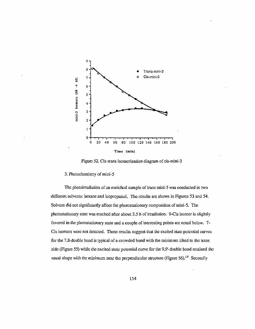
9
8 • Trans-mini-S
..... 7 0 Cis·mini·3;e'r 6
;;..... 5<il..41
e 40~
":l 3COs 2
O-t-T"""T-r-y-,-t--r-r"-r-"""''''-'r-'I''-r''''-T""''1-r"-r-'I
o 20 40 60 80 100 120 140 160 180 200
Time (min)
Figure 52. Cis-trans isomerization diagram of cis-mini-3
3. Photochemistry of mini-5
The photoirradiation of an enriched sample of trans mini-5 was conducted in two
different solvents: hexane and isopropanol. The results are shown in Figures 53 and 54.
Solvent did not significantly affect the photostationary composition of mini-5. The
photostationary state was reached after about 3.5 h of irradiation. 9-Cis isomer is slightly
favored in the photostationary state and a couple of interesting points are noted below. 7
Cis isomers were not detected. These results suggest that the excited state potential curves
for the 7,8-double bond is typical of a crowded bond with the minimum tilted to the trans
side (Figure 55) while the excited state potential curve for the 9,9'-double bond retained the
usual shape with the minimum near the perpendicular structure (Fi~ure 56).137 Secondly
154

both irradiation curves of mini-5 arc distinctly different from those of the mini-3.
Photostationary state was clearly reached after about 200 min of lrradiation. The
sigmatropic hydrogen shift and electro cyclization reactions arc not prominent during the
irradiation, This parallels the observation in thc vitamin A series that the irreversible
cyclization and degradation processes became less efficient as the polyene chain extends
longcr.!"
100
80
~.. •..S 600~
~.: 40 0e
20
400300200100o+--~--r-""""---r----"--""---"
o
Time (min) o all-trans mini-5
• 9-cis mini-S
Figure 53. Cis-trans isomerization of mini-5 in hexane
155

100
3002001000-1.---...,.....--.,..----..---,-----,----,
o
time (min)
Figure 54. Cis-trans isomerization of mini-S in isopropanol
_ c.fl---- I
.-:....:..-_-__ S1cis
p'" -Sltrans
Energy
0° 90° 180°
Figure 55. Potential curves of the C7-C8-double bond
156

Energy
ISItrans
ISIcis
9-trans
00 90°
9-cis
1800
Figure 56. Potential curves of the C9-C9'-double bond
4. Photochemistry of mini-7 and mini-9
Photoirradiation of all-trans mini-7 in DCCl3 did not result in significant
isomerization. The 1HNMR spectrum of the irradiated sample showed very little change
[rom that of the starting material. It has been known that the J3-carotene is relatively
inactive toward isomerization upon direct irradiation. The quantum yield of trans-cis
isomerization was reported to be in the order of 1O-6.1JJ The result of mini-7 indicates that
in this polyene series, the potential curves of the double bonds in the excited states
probably have a minimum distorted toward the side of trans isomer when' the number of
double bonds approaches 7 or more. Another contributing factor could be the greatly
reduced excited state life time which makes isomerization not competitively favorable.
More detailed work of the photochemistry of mini-7 and mini-9 still needs to be done to
evaluate the very small extent of isomerization of all-trans isomers.
157

References
1. Wald, G., Nature, 139, 1017 (1937).
2. Morton, R A, Salah, M. K., and Stubbs, A. L., Nature, 159, 744 (1947).
3. Betty, D. D., Vision Res., 24, 1563 (1984).
4. Provencio, I., Loew, E. R and Foster, R. G., Vision Res., 32, 2201 (1992).
5. Ito, M., Matsuoka, N., Tsukida, K. and Seki, T., Chern. Phann. Bull.. 36, 78 (1988).
6. Matsui, S., Seidou, M., Uchiyama, I., Sekiya, N., Hiraki, K., Yoshihara, K., Kito, Y.,
Biochim. Biophys. Acta, 966, 370 (1988).
7. Whitmore, A. V., and Bowmaker, J. K., J. Comp. Phys. A, 166, 103 (1989).
8. Lythgoe, J. N., and Partridge, J. C., Vision Res., 31, 361 (1991).
9. Rando, R. R, Angew. Chern. Int. Ed. Engl., 29, 461 (1990).
10. Becker R S., Photochem. Photobiol., 48, 369 (1988).
11. Schichida, Y., Matuoka, Y. S. and Yoshizawa, T., Photochem. Photobilol., 7, 221
(1984).
12. Schichida, Y., Photochem. Photobiol., 13,287 (1986).
13. Schoenlein, R. W., Peteanu, L. A, Mathies, R A and Shank, C. V., Science, 254,
412 (1991).
14. Randall, C. E, Lewis, J. W., Hug, S. J., Bjorling, S. C., Eisner-Shanas, I., Friedman,
N., Ottolenghi, M., Sheves, M. and Kliger, D. S., J. Am. Chern. Soc.. 113, 3473
(1991).
15. Liebman, P A, Parker, K. Rand Dratz, E. A, Ann. Rev. Physiol., 49,965 (1987).
16. Hurley, J. B., Ann. Rev. Physiol., 49, 793 (1987).
158

17. Hargrave, P.A., McDowell, 1. H., Curtis, D.R., Wang, J. K., Juszczak, E., Fong, S.
L., Rao, J. K. M. and Argos, P., Biophys. Struc. Mech., 9, 235 (1983).
18. Ovchinnikov, Y. A., Abdulaev, N. G., Feigina, M. Y., Artramonov, 1. D., Zolotarev,
A. S., Kostina, M. B., Bogachuk, A. K., Mirochnikov, A. 1.,Martinov, V. I. and
Kudelin, A. B., Bioorg. Khim., 8, 1011 (1982).
19. Henderson, R. and Unwin, P. N. T., Nature, 257, 28 (1975).
20. Chabre, M., Proc. Nati. Acad. Sci. USA, 75, 5471 (1978).
21. Michel-Villaz, M., Saibil, H. and Chabre, M., Proc. Nati. Acad. Sci. USA. 76, 4405
(1979).
22. Farrens, D. L. and Khorana, H. G., J. BioI. Chern., 270, 5073 (1995).
23. Hargrave P. A., McDowell, 1. H., Curtis, D. R., Wang, J. K., Juszczak E., Fong, S.
L., Rao, J. K. M. and Argos, P., Vision Res., 24, 1487 (1984).
24. Saibil, H., Chabre, M. and Worcester, D., Nature, 262,266 (1976).
25. Pistorius, A. M. A. and de Grip, W. J., Biochem. Biophys. Res. Comm., 198, 1040,
(1994).
26. Unger, V. M. and Schertler, G. F. X. Biophys. J., 68, 1776 (1995).
27. Lamba, O. P., Borchman, D. and O'brien, P. J., Biochemistry, 33, 1704 (1994)
28. Ridge, K. D., Liu, X., Lu, Z. and Khorana, H. G., Biochemistry, 34,3261 (1995).
29. Han, M. and Smith, S. 0., Biochemistry, 34, 1425 (1995).
30. Zhang, H, Lerro, K. A., Yamamoto, T., Lien, T. H., Sastry, L., Gawinowicz, M. A.
and Nakanishi, K., J. Am. Chern. Soc., 116, 10165 (1994).
31. Baldwin, J. M., The EMBO 1.,12, 1693 (1993).
32. Honig, B., Ann. Rev. Phys. Chern. 29, 31 (1978).
159

33. (a) Nakanishi, K., Pure AppI. Chern., 63, 161 (1991).
(b) Ottolenghi, M. and Sheves, M., J. Membr. BioI., 112, 193 (1989).
(c) Randall, L. E., Lewis, J. W., Hug, S. J., Bjorling, S. C, Eisner-Shanas, I.,
Friedman, N., Ottolenghi, M., Sheves, M., Kliger, D. S., J. Am. Chern. Soc., 113,
3473 (1991).
34. Blatz, P. E., Mohler, 1.H. and Navangul, H. V., Biochemistry, 11,848 (1972).
35. Honig, B., Dinur, U., Nakanishi, K., Balogh-Nair, V., Garvinowicz, M. A.,
Amaboldi, M. and Motto, M. G., 1. Am. Chern. Soc., 101,7084 (1979).
36. Koutalos, Y., Ebrey, T. G., Tsuda, M., Odashima, K., Lien, T., Park, M. H., Shimizu,
N., Derguini, F., Nakanishi, K., Gilson, H. R and Honig, B., Biochemistry, 28, 2732
(1989).
37. Wada, M., Sakurai, M., Inoue, Y., Tamura, Y. and Watanabe, Y., J. Am. Chern. Soc.,
116, 1737 (1994).
38. Frank, R R, Sakmar, T. P., Oprian, D. D. and Khorana, H. G., J. BioI. Chern., 263,
2119 (1988).
39. Jager, F., Fahmy, K., Sakmar, T. P. and Siebert, F., Biochemistry, 33, 10878 (1994).
40. Oprian, D. D., 1. Bioenerg. Biomembr., 24, 9746 (1992).
41. Hirayama, J., Imamoto, Y., Shichida, Y., Yoshizawa, T., Asato, A. E., Liu, R. S. H.
and Kamo, N., Photochem. PhotobioI., 60, 388, (1994).
42. (a) Hubbard, R and Wald, G., J. Gen. PhysioI., 36, 269, (1952).
(b) Wald, G., Brown, P. K., Hubbard, Rand Oroshnik, W., Proc. NatI. Acad. Sci. U.
S. A., 41,438 (1955); ibid, 42, 578 (1956).
43. Crouch, R., Purvin, V., Nakanishi, K. and Ebrey, T., Proc. NatI. Acad. Sci. U. S. A.,
72, 1538 (1975).
160

44. Trehan, A., Liu, R. S. H., Shichida, Y., Imamoto, Y., Nakamura, K. and Yoshizawa,
T., Bioorg. Chern., 18,30 (1990).
45. Liu, R. S. H., Matsumoto, H., Kini, A., Asato, A. E., Denny, M., Kropf, A. and
DeGrip, W. J., Tetrahedron, 40, 473 (1984).
46. Liu, R. S. H. in Carotenoid Chemistry and Biochemistry, G. Britton and T. W.
Goodwin, Eds., Pergamon Press, p 253 (1982).
47. Eyring, G., Bostick, C., Mathies, R, Fransen, R., Palings, I. and Lutgenberg, 1.,
Biochemistry, 19,2410 (1980).
48. Liu, R. S. H. and Matsumoto, H., Methods Enzymol., 81, 694 (1982).
49. Asato, A. E., Mead, D., Denny, M., Bopp, T. T. and Liu, R. S. H., J. Am. Chern.
Soc., 104,4979 (1982).
50. Asato, A. E., Denny, M., Matsumoto, H., Mirzadegan, T., Ripka, W. C., Crescitelli,
F. and Liu, R. S. H., Biochemistry, 25, 7021 (1986).
51. Asato, A. E. and Liu, R S. H., unpublished results.
52. Liu, R. S. H., Asato, A. E., Denny, M. and Mead, D., J. Am. Chern. Soc., 106, 8298
(1984).
53. Trehan, A., Mirzadegan T. and Liu, R S. H., Tetrahedron, 46,3769 (1990).
54. Knudson, C. G., Carey, S. C. and Okamura, W. H., J. Am. Chern. Soc., 102,6355
(1980).
55. Mayer, H. and Isler, 0., in Carotenoids, O. Isler, ed., Birkhauser, Basel, p 325,
(1971).
56. Pommer, H., Angew. Chern., 72, 811 (1960).
57. Isler, 0., Ronco, A., Guex, W., Hindley, N. c., Huber, W., Dialer, K. and Kofler,
M., Helv. Chim. Acta, 32, 489 (1949).
58. Oroshnik, W" 1. Am. Chern. Soc" 78, 2651 (1956).
161

59. Schwieter, U., von Planta, C., Ruegg, R and Isler, 0., Helv. Chim Acta, 45, 528
(1962).
60. Schwieter, U., Saucy, G., Montavon, M., von Planta, C., Ruegg, R. and Isler, 0.,
Helv. Chim. Acta, 45,517 (1962).
61. Oediger, H., Kabbe, H. J., Moller, F. and Eiter, K., Chern. Ber., 99, 2012 (1966).
62. Derguini, F., Balogh-Nair, V. and Nakanishi, K., Tetrahedron Lett., 51, 4899 (1979).
63. Attenburrow, J., Cameron, A. F. B., Chapman, J. H., Evans, R. M., Hems, B. A.,
Jansen, A. B. A. and Walker, T., J. Chern. Soc., 1094 (1952).
64. H. Fagle and P. Karrer, Helv. Chim. Acta, 44, 1261 (1961).
65. Kini, A. M., Ph. D. Dissertation, University of Hawaii, 1979.
66. Redel, J., Boch, J. and Chen, T., C. R. Acad. Sci., 259, 2466 (1964).
67. Jacobs, H. A. M., Berg, M. H., Brandsrna, L. and Arens, J. E, Recl. Trav. Chirn.
Pays-Bas, 84, 1113 (1965).
68. Garbers, C. F., J. Chern. Soc., 3234 (1956).
69. Karrer, P., Jucher, E. and Schick, E., Helv. Chim. Acta, 29, 704 (1946).
70. Matsui, M., Okano, S., Yamashita, K., Miyano, M., Kitamura, S., Kobayashi, A.,
Sato, T. and Mikami, R, J. Vitaminol. (Kyoto), 4, 178 (1958).
71. Pommer, H. and Stilz, W., Ger. Pat. 1,116,652 (1961); Chern. Abstr. 57, 2267
(1962).
72. Mead, D., Asato, A. E., Denny, M., Liu, R S. H., Hanzawa, T., Kobayashi, N.
Hasoda, A. and Kobayashi, Y., Tetrahedron Lett., 45, 259 (1987).
73. Mead, D., Ph. D. Dissertation, University of Hawaii, 1986.
74. Still, W. C. and Gennari, c., Tetrahedron Lett., 24, 4405 (1983).
162

75. Ramamurthy, V., Butt, Y., Yang, c., Yang, P. and Liu, R. S. H., J. arg. Chern., 38,
1247 (1973).
76. Ramamurthy, V. and Liu, R. S. H., Tetrahedron, 31, 201 (1975).
77. Matsumoto, H., Asato, A. E. and Liu, R. S. H., Photochem. Photobiol., 29, 695,
(1979).
78. Zeiger, J. and Goldsmith, T. H., Vision Res., 29, 519 (1989).
79. Azuma, M., Azuma, K. and Kito, Y., Biochim. Biophys. Acta, 295, 520 (1973).
80. Papermaster, D. and Dryer, W., Biochemistry, 13, 2438 (1974).
81. Denny, M., Colmenares, L., Unpublished material
82. Matsumoto, H., Horiuchi, K. and Yoshizawa, T., Biochirn. Biophys. Acta, 501, 257
(1978).
83. Surmatis, I. D. and Thommen, R., J. Org. Chern., 32, 180 (1967).
84. Broek, A. D., Muradin-Szweykowska, M., Courtin, J. M. L. and Lugtenburg, J.,
Reel. Trav. Chim. Pays-Bas, 102,46 (1983).
85. Asato, A. E., Unpublished results.
86. Schlatmann, D., Pot, W., and Havinga, G., Recl. Trav. Chirn. Pays-Bas, 83, 1173
(1964).
87. Frater, von G., Helv. Chirn. Acta .. 57, 2447 (1974).
88. Mead, D., Asato, A. E., Denny, M., Liu, R. S. H., Hanzawa, Y., Taguchi, T.,
Yamada, A., Kobayashi, N., Hosoda, A. and Kobayashi, Y., Tetrahedron Lett., 28,
259 (1987).
89. Zhu, Y. and Trehan, A., unpublished material.
163

90. Schwieter, U. and Isler, 0., "The Vitamins, Chemistry, Physiology, Pathology,
Methods", 2nd edition, Sebrell, Jr. W. H. and Harris, R. S., (ed.), Academic Press,
New York, p 56, 1967.
91. Li, X., Ph. D. Dissertation, University of Hawaii, 1993.
92. Silverstein, R. M., Bassler, G. C. and Morrill T. c., "Spetrometric Identification of
Organic Compounds", 4th Ed., John Wiley & Sons, Inc., p 313,1981.
93. Hamanaka, T., Mitsui, T., Ashida, T. and Kakudo, M., Acta CO'st., B28, 214 (1971).
94. Gilardi, R. D., Main, P. and Woolfson, M. M., Acta CO'st., A27, 368 (1971).
95. Simmons, C. J., Liu, R. S. H., Denny, M. and Seff, K., Acta CO'st., B37, 2197
(1981).
96. B. Honig, B. Hudson, B. D. Sykes and M. Karplus, Pmc. Nat. Acad. Sci. USA, 68,
1289 (1971).
97. Liu, R. S. H., Zingoni, J. P., Kini, P., Trammell M., Chu, D., Asato, A. E., and Bopp,
T. T., J. Org. Chern., 48, 4817 (1983).
98. Nakayama, A. T., Bopp, T. T. and Liu, R. S. H., J. Org. Chern., 49,3424 (1984).
99. H. Matsumoto, K. Horiuchi and T. Yoshizawa, Biochim. Biophys. Acta, 501, 257
(1978).
100. Liu, R. S. H. and Mirzadegan, T., J. Am. Chern. Soc., 110,8617 (1988).
101. (a) Groenendijk, C. W. T., de Grip, W. J. and Daemen, F. J. M., Anal. Biochem.,
99,304 (1979); (b) Groenendijk, C. W. T., de Grip, W. J. and Daemen, F. J. M.,
Biochim. Biophys. Acta, 617,430 (1980).
102. Trehan, A., Liu, R. S. H., Shichida, Y., Imamoto, Y., Nakamura, K. and Yoshizawa,
T., Bioorganic ChemistI:y, 18,30 (1990).
103. Trehan, A., Ph. D. Dissertation, University of Hawaii, 1989
104. Kini, A., Matsumoto, H. and Liu, R. S. H., J. Am. Chern. Soc., 101,5078 (1979).
164

105. Y. Koyama and Y. Mukai, Biornolecular Spectroscopy. Part B in Advances in
Spectroscopy, Ed, R. 1.H. Clark and R. E. Hester, John Wiley & Sons, Ltd, 21,49
(1993).
106. Ganapathy, S. and Liu, R. S. H., J. Am. Chern. Soc., 114,490 (1992).
107. Pfander, H. in Methods in Enzymology. 213, 3 (1992).
108. Nagao, A. and Olson,1. A., FASEB J, 8, 968 (1994).
109. Cogdell, R. J. and Frank, H. A., Biochirn. Biophys. Acta, 895,63 (1987).
110. Oliveros, E., Braun, A. M. and Aminian-Saghafi, T., New 1. Chern., 18,535
(1994).
Ill. McClinton-Adams, 1. L., Hart, L. L., Ann. Phannacotherapy, 28,470 (1994).
112. Knox, R. S., (ed.) Luminescence Studies of Photosynthesis. Special issue of 1.
Luminescence, 51 (1992).
113. Scita, G. in Methods in Enzymology, 213, 175 (1992).
114. Kohler, B. E., Spangler C. and Westerfield, c, J. Chern. Phys., 89, 5422 (1988).
115. Allen, M. T. and Whitten, D. G., Chern. Rev., 89, 1691 (1989).
116. Hudson, B. S. and Kohler, B. E., Chern. Phys. Lett., 14,299 (1972).
117. Gillbro, T., Andersson, P. O. and Liu, R. S. H., Photochem. Photobiol. 57,44
(1993).
118. Andersson, P.O., Gillbro, T., Asato, A. E., and Liu, R. S. H., J. Luminescence, 51,
11 (1992).
119. Andersson, P.O., Gillbro, T.; Chen, R-L, J. Chern. Phys., 00, 0000 (1995).
120. Gedye, R. N., Arora, P. C. and Deck K., Can. J. Chern., 49,1764 (1971).
121. Cainelli, G., Cardillo, G. and Orena M., J. Chern. Soc. Perkin I, 1597 (1979).
165

122. Curley, Jr., R. W., and Deluca, H. F., J. Org. Chern., 49, 1941 (1984).
123. Broek, A. D., Muradin-Szweykowska, Courtin, J. M. L. and Lugtenburg, Recl.
Trav. Chim. Pays-Bas, 102,46 (1983).
124. Sandstrom, J., "Dynamic NMR Spectroscopy", New York, Academic Press, 1982.
125. Jackman, L. M. and Cotton, F. A., "Dynamic Nuclear Magnetic Resonance
Spectroscopy", New York, Academic Press, 1975.
126. Stephenson, D. S. and Binsch, G., J. Magn. Reson., 145, 30 (1978) and 145, 32
(1978).
127. Jackman, L. M., Cotton, F. A. "Dynamic Nuclear Magnetic Resonance", Academic
Press, New York, 1975.
128. Saltie1, J., private communication.
129. Bonneau, R., Joussot-Dubien, J., Salem, L. and Yarwood, A. 1., J. Am. Chern. Soc.,
98,4329 (1976).
130. Saltie1, J., D'Agostino, J., Megarity, E. D., Metts, L., Neuberger, K. R., Wrighton,
M., Zafiriou, O. C. in "Organic Photochemistry", Chapman, O. L., Ed., Marcel
Dekker, New York, 3, 1 (1973).
131. Ramarnurthy, V., Ph. D. Dissertation, University of Hawaii, 1974.
132. (a) Craig, D., Shipman, J. J. and Fowler, R. B., J. Am. Chern. Soc., 83, 2885 (1961);
(b) Briuwer, A. M., Cornelisse, J. and Jacobs, H. J. c., 1.Photochem. Photobiol.,
42, 117 (1988).
133. Chen, R-L., Colmenares, L. U., Thiel, J. R. and Liu, R. S. H., Tetrahedron lett., 35,
7177 (1994).
134. Molnar, P., Szabolcs, J. and Radics, L., Phytochem.. 25, 195 (1986).
135. Chandler, L. A. and Schwartz, S. J., J. Food. Sci., 52, 669 (1987).
136. Li, X-Y., Chen, R-L., unpublished results.
166

137. Arai, T. and Tokumaru, K., Chern. Rev., 93,23 (1993).
138. liu and Asatophotochemistry
139. Kuki, M., Koyama, Y. and Nagae, R., J. Phys. Chern., 95, 7171 (1991).
167