
microRNA target identification by RNA pull down with biotinylated ...

Virginia Commonwealth UniversityVCU Scholars Compass
Study of Biological Complexity Publications Center for the Study of Biological Complexity
2012
A comparison of two distinct murine macrophagegene expression profiles in response to Leishmaniaamazonensis infectionChristian M. ProbstInstituto Carlos Chagas
Rodrigo A. SilvaLaboratório de Patologia e Biointervenção
Juliana P.B. MenezesLaboratório de Patologia e Biointervenção
See next page for additional authors
Follow this and additional works at: http://scholarscompass.vcu.edu/csbc_pubs
© 2012 Probst et al; licensee BioMed Central Ltd. This is an Open Access article distributed under the terms of theCreative Commons Attribution License (http://creativecommons.org/licenses/by/2.0), which permits unrestricted use,distribution, and reproduction in any medium, provided the original work is properly cited.
This Article is brought to you for free and open access by the Center for the Study of Biological Complexity at VCU Scholars Compass. It has beenaccepted for inclusion in Study of Biological Complexity Publications by an authorized administrator of VCU Scholars Compass. For moreinformation, please contact [email protected].
Downloaded fromhttp://scholarscompass.vcu.edu/csbc_pubs/11

AuthorsChristian M. Probst, Rodrigo A. Silva, Juliana P.B. Menezes, Tais F. Almeida, Ivana N. Gomes, Andréia C.Dallabona, Luiz S. Ozaki, Gregory A. Buck, Daniel P. Pavoni, Marco A. Krieger, and Patrícia S.T. Veras
This article is available at VCU Scholars Compass: http://scholarscompass.vcu.edu/csbc_pubs/11
RESEARCH ARTICLE Open Access
A comparison of two distinct murine macrophagegene expression profiles in response toLeishmania amazonensis infectionChristian M Probst1, Rodrigo A Silva2, Juliana P B Menezes2, Tais F Almeida2, Ivana N Gomes2,Andréia C Dallabona1, Luiz S Ozaki3, Gregory A Buck3, Daniela P Pavoni1, Marco A Krieger1 and Patrícia S T Veras2*
Abstract
Background: The experimental murine model of leishmaniasis has been widely used to characterize the immuneresponse against Leishmania. CBA mice develop severe lesions, while C57BL/6 present small chronic lesions underL. amazonensis infection. Employing a transcriptomic approach combined with biological network analysis, thegene expression profiles of C57BL/6 and CBA macrophages, before and after L. amazonensis infection in vitro, werecompared. These strains were selected due to their different degrees of susceptibility to this parasite.
Results: The genes expressed by C57BL/6 and CBA macrophages, before and after infection, differ greatly, bothwith respect to absolute number as well as cell function. Uninfected C57BL/6 macrophages express genes involvedin the deactivation pathway of macrophages at lower levels, while genes related to the activation of the hostimmune inflammatory response, including apoptosis and phagocytosis, have elevated expression levels. Severalgenes that participate in the apoptosis process were also observed to be up-regulated in C57BL/6 macrophagesinfected with L. amazonensis, which is very likely related to the capacity of these cells to control parasite infection.By contrast, genes involved in lipid metabolism were found to be up-regulated in CBA macrophages in responseto infection, which supports the notion that L. amazonensis probably modulates parasitophorous vacuoles in orderto survive and multiply in host cells.
Conclusion: The transcriptomic profiles of C57BL/6 macrophages, before and after infection, were shown to beinvolved in the macrophage pathway of activation, which may aid in the control of L. amazonensis infection, incontrast to the profiles of CBA cells.
BackgroundSeveral factors related to the pathogen itself greatlyinfluence the severity and clinical manifestation ofinfectious diseases, including parasite pathogenicityand virulence, as well as a variety of other factorsrelated to the host’s state of general health and geneticbackground [1-4]. Functional genomics is an importanttool to study host-pathogen interactions, since it givesinsight into the molecular mechanisms that control theonset of disease [5-7].The cutaneous leishmaniasis murine model has been
widely used to characterize the immune response
against Leishmania. The association between resistanceto Leishmania major and cell differentiation in CD4+
Th1 lymphocytes has been well documented [8,9]. Theimmune response to L. amazonensis varies in accor-dance with the genetic background of the host.L. amazonensis causes severe lesions at cutaneousinoculation sites in the highly susceptible CBA andBALB/c mouse strains [4,10,11], while this same para-site causes chronic non-healing lesions in L. major-resistant strains, such as C57BL/6, C3H and C57BL/10[10,12-14]. In response to infection by L. amazonensis,highly susceptible BALB/c mice mount a Th2-type ofimmune response, while C57BL/6 mice develop a non-Th1-type of immune response [15].Macrophages are immune cells involved in the early
events of pathogen infection [3,16]. Leishmania spp.
* Correspondence: [email protected]ório de Patologia e Biointervenção, CPqGM-FIOCRUZ, Bahia, BrazilFull list of author information is available at the end of the article
Probst et al. BMC Microbiology 2012, 12:22http://www.biomedcentral.com/1471-2180/12/22
© 2012 Probst et al; licensee BioMed Central Ltd. This is an Open Access article distributed under the terms of the Creative CommonsAttribution License (http://creativecommons.org/licenses/by/2.0), which permits unrestricted use, distribution, and reproduction inany medium, provided the original work is properly cited.

parasites are delivered to the mammal dermis in theform of metacyclic promastigotes where they are phago-cytosed [17]. Some Leishmania species, such as L. ama-zonensis, can survive and proliferate inside macrophagesby modulating host cell killing mechanisms, regardlessof microbicidal molecule production [3]. Followinguptake, the surviving promastigotes differentiate intoamastigotes and multiply within parasitophorousvacuoles [18].Several studies have demonstrated that the survival of
Leishmania spp. is associated with slight modificationsin macrophage gene expression [6,19-21]. Over the last10 years, several studies have presented evidence thatLeishmania species do not adequately induce classicalmacrophage activation [19,20]. Moreover, a recent studyfound that these parasites down- and up-regulate similarnumbers of proinflammatory response genes in humanmacrophages, as well as activate a gene that is compati-ble with an alternative phenotype [21]. Other authorshave recently demonstrated that L. amazonensis is ableto induce a transcriptional signature that resemblesdeactivation yet also appears similar to an alternativemacrophage activation signature [22]. Interestingly,these authors showed that L. amazonensis directsmacrophage response towards lipid and polyamine path-ways by activating parasite- and host tissue-protectiveprocesses [22].The role that host genetic factors play in the outcome
of pathogen infection has also been studied using micro-array analysis [23,24]. In addition, several studies havecompared the gene expression profiles of cells [23,24]and tissues [25] from a variety of mouse strains inresponse to several pathogens. However, no studies haveyet attempted to compare the transcriptional signaturesof uninfected macrophages from two distinct murinegenetic backgrounds, nor the transcriptional programsof a distinct macrophage lineage in response to a singleLeishmania species.The present study employed a transcriptomic
approach combined with biological network analysis tohighlight the differences between the responses ofmurine macrophages from two inbred mouse strains toL. amazonensis infection. C57BL/6 and CBA strainswere selected due to their divergent degrees of sus-ceptibility to this parasite [4,12]. The expression pro-files of more than 12,000 murine genes were evaluatedin each mouse strain before and after infection invitro. The authors identified the genes that were differ-entially expressed between uninfected C57BL/6 andCBA macrophages, thereby establishing baseline levelsof differential expression. We then attempted to inves-tigate modulations in macrophage gene expression,before and after infection, within a given mouse strain.We showed that the transcriptional profile of
uninfected C57BL/6 macrophages differed from that ofCBA macrophages with respect to the modulation ofgenes involved in the macrophage pathway of activa-tion. In response to infection, C57BL/6 macrophagesup-regulate genes related to controlling infection,while CBA cells up-regulate genes involved in lipidmetabolism. These findings provide evidence thatC57BL/6 macrophages’ transcriptional profiles mayhelp in the control of L. amazonensis infection, in con-trast to the profiles of CBA cells.
MethodsMiceAll experiments were performed according to the guide-lines of the Institutional Review Board on AnimalExperimentation at the Oswaldo Cruz Foundation -CPqGM/FIOCRUZ. Male and female CBA mice, 6-12weeks old, were provided by the Animal Care Facility atCPqGM/FIOCRUZ. The animals were housed underspecific pathogen-free conditions, fed commercialrations and given water ad libitum.
ParasitesThe L. amazonensis (strain MHOM/Br88/Ba-125) pro-mastigotes used in this study were grown in axenic cul-ture for up to seven passages, suspended in Schneider’scomplete medium (Gibco, Grand Island, NY, USA) sup-plemented with 10% inactivated fetal calf serum and 50μg/mL of gentamicin (Sigma, St. Louis, MO, USA). Allparasite cultures were washed three times in a salinesolution, counted, adjusted and added to macrophagecultures at a ratio of 10:1.
Macrophage culturesInflammatory peritoneal macrophages were elicitedusing a 3 mL intraperitoneal injection of 3% thioglyco-late solution (Sigma) in C57BL/6 or CBA mice. After 96h, all animals were euthanized and the elicited perito-neal macrophages were obtained as previously described[3]. The cells were suspended in complete Dulbecco’sModified Eagle’s Medium (DMEM) (Gibco) [DMEMsupplemented with 10% fetal bovine serum (Gibco), 2 g/L sodium bicarbonate (Sigma), 25 mM HEPES (Sigma),1 mM glutamine (Sigma) and 0.2% ciprofloxacin (Halex-istar, Goiania, GO, BR)] and distributed in 6-well platesat a concentration of 1 × 10⁷ macrophages per well.Cultures were subsequently incubated overnight at 37°Cin 5% CO2.
Macrophage infectionThe inflammatory peritoneal macrophage cultures wereinfected for 12 h with L. amazonensis stationary phasepromastigotes. Cell cultures were then washed twicewith saline to remove non-internalized parasites and
Probst et al. BMC Microbiology 2012, 12:22http://www.biomedcentral.com/1471-2180/12/22
Page 2 of 12
reincubated for an additional six or 24 h before eitherRNA extraction or fixation with ethanol for 20 min fol-lowed by staining with hematoxylin and eosin (H&E).Each independent experiment was repeated three timesfor microarray analysis, and each experiment was per-formed at least three times in triplicate for microscopicanalysis.
Microarray analysisTotal RNA from uninfected or L. amazonensis-infectedmacrophages was prepared using Qiagen RNeasy mini-prep columns (Qiagen, Valencia, CA, USA) in accor-dance with manufacture protocols. The integrity of eachRNA preparation was assessed using agarose gel electro-phoresis. The RNA was reverse transcribed using Super-script II (Invitrogen, Carlsbad, CA, USA) in thepresence of oligo(dT) primers linked to a T7 RNA poly-merase promoter sequence (Proligo, La Jolla, CA, USA)to prime cDNA synthesis. After second-strand synthesis,biotinylated cRNA was produced by in vitro transcrip-tion using biotinylated UTP and CTP (Bioarray high-yield RNA transcript labeling kit, Enzo Diagnostics,Farmingdale, NY, USA) and purified with RNAeasy minicolumns (Qiagen). The biotinylated cRNA was fragmen-ted at 94°C for 30 min. For probe array hybridizationand scanning, 16 μg of fragmented labeled cRNA washybridized to the Murine Genome U74v2 GeneChip®
array (Affymetrix, Santa Clara, CA, USA), which con-tains nearly 400,000 probe sets covering approximately12,000 different murine genes. Array scanning was per-formed using the Affymetrix® GeneChip Scanner 3000 7G and all images were analyzed using Microarray Analy-sis Software (Affymetrix v5.0). Experimental data areavailable online at ArrayExpress (E-MEXP-3448).
Statistical analysis of differentially expressed genesamong C57BL/6 and CBA macrophagesAll microarray data were analyzed using the gcRMAlibrary [26] from the Bioconductor project, using the Rstatistical software suite. Next, in order to identify differ-entially expressed genes, the SAM (Significance Analysesof Microarray) statistical package was used to comparethe levels of gene expression among the followinggroups: (1) uninfected C57BL/6 and CBA macrophages;(2) L. amazonensis-infected C57BL/6 macrophages anduninfected cells; (3) L. amazonensis-infected CBAmacrophages and uninfected cells; (4) L. amazonensis-infectedC57BL/6 and CBA macrophages. In order toenhance confidence in the statistical analysis of microar-ray data, experiment variables of incubation and infec-tion time were not considered when comparing geneexpression among groups (1) to (4). SAM software usesa modified t-test measurement which corrects for
multiple comparisons by means of a False DiscoveryRate (FDR) approach [27]. The q-values, or the mini-mum FDRs at which a statistical test may be called sig-nificant [28], have been provided for each differentiallyexpressed gene in Tables S1, S2 and S3 (See Additionalfile 1: Table S1; Additional file 2: Table S2 and Addi-tional file 3: Table S3, respectively). Finally, differentiallyexpressed genes were analyzed and grouped in func-tional networks using the Ingenuity Pathway Analysisprogram v8.8 (IPA-Ingenuity Systems®, http://www.ingenuity.com). Possible networks and pathways werescored and modeled considering the sets of differentiallyexpressed genes derived from the four comparisonsdescribed above. To calculate the probability of associa-tions between genes from the functional networks andpathways generated by IPA®, Fisher’s exact test wasused with a 0.05 threshold value.
Total macrophage mRNA extraction and mRNAquantification by RT-qPCRIn order to perform reverse transcriptase-quantitativepolymerase chain reactions (RT-qPCR), RNA was initi-ally extracted from uninfected or infected macrophagesusing a QIAGEN Mini Kit (RNAeasy) in accordancewith manufacturer directions. An optical density readingwas taken following extraction procedures and RNAintegrity was verified using an agarose gel. Complemen-tary DNA (cDNA) was synthesized by reverse transcrip-tion in a final volume of 20 μL containing 5 mM MgCl2(Invitrogen), PCR buffer 1× (Invitrogen), deoxyribonu-cleotide triphosphates each at 1 mM (dNTPs - Invitro-gen), 0.5 mM oligonucleotide (oligo d(T) - Invitrogen),1 UI RNase inhibitor (RNase Out - Invitrogen), 2.5 UIreverse transcriptase (MuLVRT- Invitrogen) and 1 μg ofsample RNA in RNAse-Free Distilled Water. All reac-tion conditions consisted of a single cycle at 42°C for 50min, followed by 70°C for 15 min and, finally, 4°C for atleast 5 min. Following reverse transcription, the synthe-sized cDNA was aliquoted and frozen at -20°C. ThecDNA aliquots were later thawed and amplified byqPCR in order to perform gene quantification. All reac-tions were performed in a final volume of 20 μL con-taining SYBR green® (Applied Biosystems, Foster City,CA, USA) commercial mix solution, composed of SYBRGreen I Dye, AmpliTaq Gold® DNA polymerase, dNTPswith dUTP, 10 ng cDNA, and 50 pmoles of reverse andforward primers for each evaluated gene (Invitrogen).qPCRs were run in a 7500 Real-Time PCR thermalcycler system (Applied Biosystems) and performedaccording to manufacturer’s instructions, with variationsoccurring only with respect to melting temperature(Tm) for each pair of primers. Each sample was testedtwo or three times in duplicate. Table S4 (See
Probst et al. BMC Microbiology 2012, 12:22http://www.biomedcentral.com/1471-2180/12/22
Page 3 of 12

Additional file 4: Table S4) lists the primer sequencesused for each macrophage gene amplified by RT-qPCR,as well as Tm for each pair of primers.
Analysis of mRNA quantificationGene amplification results were obtained using SequenceDetection Software v1.3 (Applied Biosystems) with dataexpressed as mean values from experiments performed induplicate. For each reaction, a serial dilution containing amixture of cDNA from both uninfected and infectedmacrophages was used to generate a standard curve forgene expression quantification. Each gene’s expressionvalues were normalized against the respective value of theconstitutive gapdh1 (glyceraldehyde 3-phosphate dehydro-genase) gene. The following comparisons of normalizedgene expression were made: (1) C57BL/6 macrophages inrelation to CBA macrophages; (2) L. amazonensis-infectedC57BL/6 macrophages in relation to uninfected cells; (3)L. amazonensis-infected CBA macrophages in relation touninfected cells. Resulting comparison values wereexpressed as mean values of log2 ± SE from the two inde-pendent experiments in comparison (1), and three inde-pendent experiments in comparisons (2) and (3), allperformed in duplicate. To determine the statistically sig-nificant differences in gene expression between all groupsusing RT-qPCR, the nonparametric Mann-Whitney testwas used with a significance level of p ≤ 0.05.
Results and discussionDifferences in transcription between uninfected C57BL/6and CBA macrophagesIn order to evaluate the influence of genetic factors on theoutcome of Leishmania infection, the gene expressionprofiles from uninfected C57BL/6 and CBA macrophageswere identified using an Affymetrix® DNAmicroarray.Firstly, among the 12,000 genes analyzed using the MurineGenome U74v2 Genechip®, a total of 208 probe sets (SeeAdditional file 1: Table S1) were found to be differentiallyexpressed between the uninfected C57BL/6 and CBAmacrophages with a 1.5 fold-change threshold and an esti-mated 5% FDR. All differential expression values are com-paratively expressed as follows: a positive/negative valueindicates that a given C57BL/6 macrophage exhibited ahigher/lower level of expression than its CBA counterpart.Of these probe sets, 148 had higher expression levels inC57BL/6 macrophages (expressed as positive values) and60 were found to be more highly expressed in CBA unin-fected cells (expressed as negative values). In order to con-firm these findings, a total of 27 genes were randomlyselected and RT-qPCR was used to verify the differencesin expression observed in the microarray analysis. Differ-ential expression was confirmed in each of the 27 genesselected, and, among these, 13 genes showed statisticallysignificant differences (Figure 1A).
Increased levels of gene expression in uninfected C57BL/6 macrophages associated with cell death and lipidmetabolismUsing IPA-Ingenuity Systems® v8.8 biological data analy-sis software, several functional networks and metabolicpathways were modeled from the differentially expressedgenes by uninfected C57BL/6 and CBA macrophages.The cell death and lipid metabolism network had thehighest probability of interrelated genes being differen-tially expressed (score 51). In this network, 17 out of the22 genes identified by microarray analysis had higherlevels of expression in C57BL/6 macrophages in compari-son to CBA macrophages (Figure 2A). Among these,some encode proteins involved in lipid metabolism: apoe(+2.69) and apoc2 (+2.47). Both apolipoprotein E (Apoe)and apolipoprotein C (Apoc) are lipoproteins, mainlycomponents of lipoprotein complexes, which are asso-ciated with proteins in plasma and the central nervoussystem [30].Apoe regulates the metabolism of lipids by directing
their transport, delivery, and distribution from one typeof tissue or cell to another [30,31]. Alternatively, Apoe isalso known to participate in the immune inflammatoryresponse by scavenging reactive oxygen species (ROS).Accordingly, some genes that encode enzymes involvedin antioxidant activity, such as sod1 (+1.34) and prdx2(+2.05) were also expressed at higher levels in C57BL/6macrophages. A previous study showed that peroxiredox-ins (Prdxs) constitute a family of multifunctional antioxi-dant thiol-dependent peroxidases, which may modulatemacrophage defense mechanisms against oxidative stressduring inflammatory or infection events [32]. In thisstudy, Bast et al. (2010) found higher levels of expressionof peroxiredoxin mRNA and Prdx2 by C57BL/6 macro-phages in response to stimulation with lipopolysaccharide(LPS) and IFN-g, compared to BALB/c macrophages,which are known to be as susceptible as CBA macro-phages to L. amazonensis. The proteins encoded byprdx2 and apoe may alternately play a role in apoptosis[33], in addition to ifi204 (+1.38), also known as ifi16,which encodes a transcriptional regulator, and gdf15(+1.51), which encodes growth differentiation factor-15.It is possible that, with respect to uninfected CBA macro-phages, the lower baseline levels of differential expressionfound among genes involved in apoptosis may affect theability of these cells to control L. amazonensis infection[3].Besides being a component of both high and very
low-density lipoproteins, Apoc is known to readily accu-mulate in amyloid fibrils, inducing macrophage inflam-matory responses, such as ROS production and TNF-aexpression [34]. It is possible that the lower apoc2expression levels found in uninfected CBA macrophagesherein might be related to the low levels of TNF-a
Probst et al. BMC Microbiology 2012, 12:22http://www.biomedcentral.com/1471-2180/12/22
Page 4 of 12

expression in IFN-g-stimulated CBA macrophages inresponse to L. amazonensis infection demonstrated by aprevious study [3].Genes such as chi3l3/chi3l4, fizz1/relm-a and arg1 are
considered to be signature markers of alternative macro-phage activation in response to IL-4 stimulation [6].Among these types of genes, chi3l3/chi3l4 (+3.028) wasfound to have increased differential expression inC57BL/6 macrophages. In addition, il10ra (-1.39), whichencodes the ligand-binding subunit of the immunereceptor for the IL-10 cytokine, is known to be involvedin macrophage deactivation, and was found to have alower level of expression in C57BL/6 macrophages.Accordingly, fcgr1a (+1.27), which encodes the high-affi-nity Fc-gamma receptor, participates in the innateimmune response by promoting the clearance of patho-gens and necrotic cells, and also was found to be morehighly expressed in C57BL/6 macrophages.By contrast, very few genes were identified as highly
expressed in CBA macrophages compared to C57BL/6(represented by negative expression values) in the celldeath and lipid metabolism network (Figure 2A), such asmt1 (-0.99), which can have a protective effect on cellsagainst apoptosis and oxidative stress responses; hal (-5.65),which participates in histidine catabolism; and pltp (-1.19),which is involved in lipid transport and metabolism.
Increased levels of gene expression in uninfected C57BL/6 macrophages associated with the cell-cell signaling andinteraction networkIPA® identified several genes as part of the cell-cell sig-naling and interaction network (score 30) (Figure 2B):c1qa (+2.95), c1qb (+5.08) and c1qc (+5.04). These genesencode components of the complement cascade and allhad higher expression levels in C57BL/6 macrophages.The classical pathway activation of complement elementsconstitutes events that are initiated by the binding ofimmune complexes to the C1 subcomponent, followedby subsequent C1q activation by serine proteases [35].Constitutive synthesis of C1q in resident peritonealmacrophages suggests that C1q expression may be linkedto the differentiation process in which blood monocytesbecome tissue macrophages [36]. Additionally, microor-ganism opsonization by C1q facilitates the phagocytosisof foreign particles during the innate immune response[37]. The production of anti-inflammatory mediatorsduring proinflammatory responses is inhibited by C1qopsonization, which is followed by the phagocytosis ofapoptotic cells [38].In sum, the authors found significant differences in
the baseline gene expression profiles of C57BL/6 macro-phages compared to those of CBA cells, which suggeststhat the higher capacity of C57BL/6 macrophages to
Figure 1 Comparison of differentially expressed genes using microarray and RT-qPCR techniques. RT-qPCR was used to verify thedifferential expression of randomly selected genes (n = 27) by uninfected C57BL/6 and CBA macrophages (A), by L. amazonensis-infected C57BL/6 macrophages in comparison to uninfected cells (n = 7) (B), and by L. amazonensis-infected CBA macrophages in comparison to uninfectedcells (n = 2) (C). Figure 1 (A-C) depicts only genes that were successfully verified using RT-qPCR. Resulting comparison values are expressed asmean values of log2 ± SE from two independent experiments in comparison (A), and three independent experiments in comparisons (B) and (C),all performed in duplicate. The nonparametric Mann-Whitney test was used for comparison between uninfected cells, and Stouffer method [29]was used to integrate the results from independent microarray and RT-qPCR analyses to determine significant differences between infected anduninfected cells (level of significance, p ≤ 0.05)
Probst et al. BMC Microbiology 2012, 12:22http://www.biomedcentral.com/1471-2180/12/22
Page 5 of 12

control L. amazonensis infection is related to the base-line transcriptional signature of these cells. Thesemacrophages have genes involved in the deactivationpathway of macrophages which are expressed at lowerlevels, as well as higher expression levels of genes thatencode proteins that play a role in the host immuneinflammatory response, including several moleculesinvolved in apoptosis in addition to phagocytic receptorsthat recognize pathogens and apoptotic cells.Similarities between the expression profiles of genes relatedto apoptosis and stress responseDifferent genes with similar functions that are involvedin specific cellular processes, e.g. apoptosis, immune andstress responses, were described as modulated byC57BL/6 and CBA macrophages. For instance, IFN-a/b-induced ifi202 gene expression was described by otherauthors as being induced in macrophages from severalmouse strains, except C57BL/6 macrophages [39]. ifi202participates in the immune response and composes thecell death and lipid metabolism network in the presentstudy, this gene was shown to have a differential expres-sion of -1.31 to -3.69 in C57BL/6 compared to CBAmacrophages. This result was confirmed using RT-qPCR, which did not detect ifi202 expression in C57BL/6 macrophages. Additionally, other members of theifi200 family, ifi203 (+0.96) and ifi204 (+1.38) geneswere more highly expressed in C57BL/6 than in CBAcells. Taken together, these findings may suggest thatdifferent genes are responsible for triggering similar cel-lular processes, despite the distinct transcriptional signa-tures inherent in C57BL/6 and CBA macrophages.
L. amazonensis infection triggers differentially expressedgenes in macrophages from different geneticbackgroundsMacrophages’ capacity to control parasite infection var-ies [3]. CBA macrophages are more susceptible to L.amazonensis infection than C57BL/6 macrophages. As
Figure 2 Networks built using differentially expressed genes inuninfected macrophages from C57BL/6 and CBA mice. C57BL/6and CBA macrophages were cultured separately and then processedfor microarray analysis as described in Materials and Methods. Thecell death and lipid metabolism network (A) and the cell-cellsignaling and interaction network (B) were modeled using IngenuityPathway Analysis software v8.8 (IPA-Ingenuity Systems®). The abovenetworks are displayed as a series of nodes (genes or gene
products) and edges (or lines, corresponding to biologicalrelationships between nodes). Nodes are displayed using shapesthat represent the functional class of the gene product as indicatedin the key. Nodes marked in green were found to be highlyexpressed in C57BL/6 macrophages in comparison to CBA. Nodesmarked in red were found to be highly expressed in CBAmacrophages compared to C57BL/6. The unmarked nodes were notidentified in our samples; however, IPA® added them to thenetworks due to their high probability of involvement in a givennetwork. The node color intensity is an indication of the degree ofup-(green) or down-(red) regulation of genes observed in thebiological network analysis from uninfected C57BL/6 macrophagescompared to CBA cells. Solid lines denote direct interactions,whereas dotted lines represent indirect interactions between thegenes represented in this network.
Probst et al. BMC Microbiology 2012, 12:22http://www.biomedcentral.com/1471-2180/12/22
Page 6 of 12

depicted in Additional file 5: Figure S1, the percentageof infected CBA macrophages (78.50 ± 0.81% n = 3) wasfound to be 30% higher than in C57BL/6 macrophages(55.44 ± 3.86% n = 3) at 24 h after infection (p < 0.05,Mann Whitney test) (See Additional file 5: Figure S1A).In addition, the number of parasites per infected cellwas also higher in CBA macrophages (3.42 ± 0.14 para-sites/cell, n = 3) than in C57BL/6 (2.00 ± 0.06 parasites/cell, n = 3, p < 0.05, Mann-Whitney test) (See Addi-tional file 5: Figure S1B). In order to analyze theresponse of macrophages to L. amazonensis infection,DNA microarray technology was used to compare dif-ferences in gene expression in response to parasiteinfection between infected and uninfected C57BL/6 orCBA macrophages. Firstly, the differential expressionbetween infected and uninfected C57BL/6 or CBAmacrophages was identified and tabulated (See Addi-tional file 2: Table S2 and Additional file 3: Table S3).In response to L. amazonensis infection, C57BL/6macrophages were observed to modulate 105 genes,while CBA macrophages modulated less than eleventimes as many genes (n = 9). Next, to confirm theseanalyses, 12 out of the 105 differentially expressed genesin C57BL/6 macrophages were randomly selected forRT-qPCR verification. Differential expression was vali-dated in seven of the 12 genes evaluated in these L.amazonensis-infected cells (Figure 1B). Conversely, onlytwo of the six randomly selected genes that were differ-entially expressed by infected CBA cells were confirmedusing RT-qPCR (Figure 1C).In contrast to the relatively small number of differen-
tially expressed genes detected in the present study,Osorio y Fortéa et al. (2009) encountered a considerablenumber of probe sets (1,248) with statistically significantdifferences in gene expression by L. amazonensis-infected BALB/c macrophages when compared to unin-fected cells. Additionally, these authors found compar-able fold-change values between the cDNA Affymetrixmicroarray analysis and the RTqPCR technique used forvalidation. There are several factors which may explainthe differences in findings between these two studies: a)the present analysis collected peritoneal inflammatorymacrophages from C57BL/6 and CBA mice, whileOsorio y Fortéa et al. (2009) used BMMj from BALB/cmice; b) stationary-phase promastigotes were used toinfect peritoneal macrophages in the present study,while Osorio y Fortéa et al. (2009) infected BMMj withamastigote forms of this same parasite; c) different ver-sions of the Affymetrix gene chip were used in eachstudy.However, Zhang S. et al. (2010) showed that infection
of BMMj with L. mexicana, a parasite species closelyrelated to L. amazonensis, resulted in minimal changesin gene expression, which corroborates the findings of
the present study. Furthermore, other reports have con-sistently described the global transcriptome of macro-phages in response to Leishmania spp. infection in asimilar fashion [6,19,20,40].Genes involved in the host inflammatory response andapoptosis are modulated in C57BL/6 macrophages inresponse to L. amazonensis infectionIPA® was used to model pathways and networks of thedifferentially expressed genes by C57BL/6 macrophagesin response to L. amazonensis infection, in order toinfer relationships among these genes by consideringtheir potential involvement in the course and outcomeof parasite infection in accordance with host geneticbackground. To this end, IPA® built the cell morphol-ogy and immunological disease network containing 35genes with the highest probability of being modulatedtogether as a result of infection (score 40, Figure 3A). Inthis network, 17 genes were down-modulated in infectedmacrophages, including: g6pd (- 2.89), involved in stressoxidative response; ctcs (-2.80) which participates inimmune response and proteolysis; sec61b (-3.03), whichparticipates in protein translocation at the endoplasmicreticulum; Rab7 (-2.25), which encodes a small GTPaseinvolved in membrane trafficking during the late endo-some maturation process; Rhogam (-2.43) known to beinvolved in cell signaling, adhesion and migration; vav1(-2.49) and map2k5 (-2.14) which both encode proteinsthat participate in cell signaling. Only three genes werefound to be up-regulated: map4k4 (+2.08), which parti-cipates in the ubuquitination process; tax1bp1 (+2.12),which encodes a protein involved in proliferation andcellular metabolism; and arg1 (+3.16), which encodesarginase 1 (Arg1), known to be involved in cell signalingand stress response.Very recently, Shweash, M. et al. (2011) showed that
L. mexicana promastigotes provoke higher levels ofArg1 expression, as well as activation of the MAPkinase-signaling pathway in C57BL/6 macrophages [41].Additionally, Wilmanski, J. et al. (2007) revealed thatthe silencing of Map4k4 in macrophages in vivo pro-tected mice from LPS-induced lethality by inhibitingpro-inflammatory molecules, such as TNF-a and inter-leukin-1b production [42]. Interestingly, these sameauthors reported that, in comparison with wild-typemice, the glucose-6-phosphate dehydrogenase (G6pd)-deficient mice (g6pd-/-) treated with LPS producedgreater levels of interleukin (IL)-1b, IL-6, and IL-10 intheir sera and peritoneal cavities [42]. These findings areconsistent with the data in the present study withrespect to the down-regulation of g6pd (-2.89) and up-regulation map4k4 (+2.08) in infected C57BL/6 macro-phages compared to uninfected cells. Taken together,these findings support the notion that the modulation ofthese genes involved in the host inflammatory response
Probst et al. BMC Microbiology 2012, 12:22http://www.biomedcentral.com/1471-2180/12/22
Page 7 of 12
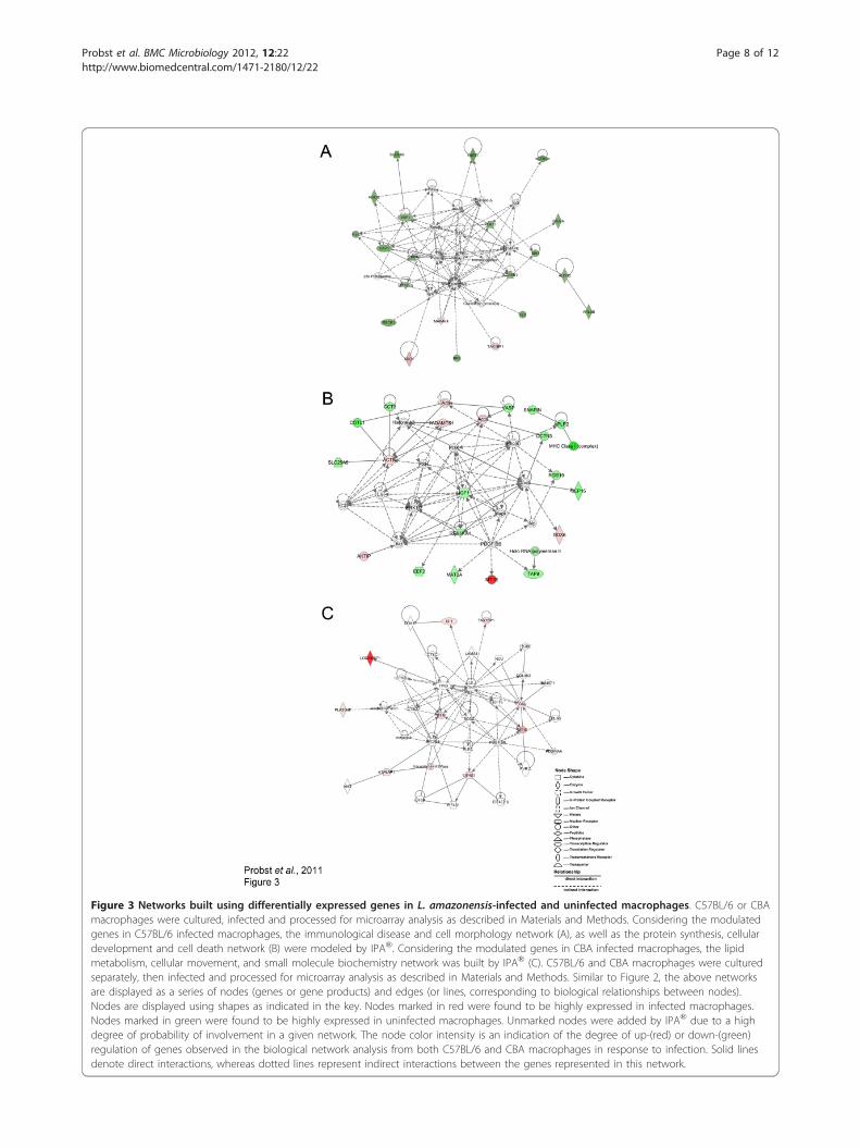
Figure 3 Networks built using differentially expressed genes in L. amazonensis-infected and uninfected macrophages. C57BL/6 or CBAmacrophages were cultured, infected and processed for microarray analysis as described in Materials and Methods. Considering the modulatedgenes in C57BL/6 infected macrophages, the immunological disease and cell morphology network (A), as well as the protein synthesis, cellulardevelopment and cell death network (B) were modeled by IPA®. Considering the modulated genes in CBA infected macrophages, the lipidmetabolism, cellular movement, and small molecule biochemistry network was built by IPA® (C). C57BL/6 and CBA macrophages were culturedseparately, then infected and processed for microarray analysis as described in Materials and Methods. Similar to Figure 2, the above networksare displayed as a series of nodes (genes or gene products) and edges (or lines, corresponding to biological relationships between nodes).Nodes are displayed using shapes as indicated in the key. Nodes marked in red were found to be highly expressed in infected macrophages.Nodes marked in green were found to be highly expressed in uninfected macrophages. Unmarked nodes were added by IPA® due to a highdegree of probability of involvement in a given network. The node color intensity is an indication of the degree of up-(red) or down-(green)regulation of genes observed in the biological network analysis from both C57BL/6 and CBA macrophages in response to infection. Solid linesdenote direct interactions, whereas dotted lines represent indirect interactions between the genes represented in this network.
Probst et al. BMC Microbiology 2012, 12:22http://www.biomedcentral.com/1471-2180/12/22
Page 8 of 12

trigger the production of significant amounts of pro-inflammatory cytokines, which is related to the capacityof C57BL/6 macrophages to control L. amazonensisinfection.The second network modeled by IPA® was the protein
synthesis, cellular development and cell death network(score 38, Figure 3B). This network contains 19 out ofthe 35 genes that were modulated by C57BL/6 macro-phages in response to L. amazonensis infection. Most ofthese genes (14/19) were found to be down-regulated ininfected cells, including: vasp (-2.06), involved in actinfilament organization; snapin (-2.28), which participatesin intracellular protein transport and exocytosis; aplp2(-2.61) and rgs19 (-2.27), which encode proteins fromthe G protein signaling pathway; igf1 (-2.01), involved incell proliferation and apoptosis; eef2 (-2.20), whichencodes a protein implicated in transcription processes.A total of five genes (5/19) were up-regulated ininfected C57BL/6 macrophages compared to uninfectedcells, including: mt1e (+9.53), involved in apoptosis andoxidative stress response; ddx6 (+2.24), involved in cellreplication; actb (+1.99), which participates in intracellu-lar transport and endocytosis; aktip (+2.21), whichencodes a protein that participates in intracellular trans-port and apoptosis; adamts1 (+2.07), involved in anintegrin signaling pathway, as well as cellular migration.In both of the networks modeled by IPA® pertaining
to infected C57BL/6 macrophages, namely the cell mor-phology and immunological disease network, as well asthe protein synthesis, cellular development and celldeath network, many genes involved in apoptosis werefound to be up-regulated. This finding is consistent withthe uninfected C57BL/6 macrophage expression profile,which also found up-regulation of genes involved inapoptosis (Figure 3A, B) and is very likely related to thecapacity of C57BL/6 macrophages to control parasiteinfection. This hypothesis is also supported by previousstudies which have described the inhibition of apoptosisin host cells using several susceptibility models of L.donovani [42,43], as well as L. major [44,45] and L.amazonensis [22] infection.Genes involved in the lipid metabolism, cellular movement,and small molecule biochemistry network are up-regulatedin CBA macrophages in response to L. amazonensisinfectionConsidering L. amazonensis infection in CBA macro-phages IPA® modeled the lipid metabolism, cellularmovement, and small molecule biochemistry network(score 26) containing 35 genes with the highest prob-ability of being modulated together as a result of infec-tion (Figure 3C). Nine out of these 35 genes were foundto be up-regulated under infection in CBA cells:loc340571 (similar to hsiah1, +13.00), tax1bp1 (+2.70),vacuolar H + ATPase, mt1f (+2.84) and mt1e (+5.19),
which are all involved in apoptosis, while the latter twoare additionally known to play a role in the oxidativestress response; sf1 (+2.13), which is implicated in tran-scriptional regulation and splicing processes; pla2g4f(+2.08), which is involved in chemotaxis and cellularmigration; itgav (+2.30), which participates in cell adhe-sion; and eif4g1 (+2.45), that encodes a protein whichparticipates in translation process regulation.In accordance with the present findings, the up-regu-
lation of genes involved in the lipid metabolism processhas been recently described in BALB/c macrophages [5].Osorio y Fortéa et al. (2009) suggest that collaborationsamong these genes likely act to facilitate the survival ofL. amazonensis inside susceptible macrophages by wayof a mechanism involved in the biogenesis of large L.amazonensis-induced parasitophorous vacuoles in bothBALB/c and CBA macrophages.Comparison of differential gene expression by C57BL/6 andCBA macrophages in response to L. amazonensis infectionTo gain deeper insight into the differences between therespective responses of C57BL/6 and CBA macrophagesto infection, the authors attempted to identify specificgenes observed to be significantly modulated in a diver-gent pattern as a result of L. amazonensis infection.However, the baseline gene expression signatures mea-sured prior to infection present a challenge to this typeof analysis, as inherent transcriptomic differences mayinterfere with the accurate identification of differentiallyexpressed gene sets. Firstly, all gene expression valueswere normalized by subtracting the expression levels byinfected macrophages from the corresponding meanexpression levels (log2-scale) by uninfected cells within agiven mouse strain. Thereafter, a direct comparison ofnormalized gene expression levels was performed usingSAM analysis to identify the genes that were differen-tially expressed between these two mouse strains.Finally, IPA® was used to highlight possible connec-
tions between C57BL/6 and CBA macrophagesresponses to L. amazonensis infection. Networks wereconstructed from the total number of differentiallyexpressed genes (n = 114), considering both strains ofmice. The cell cycle network (See Additional file 6: Fig-ure S2) had the highest probability of interrelated genesbeing modulated together. This network contains 35genes (score 36), with 16 out of the 114 genes that weremodulated by either C57BL/6 or CBA macrophages inresponse to L. amazonensis. Ten of the 16 modulatedgenes encode proteins involved in several cellular pro-cesses: usp3, which encodes an enzyme involved in ubi-quitination; phb and polr2a, which encode proteinsimplicated in the transcription process; elf4b, involvedin the translational process; gstp1, which participates indetoxification; rps6ka1 and sipa1, both involved in cellu-lar signaling; cd72, s1pr2 and ptafr, which encode
Probst et al. BMC Microbiology 2012, 12:22http://www.biomedcentral.com/1471-2180/12/22
Page 9 of 12

surface receptors. Of these, cd72, s1pr2 and ptafr werefound to be up-regulated in C57BL/6 macrophagesinfected with L. amazonensis (data not shown). Thesegenes encode receptors, which are expressed on macro-phage surfaces. Moreover, the modulation of thesereceptors and subsequent down-regulation of themacrophage proinflammatory response has been pre-viously described [46,47] and is in accordance with theability of C57BL/6 macrophages to control L. amazo-nensis infection [3].Cd72 has been described as a costimulatory molecule
found to be up-regulated in macrophages during the acti-vation of a Th1-type immune response [48]. Cd72 parti-cipates in the activation of a pro-inflammatory responsein the lungs of aging mice and was also found to be asso-ciated with an increase in the number of CD4, CD8 andB cells, as well as macrophages. s1pr2 encodes the sphin-gosine-1-phosphate receptor-2 (S1pr2), involved in therecognition of sphingosine-1-phosphate, a biologicallyactive sphingolipid that causes pleiotropic effects inmacrophages, and is central to the development of ather-osclerosis [48]. Evidence shows that S1pr2 is involved inmacrophage retention at the site of atherosclerotic pla-que inflammation [49]. The authors suggest furtherinvestigation into the role played by both Cd72 andS1pr2 in L. amazonensis infection.The other gene found to be up-regulated in C57BL/6
infected macrophages was ptafr, which encodes thereceptor for lipid mediator platelet-activating factor (Paf)and is implicated in a number of pathological conditionscharacterized by tissue inflammation [50]. The role Ptafrplays in protozoan infections has previously been evalu-ated [51,52]. Ptafr-/- mice of C57BL/6 background werefound to be more susceptible to infection by L. amazo-nensis than in wild-type controls, as evidenced by bothlesion size and parasite number at the site of infection.These findings are associated with the inefficient produc-tion of immune mediators, including IFN-g, Ccl5 andnitric oxide synthase-2 mRNA, as well as being associatedwith higher levels of arginase-1 mRNA and elevatedamounts of antibodies. These authors concluded that sig-naling through the Ptafr is essential for the murine hostto drive an immune response towards controllingL. amazonensis infection [53]. The up-regulation of Ptafrin L. amazonensis-infected C57BL/6 macrophagesobserved in the present study is consistent with the abil-ity of these cells to control parasite infection, as observedherein.
ConclusionIn conclusion, the present study represents an initialattempt at making direct comparisons between the glo-bal gene expression profiles from two distinct strains ofuninfected mouse macrophages. Our analysis revealed
that the transcriptional profile of uninfected C57BL/6macrophages was markedly different from that of CBAmacrophages. We also found that C57BL/6 macrophagesexpress higher levels of genes involved in the hostimmune inflammatory response and apoptosis, as wellas others that encode for phagocytic receptors thatrecognize pathogens and apoptotic cells. These cellswere also found to down-regulate genes involved in thedeactivation pathway of macrophages. In response toinfection, C57BL/6 macrophages continued to up-regu-late genes involved in apoptosis, as was similarlyobserved in uninfected cells. Finally, the authors found alow number of genes, which were related to lipid meta-bolism, up-regulated by CBA macrophages in responseto L. amazonensis infection. Collaboration among thesegenes likely facilitates the survival of L. amazonensisinside susceptible macrophages by way of a mechanisminvolved in the biogenesis of large L. amazonensis-induced parasitophorous vacuoles. Taken together, thesefindings may aid in the understanding of C57BL/6macrophages’ greater capacity to control L. amazonensisinfection in comparison to CBA cells. However, themechanism by which these differentially expressed genesaffect the course of Leishmania infection remainsunclear. Further studies should be conducted to investi-gate the influence of baseline gene expression signatureson the outcome of L. amazonensis infection with respectto host genetic background.
Additional material
Additional file 1: Table S1. Differentially expressed genes in uninfectedmacrophages from C57BL/6 vs CBA mice.
Additional file 2: Table S2. Expressed genes in L. amazonensis-infectedC57BL/6 macrophages.
Additional file 3: Table S3. Expressed genes in L. amazonensis-infectedCBA macrophages.
Additional file 4: Table S4. List of primers used in RT-qPCRamplification of gene expression in uninfected and L. amazonensis-infected C57BL/6 and CBA macrophages.
Additional file 5: Figure S1. Comparative analysis of the kinetics ofinfection by L. amazonensis in C57BL/6 and CBA. C57BL/6 or CBAinflammatory peritoneal macrophages were plated (2 × 10⁵/mL) for 24 hand infected with L. amazonensis stationary phase promastigotes at aratio of 10:1 (parasite to macrophage). After 12 h, cells were washed,reincubated for additional 6 or 24 h and then fixed with ethanol for 20min. After H&E staining, the percentage of infected cells (A) and theparasite numbers per macrophage (B) were quantified using lightmicroscopy at each time interval. Results are representative of twoindependent experiments performed in quadruplicate ± SD. (Mann-Whitney *p = 0.05).
Additional file 6: Figure S2. Network built using differentially expressedgenes in L. amazonensis-infected macrophages from C57BL/6 and CBAmice. C57BL/6 and CBA macrophages were cultured separately, theninfected and processed for microarray analysis as described in Materialsand Methods. The cell cycle network was modeled using IPA®®. Genesmarked in gray represent those found to be differentially expressedbetween C57BL/6 and CBA infected macrophages, while unmarkedgenes were added by IPA®® due to a high probability of involvement in
Probst et al. BMC Microbiology 2012, 12:22http://www.biomedcentral.com/1471-2180/12/22
Page 10 of 12

this network. Similar to Figure 2, the above network is displayed as aseries of nodes (genes or gene products) and edges (or lines,corresponding to biological relationships between nodes). Nodes aredisplayed using shapes as indicated in the key. Solid lines denote directinteractions, whereas dotted lines represent indirect interactions betweenthe genes represented in this network.
AcknowledgementsThe authors would like to thank Andris K. Walter for providing Englishrevision and consulting services.DisclosureThe authors declare that there are no conflicts of interest exist in thepresent study.Financial supportThis work was supported by grants and fellowships from FAPESB (Fundaçãode Amparo a Pesquisa no estado da Bahia), CAPES (Coordenação deAperfeiçoamento de Pessoal de Nível Superior) and CNPq (ConselhoNacional de Pesquisa e Desenvolvimento). Veras, PST holds a grant fromCNPq for productivity in research (306672/2008-1).
Author details1Laboratório de Genômica Funcional, Instituto Carlos Chagas, ICC-FIOCRUZ,Paraná, Brazil. 2Laboratório de Patologia e Biointervenção, CPqGM-FIOCRUZ,Bahia, Brazil. 3Center for the Study of Biological Complexity, VirginiaCommonwealth University, Richmond, VA, USA.
Authors’ contributionsCMP performed all bioinformatics and statistical analyses, and drafted themanuscript, RAS performed most of the RT-qPCR experiments, and helpedto draft the manuscript, ACD and DPP designed and performed some of theRT-qPCR experiments, JPBM conducted building of the networks using IPA,helped to draft the manuscript, and contributed to the discussion section,TFA and ING carried out the experiments involving tissue culture and RNAextractions, LSO and GAB contributed to the results and discussion section,MAK participated in the design of the study and microarray experiments,PSTV conceived of the study, performed the microarray experiments,participated in the study design and coordination, as well as helped to draftthe manuscript. All authors read and approved the final manuscript.
Received: 27 June 2011 Accepted: 9 February 2012Published: 9 February 2012
References1. Bogdan C, Gessner A, Solbach W, Rollinghoff M: Invasion, control and
persistence of Leishman parasites. Curr Opin Immunol 1996, 8(4):517-525.2. Garg R, Dube A: Animal models for vaccine studies for visceral
Leishmaniasis. Indian J Med Res 2006, 123(3):439-454.3. Gomes IN, Calabrich AF, Tavares Rda S, Wietzerbin J, de Freitas LA, Veras PS:
Differential properties of CBA/J mononuclear phagocytes recoveredfrom an inflammatory site and probed with two different species ofLeishmania. Microbes Infect 2003, 5(4):251-260.
4. Lemos de Souza V, Ascencao Souza J, Correia Silva TM, Sampaio TavaresVeras P, Rodrigues de-Freitas LA: Different Leishmania species determinedistinct profiles of immune and histopathological responses in CBAmice. Microbes Infect 2000, 2(15):1807-1815.
5. Osorio y Fortea J, Prina E, de La Llave E, Lecoeur H, Lang T, Milon G:Unveiling pathways used by Leishmania amazonensis amastigotes tosubvert macrophage function. Immunol Rev 2007, 219:66-74.
6. Zhang S, Kim CC, Batra S, McKerrow JH, Loke P: Delineation of diversemacrophage activation programs in response to intracellular parasitesand cytokines. PLoS Negl Trop Dis 2010, 4(3):e648.
7. Jenner RG, Young RA: Insights into host responses against pathogensfrom transcriptional profiling. Nat Rev Microbiol 2005, 3(4):281-294.
8. Reiner SL, Locksley RM: The regulation of immunity to Leishmania major.Annu Rev Immunol 1995, 13:151-177.
9. Scharton-Kersten T, Scott P: The role of the innate immune response inTh1 cell development following Leishmania major infection. J Leukoc Biol1995, 57(4):515-522.
10. Abreu-Silva AL, Calabrese KS, Cupolilo SM, Cardoso FO, Souza CS, Goncalvesda Costa SC: Histopathological studies of visceralized Leishmania(Leishmania) amazonensis in mice experimentally infected. Vet Parasitol2004, 121(3-4):179-187.
11. Norsworthy NB, Sun J, Elnaiem D, Lanzaro G, Soong L: Sand fly salivaenhances Leishmania amazonensis infection by modulating interleukin-10 production. Infect Immun 2004, 72(3):1240-1247.
12. Jones DE, Ackermann MR, Wille U, Hunter CA, Scott P: Early enhanced Th1response after Leishmania amazonensis infection of C57BL/6 interleukin-10-deficient mice does not lead to resolution of infection. Infect Immun2002, 70(4):2151-2158.
13. Maioli TU, Takane E, Arantes RM, Fietto JL, Afonso LC: Immune responseinduced by New World Leishmania species in C57BL/6 mice. Parasitol Res2004, 94(3):207-212.
14. Rosas LE, Keiser T, Barbi J, Satoskar AA, Septer A, Kaczmarek J, Lezama-Davila CM, Satoskar AR: Genetic background influences immuneresponses and disease outcome of cutaneous L. mexicana infection inmice. Int Immunol 2005, 17(10):1347-1357.
15. Afonso LC, Scott P: Immune responses associated with susceptibility ofC57BL/10 mice to Leishmania amazonensis. Infect Immun 1993,61(7):2952-2959.
16. Prina E, Jouanne C, de Souza Lao S, Szabo A, Guillet JG, Antoine JC:Antigen presentation capacity of murine macrophages infected withLeishmania amazonensis amastigotes. J Immunol 1993, 151(4):2050-2061.
17. Courret N, Lang T, Milon G, Antoine JC: Intradermal inoculations of lowdoses of Leishmania major and Leishmania amazonensis metacyclicpromastigotes induce different immunoparasitic processes and status ofprotection in BALB/c mice. Int J Parasitol 2003, 33(12):1373-1383.
18. Veras PS, Topilko A, Gouhier N, Moreau MF, Rabinovitch M, Pouchelet M:Fusion of Leishmania amazonensis parasitophorous vacuoles withphagosomes containing zymosan particles: cinemicrographic andultrastructural observations. Braz J Med Biol Res 1996, 29(8):1009-1018.
19. Buates S, Matlashewski G: General suppression of macrophage geneexpression during Leishmania donovani infection. J Immunol 2001,166(5):3416-3422.
20. Chaussabel D, Semnani RT, McDowell MA, Sacks D, Sher A, Nutman TB:Unique gene expression profiles of human macrophages and dendriticcells to phylogenetically distinct parasites. Blood 2003, 102(2):672-681.
21. Rodriguez NE, Chang HK, Wilson ME: Novel program of macrophage geneexpression induced by phagocytosis of Leishmania chagasi. Infect Immun2004, 72(4):2111-2122.
22. Osorio y Fortea J, de La Llave E, Regnault B, Coppee JY, Milon G, Lang T,Prina E: Transcriptional signatures of BALB/c mouse macrophageshousing multiplying Leishmania amazonensis amastigotes. BMC Genomics2009, 10:119.
23. Banus S, Vandebriel RJ, Pennings JL, Gremmer ER, Wester PW, vanKranen HJ, Breit TM, Demant P, Mooi FR, Hoebee B, et al: Comparativegene expression profiling in two congenic mouse strains followingBordetella pertussis infection. BMC Microbiol 2007, 7:88.
24. Morrison LJ, McLellan S, Sweeney L, Chan CN, MacLeod A, Tait A,Turner CM: Role for parasite genetic diversity in differential hostresponses to Trypanosoma brucei infection. Infect Immun 2010,78(3):1096-1108.
25. Borjabad A, Brooks AI, Volsky DJ: Gene expression profiles of HIV-1-infected glia and brain: toward better understanding of the role ofastrocytes in HIV-1-associated neurocognitive disorders. J NeuroimmunePharmacol 2010, 5(1):44-62.
26. Wu Z, Irizarry RA: Stochastic models inspired by hybridization theory forshort oligonucleotide arrays. J Comput Biol 2005, 12(6):882-893.
27. Hochberg Y, Benjamini Y: More powerful procedures for multiplesignificance testing. Stat Med 1990, 9(7):811-818.
28. Storey JD: A direct approach to false discovery rates. J R Stat Soc Ser B2002, 64(Part 3):479-498.
29. Wang F, Okamoto Y, Inoki I, Yoshioka K, Du W, Qi X, Takuwa N, Gonda K,Yamamoto Y, Ohkawa R, et al: Sphingosine-1-phosphate receptor-2deficiency leads to inhibition of macrophage proinflammatory activitiesand atherosclerosis in apoE-deficient mice. J Clin Invest 2010,120(11):3979-3995.
30. Stouffer SA, Suchman EA, DeVinney LC, Star SA, Williams RMJ: In TheAmerican Soldier. Volume 1. Princeton: Princeton University Press; 1949.
Probst et al. BMC Microbiology 2012, 12:22http://www.biomedcentral.com/1471-2180/12/22
Page 11 of 12

31. Mahley RW, Rall SC Jr: Apolipoprotein E: far more than a lipid transportprotein. Annu Rev Genomics Hum Genet 2000, 1:507-537.
32. Mahley RW: Apolipoprotein E: cholesterol transport protein withexpanding role in cell biology. Sci 1988, 240(4852):622-630.
33. Bast A, Fischer K, Erttmann SF, Walther R: Induction of peroxiredoxin Igene expression by LPS involves the Src/PI3K/JNK signalling pathway.Biochim Biophys Acta 2010, 1799(5-6):402-410.
34. Grainger DJ, Reckless J, McKilligin E: Apolipoprotein E modulates clearanceof apoptotic bodies in vitro and in vivo, resulting in a systemicproinflammatory state in apolipoprotein E-deficient mice. J Immunol2004, 173(10):6366-6375.
35. Medeiros LA, Khan T, El Khoury JB, Pham CL, Hatters DM, Howlett GJ,Lopez R, O’Brien KD, Moore KJ: Fibrillar amyloid protein present inatheroma activates CD36 signal transduction. J Biol Chem 2004,279(11):10643-10648.
36. Arlaud GJ, Gaboriaud C, Thielens NM, Rossi V, Bersch B, Hernandez JF,Fontecilla-Camps JC: Structural biology of C1: dissection of a complexmolecular machinery. Immunol Rev 2001, 180:136-145.
37. Armbrust T, Nordmann B, Kreissig M, Ramadori G: C1Q synthesis by tissuemononuclear phagocytes from normal and from damaged rat liver: up-regulation by dexamethasone, down-regulation by interferon gamma,and lipopolysaccharide. Hepatol 1997, 26(1):98-106.
38. Brown JS, Hussell T, Gilliland SM, Holden DW, Paton JC, Ehrenstein MR,Walport MJ, Botto M: The classical pathway is the dominant complementpathway required for innate immunity to Streptococcus pneumoniaeinfection in mice. Proc Natl Acad Sci USA 2002, 99(26):16969-16974.
39. Roos A, Xu W, Castellano G, Nauta AJ, Garred P, Daha MR, van Kooten C:Mini-review: A pivotal role for innate immunity in the clearance ofapoptotic cells. Eur J Immunol 2004, 34(4):921-929.
40. Gribaudo G, Riera L, Hertel L, Landolfo S: In vitro and in vivo expressionanalysis of the interferon-inducible 203 gene. J Interferon Cytokine Res1999, 19(2):129-136.
41. Gregory DJ, Sladek R, Olivier M, Matlashewski G: Comparison of the effectsof Leishmania major or Leishmania donovani infection on macrophagegene expression. Infect Immun 2008, 76(3):1186-1192.
42. Shweash M, Adrienne McGachy H, Schroeder J, Neamatallah T, Bryant CE,Millington O, Mottram JC, Alexander J, Plevin R: Leishmania mexicanapromastigotes inhibit macrophage IL-12 production via TLR-4dependent COX-2, iNOS and arginase-1 expression. Mol Immunol 2011,48(15-16):1800-1808.
43. Wilmanski J, Villanueva E, Deitch EA, Spolarics Z: Glucose-6-phosphatedehydrogenase deficiency and the inflammatory response to endotoxinand polymicrobial sepsis. Crit Care Med 2007, 35(2):510-518.
44. Engwerda CR, Ato M, Cotterell SE, Mynott TL, Tschannerl A, Gorak-Stolinska PM, Kaye PM: A role for tumor necrosis factor-alpha inremodeling the splenic marginal zone during Leishmania donovaniinfection. Am J Pathol 2002, 161(2):429-437.
45. Moore KJ, Matlashewski G: Intracellular infection by Leishmania donovaniinhibits macrophage apoptosis. J Immunol 1994, 152(6):2930-2937.
46. Conceicao-Silva F, Hahne M, Schroter M, Louis J, Tschopp J: The resolutionof lesions induced by Leishmania major in mice requires a functional Fas(APO-1, CD95) pathway of cytotoxicity. Eur J Immunol 1998, 28(1):237-245.
47. Aga E, Katschinski DM, van Zandbergen G, Laufs H, Hansen B, Muller K,Solbach W, Laskay T: Inhibition of the spontaneous apoptosis ofneutrophil granulocytes by the intracellular parasite Leishmania major. JImmunol 2002, 169(2):898-905.
48. Sinclair NR: Why so many coinhibitory receptors? Scand J Immunol 1999,50(1):10-13.
49. Agostini C, Trentin L, Perin A, Facco M, Siviero M, Piazza F, Basso U,Adami F, Zambello R, Semenzato G: Regulation of alveolar macrophage-Tcell interactions during Th1-type sarcoid inflammatory process. Am JPhysiol 1999, 277(2 Pt 1):L240-250.
50. Skoura A, Michaud J, Im DS, Thangada S, Xiong Y, Smith JD, Hla T:Sphingosine-1-phosphate receptor-2 function in myeloid cells regulatesvascular inflammation and atherosclerosis. Arterioscler Thromb Vasc Biol2011, 31(1):81-85.
51. Keely S, Glover LE, Weissmueller T, MacManus CF, Fillon S, Fennimore B,Colgan SP: Hypoxia-inducible factor-dependent regulation of platelet-activating factor receptor as a route for gram-positive bacterialtranslocation across epithelia. Mol Biol Cell 2010, 21(4):538-546.
52. Santiago HC, Braga Pires MF, Souza DG, Roffe E, Cortes DF, Tafuri WL,Teixeira MM, Vieira LQ: Platelet activating factor receptor-deficient micepresent delayed interferon-gamma upregulation and high susceptibilityto Leishmania amazonensis infection. Microb Infect 2006, 8(11):2569-2577.
53. Talvani A, Santana G, Barcelos LS, Ishii S, Shimizu T, Romanha AJ, Silva JS,Soares MB, Teixeira MM: Experimental Trypanosoma cruzi infection inplatelet-activating factor receptor-deficient mice. Microb Infect 2003,5(9):789-796.
doi:10.1186/1471-2180-12-22Cite this article as: Probst et al.: A comparison of two distinct murinemacrophage gene expression profiles in response to Leishmaniaamazonensis infection. BMC Microbiology 2012 12:22.
Submit your next manuscript to BioMed Centraland take full advantage of:
• Convenient online submission
• Thorough peer review
• No space constraints or color figure charges
• Immediate publication on acceptance
• Inclusion in PubMed, CAS, Scopus and Google Scholar
• Research which is freely available for redistribution
Submit your manuscript at www.biomedcentral.com/submit
Probst et al. BMC Microbiology 2012, 12:22http://www.biomedcentral.com/1471-2180/12/22
Page 12 of 12


