A comparison of heart rate variability, n-3 PUFA status ...
17
A comparison of heart rate variability, n-3 PUFA status and lipid mediator profile in age- and BMI-matched middle-aged vegans and omnivores Ana M. Pinto 1 , Thomas A. B. Sanders 1 , Alexandra C. Kendall 2 , Anna Nicolaou 2 , Robert Gray 1 , Haya Al-Khatib 1 and Wendy L. Hall 1 * 1 Diabetes & Nutritional Sciences Division, King’s College London, Faculty of Life Sciences & Medicine, Franklin-Wilkins Building, 150 Stamford Street, London SE1 9NH, UK 2 School of Health Sciences, Division of Pharmacy and Optometry, Faculty of Biology, Medicine and Health, The University of Manchester, Stopford Building, Oxford Road, Manchester M13 9PT, UK (Submitted 5 September 2016 – Final revision received 8 February 2017 – Accepted 22 February 2017) Abstract Low heart rate variability (HRV) predicts sudden cardiac death. Long-chain (LC) n-3 PUFA (C20–C22) status is positively associated with HRV. This cross-sectional study investigated whether vegans aged 40–70 years (n 23), whose diets are naturally free from EPA (20 : 5n-3) and DHA (22 : 6n-3), have lower HRV compared with omnivores (n 24). Proportions of LC n-3 PUFA in erythrocyte membranes, plasma fatty acids and concentrations of plasma LC n-3 PUFA-derived lipid mediators were significantly lower in vegans. Day-time interbeat intervals (IBI), adjusted for physical activity, age, BMI and sex, were significantly shorter in vegans compared with omnivores (mean difference -67 ms; 95 % CI -130, -3·4, P < 0·05), but there were no significant differences over 24 h or during sleep. Vegans had higher overall HRV, measured as 24 h standard deviation of normal-to-normal intervals (SDNN) (mean adjusted difference 27 ms; 95 % CI 1, 52, P = 0·039). Conversely, vegans presented with decreased 8 h day-time HRV: mean adjusted difference in SDNN -20 ms; 95 % CI -37, -3, P = 0·021, with no differences during nocturnal sleep. Day-time parameters of beat-to-beat HRV (root of the mean of the sum of the squares of differences between adjacent normal-to-normal intervals, percentage of adjacent normal-to-normal intervals that differ by >50 % and high-frequency power) were similarly lower in vegans, with no differences during sleep. In conclusion, vegans have higher 24 h SDNN, but lower day-time HRV and shorter day- time IBI relative to comparable omnivores. Vegans may have reduced availability of precursor markers for pro-resolving lipid mediators; it remains to be determined whether there is a direct link with impaired cardiac function in populations with low-n-3 status. Key words: Vegans: n-3 PUFA: CHD: Heart rate variability: Inflammation: Eicosanoids: Lipid mediators The longer chain (LC) n-3 PUFA (C20–C22), EPA (20 : 5n-3) and DHA (22 : 6n-3), are mainly derived from seafood, although small amounts are provided by meat, eggs and dairy products. Consequently, vegans consume a diet devoid of 20 : 5n-3 and 22 : 6n-3 (1) . The main n-3 PUFA in vegan diets is α-linolenic acid (ALA; 18 : 3n-3), derived from plant foods, particularly soya and seed oils such as rapeseed oil. (LC) n-3 PUFA as percentages of total fatty acids in blood fractions are, in vegans, only a third of the level in meat- and fish-eaters (2) . 20 : 5n-3 and 22 : 6n-3 can be endogenously synthesised from 18 : 3n-3 by desaturation and elongation enzymes, but the rate of this conversion is restricted to a narrow range in adults (3) . Stable isotope studies have suggested that conversion of 18 : 3n-3 to 22:6n-3 can vary from undetectable amounts up to 4 % in men, and 9 % in women (4) . An observational study suggested that conversion from dietary 18 : 3n-3 to LC n-3 PUFA might be increased in non-fish-eaters (5) . Furthermore, dietary supplementation of vegans with 18 : 3n-3 has not been found to increase 22 : 6n-3 in blood lipids, including plasma choline phosphoglycerides and platelet phosphoglycerides (6,7) and erythrocytes, platelets and plasma cholesteryl esters, phospholipids and TAG (6,7) . It is unclear whether the lack of 20 : 5n-3 and 22 : 6n-3 intake in vegans has adverse effects on cardiovascular health (8,9) especially as BMI (10,11) , blood cholesterol (12,13) and blood pressure (14,15) are lower than in meat-eaters. LC n-3 PUFA, especially 22 : 6n-3, are rapidly incorporated into cellular lipids, primarily membrane phospholipids, in a variety of cells including cardiomyocytes and neural tissue, Abbreviations: %E, percentage of energy; HDHA, hydroxydocosahexaenoic acid; HF, high-frequency power; HR, heart rate; HRV, heart rate variability; IBI, interbeat intervals; LC, long chain; LF, low-frequency power; pNN50, percentage of adjacent normal-to-normal intervals that differ by >50 %; RMSSD, root of the mean of the sum of the squares of differences between adjacent normal-to-normal intervals; SDANN, standard deviation of the average 5-min normal-to- normal intervals; SDNN, standard deviation of normal-to-normal intervals; SPM; specialised pro-resolving lipid mediators; VLF, very-low-frequency power. * Corresponding author: W. L. Hall, email [email protected] British Journal of Nutrition (2017), 117, 669–685 doi:10.1017/S0007114517000629 © The Authors 2017 Downloaded from https://www.cambridge.org/core. IP address: 65.21.228.167, on 01 Dec 2021 at 19:01:50, subject to the Cambridge Core terms of use, available at https://www.cambridge.org/core/terms. https://doi.org/10.1017/S0007114517000629
Transcript of A comparison of heart rate variability, n-3 PUFA status ...

A comparison of heart rate variability, n-3 PUFA status and lipid mediatorprofile in age- and BMI-matched middle-aged vegans and omnivores
Ana M. Pinto1, Thomas A. B. Sanders1, Alexandra C. Kendall2, Anna Nicolaou2, Robert Gray1,Haya Al-Khatib1 and Wendy L. Hall1*1Diabetes & Nutritional Sciences Division, King’s College London, Faculty of Life Sciences & Medicine, Franklin-WilkinsBuilding, 150 Stamford Street, London SE1 9NH, UK2School of Health Sciences, Division of Pharmacy and Optometry, Faculty of Biology, Medicine and Health, The Universityof Manchester, Stopford Building, Oxford Road, Manchester M13 9PT, UK
(Submitted 5 September 2016 – Final revision received 8 February 2017 – Accepted 22 February 2017)
AbstractLow heart rate variability (HRV) predicts sudden cardiac death. Long-chain (LC) n-3 PUFA (C20–C22) status is positively associated with HRV.This cross-sectional study investigated whether vegans aged 40–70 years (n 23), whose diets are naturally free from EPA (20 : 5n-3) and DHA(22 : 6n-3), have lower HRV compared with omnivores (n 24). Proportions of LC n-3 PUFA in erythrocyte membranes, plasma fatty acids andconcentrations of plasma LC n-3 PUFA-derived lipid mediators were significantly lower in vegans. Day-time interbeat intervals (IBI), adjustedfor physical activity, age, BMI and sex, were significantly shorter in vegans compared with omnivores (mean difference −67ms; 95% CI−130, −3·4, P< 0·05), but there were no significant differences over 24 h or during sleep. Vegans had higher overall HRV, measured as 24 hstandard deviation of normal-to-normal intervals (SDNN) (mean adjusted difference 27ms; 95% CI 1, 52, P= 0·039). Conversely, veganspresented with decreased 8 h day-time HRV: mean adjusted difference in SDNN −20ms; 95% CI −37, −3, P= 0·021, with no differences duringnocturnal sleep. Day-time parameters of beat-to-beat HRV (root of the mean of the sum of the squares of differences between adjacentnormal-to-normal intervals, percentage of adjacent normal-to-normal intervals that differ by >50% and high-frequency power) were similarlylower in vegans, with no differences during sleep. In conclusion, vegans have higher 24 h SDNN, but lower day-time HRV and shorter day-time IBI relative to comparable omnivores. Vegans may have reduced availability of precursor markers for pro-resolving lipid mediators;it remains to be determined whether there is a direct link with impaired cardiac function in populations with low-n-3 status.
Key words: Vegans: n-3 PUFA: CHD: Heart rate variability: Inflammation: Eicosanoids: Lipid mediators
The longer chain (LC) n-3 PUFA (C20–C22), EPA (20 : 5n-3) andDHA (22 : 6n-3), are mainly derived from seafood, althoughsmall amounts are provided by meat, eggs and dairy products.Consequently, vegans consume a diet devoid of 20 : 5n-3 and22 : 6n-3(1). The main n-3 PUFA in vegan diets is α-linolenic acid(ALA; 18 : 3n-3), derived from plant foods, particularly soya andseed oils such as rapeseed oil. (LC) n-3 PUFA as percentages oftotal fatty acids in blood fractions are, in vegans, only a third ofthe level in meat- and fish-eaters(2). 20 : 5n-3 and 22 : 6n-3 canbe endogenously synthesised from 18 : 3n-3 by desaturationand elongation enzymes, but the rate of this conversion isrestricted to a narrow range in adults(3). Stable isotope studieshave suggested that conversion of 18 : 3n-3 to 22 : 6n-3 canvary from undetectable amounts up to 4% in men, and 9%
in women(4). An observational study suggested that conversionfrom dietary 18 : 3n-3 to LC n-3 PUFA might be increased innon-fish-eaters(5). Furthermore, dietary supplementation ofvegans with 18 : 3n-3 has not been found to increase 22 : 6n-3 inblood lipids, including plasma choline phosphoglycerides andplatelet phosphoglycerides(6,7) and erythrocytes, platelets andplasma cholesteryl esters, phospholipids and TAG(6,7). It isunclear whether the lack of 20 : 5n-3 and 22 : 6n-3 intake invegans has adverse effects on cardiovascular health(8,9)
especially as BMI(10,11), blood cholesterol(12,13) and bloodpressure(14,15) are lower than in meat-eaters.
LC n-3 PUFA, especially 22 : 6n-3, are rapidly incorporatedinto cellular lipids, primarily membrane phospholipids, in avariety of cells including cardiomyocytes and neural tissue,
Abbreviations: %E, percentage of energy; HDHA, hydroxydocosahexaenoic acid; HF, high-frequency power; HR, heart rate; HRV, heart rate variability;IBI, interbeat intervals; LC, long chain; LF, low-frequency power; pNN50, percentage of adjacent normal-to-normal intervals that differ by >50%; RMSSD, rootof the mean of the sum of the squares of differences between adjacent normal-to-normal intervals; SDANN, standard deviation of the average 5-min normal-to-normal intervals; SDNN, standard deviation of normal-to-normal intervals; SPM; specialised pro-resolving lipid mediators; VLF, very-low-frequency power.
* Corresponding author: W. L. Hall, email [email protected]
British Journal of Nutrition (2017), 117, 669–685 doi:10.1017/S0007114517000629© The Authors 2017
Dow
nloaded from https://w
ww
.cambridge.org/core . IP address: 65.21.228.167 , on 01 D
ec 2021 at 19:01:50 , subject to the Cambridge Core term
s of use, available at https://ww
w.cam
bridge.org/core/terms . https://doi.org/10.1017/S0007114517000629

thereby influencing membrane properties and function ofmembrane proteins. Fish oil consumption reduces heart rate(HR) in humans(16). Increasing LC n-3 PUFA content in cardio-myocyte membranes by 3 weeks of dietary fish oil in rabbitsdecreases HR in isolated hearts, and reduces pacemaker activityand pacemaker current in sinoatrial node cells(17); mechanismsare likely to be related to increased membrane fluidity anddirect interaction with a hyperpolarisation-activated If channelprotein(17), altering ion channel currents and reducing intrinsicpacemaker rate, reviewed by Billman(18).Raised HR are associated with a high degree of sympathetic
activity and suppressed parasympathetic activity (vagal activityslows HR) resulting in low heart rate variability (HRV); a reducedcapacity to self-regulate the HR in response to physio-logical demands. Low HRV is associated with mortality after amyocardial infarction(19–21), risk of sudden death in patients withCHD(22), and risk of cardiac events in the general population(23).Higher n-3 PUFA tissue status or fish consumption has beenpositively associated with HRV(24,25). Since HRV is under thecontrol of the autonomic nervous system, regulation of HR maybe influenced by n-3 PUFA status of neuronal and cardiac tissue.The brain is particularly rich in 22 : 6n-3, and incorporation ofdietary LC n-3 PUFA into neuronal tissue influences geneexpression, membrane protein signalling, neurotransmission andsignal transduction pathways(26). This may influence autonomicfunction by enhancing parasympathetic and/or reducing sym-pathetic activity, thus reducing HR and increasing HRV. There-fore, impairment of cardiac autonomic function due to depletedLC n-3 PUFA content in the central or peripheral nervous tissuewould reduce the responsivity of the heart.A further mechanism whereby cardiac function may be
modulated by neuronal LC n-3 PUFA status is via the productionof eicosanoids and related PUFA-derived lipid mediators that mayreduce inflammation and terminate (‘resolve’) acute inflammatoryevents, preventing further neuronal tissue damage. PUFA canbe oxygenated into numerous bioactive lipid mediators(27),and some of the 20 : 5n-3- and 22 : 6n-3-derived species act asprecursors of the specialised pro-resolving lipid mediators (SPM),resolvins, protectins and maresin, which are autocoid substancesactively involved in the resolution of local inflammation(27–29).Neuroprotectin D1 is a neuroprotective lipid mediator derivedfrom 22 : 6n-3 which might be particularly relevant to thepreservation of optimal cardiac autonomic function(30).This study aims to compare HRV between vegans and age/
sex/BMI-matched omnivores, representing populations withlow and adequate tissue n-3 PUFA status, respectively. Theprimary hypothesis of the study is that vegans have higherHR/shorter interbeat intervals (IBI) and lower HRV comparedwith omnivores. Exploratory analysis of plasma 20 : 5n-3- and22 : 6n-3-derived lipid mediator concentrations was conductedin order to provide mechanistic hypothesis-generating data thatmay help explain differences in HR/IBI/HRV between low andhigh LC n-3 PUFA status groups.
Methods
The present study was conducted according to the guidelines laiddown in the Declaration of Helsinki, and all procedures involving
human subjects were approved by the research ethics committeeof King’s College London (BDM/12/13-84). Written informedconsent was obtained from each subject. In all, twenty-threehealthy, non-smoking men and women, aged 40–70 years whohad been following a vegan diet for at least 2 years were com-pared with 24 age- and BMI-matched healthy participants whofollowed a mixed diet including meat, fish, eggs and dairy-containing foods (omnivores). Primary outcome variables wereHR/IBI and time-domain parameters of different components ofHRV: standard deviation of normal-to-normal intervals (SDNN):the most commonly reported marker of HRV and an indication ofoverall HRV, mainly determined by day/night differences) androot square root of the mean of the sum of the squares of dif-ferences between adjacent normal-to-normal intervals (RMSSD);an indicator of beat-to-beat, respiration-driven variability repre-senting parasympathetic cardiac regulation). Secondary outcomevariables included: other time and frequency-domain and non-linear parameters of HRV, erythrocyte and plasma fatty acidcomposition, plasma oxygenated lipid mediator profile, fastingplasma lipid profile, vitamin B12, serum 25-hydroxyvitamin D,IL-6, fasting plasma glucose, blood pressure, body compositionand background diet in order to compare risk factors for CVD invegans and omnivores. A sample size of twenty-three in eachgroup has a 80% power to detect a difference between SDNNmeans of 25ms and between RMSSD means of 15ms with asignificance level of 0·05 (two-tailed), based on SD of 30 and18ms, respectively, obtained from sleep-time HRV recordings ina previous cohort of middle-aged to older healthy men andwomen(31). Participants were recruited by distributing adverts tovegan organisations and societies. Omnivore participants wererecruited through internal and external email circulars and postersamongst university students and staff. The study was also pro-moted via social media, flyer distributions to vegan restaurantsand vegan food shops throughout London, and at various veganfood events. Volunteers who responded to advertisements weregiven more information about the study, completed an initialeligibility questionnaire via telephone or email and, if eligible,were provided with a study information sheet. Exclusion criteriaincluded a reported history of CVD, diabetes, cancer (excludingbasal cell carcinoma) in the past 5 years, chronic renal, liver orinflammatory bowel disease, history of drug or alcohol abuse(previous weekly alcohol intake >60 units/men or 50 units/women), current self-reported weekly alcohol intake exceedingtwenty-eight units, current use of marine n-3 supplements,pregnancy, weight change of >3kg in the previous 2 months, andBMI <18·5 and >35kg/m2. Vegan subjects were enrolled on thestudy along with omnivore controls, aiming to match for sex, age(±5 years) and BMI (±2kg/m2). A validated FFQ(32) was used toverify self-classification of dietary status of eligible volunteers andto provide supplementary information on habitual dietary intakes.Analysis was carried out using an Excel spreadsheet that incor-porated additional food composition data on LC n-3 PUFA con-tents of foods other than fish (meat, dairy products and eggs).
Participants attended one study visit, which took place inthe morning. Volunteers were instructed to fast for 12h before thevisit and consume nothing but water until attending the clinic.Once written informed consent was obtained, seated bloodpressure was measured in triplicate using an A&D
670 A. M. Pinto et al.
Dow
nloaded from https://w
ww
.cambridge.org/core . IP address: 65.21.228.167 , on 01 D
ec 2021 at 19:01:50 , subject to the Cambridge Core term
s of use, available at https://ww
w.cam
bridge.org/core/terms . https://doi.org/10.1017/S0007114517000629

Medical UA-767Plus upper arm automatic blood pressure monitor(A&D Instruments Limited), in accordance with guidelines fromthe British Hypertension Society. Height, body weight and per-centage body fat and waist circumference (WC) were measuredusing a stadiometer, a Tanita weighing scale (model: BC-418 MA;Tanita UK Ltd) and a tape measure, respectively. Participantscompleted the FFQ, which was checked for completeness andany missing data verified directly with the participant. Fastingplasma glucose and serum lipids, serum liver function markersand whole blood haematology was analysed on the same day infresh blood samples, and further plasma aliquots were frozen at−70°C until analysis of fatty acid and lipid mediator profiles couldtake place. Erythrocytes were washed with saline and lysed. Theerythrocyte lysate was de-proteinised in the presence of butylatedhydroxytoluene, chloroform was added to extract lipids thencentrifuged as previously described(33); supernatant was frozen at−20°C until analysis for fatty acid composition could be con-ducted(33). An Actiheart monitor was fitted on the chest (CamN-tech Ltd), which they wore for 24h. A diary was provided duringthe recording period to keep a register of all the daily activities(activities/exercise, meals or naps).
Heart rate variability measurements
IBI and continuous HR were measured for approximately 24 husing Actiheart monitors, which are small, light-weight (<10 g)waterproof devices that also contain piezoelectric sensors torecord acceleration in the vertical plane (counts per minute) asa measure of physical activity(34). Before the monitor could befitted, the area of skin was prepared including shaving of chesthair where required, using alcohol wipes to clean and dry theskin and use of an abrasive pad (UnilectTM) to remove the toplayer of skin cells. Two electrocardiogram (ECG) electrodes(SP-50, 50mm round; Pulse Medical) were placed on the chestto fit the Actiheart monitor. A short signal test involving a 5minwalk was performed before programming for the 24 h recordingto confirm adequate signal:noise ratio. Data processing of the24 h IBI recordings was carried out using the Actiheart software(version 4.0.91; CamNtech Ltd) and Kubios HRV analysis soft-ware (Biosignal Analysis and Medical Imaging Group, Depart-ment of Physics, University of Kuopio)(35). HRV, HR/IBI andaccelerometry data(34) were analysed for the full length ofrecording time (minimum of 18 h, up to 24 h). Further analysiswas carried on a standardised day-time period of 8 h and sleep-time period of 2 h to remove the influence of variability inrecording duration on HRV parameters. HRV outcomes inclu-ded time and frequency-domain parameters; time-domainparameters are based on the time intervals between adjacentQRS (Q, R and S being points on the R wave seen on an ECGduring ventricular depolarisation, and R being the peak upwarddeflection) complexes (normal-to-normal (NN) intervals)whereas frequency-domain parameters employ power spectralanalysis of NN intervals to determine the power (variance)within frequency bands(36). Time-domain parameters includedSDNN, standard deviation of the average normal-to-normalintervals in 5min segments of the whole recording (SDANN),RMSSD, the percentage of adjacent normal-to-normal intervalsthat differed by >50% (pNN50) and triangular index (TI), the
integral of the density distribution (the number of all NNintervals) divided by the maximum of the density distribution.Frequency-domain parameters included high-frequency (HF),low-frequency (LF) and very-low-frequency (VLF) power, andthe ratio of the LF and HF band powers (LF:HF). A non-linearparameter using Poincaré plots of short-term variability (SD1)against long-term variability (SD2) was also calculated as ameasure of complexity of HRV distribution over the duration ofthe recording. SDNN, LF and TI represent overall variability.Short-term (beat-to-beat) components of HRV include RMSSD,pNN50 and HF. SDANN and VLF reflect longer-phase compo-nents of variability.
Fatty acid analysis
Proportions of fatty acids in whole plasma and erythrocytemembranes were analysed by GC (Agilent 7890A GC; AgilentTechnologies) with a BPX70 GC column (length 25m, internaldiameter 0·32mm, film thickness 0·25 μm) custom designed forseparation of fatty acid methyl esters (SGE Analytic Science)following transesterification, as previously described(37), butsubstituting toluene for benzene and using pentadecanoic acidas an internal standard. Since total plasma concentrations offatty acids differ in vegans compared with omnivores, individualplasma fatty acids were compared between groups asweight percentages of the sum of fatty acids (% weight)(38) TheOmega-3 Index was defined as the sum of % weight EPA +DHAin erythrocytes.
Mediator lipidomics
Prostanoids and hydroxy fatty acids derived from dihomo-γ-linolenic acid (20 : 3n-6), linoleic acid (LA; 18 : 2n-6), 18 : 3n-3,arachidonic acid (AA; 20 : 4n-6), 20 : 5n-3 and 22 : 6n-3 wereextracted from plasma and analysed by ultraperformance liquidchromatography with electrospray ionisation and tandem MS aspreviously described(39). In brief, samples were extracted in15% (v/v) methanol, and internal standards were added (20 ngeach of PGB2-d4, 12-hydroxyeicosatetraenoic acid (HETE)-d8,8,9-epoxyeicosatetraenoic acid-d11 and 8(9)-dihydroeicosate-traenoic acid-d11. Lipid extracts were semi-purified using solidphase extraction (C18-E cartridges; 500mg, 6ml; Phenomenex),and dried under N, before reconstitution in ethanol foranalysis. Chromatographic separation was performed on a C18column (Acquity UPLC BEH, 1·7 μm, 2·1× 50mm; Waters)using a gradient of acidified acetonitrile and water (AcquityUltraperformance Liquid Chromatography and Xevo triplequadrupole mass spectrometer; Waters). Analytes wererecorded using multiple reaction monitoring assays, using thetransitions reported in Astarita et al.(27) and quantified usingcalibration lines constructed with commercially available stan-dards (Cayman).
Blood biochemistry analysis
Blood samples were collected into fluoride oxalate tubes forglucose analysis and SST™ II tubes for TAG, total cholesterol,and HDL-cholesterol, vitamin B12, 25-hydroxy vitamin D and
Heart rate variability in vegans 671
Dow
nloaded from https://w
ww
.cambridge.org/core . IP address: 65.21.228.167 , on 01 D
ec 2021 at 19:01:50 , subject to the Cambridge Core term
s of use, available at https://ww
w.cam
bridge.org/core/terms . https://doi.org/10.1017/S0007114517000629

IL-6 analysis; plasma and serum were stored frozen at −40°Cuntil analysis (Becton Dickinson). Analyses of full blood counts,plasma glucose, serum lipids, vitamins and IL-6 were deter-mined by a clinical pathology accredited clinical biochemistrylaboratory (ViaPath, Kings College Hospital). Glucose and lipidswere analysed following enzymatic methods using reagentssupplied by Bayer Diagnostics Europe Ltd (Bayer House) usingan ADVIA 2400 analyser (Siemans Healthcare Diagnostics). IL-6was analysed using a high-sensitivity cytokine chip array assay(Human cytokine HS X biochip; Randox Laboratories Limited).Serum vitamin D and B12 concentrations were analysed usingthe ADVIA Centaur total vitamin D and vitamin B12 immuno-assays (Siemens Healthcare Diagnostics Ltd).
Statistical analysis
Statistical analyses were performed using IBM SPSS Statistics21.0 (Statistical Product and Service Solutions; IBM Corp.).χ2 tests for categorical variables and independent samples t testfor continuous variables were used to assess the differencesbetween vegan and omnivore subjects’ characteristics, dietaryintakes, erythrocyte and plasma fatty acids and lipid mediators.Non-normally distributed data were normalised by naturallogarithm (LN) (results shown as geometric means and 95% CI)before analysis by independent t test. If LN transformation failedto yield a normal distribution, a Mann–Whitney U test wasapplied to compare groups (results shown as medians withlower and upper quartiles). In the case of lipid mediators,results from non-normally distributed data analysed by Mann–Whitney U test were shown as medians with minimum andmaximum values due to the proportion of undetectableconcentrations of LC n-3 PUFA-derived mediators in omnivoresas well as vegans.For HRV analysis, normally distributed raw data or LN trans-
formed data were analysed by univariate ANCOVA, adjusted forsex, age, BMI and, in the case of day-time and 24h data, physicalactivity (accelerometry data). Results are expressed as estimatedmarginal means (95% CI), adjusted for sex, age, BMI and 24hactivity for 24 h HRV and sleep-time – day-time HRV, or sex, age,BMI and 8h activity for 8 h day-time HRV, or adjusted for sex,age and BMI only for 2 h sleep-time. Estimated marginal meansand 95% CI from data that were LN transformed before analysisby ANCOVA were back-transformed and expressed as geometricmeans and 95% CI. Data that could not be normalised by LNtransformation were analysed using Mann–Whitney U test andsignificance values are presented unadjusted, with results shownas medians (lower and upper quartiles).
Results
Participant characteristics
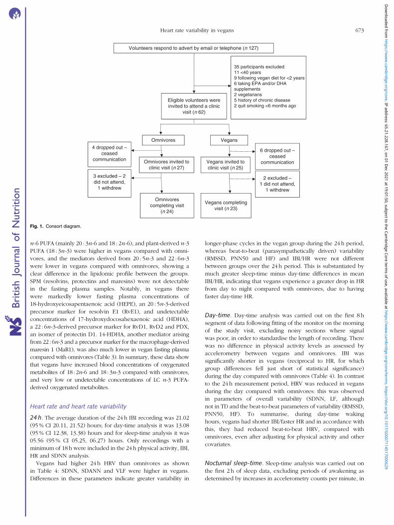
Fig. 1 shows the flow of participants through the study. Subjectcharacteristics of the forty-seven participants who completedthe study are presented in Table 1. The mean ages of vegans(eight men, fifteen women) and omnivores (twelve men, twelvewomen) were 49 (SD 8) and 54 (SD 9) years, respectively, andthere were no significant differences in mean age or BMI, or
distributions of sex between groups, although the sex dis-tributions were not fully balanced across groups. Furthermorethere were no significant differences in other markers of bodycomposition (% body fat, WC). Seated resting HR was onaverage 7 beats per minute higher and systolic blood pressurewas 7mmHg lower in vegans compared with omnivores; therewas no difference in mean seated diastolic blood pressure(Table 1). Fasting serum total and LN serum LDL-cholesterolconcentrations were lower in vegans compared with omni-vores, but there were no differences in mean fasting plasmaglucose, LN serum TAG, serum HDL-cholesterol, serum vitaminB12, 25-hydroxy vitamin D or IL-6 concentrations, nor blood Hbconcentrations, indicating that the vegan group did not differ invitamin D status and were likely to be taking dietary vitamin B12
supplements. Analysis of FFQ (Table 1) showed that 61% ofvegans reported taking vitamin B12 supplements, and suggestedthat vegans and omnivores had comparable total energy andpercentage of energy (%E) as fat intakes. Omnivores reportedsignificantly higher protein (%E), SFA (%E) and food-derivedvitamin B12 (μg) intakes and vegans had significantly highercarbohydrate (%E), total PUFA (%E) and 18 : 2n-6 (g) intakes.There were no differences in reported 18 : 3n-3 (g) intakes. Asexpected, vegans reported no dietary intake of 20 : 5n-3 and22 : 6n-3, hence omnivores obtained significantly higher intakeof these fatty acids, with estimated median intakes of 0·14(interquartile range (IQR) 0·09, 0·24) and 0·45 (IQR 0·30,0·81) g/d for 20 : 5n-3 and 22 : 6n-3, respectively. A subset(twelve omnivore and eight vegan participants) completed4-d food diaries (data not shown); analyses of these supportedthe FFQ data.
Fatty acid and lipid mediator profiles
Vegans had a significantly higher proportion of plasma anderythrocyte 18 : 2n-6, plasma 18 : 3n-3 and erythrocyte 20 : 3 n-6compared with omnivores (Table 2). Both whole plasma anderythrocyte membrane proportions of 20 : 5n-3 and 22 : 6n-3,plasma palmitic acid (16 : 0), and erythrocyte docosapentaenoicacid n-3 (22 : 5n-3) and palmitoleic acid (16 : 1n-7) were sig-nificantly lower in vegans compared with omnivores. Veganshad a significantly lower Omega-3 Index, with a geometricmean of 2·7% compared with 5·4% in omnivores, althoughboth groups would be considered below the proposedOmega-3 Index cut-off of >8% for optimal CVD protection(8).Erythrocyte 18 : 2n-6:18 : 3n-3 ratios were inversely correlatedwith erythrocyte 20 : 5n-3 contents in vegans (r −0·541,P= 0·008, n 23), but not 22 : 5n-3 or 22 : 6n-3 contents; with nosignificant correlations in the erythrocyte lipids of omnivores. Inplasma, the ratio of 18 : 2n-6:18 : 3n-3 was inversely correlatedwith plasma 22 : 5n-3 (r −0·576, P= 0·004, n 23) and 22 : 6n-3(r −0·498, P= 0·016, n 23) in vegans and plasma 22 : 5n-3 onlyin omnivores (r −0·474, P= 0·019, n 24).
Table 3 shows n-3 and n-6 PUFA-derived lipid mediatorsevaluated in the fasting plasma of the two study groups.A complete diagrammatic list of all lipid mediators includedin the analysis protocol, including those that were and werenot detectable in the plasma of this study population is givenin Fig. 2(a) and (b). In general, the lipid mediators derived from
672 A. M. Pinto et al.
Dow
nloaded from https://w
ww
.cambridge.org/core . IP address: 65.21.228.167 , on 01 D
ec 2021 at 19:01:50 , subject to the Cambridge Core term
s of use, available at https://ww
w.cam
bridge.org/core/terms . https://doi.org/10.1017/S0007114517000629
n-6 PUFA (mainly 20 : 3n-6 and 18 : 2n-6), and plant-derived n-3PUFA (18 : 3n-3) were higher in vegans compared with omni-vores, and the mediators derived from 20 : 5n-3 and 22 : 6n-3were lower in vegans compared with omnivores, showing aclear difference in the lipidomic profile between the groups.SPM (resolvins, protectins and maresins) were not detectablein the fasting plasma samples. Notably, in vegans therewere markedly lower fasting plasma concentrations of18-hydroxyeicosapentaenoic acid (HEPE), an 20 : 5n-3-derivedprecursor marker for resolvin E1 (RvE1), and undetectableconcentrations of 17-hydroxydocosahexaenoic acid (HDHA),a 22 : 6n-3-derived precursor marker for RvD1, RvD2 and PDX,an isomer of protectin D1. 14-HDHA, another mediator arisingfrom 22 : 6n-3 and a precursor marker for the macrophage-derivedmaresin 1 (MaR1), was also much lower in vegan fasting plasmacompared with omnivores (Table 3). In summary, these data showthat vegans have increased blood concentrations of oxygenatedmetabolites of 18 : 2n-6 and 18 : 3n-3 compared with omnivores,and very low or undetectable concentrations of LC n-3 PUFA-derived oxygenated metabolites.
Heart rate and heart rate variability
24 h. The average duration of the 24 h IBI recording was 21.02(95% CI 20.11, 21.52) hours; for day-time analysis it was 13.08(95% CI 12.38, 13.38) hours and for sleep-time analysis it was05.56 (95% CI 05.25, 06.27) hours. Only recordings with aminimum of 18 h were included in the 24 h physical activity, IBI,HR and SDNN analysis.Vegans had higher 24 h HRV than omnivores as shown
in Table 4: SDNN, SDANN and VLF were higher in vegans.Differences in these parameters indicate greater variability in
longer-phase cycles in the vegan group during the 24 h period,whereas beat-to-beat (parasympathetically driven) variability(RMSSD, PNN50 and HF) and IBI/HR were not differentbetween groups over the 24 h period. This is substantiated bymuch greater sleep-time minus day-time differences in meanIBI/HR, indicating that vegans experience a greater drop in HRfrom day to night compared with omnivores, due to havingfaster day-time HR.
Day-time. Day-time analysis was carried out on the first 8 hsegment of data following fitting of the monitor on the morningof the study visit, excluding noisy sections where signalwas poor, in order to standardise the length of recording. Therewas no difference in physical activity levels as assessed byaccelerometry between vegans and omnivores. IBI wassignificantly shorter in vegans (reciprocal to HR, for whichgroup differences fell just short of statistical significance)during the day compared with omnivores (Table 4). In contrastto the 24 h measurement period, HRV was reduced in vegansduring the day compared with omnivores: this was observedin parameters of overall variability (SDNN, LF, althoughnot in TI) and the beat-to-beat parameters of variability (RMSSD,PNN50, HF). To summarise, during day-time wakinghours, vegans had shorter IBI/faster HR and in accordance withthis, they had reduced beat-to-beat HRV, compared withomnivores, even after adjusting for physical activity and othercovariates.
Nocturnal sleep-time. Sleep-time analysis was carried out onthe first 2 h of sleep data, excluding periods of awakening asdetermined by increases in accelerometry counts per minute, in
Volunteers respond to advert by email or telephone (n 127)
35 participants excluded:11 <40 years9 following vegan diet for <2 years6 taking EPA and/or DHAsupplements2 vegetarians5 history of chronic disease2 quit smoking <6 months ago
Eligible volunteers wereinvited to attend a clinic
visit (n 62)
Omnivores Vegans
6 dropped out –ceased
communication
2 excluded –1 did not attend,
1 withdrew
Vegans completingvisit (n 23)
Omnivorescompleting visit
(n 24)
3 excluded – 2did not attend,
1 withdrew
4 dropped out –ceased
communication Omnivores invited toclinic visit (n 27)
Vegans invited toclinic visit (n 25)
Fig. 1. Consort diagram.
Heart rate variability in vegans 673
Dow
nloaded from https://w
ww
.cambridge.org/core . IP address: 65.21.228.167 , on 01 D
ec 2021 at 19:01:50 , subject to the Cambridge Core term
s of use, available at https://ww
w.cam
bridge.org/core/terms . https://doi.org/10.1017/S0007114517000629
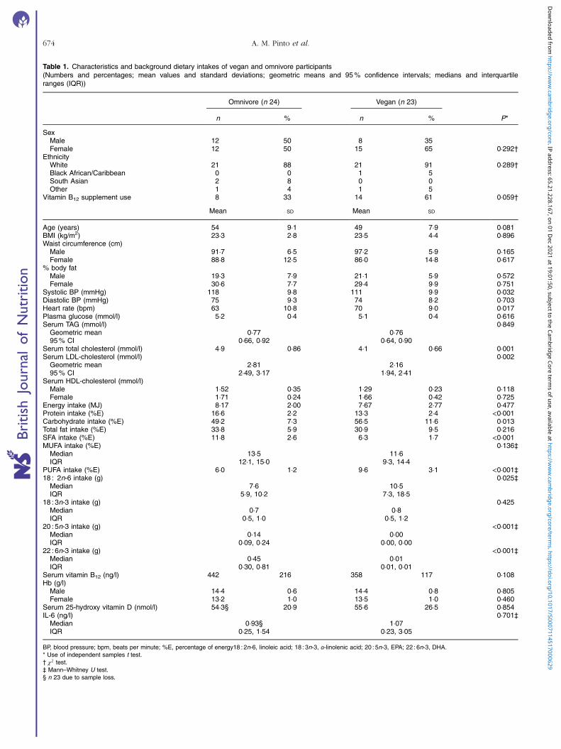
Table 1. Characteristics and background dietary intakes of vegan and omnivore participants(Numbers and percentages; mean values and standard deviations; geometric means and 95% confidence intervals; medians and interquartileranges (IQR))
Omnivore (n 24) Vegan (n 23)
n % n % P*
SexMale 12 50 8 35Female 12 50 15 65 0·292†
EthnicityWhite 21 88 21 91 0·289†Black African/Caribbean 0 0 1 5South Asian 2 8 0 0Other 1 4 1 5
Vitamin B12 supplement use 8 33 14 61 0·059†
Mean SD Mean SD
Age (years) 54 9·1 49 7·9 0·081BMI (kg/m2) 23·3 2·8 23·5 4·4 0·896Waist circumference (cm)
Male 91·7 6·5 97·2 5·9 0·165Female 88·8 12·5 86·0 14·8 0·617
% body fatMale 19·3 7·9 21·1 5·9 0·572Female 30·6 7·7 29·4 9·9 0·751
Systolic BP (mmHg) 118 9·8 111 9·9 0·032Diastolic BP (mmHg) 75 9·3 74 8·2 0·703Heart rate (bpm) 63 10·8 70 9·0 0·017Plasma glucose (mmol/l) 5·2 0·4 5·1 0·4 0·616Serum TAG (mmol/l) 0·849
Geometric mean 0·77 0·7695% CI 0·66, 0·92 0·64, 0·90
Serum total cholesterol (mmol/l) 4·9 0·86 4·1 0·66 0·001Serum LDL-cholesterol (mmol/l) 0·002
Geometric mean 2·81 2·1695% CI 2·49, 3·17 1·94, 2·41
Serum HDL-cholesterol (mmol/l)Male 1·52 0·35 1·29 0·23 0·118Female 1·71 0·24 1·66 0·42 0·725
Energy intake (MJ) 8·17 2·00 7·67 2·77 0·477Protein intake (%E) 16·6 2·2 13·3 2·4 <0·001Carbohydrate intake (%E) 49·2 7·3 56·5 11·6 0·013Total fat intake (%E) 33·8 5·9 30·9 9·5 0·216SFA intake (%E) 11·8 2·6 6·3 1·7 <0·001MUFA intake (%E) 0·136‡
Median 13·5 11·6IQR 12·1, 15·0 9·3, 14·4
PUFA intake (%E) 6·0 1·2 9·6 3·1 <0·001‡18 : 2n-6 intake (g) 0·025‡
Median 7·6 10·5IQR 5·9, 10·2 7·3, 18·5
18 : 3n-3 intake (g) 0·425Median 0·7 0·8IQR 0·5, 1·0 0·5, 1·2
20 : 5n-3 intake (g) <0·001‡Median 0·14 0·00IQR 0·09, 0·24 0·00, 0·00
22 : 6n-3 intake (g) <0·001‡Median 0·45 0·01IQR 0·30, 0·81 0·01, 0·01
Serum vitamin B12 (ng/l) 442 216 358 117 0·108Hb (g/l)
Male 14·4 0·6 14·4 0·8 0·805Female 13·2 1·0 13·5 1·0 0·460
Serum 25-hydroxy vitamin D (nmol/l) 54·3§ 20·9 55·6 26·5 0·854IL-6 (ng/l) 0·701‡
Median 0·93§ 1·07IQR 0·25, 1·54 0·23, 3·05
BP, blood pressure; bpm, beats per minute; %E, percentage of energy18 : 2n-6, linoleic acid; 18 : 3n-3, α-linolenic acid; 20 : 5n-3, EPA; 22 : 6n-3, DHA.* Use of independent samples t test.† χ2 test.‡ Mann–Whitney U test.§ n 23 due to sample loss.
674 A. M. Pinto et al.
Dow
nloaded from https://w
ww
.cambridge.org/core . IP address: 65.21.228.167 , on 01 D
ec 2021 at 19:01:50 , subject to the Cambridge Core term
s of use, available at https://ww
w.cam
bridge.org/core/terms . https://doi.org/10.1017/S0007114517000629

order to standardise the length of recording. Longer segmentswere not available for all participants and therefore were notincluded in the analysis. There were no significant differencesfor any of the parameters between groups:
Nocturnal sleep–time–day–time differencesin HR/IBI and beat–to–beat HRV.
Circadian changes are a key determinant of variability in HRover 24 h, measured as 24 h SDNN. Differences in meannocturnal sleep-time and day-time IBI are a significant factor inthe size of the SDNN value. As described above, the sleep-timeminus day-time differences in HR/IBI were statisticallysignificant, with the mean decrease in HR/increase in IBIfrom day-time to sleep-time being distinctly larger in vegans
Table 2. Plasma and erythrocyte fatty acid composition in vegan and omnivore participants (n 47)(Mean values and 95% confidence intervals; mean differences (vegan) and 95% confidence intervals (omnivore); geometric means and 95% confidenceintervals; medians and interquartile ranges (IQR))
Omnivore (n 24) Vegan (n 23) Difference between groups
Plasma and erythrocyte fatty acids (weight %) Mean 95% CI Mean 95% CI Mean difference 95% CI P*
Plasma16 : 0 20·8 20·4, 21·3 19·3 18·6, 20·0 −1·49 −2·31, −0·67 0·00116 : 1n-7 1·79 1·48, 2·10 1·11 0·91, 1·32 −0·67 −1·04, −0·31 0·00118 : 0 7·62 7·30, 7·94 7·60 7·15, 8·04 −0·02 −0·55, 0·50 0·93118 : 1n-9 18·5 17·6, 19·3 18·9 17·9, 19·8 0·37 −0·89, 1·62 0·55918 : 2n-6 27·1 26·0, 28·2 33·1 31·9, 34·4 6·06 4·43, 7·68 <0·00118 : 3n-3 0·006Geometric mean 0·53 0·71 1·34†95% CI 0·48, 0·59 0·59, 0·85 1·09, 1·64
20 : 3n-6 1·42 1·28, 1·55 1·42 1·28, 1·57 0·01 −0·18, 0·20 0·95220 : 4n-6 6·68 6·12, 7·25 6·55 5·94, 7·16 −0·13 −0·94, 0·68 0·74520 : 5n-3 <0·001Geometric mean 1·03 0·47 0·46†95% CI 0·79, 1·34 0·40, 0·55 0·34, 0·62
22 : 4n-6 0·036Geometric mean 0·20 0·23 1·14†95% CI 0·19, 0·21 0·21, 0·25 1·01, 1·28
22 : 5n-6 0·26 0·21, 0·30 0·20 0·15, 0·26 −0·05 −0·12, 0·02 0·14622 : 5n-3 0·59 0·53, 0·64 0·51 0·44, 0·59 −0·07 −0·16, 0·02 0·11322 : 6n-3 <0·001Geometric mean 2·23 0·91 0·41†95% CI 1·94, 2·57 0·80, 1·05 0·34, 0·49
Erythrocyte16 : 0 16·8 15·4, 18·2 17·6 16·7, 18·6 0·81 −0·87, 2·48 0·33716 : 1n-7 0·016‡Median 0·41 0·31 –
IQR 0·30, 1·57 0·21, 0·50 –
18 : 0 0·135Geometric mean 15·6 16·3 1·05†95% CI 14·9, 16·3 15·6, 17·1 0·99, 1·14
18 : 1n-9 15·7 15·1, 16·3 15·4 14·7, 16·2 −0·23 −1·15, 0·68 0·60918 : 2n-6 11·7 11·0, 12·3 13·3 12·5, 14·1 1·64 0·64, 2·64 0·00218 : 3n-3 0·610Geometric mean 0·34 0·32 0·92†95% CI 0·26, 0·45 0·27, 0·38 0·67, 1·27
20 : 3n-6 0·042Geometric mean 1·78 2·02 1·13†95% CI 1·64, 1·94 1·84, 2·22 1·01, 1·28
20 : 4n-6 15·9 14·9, 16·9 15·6 14·4, 16·9 −0·27 −1·82, 1·27 0·72520 : 5n-3 1·26 1·07, 1·45 0·67 0·52, 0·81 −0·59 −0·83, −0·36 <0·00122 : 4n-6 2·75 2·47, 3·03 3·83 3·50, 4·16 1·08 0·66, 1·50 <0·00122 : 5n-6 0·38 0·27, 0·49 0·52 0·40, 0·64 0·14 −0·02, 0·30 0·07822 : 5n-3 2·62 2·36, 2·88 2·15 1·94, 2·36 −0·47 −0·80, −0·15 0·00522 : 6n-3 <0·001Geometric mean 4·19 2·07 0·49†95% CI 3·63, 4·83 1·85, 2·32 0·41, 0·59
Omega-3 Index <0·001Geometric mean 5·42 2·71 0·50†95% CI 4·73, 6·20 2·40, 3·05 0·42, 0·60
16 : 0, palmitic acid; 16 : 1n-7, palmitoleic acid; 18 : 0, stearic acid; 18 : 1n-9, oleic acid; 18 : 2n-6, linoleic acid; 18 : 3n-3, α-linolenic acid; 20 : 3n-6, dihomo-γ-linolenic acid; 20 : 4n-6,arachidonic acid; 20 : 5n-3, EPA; 22 : 5n-3, docosapentaenoic acid n-3; 22 : 6n-3, DHA.
* P value obtained using independent samples t test.† Exponents of mean differences in Ln values (the ratio of the geometric mean in vegans:that in omnivores, with 95% CI of the geometric mean ratios).‡ Use of Mann–Whitney U test where data remained not normally distributed following LN transformation. Total plasma fatty acid concentrations were (geometric means with
95% CI): omnivores (1869mg/l, 1660, 2104; n 24), vegans (1998mg/l, 1755, 2274; n 23); there were no significant differences between groups.
Heart rate variability in vegans 675
Dow
nloaded from https://w
ww
.cambridge.org/core . IP address: 65.21.228.167 , on 01 D
ec 2021 at 19:01:50 , subject to the Cambridge Core term
s of use, available at https://ww
w.cam
bridge.org/core/terms . https://doi.org/10.1017/S0007114517000629

Table 3. Plasma concentrations of n-6 and n-3 PUFA-derived lipid mediators in vegan and omnivore participants (n 47)(Mean values and 95% confidence intervals; geometric means and 95% confidence intervals; medians and minimum and maximum values)
Omnivore (n 24) Vegan (n 23)
Compounds (ng/l) Mean 95% CI Mean 95% CI P*
n-6 PUFA derived20 : 3n-6-derived15-HETrE 51·82 36·94, 66·70 53·87 39·78, 67·96 0·53313,14-dihydro-15-keto PGE1 0·519
Geometric mean 17·45 13·9395% CI 9·52, 31·99 7·82, 24·78
13,14-dihydro PGE1 0·001†Median 0·000 0·000Minimum and maximum values 0·000, 4·332 0·000, 76·928
18 : 2n-6 derived9-HODE <0·001
Geometric mean 2433 504595% CI 1982, 2988 4067, 6260
9 OxoODE <0·001Geometric mean 477 99495% CI 398, 571 771, 1282
13-HODE <0·001Geometric mean 3320 653695% CI 2717, 4056 5483, 7791
13 OxoODE 335 291, 379 537 467, 606 <0·00112,13-EpOME 389 326, 451 769 622, 917 <0·00112,13-DiHOME 2820 2159, 3480 5544 4527, 6561 <0·0019,10-EpOME <0·001
Geometric mean 258 42695% CI 220, 303 351, 518
9,10-DiHOME <0·001Geometric mean 3199 740095% CI 2482, 4125 5910, 9267
Trans-EKODE <0·001Geometric mean 300 56095% CI 248, 362 425, 738
20 : 4n-6 derived6-keto PGF1α <0·001†
Median 8·20 0·000Minimum and maximum values 0·000, 59·14 0·000, 4·49
13,14-dihydro PGF2α <0·001Geometric mean 48·4 20·095% CI 34·57, 67·91 14·82, 26·97
13,14-dihydro-15-keto PGF2α 0·016†Median 0·000 0·000Minimum and maximum values 0·000, 25·56 0·000, 12·79
13,14-dihydro-15-keto PGE2 0·459Geometric mean 3·67 3·0795% CI 2·584, 5·217 2·172, 4·347
TXB2 0·968Geometric mean 10·3 10·595% CI 7·57, 14·14 6·08, 18·05
5-HETE 0·738Geometric mean 124 13295% CI 96·4, 160·4 103·2, 168
8-HETE 70·2 56·77, 83·57 74·5 62·33, 86·60 0·6269-HETE 0·478†
Median 12·82 0·000Minimum and maximum values 0·00, 70·99 0·000, 70·99
11-HETE 62·3 49·55, 75·13 59·7 49·68, 69·64 0·73612-HETE 0·580†
Median 146·48 143·6Minimum and maximum values 96·76, 1214·43 82·20, 1625·76
15-HETE 174 145, 203 186 158, 214 0·53320-HETE 0·896
Geometric mean 284 29095% CI 232, 348 229, 367
5,6-DHET 0·635Geometric mean 55·4 52·095% CI 47·09, 65·19 41·29, 65·37
676 A. M. Pinto et al.
Dow
nloaded from https://w
ww
.cambridge.org/core . IP address: 65.21.228.167 , on 01 D
ec 2021 at 19:01:50 , subject to the Cambridge Core term
s of use, available at https://ww
w.cam
bridge.org/core/terms . https://doi.org/10.1017/S0007114517000629
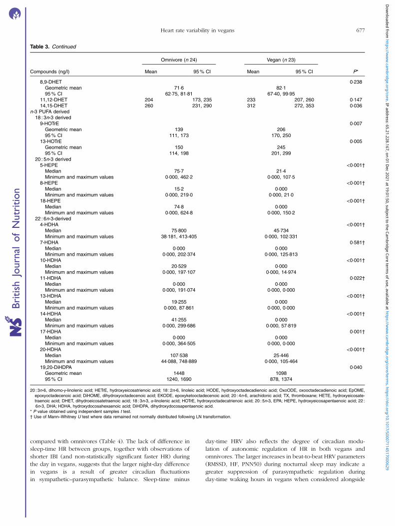
compared with omnivores (Table 4). The lack of difference insleep-time HR between groups, together with observations ofshorter IBI (and non-statistically significant faster HR) duringthe day in vegans, suggests that the larger night-day differencein vegans is a result of greater circadian fluctuationsin sympathetic–parasympathetic balance. Sleep-time minus
day-time HRV also reflects the degree of circadian modu-lation of autonomic regulation of HR in both vegans andomnivores. The larger increases in beat-to-beat HRV parameters(RMSSD, HF, PNN50) during nocturnal sleep may indicate agreater suppression of parasympathetic regulation duringday-time waking hours in vegans when considered alongside
Table 3. Continued
Omnivore (n 24) Vegan (n 23)
Compounds (ng/l) Mean 95% CI Mean 95% CI P*
8,9-DHET 0·238Geometric mean 71·6 82·195% CI 62·75, 81·81 67·40, 99·95
11,12-DHET 204 173, 235 233 207, 260 0·14714,15-DHET 260 231, 290 312 272, 353 0·036
n-3 PUFA derived18 : 3n-3 derived9-HOTrE 0·007
Geometric mean 139 20695% CI 111, 173 170, 250
13-HOTrE 0·005Geometric mean 150 24595% CI 114, 198 201, 299
20 : 5n-3 derived5-HEPE <0·001†
Median 75·7 21·4Minimum and maximum values 0·000, 462·2 0·000, 107·5
8-HEPE <0·001†Median 15·2 0·000Minimum and maximum values 0·000, 219·0 0·000, 21·0
18-HEPE <0·001†Median 74·8 0·000Minimum and maximum values 0·000, 624·8 0·000, 150·2
22 : 6n-3-derived4-HDHA <0·001†
Median 75·800 45·734Minimum and maximum values 38·181, 413·405 0·000, 102·331
7-HDHA 0·581†Median 0·000 0·000Minimum and maximum values 0·000, 202·374 0·000, 125·813
10-HDHA <0·001†Median 20·529 0·000Minimum and maximum values 0·000, 197·107 0·000, 14·974
11-HDHA 0·022†Median 0·000 0·000Minimum and maximum values 0·000, 191·074 0·000, 0·000
13-HDHA <0·001†Median 19·255 0·000Minimum and maximum values 0·000, 87·861 0·000, 0·000
14-HDHA <0·001†Median 41·255 0·000Minimum and maximum values 0·000, 299·686 0·000, 57·819
17-HDHA 0·001†Median 0·000 0·000Minimum and maximum values 0·000, 364·505 0·000, 0·000
20-HDHA <0·001†Median 107·538 25·446Minimum and maximum values 44·088, 748·889 0·000, 105·464
19,20-DiHDPA 0·040Geometric mean 1448 109895% CI 1240, 1690 878, 1374
20 : 3n-6, dihomo-γ-linolenic acid; HETrE, hydroxyeicosatrienoic acid; 18 : 2n-6, linoleic acid; HODE, hydroxyoctadecadienoic acid; OxoODE, oxooctadecadienoic acid; EpOME,epoxyoctadecenoic acid; DiHOME, dihydroxyoctadecenoic acid; EKODE, epoxyketooctadecenoic acid; 20 : 4n-6, arachidonic acid; TX, thromboxane; HETE, hydroxyeicosate-traenoic acid; DHET, dihydroeicosatetraenoic acid; 18 : 3n-3, α-linolenic acid; HOTrE, hydroxyoctadecatrienoic acid; 20 : 5n-3, EPA; HEPE, hydroxyeicosapentaenoic acid; 22 :6n-3, DHA; HDHA, hydroxydocosahexaenoic acid; DiHDPA, dihydroxydocosapentaenoic acid.
* P value obtained using independent samples t test.† Use of Mann–Whitney U test where data remained not normally distributed following LN transformation.
Heart rate variability in vegans 677
Dow
nloaded from https://w
ww
.cambridge.org/core . IP address: 65.21.228.167 , on 01 D
ec 2021 at 19:01:50 , subject to the Cambridge Core term
s of use, available at https://ww
w.cam
bridge.org/core/terms . https://doi.org/10.1017/S0007114517000629

9-OxoODE
13-OxoODE 9,10-
DiHOME
9-HODE
13-HODE 9,10-
EpOME 12,13-DiHOME
12,13-EpOME
LA18 : 2n – 6
Trans-EKODE
13,14dihydroPGE1
13,14dihydro-15-keto PGE1
PGE1DGLA
20 : 3n – 6
13,14dihydroPGF1�
13,14dihydro-15-keto PGF1�
PGF1�
PGD1
15-HETrE
8-iso-PGF2�
LTB4
5-HETE 8-
HETE
5,15-DiHETE HXA3
12-HETE
8,15-DiHETE
15-HETE
AA20 : 4n – 6
14(15)-EET
14,15-DHET
11(12)-EET
11,12-DHET
8,9-DHET
8(9)-EET
5(6)-EET
5,6-DHET
9-HETE
20-HETE
PGI2
6-ketoPGF1�
PGF2�
11-HETE
TXB2
PGE2PGD2
15-ketoPGE2
13,14-dihydro-15-keto PGE2
15d PGJ2
Δ12 PGJ2
PGJ2
13,14-dihydroPGF2�
13,14-dihydro-15-keto PGF2�
9-HOTrE
13-HOTrE
ALA18 : 3n – 3
TXB3
PGE3PGF3�
PGD3
EPA20 : 5n – 3
5-HEPE
12-HEPE
15-HEPE
16(17)-EpDPE
19(20)-EpDPE
4-HDHA
7-HDHA
11-HDHA
14-HDHA MaR1
PDX
RvD1
RvD2
17-HDHA
13-HDHA20-
HDHA
10-HDHA
11-HEPE
8-HEPE
9-HEPE
18-HEPE
RvE1
DHA22 : 6n – 3
19,20-DiHDPA
(a)
(b)
Fig. 2. (For caption see following page)
678 A. M. Pinto et al.
Dow
nloaded from https://w
ww
.cambridge.org/core . IP address: 65.21.228.167 , on 01 D
ec 2021 at 19:01:50 , subject to the Cambridge Core term
s of use, available at https://ww
w.cam
bridge.org/core/terms . https://doi.org/10.1017/S0007114517000629

the shorter mean day-time IBI in this group compared withomnivores.
Discussion
Low HRV is associated with mortality after a myocardialinfarction(21,40,41) and risk of cardiac events in the generalpopulation(23). Associations between increased n-3 PUFAconsumption and higher HRV(42–45), and lower HR(16), suggeststhat populations with very low n-3 PUFA tissue status might beat greater risk of arrhythmic events or sudden cardiac death.Vegetarians/vegans in the Adventist Health Study(46), EuropeanProspective Investigation into Cancer and Nutrition (EPIC)-Oxford cohort(47) and five combined cohorts(48) have beenreported to have lower risk of CHD than non-vegetarians.However, a recent study of two combined population cohorts(EPIC-Oxford and the earlier Oxford Vegetarian Study cohort)reported similar rates of all-cause mortality and no cleardifferences between vegans and comparable regular meat-eaters, fish-eaters and vegetarians in mortality from CHD up tothe age of 90 years(49), despite the fact that vegan populationshave lower CHD risk factors such as blood pressure(14,15),plasma lipids(12,13) and lower BMI (10,11) compared with popu-lations that eat foods of animal origin. Although the latterfindings do not preclude a lower risk of premature CHD invegans, the notion that cardiovascular health of elderly vegansmight be further optimised by increased intakes of dietary LCn-3 PUFA remains a possibility.We hypothesised that a population with low tissue LC n-3
PUFA status would have higher HR and lower HRV, and veganswere chosen as a clearly defined group that could be consideredas a model to test this hypothesis. As expected, we observedmarked differences between vegans and omnivores in their tis-sue n-3 PUFA status, as represented by erythrocyte lipid fattyacid composition (an indicator of longer-term PUFA intake dueto the 4-month lifespan of an average erythrocyte(50)). Thesefindings were supported by differences in the plasma fatty acidcomposition and self-reported dietary LC n-3 PUFA intakes. Theaverage erythrocyte Omega-3 Index in the omnivore group waslower than indices reported previously for a meat- and fish-eating UK population(51–53), but differences between the groupsstudied here were clear-cut. Inverse relationships were observedbetween erythrocyte 18 : 2n-6:18 : 3n-3 ratios and erythrocyte20 : 5n-3 in the vegan group. This supports existing evidence thathigher dietary intakes of 18 : 2n-6, an n-6 PUFA which is abun-dant in omnivore diets but even more so in vegan/vegetariandiets(5), may inhibit conversion of 18 : 3n-3 to LC n-3 PUFA(54).The observed group differences in HR and HRV were more
complex than hypothesised, mainly due to divergence in
night/day differences. Differences in all primary outcomevariables – HR/IBI, SDNN (overall HRV), and RMSSD (beat-to-beat HRV) – were observed between groups but the natureof the difference depended on whether analysis was carried outover the full 24 h or only during day-time waking hours. In linewith the hypothesis, mean day-time HR was higher/IBI shorterand overall (SDNN) and beat-to-beat HRV (RMSSD, PNN50%,HF) was lower in vegans, even following adjustment for phy-sical activity during the same 8 h period. These observationsmight indicate that low n-3 status could lead to either a pre-dominance of sympathetic regulation, a greater withdrawal ofparasympathetic activity, or possibly, due to depletion of LC n-3PUFA in cardiomyocyte membranes, there is a greater stimu-lation of pacemaker activity despite a normal level of sympa-thetic neural transmission during waking hours. However, it isalso possible that the differences in HRV observed in vegansand omnivores are unrelated to LC n-3 PUFA tissue status; thiswould require investigation with a dietary intervention trial.A recent review on n-3 fatty acids and effects on HR and HRVhas argued that, according to evidence from animal models, it ismore likely that 22 : 6n-3 is acting to reduce HR via modulationof pacemaker activity rather than changes in cardiac autonomicneural regulation(18), although the role of the 22 : 6n-3-derivedSPM, neuroprotectin D1 (PD1), in protecting the nervoussystem from inflammation-related injury shows that 22 : 6n-3-dependent physiological mechanisms exist in synapses andneural circuits in order to sustain neuronal function(55,56). Thestable precursor to PD1 and RvD1, 17-HDHA , was not detectedin the fasting plasma of any vegan subjects, whereas nine out oftwenty-four omnivores had detectable concentrations. Therewere also marked differences in concentrations of LC n-3 PUFA-derived precursor markers to RvE1 (from 18-HEPE) and MaR1(from 14-HDHA). Venous blood plasma concentrations of lipidmediators in whole fasting plasma are likely to be an insensitivemarker of capacity for autacoid release and activity in specificsites of inflamed tissue. Nevertheless, higher circulating plasmaconcentrations of SPM precursor markers may indicate ease ofbioavailability for conversion to SPM at times of need, whichpresents clear functional implications for populations with lowtissue 20 : 5n-3 and 22 : 6n-3 stores.
Although the vegan group were not deficient in other nutri-ents that are related to HRV, such as vitamin D(57) and vitaminB12
(58), the nature of the study design means that we cannotexclude the influence of other dietary or lifestyle factorsassociated with the vegan lifestyle. The vegans reported almosthalf the intake of SFA (%E) as the omnivores, in agreementwith results reported in larger vegan populations(59), andcorrespondingly lower amounts of 16 : 0 as a proportion oftotal plasma fatty acids and lower serum concentrations of
Fig. 2. (For figure see previous page) (a) Schematic outline of oxygenated species derivatives of n-6 PUFA, linoleic acid (LA; 18 : 2n-6), dihomo-γ-linolenic acid (DGLA;20 : 3n-6) and arachidonic acid (AA; 20 : 4n-6), analysed in blood plasma of vegans and omnivores. 20: 3n-6 (DGLA)-, 18: 2n-6 (LA)- and 20: 4n-6 (AA)-derived lipidmediators assayed in study participants’ plasma. , Higher in vegans, , higher in omnivores, , no difference, , not detected or below the limit of detection.OxoODE, oxooctadecadienoic acid; HODE, hydroxyoctadecadienoic acid; EpOME, epoxyoctadecenoic acid; DiHOME, dihydroxyoctadecenoic acid; EKODE,epoxyketooctadecenoic acid; TX, thromboxane; HETE, hydroxyeicosatetraenoic acid; EET, epoxyeicosatetraenoic acid; DHET, dihydroeicosatetraenoic acid; HETrE,hydroxyeicosatrienoic acid; DiHETE,dihydroxyeicosatetraenoic acid; LT, leukotriene; HX, hepoxilin. (b) Schematic outline of oxygenated species derivatives of n-3PUFA, α-linolenic acid (ALA; 18 : 3n-3), EPA (20 : 5n-3) and DHA (22 : 6n-3), analysed in blood plasma of vegans and omnivores. 18 : 3n-3 (ALA)-, 20 : 5n-3 (EPA)- and22 : 6n-3 (DHA)-derived lipid mediators assayed in study participants’ plasma. , Higher in vegans, , higher in omnivores, , no difference, , not detected or belowthe limit of detection. HOTrE, hydroxyoctadecatrienoic acid; TX, thromboxane; HEPE, hydroxyeicosapentaenoic acid; DiHDPA,dihydroxydocosapentaenoic acid;Rv, resolvin; EpDPE, epoxydocosapentaenoic acid; HDHA, hydroxydocosahexaenoic acid; MaR, maresin; PD, protectin D.
Heart rate variability in vegans 679
Dow
nloaded from https://w
ww
.cambridge.org/core . IP address: 65.21.228.167 , on 01 D
ec 2021 at 19:01:50 , subject to the Cambridge Core term
s of use, available at https://ww
w.cam
bridge.org/core/terms . https://doi.org/10.1017/S0007114517000629

Table 4. Physical activity, heart rate and heart rate variability parameters of vegan and omnivore participants over 24 h, day-time and sleep-time,with sleep – day differences (n 47)*(Estimated marginal means and 95% confidence intervals; geometric means and 95% confidence intervals; medians and interquartile ranges (IQR))
Omnivore (n 24) Vegan (n 22†)
Mean 95% CI Mean 95% CI P‡
24-h measurementActivity, IBI and HRPhysical activity (cpm)§ 0·181
Geometric mean 236 28595% CI 196, 284 233, 348
IBI (ms)§ 845 798, 892 811 760, 861 0·336HR (bpm)§ 75 71, 79 78 74, 83 0·210
Time-domain HRV parametersTI 38 33, 42 41 36, 46 0·294SDNN (ms)§ 145 129, 162 172 154, 189 0·039SDANN (ms) 128 114, 143 155 139, 170 0·018RMSSD (ms) 0·905
Geometric mean 35 3595% CI 31, 40 30, 40
PNN50 (%) 0·299Geometric mean 8·9 7·095% CI 6·5, 12·2 5·0, 9·7
Frequency-domain HRV parametersLF (ms2) 0·130
Geometric mean 971 76195% CI 786, 1198 608, 953
HF (ms2) 0·567Geometric mean 350 30995% CI 262, 467 227, 421
VLF (ms2) 0·022Geometric mean 12 619 17 98095% CI 10351, 15 379 14 547, 22 204
Non-linear methodsSD1:SD2 (Poincaré ratio) 0·13 0·11, 0·14 0·11 0·10, 0·12 0·051
Day-time (8 h) measurementActivity, IBI and HRPhysical activity (cpm) 0·935
Geometric mean 437 44395% CI 347, 552 345, 570
IBI (ms) 787 745, 830 721 675, 766 0·039HR (bpm) 80 76, 84 86 81, 91 0·062
Time-domain HRV parametersTI 31 28, 34 27 24, 31 0·135SDNN (ms) 121 109, 132 101 89, 113 0·021SDANN (ms) 100 89, 111 84 73, 96 0·056RMSSD (ms) 34 30, 38 25 20, 30 0·009PNN50 (%) 0·001
Geometric mean 6·6 2·795% CI 4·6, 9·4 1·8, 3·9
Frequency-domain HRV parametersLF (ms2) 908 785, 1032 628 495, 761 0·004HF (ms2) 0·002
Geometric mean 260 13595% CI 198, 342 100, 181
VLF (ms2) 0·078Geometric mean 8325 647095% CI 6894, 10 052 5277, 7929
Non-linear methodsSD1:SD2 (Poincaré ratio) 0·15 0·13, 0·16 0·13 0·11, 0·15 0·125
Sleep-time (2 h) measurementIBI and HRIBI (ms) 965 903, 1026 991 925, 1056 0·568HR (bpm) 64 61, 68 62 59, 66 0·475
Time-domain HRV parametersTI 19 16, 22 18 16, 21 0·762SDNN (ms) 78 68, 88 85 75, 96 0·324SDANN (ms) 0·717
Geometric mean 42 4495% CI 36, 51 37, 54
680 A. M. Pinto et al.
Dow
nloaded from https://w
ww
.cambridge.org/core . IP address: 65.21.228.167 , on 01 D
ec 2021 at 19:01:50 , subject to the Cambridge Core term
s of use, available at https://ww
w.cam
bridge.org/core/terms . https://doi.org/10.1017/S0007114517000629

LDL-cholesterol. However, these differences are less likely toexert a major influence on cardiac electrophysiology. Animalstudies have shown that PUFA feeding decreased vulnerabilityto arrhythmia compared with high SFA feeding without anyreduction in the proportion of membrane SFA, and high-MUFAfeeding did not reduce arrhythmia compared with high-SFAdiets(60). This suggests that SFA membrane composition is not amajor determinant of vulnerability to arrhythmias and additionof LC n-3 PUFA (replacing mainly 18 : 1 and n-6 PUFA) might bethe most important determinant. In fact our small cross-sectional study showed that erythrocyte SFA proportions werenot different between groups and that vegans had lower day-time HRV, and therefore potentially a greater risk of arrhythmiaif there was also coronary atherosclerosis present, despite lowerSFA intake.There may be other explanations for higher HR and reduced
HRV in vegans that are not related to n-3 PUFA status and were
not measured as part of this study, for example, susceptibility topsychological stress (although reduced self-reported stressand anxiety has been observed in 109 vegans compared with228 omnivores(61)), and job-related activities, and possiblyfrequency/duration of using a bicycle (which would not havebeen detected by accelerometry). The effects of physicalactivity on HRV depend on the type and intensity of activityinvolved, but higher parasympathetically regulated HRV para-meters are associated with greater levels of habitual physicalactivity(62). As parasympathetically regulated HRV parameterswere lower in vegans during the day-time, then it suggests thateither habitual physical activity levels were lower in vegans orsome other factor associated with vegan diet and lifestyle, suchas the depletion in tissue 20 : 5n-3 + 22 : 6n-3 content, counter-acted the effect of habitual physical activity levels.
No differences were observed between groups duringa standardised 2 h sleep period. Previous research from our
Table 4. Continued
Omnivore (n 24) Vegan (n 22†)
Mean 95% CI Mean 95% CI P‡
RMSSD (ms) 0·214Geometric mean 38 4495% CI 32, 45 37, 53
PNN50 (%) 0·826Geometric mean 10·9 11·795% CI 7·0, 16·9 7·3, 18·7
Frequency-domain HRV parametersLF (ms2) 0·925
Geometric mean 882 90295% CI 646, 1205 647, 1256
HF (ms2) 0·601Geometric mean 403 46495% CI 280, 580 315, 684
VLF (ms2) 0·304Geometric mean 2881 351995% CI 2215, 3744 2661, 3744
Non-linear methodsSD1:SD2 (Poincaré ratio) 0·740
Geometric mean 0·27 0·2895% CI 0·23, 0·32 0·24, 0·33
Sleep-time – day-time differencesIBI and HRIBI (ms) 183 139, 227 270 223, 317 0·012HR (bpm) −16 −19, −13 −24 −28, −20 0·003
Time-domain beat-to-beat HRV parametersRMSSD (ms) 8 1, 14 22 15, 29 0·006PNN50 (%) 6·4 0·9, 11·8 14·2 8·5, 20·1 0·058
Frequency-domain beat-to-beat HRV parametersHF (ms2) 0·095¶
Median 127 234IQR −12, 417 84, 988
IBI, interbeat interval (also known as RR interval), the time interval between R spikes of the QRS complex of the electrocardiogram; HR, heart rate; Cpm, counts per minute; bpm,beats per minute; HRV, heart rate variability; NN, normal-to-normal; TI, triangular index (total number of all NN intervals divided by the height of the histogram of all NN intervals);SDNN, standard deviation of all NN intervals (NN intervals, similar to R-R, but on normalised IBI data); SDANN, standard deviation of the averaged NN intervals, calculated from5min epochs; RMSSD, the square root of the mean of the sum of squares of differences between adjacent NN intervals; PNN50, percentage of adjacent NN intervals that differedby >50%; LF, low-frequency power; HF, high-frequency power; VLF, very-low-frequency power; SD1:SD2, the ratio of the SD of beat-to-beat IBI variability (SD1) against the SD oflong-term IBI variability (SD2).
* Adjusted for sex, age, BMI and 24 h activity for 24 h HRV and sleep-time – day-time HRV, or 8 h activity for 8 h day-time, and adjusted for sex, age and BMI only for sleep-time.Sleep-time – day-time represents HR/IBI and beat-to-beat HRV during a standardised 2 h nocturnal sleep period minus a standardized 8 h day-time period, to indicate thedifference between night and day.
† Missing data from one subject due to unusable day-time HRV recording.‡ P value obtained using ANCOVA for normally distributed raw or LN transformed data (adjusted for sex, age, BMI and activity for 24h, day-time and sleep-time – day-time
differences, and adjusted for sex, age and BMI only for sleep-time), except for sleep-time – day-time differences in HF.§ Only recordings >18h included for 24 h physical activity, SDNN, IBI and HR data analysis, n 21 for omnivores and n 19 for vegans.¶ Use of an unadjusted non-parametric test, the Mann–Whitney U test, where data remained not normally distributed following LN transformation.
Heart rate variability in vegans 681
Dow
nloaded from https://w
ww
.cambridge.org/core . IP address: 65.21.228.167 , on 01 D
ec 2021 at 19:01:50 , subject to the Cambridge Core term
s of use, available at https://ww
w.cam
bridge.org/core/terms . https://doi.org/10.1017/S0007114517000629

group showed increased longer-phase HRV (SDANN and VLF)in a middle-aged population at moderate risk of CVD duringnocturnal sleep following 12 months fish oil supplementation atdoses of 0·45–1·8 g/d LC n-3 PUFA compared with a refinedolive oil placebo(31). Consistent with this, fish consumption waspositively related to VLF in a large cohort of older adults(25).Low VLF is associated with increased risk of mortality post-myocardial infarction, particularly arrhythmic death(40). SinceSDANN and VLF represent slowly changing periodic variabilityin HR in response to thermoregulatory and hormonal shifts thatmay particularly occur during sleep, then it is likely that the 2 hstandardised period in the current study was too short todetect longer-phase differences in HRV between vegans andomnivores during sleep.Contrasting observations were made for longer-phase
components of 24 h HRV, which represent changes in HRover sustained periods in response to periodic fluctuations inneurohormonal and circadian physiology rather than beat-to-beat variability. These components of HRV (SDNN, SDANN andVLF) were higher, and Poincaré ratio was lower, over 24 h invegans compared with omnivores; this may represent morepronounced neurohormonal rhythms in vegans, or they mayjust reflect the higher HR and reduced HRV experienced byvegans during waking hours relative to sleep-time due toreasons discussed above.Sub-clinical markers of inflammation have been linked to risk
of cardiovascular events, vascular inflammation being the key,self-amplifying component of atherogenesis(63–65). Resolution ofacute inflammatory responses is a critical, programmed factor intissue repair and prevention of further pathological changes totissues. SPM derived from LC n-3 PUFA take over from theinitiating lipid mediators, prostaglandins and leukotrienes, duringthe neutrophil-monocyte sequence, and play a functional role inending acute inflammatory events by inhibition of neutrophilinflux to the site of trauma, counter-regulating pro-inflammatorycytokines, and stimulating resolving macrophages to clear theproducts of the inflammatory response, thereby allowing theinjured area to heal(66). In theory, low tissue availability of 20 :5n-3, 22 : 5n-3 and 22 : 6n-3 could compromise resolution ofacute inflammatory events increasing risk of chronic inflamma-tion, although this is purely speculative at present. Increasedcirculating concentrations of RvE1 and precursor markers ofresolvins (18-HEPE, 17-HDHA), and maresins (14-HDHA) havebeen demonstrated following n-3 PUFA supplementation(67), thesame precursor markers that were found to be different in ourcomparison of vegans and omnivores. Our data show that apopulation with no dietary intake of marine n-3 PUFA havemuch lower or zero fasting plasma concentrations of these SPMprecursor markers. It is not known whether individuals with lown-3 status have increased rates of 20 : 5n-3-/22 : 6n-3-derivedmediator turnover as an adaptive mechanism to avoid compro-mising SPM availability. If this were the case, then it would beexpected that having low pools of SPM precursors would haveno functional consequences in vegans. Future research in thisarea should address whether populations with low-n-3 status aremore at risk of having a pro-inflammatory profile.Vegans had greater concentrations of 18 : 3n-3- and 18 :
2n-6-derived lipid mediators that have a variety of deleterious
and cytoprotective effects(68,69). In the case of 18 : 2n-6, this islikely to be due to higher dietary intakes, as supported by FFQestimates, proportions of total plasma fatty acids, and incor-poration into erythrocyte membrane lipids(68,70). Althoughplasma 18 : 3n-3 proportions of total fatty acids were higher invegans, reported dietary intakes were not different; however,FFQ estimates of intakes are likely to underestimate true intakesdue to incomplete food composition data. Vegans also hadlower concentrations of markers of AA-derived prostanoidproduction (6-keto PGF1α – a marker of PGI2 synthesis, and13,14-dihydro PGF2α,/13,14-dihydro-15-keto PGF2α – markers ofPGF2α production). There were no differences between groupsfor a range of AA-derived LOX-catalysed mediators (HETE),suggesting that the lipid mediator profile of vegans may notnecessarily be entirely pro-inflammatory relative to omnivores.Few of these lipid mediators have been fully characterisedregarding their functional effects, and evidence in animal andcell models to date suggests that 18 : 2n-6- and 20 : 4n-6-derivedlipid mediators comprise a complex array of diverse bioactivemolecules that induce a range of physiological effects in varioustissues(71–73).
Previous work has also demonstrated that circulating pro-inflammatory cytokines may be reduced by fish oil supple-mentation, as reviewed by Calder(74,75). We included a measureof low grade inflammation, IL-6, in our comparison betweenvegans and omnivores, but found no differences betweengroups. However, this does not necessarily indicate that thereare no differences between groups in their capacity to inhibit orresolve acute inflammatory events since circulating cytokineconcentrations have limited utility as biomarkers of inflamma-tion that may be occurring in localised areas of tissue. Previousstudies have shown that serum IL-6 concentrations wereinversely correlated with HRV in men with renal disease(76), themetabolic syndrome(77) and young healthy subjects(78),although not not all studies agree(79). Down-regulation ofinflammatory cytokine gene expression plus increasedproduction of pro-resolving lipid mediators are two potentialmechanisms whereby cardiac function might possibly bepreserved by increased 20 : 5n-3 and 22 : 6n-3 intakes, byreducing inflammatory tissue damage in the brain andautonomic nerves, and also in the heart tissue itself.
Limitations of the present study
The cross-sectional design limits our findings to beingexploratory in nature and the associations between low n-3status and reduced HRV require confirmation by a randomisedcontrolled trial of 20 : 5n-3 + 22 : 6n-3 supplementation in apopulation with an Omega-3 Index of <3%. The samplepopulation size is small and although statistical power calcula-tions were conducted for the primary HRV outcomes, the studymay be underpowered to detect group differences in othermore variable outcomes such as beat-to-beat HR. Multiplestatistical testing was carried out to explore group differences inshort and long term, and time and frequency-domain HRV,increasing the risk of generating false-positive results. There isno agreed upon method for correcting statistical analyses thatinvolve the full set of HRV measures, but the data set represents
682 A. M. Pinto et al.
Dow
nloaded from https://w
ww
.cambridge.org/core . IP address: 65.21.228.167 , on 01 D
ec 2021 at 19:01:50 , subject to the Cambridge Core term
s of use, available at https://ww
w.cam
bridge.org/core/terms . https://doi.org/10.1017/S0007114517000629

groupings of related outcomes rather than a large collection ofdisparate variables. The data presented here are consistentwhen comparing variables that represent similar physiologicalphenomenon. For example, there are two time-domain(RMSSD, pNN50) and one frequency-domain (HF) parametersof beat-to-beat variability. These are all vagally regulated and allshow consistently that day-time beat-to-beat HRV is lower invegans compared with omnivores. Therefore, although type Ierrors cannot be ruled out with complete certainty, it is reas-suring that statistically significant differences between groupsare supported by analogous parameters. The stated aim was tomatch groups for age, sex and BMI, but matching for sex wasnot wholly achieved. Any influence of this imbalance in sexdistribution on HRV results was minimised by adjusting for age,sex and BMI, in addition to activity levels for 24 h and day-timeHRV, in the statistical model. Technical problems in obtaininggood quality sleep-time HRV data limited the standardisedduration of nocturnal HRV to 2 h which may have led to effectson longer phase HRV parameters being missed. However, thefact that HRV was lower and mean IBI was shorter in vegansonly during the day, and not over the whole 24 h period, sug-gests that there may be a diet-mental stress interaction duringwaking hours that resulted in a greater degree of sympatheticnervous system activity relative to parasympathetic activity.Future studies could investigate this further by measuringHRV responses under controlled mental stress conditions inpopulations with very low Omega-3 Indices compared withpopulations with optimum Omega-3 Indices.
Summary
The differences observed in parameters of cardiac electro-physiology and circulating lipid mediator concentrationsbetween vegans and omnivores may contribute to the sumeffect of diet and lifestyle on CVD risk. The lower availability ofLC n-3 PUFA-derived lipid mediators in vegans may influenceanti-inflammatory capacity, although other differences in LA-and ALA-derived mediators feed into an array of disparateinflammatory pathways and the sum effect is difficult to predict.Crucially, this study presents novel information on associationsbetween free-living, unsupplemented dietary PUFA intakeswith lipid mediator profiles in humans.
Acknowledgements
The authors thank Anne-Catherine Perz, MaryJo Searle andCatherine Kidd (King’s College London) for assistance in bloodsample processing.This research was supported by King’s College London and
received no specific grant from any funding agency, commer-cial or not-for-profit sectors.W. L. H., T. A. B. S. and A. M. P. conceived the research
question and devised the study. A. M. P. conducted the studyand analysed the data, with the assistance of H. A.-K. A. N.,A. C. K. and R. G. provided lipidomic analytical expertise.All authors contributed to writing and editing the manuscript.The authors declare that there are no conflicts of interest.
References
1. Sanders TA & Roshanai F (1992) Platelet phospholipid fattyacid composition and function in vegans compared withage- and sex-matched omnivore controls. Eur J Clin Nutr 46,823–831.
2. Sanders TA (2009) DHA status of vegetarians. ProstaglandinsLeukot Essent Fatty Acids 81, 137–141.
3. Brenna JT, Salem N, Sinclair AJ, et al. (2009) alpha-Linolenicacid supplementation and conversion to n-3 long-chainpolyunsaturated fatty acids in humans. ProstaglandinsLeukot Essent Fatty Acids 80, 85–91.
4. Burdge GC & Calder PC (2006) Dietary alpha-linolenic acidand health-related outcomes: a metabolic perspective. NutrRes Rev 19, 26–51.
5. Welch AA, Shakya-Shrestha S, Lentjes MA, et al. (2010) Dietaryintake and status of n-3 polyunsaturated fatty acids in apopulation of fish-eating and non-fish-eating meat-eaters,vegetarians, and vegans and the product-precursor ratio[corrected] of α-linolenic acid to long-chain n-3 poly-unsaturated fatty acids: results from the EPIC-Norfolk cohort.Am J Clin Nutr 92, 1040–1051.
6. Sanders TA & Younger KM (1981) The effect of dietarysupplements of omega 3 polyunsaturated fatty acids on thefatty acid composition of platelets and plasma choline phos-phoglycerides. Br J Nutr 45, 613–616.
7. Fokkema MR, Brouwer DA, Hasperhoven MB, et al. (2000)Short-term supplementation of low-dose gamma-linolenicacid (GLA), alpha-linolenic acid (ALA), or GLA plus ALA doesnot augment LCP omega 3 status of Dutch vegans to anappreciable extent. Prostaglandins Leukot Essent Fatty Acids63, 287–292.
8. Harris WS & Von Schacky C (2004) The Omega-3 Index:a new risk factor for death from coronary heart disease?Prev Med 39, 212–220.
9. Sanders TA (2014) Plant compared with marine n-3 fatty acideffects on cardiovascular risk factors and outcomes: what isthe verdict? Am J Clin Nutr 100, Suppl. 1, 453S–458S.
10. Rosell M, Appleby P, Spencer E, et al. (2006) Weight gain over5 years in 21,966 meat-eating, fish-eating, vegetarian, andvegan men and women in EPIC-Oxford. Int J Obes (Lond) 30,1389–1396.
11. Sanders TA, Ellis FR & Dickerson JW (1978) Studies of vegans:the fatty acid composition of plasma choline phosphoglycer-ides, erythrocytes, adipose tissue, and breast milk, and someindicators of susceptibility to ischemic heart disease in vegansand omnivore controls. Am J Clin Nutr 31, 805–813.
12. Thorogood M, Carter R, Benfield L, et al. (1987) Plasmalipids and lipoprotein cholesterol concentrations in peoplewith different diets in Britain. BMJ (Clin Res Ed) 295,351–353.
13. Bradbury KE, Crowe FL, Appleby PN, et al. (2014) Serumconcentrations of cholesterol, apolipoprotein A-I andapolipoprotein B in a total of 1694 meat-eaters, fish-eaters,vegetarians and vegans. Eur J Clin Nutr 68, 178–183.
14. Appleby PN, Davey GK & Key TJ (2002) Hypertension andblood pressure among meat eaters, fish eaters, vegetariansand vegans in EPIC-Oxford. Public Health Nutr 5,645–654.
15. Pettersen BJ, Anousheh R, Fan J, et al. (2012) Vegetarian dietsand blood pressure among white subjects: results from theAdventist Health Study-2 (AHS-2). Public Health Nutr 15,1909–1916.
16. Mozaffarian D, Geelen A, Brouwer IA, et al. (2005) Effect offish oil on heart rate in humans: a meta-analysis of rando-mized controlled trials. Circulation 112, 1945–1952.
Heart rate variability in vegans 683
Dow
nloaded from https://w
ww
.cambridge.org/core . IP address: 65.21.228.167 , on 01 D
ec 2021 at 19:01:50 , subject to the Cambridge Core term
s of use, available at https://ww
w.cam
bridge.org/core/terms . https://doi.org/10.1017/S0007114517000629

17. Verkerk AO, den Ruijter HM, Bourier J, et al. (2009) Dietaryfish oil reduces pacemaker current and heart rate in rabbit.Heart Rhythm 6, 1485–1492.
18. Billman GE (2013) The effects of omega-3 polyunsaturatedfatty acids on cardiac rhythm: a critical reassessment.Pharmacol Ther 140, 53–80.
19. Vaishnav S, Stevenson R, Marchant B, et al. (1994) Relationbetween heart rate variability early after acute myocardialinfarction and long-term mortality. Am J Cardiol 73, 653–657.
20. Anonymous (1996) Heart rate variability. Standards of mea-surement, physiological interpretation, and clinical use. TaskForce of the European Society of Cardiology and the NorthAmerican Society of Pacing and Electrophysiology. Eur Heart J17, 354–381.
21. Quintana M, Storck N, Lindblad LE, et al. (1997) Heart ratevariability as a means of assessing prognosis after acutemyocardial infarction. A 3-year follow-up study. Eur Heart J18, 789–797.
22. Lombardi F, Mäkikallio TH, Myerburg RJ, et al. (2001) Suddencardiac death: role of heart rate variability to identify patientsat risk. Cardiovasc Res 50, 210–217.
23. Tsuji H, Larson MG, Venditti FJ Jr, et al. (1996) Impact ofreduced heart rate variability on risk for cardiac events. TheFramingham Heart Study. Circulation 94, 2850–2855.
24. Christensen JH (2011) Omega-3 polyunsaturated fatty acidsand heart rate variability. Front Physiol 2, 84.
25. Mozaffarian D, Stein PK, Prineas RJ, et al. (2008) Dietary fishand omega-3 fatty acid consumption and heart rate variabilityin US adults. Circulation 117, 1130–1137.
26. Dyall SC & Michael-Titus AT (2008) Neurological benefits ofomega-3 fatty acids. Neuromolecular Med 10, 219–235.
27. Astarita G, Kendall AC, Dennis EA, et al. (2015) Targetedlipidomic strategies for oxygenated metabolites of poly-unsaturated fatty acids. Biochim Biophys Acta 1851, 456–468.
28. Serhan CN & Petasis NA (2011) Resolvins and protectins ininflammation resolution. Chem Rev 111, 5922–5943.
29. Serhan CN, Yang R, Martinod K, et al. (2009) Maresins: novelmacrophage mediators with potent antiinflammatory andproresolving actions. J Exp Med 206, 15–23.
30. Marcheselli VL, Mukherjee PK, Arita M, et al. (2010) Neuro-protectin D1/protectin D1 stereoselective and specific bindingwith human retinal pigment epithelial cells and neutrophils.Prostaglandins Leukot Essent Fatty Acids 82, 27–34.
31. Hall WL, Hay G, Maniou Z, et al. (2013) Effect of low doses oflong chain n-3 polyunsaturated fatty acids on sleep-time heartrate variability: a randomized, controlled trial. Int J Cardiol168, 4439–4442.
32. Bingham SA, Gill C, Welch A, et al. (1997) Validation ofdietary assessment methods in the UK arm of EPIC usingweighed records, and 24-hour urinary nitrogen and potassiumand serum vitamin C and carotenoids as biomarkers. Int JEpidemiol 26, Suppl. 1, S137–S151.
33. Sanders TA, Lewis F, Slaughter S, et al. (2006) Effect of varyingthe ratio of n-6 to n-3 fatty acids by increasing the dietaryintake of alpha-linolenic acid, eicosapentaenoic and doc-osahexaenoic acid, or both on fibrinogen and clotting factorsVII and XII in persons aged 45-70 y: the OPTILIP study. Am JClin Nutr 84, 513–522.
34. Brage S, Brage N, Franks PW, et al. (2005) Reliability andvalidity of the combined heart rate and movement sensorActiheart. Eur J Clin Nutr 59, 561–570.
35. Tarvainen MP, Niskanen J-P, Lipponen JA, et al. (2014) KubiosHRV – Heart rate variability analysis software. ComputerMethods Programs Biomed 113, 210–220.
36. Anonymous (1996) Heart rate variability: standards of mea-surement, physiological interpretation and clinical use. Task
Force of the European Society of Cardiology and the NorthAmerican Society of Pacing and Electrophysiology. Circula-tion 93, 1043–1065.
37. Lepage G & Roy CC (1986) Direct transesterification of allclasses of lipids in a one-step reaction. J Lipid Res 27,114–120.
38. Rosell MS, Lloyd-Wright Z, Appleby PN, et al. (2005) Long-chain n-3 polyunsaturated fatty acids in plasma in Britishmeat-eating, vegetarian, and vegan men. Am J Clin Nutr 82,327–334.
39. Massey KA & Nicolaou A (2013) Lipidomics of oxidizedpolyunsaturated fatty acids. Free Radic Biol Med 59, 45–55.
40. Bigger JT, Fleiss JL, Steinman RC, et al. (1992) Frequencydomain measures of heart period variability and mortality aftermyocardial infarction. Circulation 85, 164–171.
41. Vaishnav S, Stevenson R, Marchant B, et al. (1994) Relationbetween heart rate variability early after acute myocardialinfarction and long-term mortality. Am J Cardiol 73, 653–657.
42. Christensen JH, Christensen MS, Dyerberg J, et al. (1999)Heart rate variability and fatty acid content of blood cellmembranes: a dose-response study with n-3 fatty acids. Am JClin Nutr 70, 331–337.
43. Christensen JH, Skou HA, Madsen T, et al. (2001) Heart ratevariability and n-3 polyunsaturated fatty acids in patients withdiabetes mellitus. J Intern Med 249, 545–552.
44. Villa B, Calabresi L, Chiesa G, et al. (2002) Omega-3 fatty acidethyl esters increase heart rate variability in patients withcoronary disease. Pharmacol Res 45, 475.
45. Xin W, Wei W & Li XY (2013) Short-term effects of fish-oilsupplementation on heart rate variability in humans: a meta-analysis of randomized controlled trials. Am J Clin Nutr 97,926–935.
46. Fraser GE (2005) A comparison of first event coronary heartdisease rates in two contrasting California populations. J NutrHealth Aging 9, 53–58.
47. Crowe FL, Appleby PN, Travis RC, et al. (2013) Risk of hos-pitalization or death from ischemic heart disease amongBritish vegetarians and nonvegetarians: results from the EPIC-Oxford cohort study. Am J Clin Nutr 97, 597–603.
48. Key TJ, Fraser GE, Thorogood M, et al. (1999) Mortality invegetarians and nonvegetarians: detailed findings from acollaborative analysis of 5 prospective studies. Am J Clin Nutr70, 516S–524S.
49. Appleby PN, Crowe FL, Bradbury KE, et al. (2016) Mortality invegetarians and comparable nonvegetarians in the UnitedKingdom. Am J Clin Nutr 103, 218–230.
50. Patterson AC, Metherel AH, Hanning RM, et al. (2014) Thepercentage of DHA in erythrocytes can detect non-adherenceto advice to increase EPA and DHA intakes. Br J Nutr 111,270–278.
51. Sanders TAB, Hall WL, Maniou Z, et al. (2011) Effect of lowdoses of long-chain n-3 PUFAs on endothelial function andarterial stiffness: a randomized controlled trial. Am J Clin Nutr94, 973–980.
52. Walker CG, Browning LM, Mander AP, et al. (2014) Age andsex differences in the incorporation of EPA and DHA intoplasma fractions, cells and adipose tissue in humans. Br J Nutr111, 679–689.
53. Reidlinger DP, Darzi J, Hall WL, et al. (2015) How effective arecurrent dietary guidelines for cardiovascular disease preven-tion in healthy middle-aged and older men and women?A randomized controlled trial. Am J Clin Nutr 101,922–930.
54. Emken EA, Adlof RO & Gulley RM (1994) Dietary linoleic acidinfluences desaturation and acylation of deuterium-labeled
684 A. M. Pinto et al.
Dow
nloaded from https://w
ww
.cambridge.org/core . IP address: 65.21.228.167 , on 01 D
ec 2021 at 19:01:50 , subject to the Cambridge Core term
s of use, available at https://ww
w.cam
bridge.org/core/terms . https://doi.org/10.1017/S0007114517000629

linoleic and linolenic acids in young adult males. BiochimBiophys Acta 1213, 277–288.
55. Bazan NG, Molina MF & Gordon WC (2011) Docosahexaenoicacid signalolipidomics in nutrition: significance in aging, neu-roinflammation, macular degeneration, Alzheimer’s, and otherneurodegenerative diseases. Annu Rev Nutr 31, 321–351.
56. Bazan NG, Musto AE & Knott EJ (2011) Endogenous signalingby omega-3 docosahexaenoic acid-derived mediators sustainshomeostatic synaptic and circuitry integrity. Mol Neurobiol 44,216–222.
57. Mann MC, Hollenberg MD, Hanley DA, et al. (2015) VitaminD, the autonomic nervous system, and cardiovascular risk.Physiol Rep 3, e12349.
58. Sucharita S, Dwarkanath P, Thomas T, et al. (2014) Lowmaternal vitamin B12 status during pregnancy is associatedwith reduced heart rate variability indices in young children.Matern Child Nutr 10, 226–233.
59. Schmidt JA, Rinaldi S, Ferrari P, et al. (2015) Metabolic profilesof male meat eaters, fish eaters, vegetarians, and vegans fromthe EPIC-Oxford cohort. Am J Clin Nutr 102, 1518–1526.
60. McLennan PL (1993) Relative effects of dietary saturated,monounsaturated, and polyunsaturated fatty acids on cardiacarrhythmias in rats. Am J Clin Nutr 57, 207–212.
61. Beezhold B, Radnitz C, Rinne A, et al. (2015) Vegans report lessstress and anxiety than omnivores. Nutr Neurosci 18, 289–296.
62. Buchheit M, Simon C, Charloux A, et al. (2005) Heart ratevariability and intensity of habitual physical activity in middle-aged persons. Med Sci Sports Exerc 37, 1530–1534.
63. Ross R (1993) The pathogenesis of atherosclerosis: a pers-pective for the 1990s. Nature 362, 801–809.
64. Ridker PM, Cushman M, Stampfer MJ, et al. (1997) Inflam-mation, aspirin, and the risk of cardiovascular disease inapparently healthy men. N Engl J Med 336, 973–979.
65. Passacquale G, Di Giosia P & Ferro A (2016) The role ofinflammatory biomarkers in developing targeted cardio-vascular therapies: lessons from the cardiovascular inflam-mation reduction trials. Cardiovasc Res 109, 9–23.
66. Serhan CN (2014) Pro-resolving lipid mediators are leads forresolution physiology. Nature 510, 92–101.
67. Barden A, Mas E, Croft KD, et al. (2014) Short-term n-3 fatty acidsupplementation but not aspirin increases plasma proresolvingmediators of inflammation. J Lipid Res 55, 2401–2407.
68. Ramsden CE, Ringel A, Feldstein AE, et al. (2012) Loweringdietary linoleic acid reduces bioactive oxidized linoleic acidmetabolites in humans. Prostaglandins Leukot Essent FattyAcids 87, 135–141.
69. Kumar N, Gupta G, Anilkumar K, et al. (2016)15-Lipoxygenase metabolites of α-linolenic acid, [13-(S)-HPOTrE and 13-(S)-HOTrE], mediate anti-inflammatory effectsby inactivating NLRP3 inflammasome. Sci Rep 6, 31649.
70. Caligiuri SP, Rodriguez-Leyva D, Aukema HM, et al. (2016)Dietary flaxseed reduces central aortic blood pressure withoutcardiac involvement but through changes in plasma oxylipins.Hypertension 68, 1031–1038.
71. Choque B, Catheline D, Rioux V, et al. (2014) Linoleic acid:between doubts and certainties. Biochimie 96, 14–21.
72. Harizi H, Corcuff JB & Gualde N (2008) Arachidonic-acid-derived eicosanoids: roles in biology and immunopathology.Trends Mol Med 14, 461–469.
73. Nicolaou A, Mauro C, Urquhart P, et al. (2014) Poly-unsaturated fatty acid-derived lipid mediators and T cellfunction. Front Immunol 5, 75.
74. Calder PC (2015) Marine omega-3 fatty acids and inflamma-tory processes: effects, mechanisms and clinical relevance.Biochim Biophys Acta 1851, 469–484.
75. Calder PC (2010) Omega-3 fatty acids and inflammatoryprocesses. Nutrients 2, 355–374.
76. Psychari SN, Sinos L, Iatrou C, et al. (2005) Relations ofinflammatory markers to lipid levels and autonomic tone inpatients with moderate and severe chronic kidney disease andin patients under maintenance hemodialysis. Clin Nephrol 64,419–427.
77. Brunner EJ, Hemingway H, Walker BR, et al. (2002) Adreno-cortical, autonomic, and inflammatory causes of the metabolicsyndrome: nested case-control study. Circulation 106,2659–2665.
78. Sloan RP, McCreath H, Tracey KJ, et al. (2007) RR intervalvariability is inversely related to inflammatory markers: theCARDIA study. Mol Med 13, 178–184.
79. Owen N & Steptoe A (2003) Natural killer cell and pro-inflammatory cytokine responses to mental stress: associationswith heart rate and heart rate variability. Biol Psychol 63,101–115.
Heart rate variability in vegans 685
Dow
nloaded from https://w
ww
.cambridge.org/core . IP address: 65.21.228.167 , on 01 D
ec 2021 at 19:01:50 , subject to the Cambridge Core term
s of use, available at https://ww
w.cam
bridge.org/core/terms . https://doi.org/10.1017/S0007114517000629