A comparison of efficacy and safety of sedation between … · A comparison of efficacy and safety...
37
A comparison of efficacy and safety of sedation between dexmedetomidine- remifentanil and propofol-remifentanil during endoscopic submucosal dissection Namo Kim Department of Medicine The Graduate School, Yonsei University
-
Upload
hoangkhuong -
Category
Documents
-
view
230 -
download
0
Transcript of A comparison of efficacy and safety of sedation between … · A comparison of efficacy and safety...
A comparison of efficacy and safety of
sedation between dexmedetomidine-
remifentanil and propofol-remifentanil
during endoscopic submucosal dissection
Namo Kim
Department of Medicine
The Graduate School, Yonsei University
A comparison of efficacy and safety of
sedation between dexmedetomidine-
remifentanil and propofol-remifentanil
during endoscopic submucosal dissection
Directed by Professor Kyeong Tae Min
The Master's Thesis
submitted to the Department of Medicine
the Graduate School of Yonsei University
in partial fulfillment of the requirements for the
degree of Master of Medical Science
Namo Kim
June 2014
This certifies that the Master's Thesis
of Namo Kim is approved.
Thesis Supervisor : Kyeong Tae Min
---------------------------
Young Chul Yoo
--------------------------
Sang Kil Lee
The Graduate School
Yonsei University
June 2014
ACKNOWLEDGEMENTS
This dissertation could not be fructified without the
supports, advices, and encouragements of many people and
colleagues. I would like to express my gratitude to those who
have contributed to this work in various ways.
First of all, I’d like to thank all patients who generously
decided to participate in this study.
I would also like to express sincere gratitude to my
academic advisor, Professor Kyeong Tae Min, who, despite
my lack of knowledge and experience on a thesis at the start
of the masters degree program, provided the best tutelage and
support possible and allowed me to develop.
Likewise, I sincerely thank Professor Young Chul Yoo,
who gave me advice and guidance in many ways during the
writing of this paper and Professor Sang Kil Lee, who
encouraged and gave me constructive advice throughout my
years at graduate school.
Lastly, my family always gave me love, support and
encouragement, and I would like to thank all of them.

TABLE OF CONTENTS
ABSTRACT ........................................................................................... 1
I. INTRODUCTION ............................................................................... 3
II. MATERIALS AND METHODS ....................................................... 4
1. Patient and sedation protocol .......................................................... 4
2. Methods ......................................................................................... 10
가. Assessment of procedural performance .................................. 10
나. Assessment of patient safety ................................................... 12
다. Statistical analysis ................................................................... 12
III. RESULTS ....................................................................................... 13
IV. DISCUSSION ................................................................................ 22
REFERENCES ..................................................................................... 27
ABSTRACT (IN KOREAN) ................................................................ 30

LIST OF FIGURES
Figure 1. Changes of hemodynamic variables and SpO2
during ESD ........................................................................... 21
LIST OF TABLES
Table 1. Modified Observer’s Assessment of
Alertness/Sedation (MOAA/S) .............................................. 7
Table 2. Modified Aldrete scoring system ............................. 9
Table 3. Evaluation of gastric peristalsis ............................. 11
Table 4. Patient characteristics ............................................. 14
Table 5. Tumor characteristics ............................................. 15
Table 6. Drugs used for ESD ............................................... 17
Table 7. Efficacy of procedural performance ...................... 19
1
ABSTRACT
A comparison of efficacy and safety of sedation between
dexmedetomidine-remifentanil and propofol-remifentanil during
endoscopic submucosal dissection
Namo Kim
Department of Medicine
The Graduate School, Yonsei University
(Directed by Professor Kyeong Tae Min)
Introduction: Endoscopic submucosal dissection (ESD) requires
adequate sedation and pain control, for which the short acting drugs such
as propofol and remifentanil are recommended. Dexmedetomidine has
sedative and analgesic effects and suppresses gastrointestinal motility,
which might be critical during ESD. We compared the efficacy and
safety of sedation between dexmedetomidine-remifentanil and propofol-
remifentanil for use during ESD.
Method: Fifty-nine patients scheduled for ESD were randomly
2
allocated into a dexmedetomidine-remifentanil (DR) group or a
propofol-remifentanil (PR) group. To control patient anxiety,
dexmedetomidine or propofol was infused to maintain a score of 4–5 on
the Modified Observer’s Assessment of Alertness/Sedation scale.
Remifentanil was infused continuously at a rate of 6 μg/h/kg in both
groups. The ease of advancing the scope into the throat, gastric motility
grading, and satisfaction of the endoscopist and patient were assessed.
Hemodynamic variables and hypoxemic events were compared to
evaluate patient safety.
Results: Demographic data were comparable between the groups. The
hemodynamic variables and pulse oximetry values were stable during
the procedure in both groups despite a lower heart rate in the DR group.
No desaturation events occurred in either group. Although advancing the
scope into the throat was easier in the PR group (“very easy” 24.1% vs.
56.7%, P = 0.01), gastric motility was more suppressed in the DR group
(“no + mild” 96.6% vs. 73.3%, P = 0.013). The endoscopists felt that the
procedure was more favorable in the DR group (“very good + good”
100% vs. 86.7%, P = 0.042), whereas patient satisfaction scores were
comparable between the groups.
Conclusions: The efficacy and safety of dexmedetomidine and
remifentanil were comparable to propofol and remifentanil during ESD.
However, endoscopists favored dexmedetomidine perhaps due to lower
gastric motility.
---------------------------------------------------------------------------------------
Key words: dexmedetomidine, efficacy, safety, peristalsis, endoscopic
submucosal dissection
3
A comparison of efficacy and safety of sedation between
dexmedetomidine-remifentanil and propofol-remifentanil during
endoscopic submucosal dissection
Namo Kim
Department of Medicine
The Graduate School, Yonsei University
(Directed by Professor Kyeong Tae Min)
I. INTRODUCTION
Endoscopic submucosal dissection (ESD) is associated with greater and
longer patient discomfort and pain than conventional endoscopic procedures. It
is essential to address this issue with ESD1. Propofol has been widely used for
endoscopic procedures2, as it is safe and effective3, and is associated with
shorter recovery time and better sedation and amnesia level without an
4
increased risk for cardiopulmonary complications4 than other traditional
sedatives. However, in addition to the dose-dependent respiratory depression of
propofol, aspiration pneumonia occurs with an incidence of 2.3% following
ESD5. Moreover, it is difficult to control sedation depth with propofol6;
however, its use in combination with other analgesics can offset these
complications by reducing the dose of propofol7.
Dexmedetomidine, a selective α2-adrenoceptor agonist with sedative and
analgesic effects, has been successfully used during colonoscopy8 and ESD9.
Dexmedetomidine suppresses gastrointestinal motility and inhibits gastric
emptying in healthy volunteers10 whereas propofol does not11. Suppressing
gastric motility may be crucial for successful ESD.
In this study, we compared the procedural efficacy and patient safety of the
use of dexmedetomidine-remifentanil versus propofol-remifentanil during ESD.
II. MATERIALS AND METHODS
1. Patient and sedation protocol
This study was approved by the Institutional Review Board of Severance
Hospital, Yonsei University Health System (ref: 4-2012-0621) and was
5
registered at http://ClinicalTrials.gov (ref: NCT01920113). Written informed
consent was obtained from all patients before the procedure. Sixty patients
aged > 20 years belonging to American Society of Anesthesiology
classification I–III and scheduled for ESD were enrolled in this prospective,
randomized, and endoscopist-blind study from September 2012 to January
2013. Patients with end-organ diseases (i.e., heart failure, respiratory failure,
hepatic failure, or renal failure), known drug allergies, or a history of drug
abuse were excluded.
The patients were randomly assigned to the dexmedetomidine-remifentanil
group (DR group, n = 30) or the propofol-remifentanil (PR group, n = 30)
group using a random number table provided by www.random.org. Among the
60 patients, data for 59 patients (29 patients in the DR group and 30 patients in
the PR group) were analyzed because surgical removal was considered in one
patient.
Both the endoscopists and patients were blinded to the sedation protocol.
None of the patients were pre-medicated. The level of sedation in both groups
was targeted to a score of 4–5 on the Modified Observer’s Assessment of
Alertness/Sedation scale (MOAA/S, Table 1) for minimal sedation during the
entire procedure. For the DR group, a bolus dose of 0.5 μg/kg
dexmedetomidine (Precedex®, Abbott, Istanbul, Turkey) was injected
6
intravenously for 5 min before starting the procedure. Thereafter, a continuous
infusion dose of 0.3–0.7 μg/h/kg was given. For the PR group, a bolus injection
of 0.5 mg/kg propofol was followed by continuous infusion at a rate of 30
μg/min/kg (Pofol®, Dongkook Pharm. Co. Ltd., Seoul, Korea) using an
infusion pump (Syringe Pump TE-331, Terumo, Tokyo, Japan). In both groups,
remifentanil (Ultiva®, GlaxoSmithKline, Co. Ltd., Genval, Belgium) was
infused continuously at the rate of 6 μg/h/kg beginning 5 min before
commencing the procedure.
We monitored the MOAA/S scale score continuously. If the score was 6 or
the patient wanted deeper sedation, a bolus of 10 mg propofol was
administered. If the patient complained of pain during the procedure, 0.1 μg/kg
remifentanil bolus was administered, and its infusion rate was increased by 0.5
μg/h/kg.
7
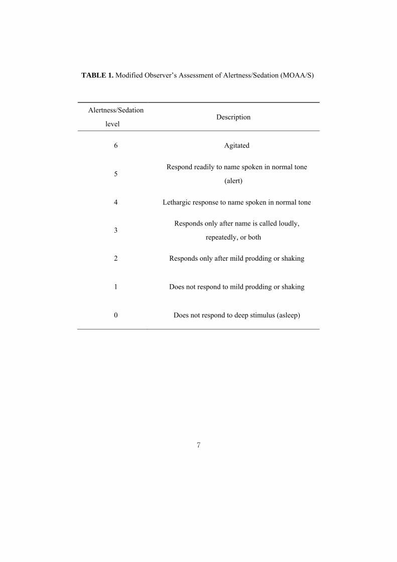
TABLE 1. Modified Observer’s Assessment of Alertness/Sedation (MOAA/S)
Alertness/Sedation
level Description
6 Agitated
5 Respond readily to name spoken in normal tone
(alert)
4 Lethargic response to name spoken in normal tone
3 Responds only after name is called loudly,
repeatedly, or both
2 Responds only after mild prodding or shaking
1 Does not respond to mild prodding or shaking
0 Does not respond to deep stimulus (asleep)

8
Hartman’s solution was administered at a rate of 3–5 mL/kg/h, and 2 L/min
oxygen was given through a nasal cannula.
Oxygen saturation (SpO2), systolic and diastolic blood pressure (SBP and
DBP), electrocardiogram (ECG), and heart rate (HR) were monitored
continuously and recorded at 5-min intervals.
The MOAA/S scale score was recorded as follows: just before the procedure
(baseline, T0); 1 min after induction of sedation (1 min after a 5 min loading of
dexmedetomidine in the DR group and 1 min after the propofol bolus injection
in the PR group, T1); as the endoscope was passed into the esophagus (T2); as
the tumor margin was marked by argon plasma coagulation (T3); 5 min after
normal saline containing epinephrine (0.01 mg/mL) injection was given in the
gastric submucosa (T4); at dissection of the gastric tumor region from the
gastric submucosa (T5); once bleeding control was performed at the gastric bed
after dissection (T6); and at the end of the procedure (T7).
Butylscopolamine (20 mg) was administered to suppress gastric motility
during the procedure at the request of the endoscopist.
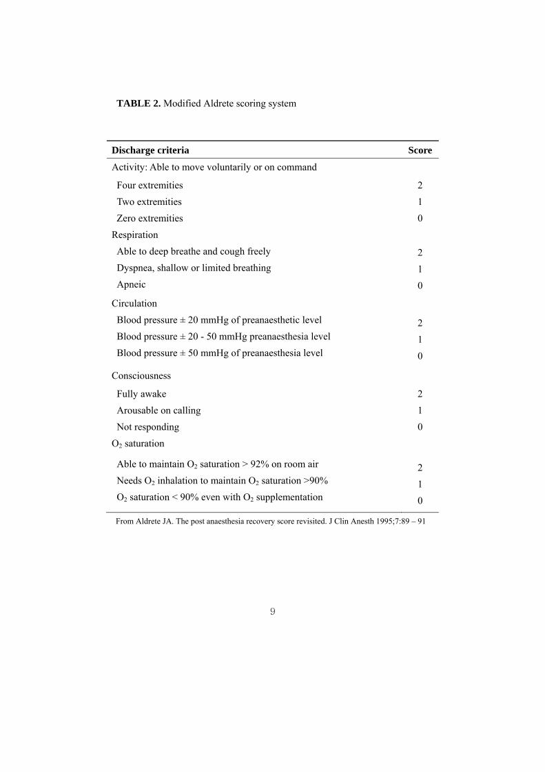
The discharge Aldrete score (Table 2) was recorded to document the
patient’s general status at the end of the procedure.
All patients were observed in the post-anesthetic care unit (PACU) until their
discharge Aldrete score reached 10.
9
TABLE 2. Modified Aldrete scoring system
Discharge criteria Score
Activity: Able to move voluntarily or on command
Four extremities
Two extremities
Zero extremities
2
1
0
Respiration
Able to deep breathe and cough freely
Dyspnea, shallow or limited breathing
Apneic
2
1
0
Circulation
Blood pressure ± 20 mmHg of preanaesthetic level
Blood pressure ± 20 - 50 mmHg preanaesthesia level
Blood pressure ± 50 mmHg of preanaesthesia level
2
1
0
Consciousness
Fully awake
Arousable on calling
Not responding
2
1
0
O2 saturation
Able to maintain O2 saturation > 92% on room air
Needs O2 inhalation to maintain O2 saturation >90%
O2 saturation < 90% even with O2 supplementation
2
1
0
From Aldrete JA. The post anaesthesia recovery score revisited. J Clin Anesth 1995;7:89 – 91
10
2. Methods
가. Assessment of procedural performance
The ease of advancing the scope through the throat (four grades: very easy,
easy, slight difficulty, and difficult), gastric motility12 (no, mild, moderate, and
vigorous) (Table 3), and procedural satisfaction (very good, good, fair, and
bad) were evaluated by the endoscopist. Patients were also asked about their
satisfaction with the procedure (very good, good, bearable, and unbearable)
before discharge from the PACU. The total amount of butylscopolamine used
was recorded.
11
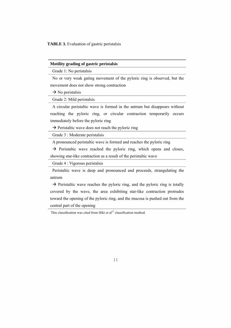
TABLE 3. Evaluation of gastric peristalsis
Motility grading of gastric peristalsis
Grade 1: No peristalsis
No or very weak gating movement of the pyloric ring is observed, but the
movement does not show strong contraction
No peristalsis
Grade 2: Mild peristalsis
A circular peristaltic wave is formed in the antrum but disappears without
reaching the pyloric ring, or circular contraction temporarily occurs
immediately before the pyloric ring
Peristaltic wave does not reach the pyloric ring
Grade 3 : Moderate peristalsis
A pronounced peristaltic wave is formed and reaches the pyloric ring
Peristaltic wave reached the pyloric ring, which opens and closes,
showing star-like contraction as a result of the peristaltic wave
Grade 4 : Vigorous peristalsis
Peristaltic wave is deep and pronounced and proceeds, strangulating the
antrum
Peristaltic wave reaches the pyloric ring, and the pyloric ring is totally
covered by the wave, the area exhibiting star-like contraction protrudes
toward the opening of the pyloric ring, and the mucosa is pushed out from the
central part of the opening
This classification was cited from Hiki et al12 classification method.
12
나. Assessment of patient safety
Hemodynamic variables of SBP and DBP, HR, and SpO2 were compared
when measuring the MOAA/S score.
All respiratory (apnea and desaturation) and hemodynamic (hypertension,
hypotension, tachycardia, or bradycardia; defined as a change in baseline value
of more than 20%) adverse events were recorded. Apnea was defined as not
breathing spontaneously for at least 20 s. Desaturation was defined as SpO2 <
90%. We managed adverse respiratory events with a jaw thrust, mask
ventilation, or by increasing oxygen flow. Ephedrine, nicardipine, atropine, or
esmolol were administered for adverse hemodynamic events. The total amount
of sedative drug, remifentanil, and sedation were recorded.
다. Statistical Analysis
Continuous variables are presented as means ± standard deviations and
dichotomous variables are given as numbers (percentages). Continuous
variables were compared using an unpaired Student’s t-test. Dichotomous
variables were compared using the chi-squared or Fisher exact tests, as
appropriate. Repeated measured variables such as the MOAA/S scale score,
13
SpO2, SBP, DBP, and HR were analyzed using a linear mixed model with
patient indicator as a random effect and group, time, and group × time as fixed
effects. When the interaction of group, time, or group × time of the variables
was significant, a post-hoc analysis with Bonferroni correction was used for
multiple comparisons. All statistical tests were two-tailed. P-values < 0.05 were
considered significant. All statistical analyses were performed using SPSS
software ver. 19.0 (SPSS Inc., Chicago, IL, USA).
Calculation of sample size was adopted by the previous study13 which
compared the efficacy and safety of dexmedetomidine with propofol TCI
during endoscopic esophageal intervention. In which, 32 patients per group
(α=0.05, 1-β=0.8 and 20% drop out) were calculated. Therefore, we intended to
enroll 30 patients per group with a 10% of drop out rate.
III. RESULTS
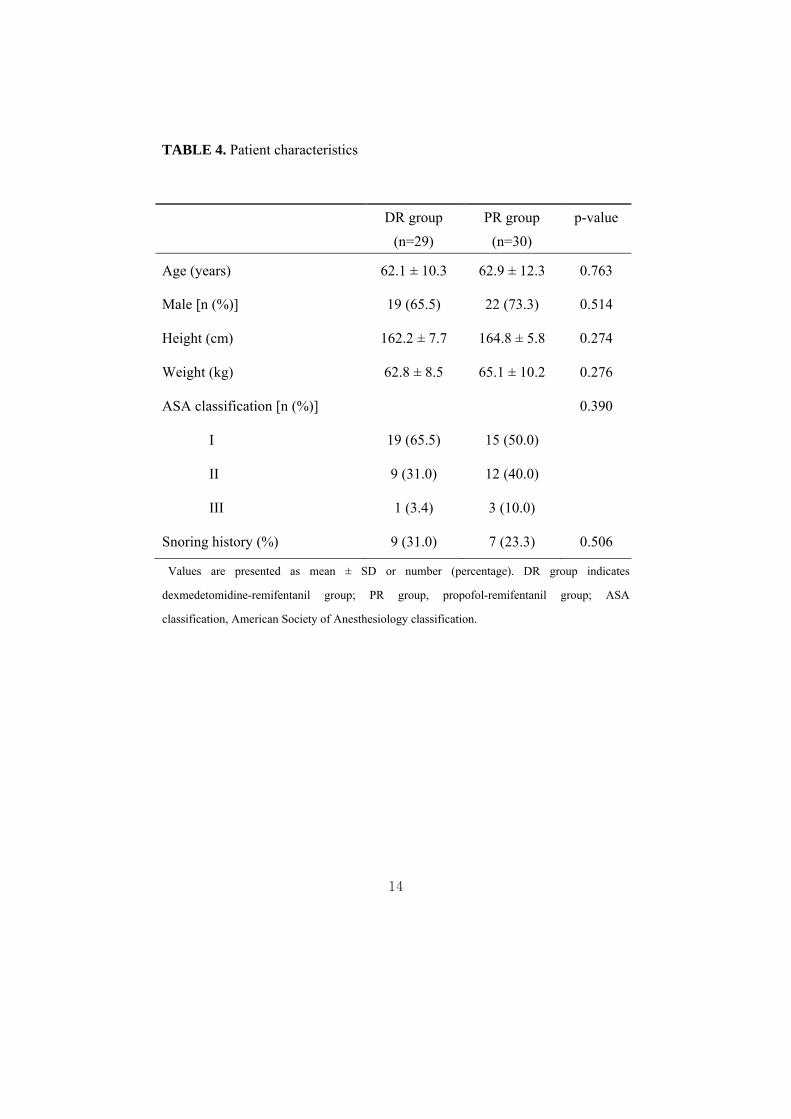
No significant differences were observed in patient demographic data such as
age, sex ratio, height, weight, snoring history, ASA classification, or sedation
duration (Table 4).
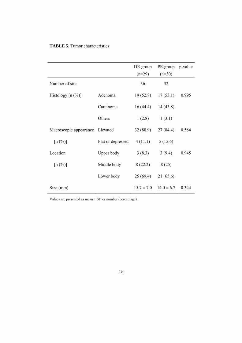
Tumor characteristics, including histology, macroscopic appearance, location
and size measured by the endoscopist were similar between the groups (Table 5).
14
TABLE 4. Patient characteristics
DR group
(n=29)
PR group
(n=30)
p-value
Age (years) 62.1 ± 10.3 62.9 ± 12.3 0.763
Male [n (%)] 19 (65.5) 22 (73.3) 0.514
Height (cm) 162.2 ± 7.7 164.8 ± 5.8 0.274
Weight (kg) 62.8 ± 8.5 65.1 ± 10.2 0.276
ASA classification [n (%)] 0.390
I 19 (65.5) 15 (50.0)
II 9 (31.0) 12 (40.0)
III 1 (3.4) 3 (10.0)
Snoring history (%) 9 (31.0) 7 (23.3) 0.506
Values are presented as mean ± SD or number (percentage). DR group indicates
dexmedetomidine-remifentanil group; PR group, propofol-remifentanil group; ASA
classification, American Society of Anesthesiology classification.
15
TABLE 5. Tumor characteristics
DR group
(n=29)
PR group
(n=30)
p-value
Number of site
36 32
Histology [n (%)] Adenoma 19 (52.8) 17 (53.1) 0.995
Carcinoma 16 (44.4) 14 (43.8)
Others 1 (2.8) 1 (3.1)
Macroscopic appearance Elevated 32 (88.9) 27 (84.4) 0.584
[n (%)] Flat or depressed 4 (11.1) 5 (15.6)
Location Upper body 3 (8.3) 3 (9.4) 0.945
[n (%)] Middle body 8 (22.2) 8 (25)
Lower body 25 (69.4) 21 (65.6)
Size (mm) 15.7 ± 7.0 14.0 ± 6.7 0.344
Values are presented as mean ± SD or number (percentage).
16
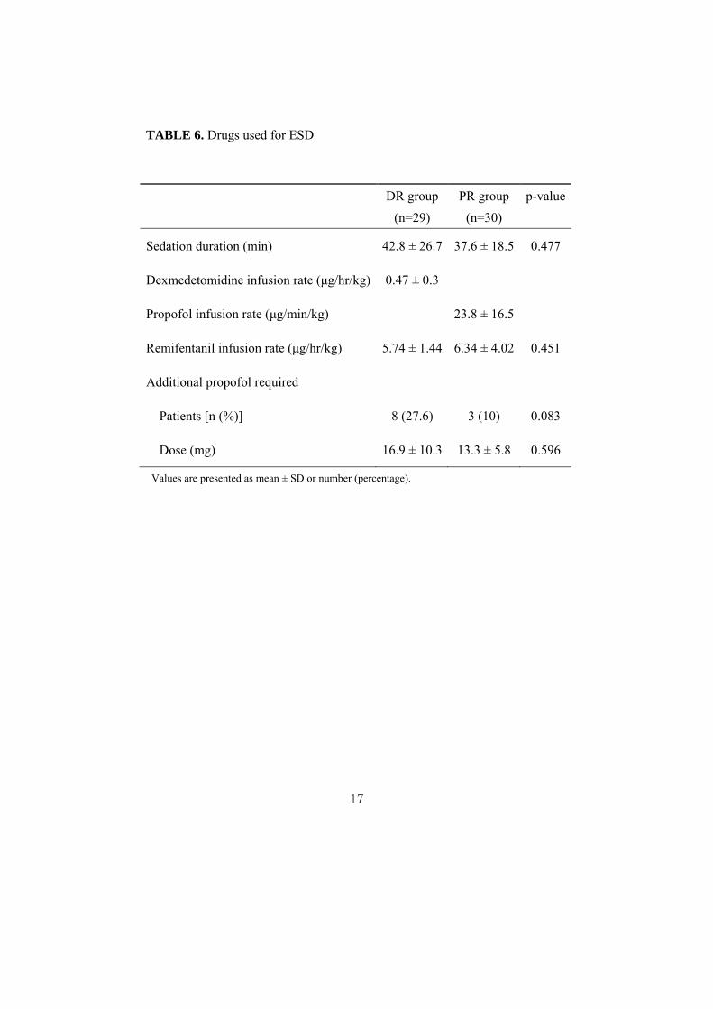
Sedation duration was similar in the groups (P = 0.477). Dexmedetomidine in
the DR group and propofol in the PR group were infused at rates of 0.47 ± 0.3
μg/h/kg and 23.8 ± 16.5 μg/min/kg, respectively. The infusion rates of
remifentanil were 5.74 ± 1.44 μg/h/kg and 6.34 ± 4.02 μg/h/kg in the DR and
PR groups, respectively (P = 0.451). Additional propofol requirements were
16.9 ± 10.3 mg in 8 patients of DR group and 13.3 ± 5.8 mg in 3 patients of PR
group (P = 0.081) (Table 6).
17
TABLE 6. Drugs used for ESD
DR group
(n=29)
PR group
(n=30)
p-value
Sedation duration (min) 42.8 ± 26.7 37.6 ± 18.5 0.477
Dexmedetomidine infusion rate (μg/hr/kg) 0.47 ± 0.3
Propofol infusion rate (μg/min/kg) 23.8 ± 16.5
Remifentanil infusion rate (μg/hr/kg) 5.74 ± 1.44 6.34 ± 4.02 0.451
Additional propofol required
Patients [n (%)] 8 (27.6) 3 (10) 0.083
Dose (mg) 16.9 ± 10.3 13.3 ± 5.8 0.596
Values are presented as mean ± SD or number (percentage).
18
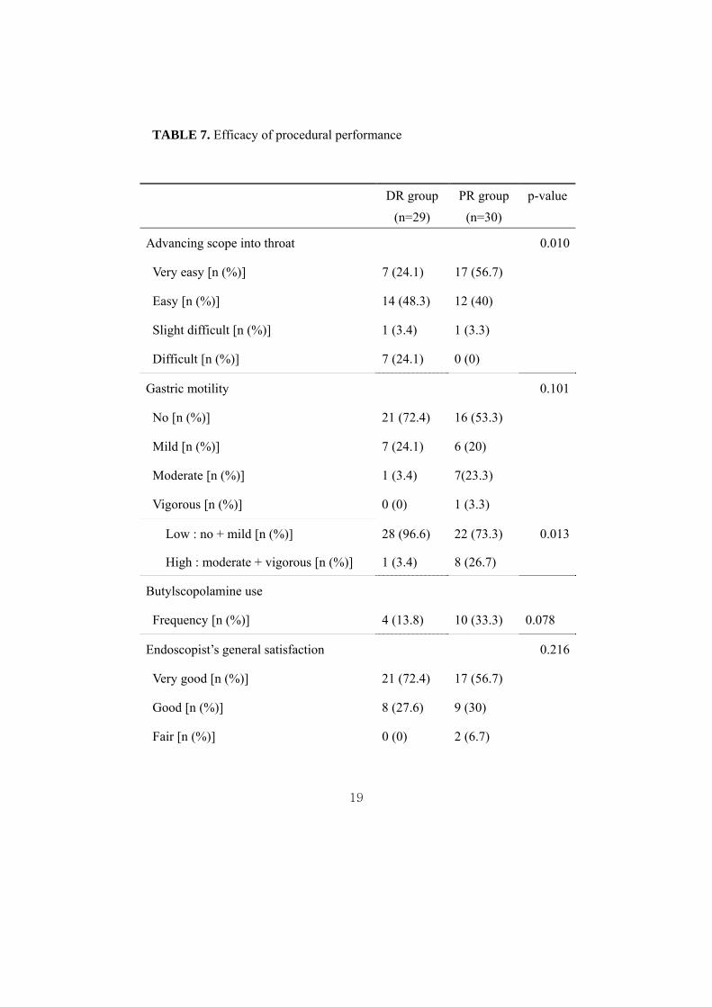
Although the endoscope was more easily advanced through the throat in the
PR group than in the DR group (P = 0.01), low-grade gastric motility (no or
mild) was more frequent in the DR group (96.6% vs. 73.3%, P = 0.013). The
butylscopolamine was administered in 10 patients of PR group compared with
4 patients of DR group (P = 0.078).
While the endoscopists were satisfied with the procedural performance and
judged the procedures as favorable (P = 0.042) in all patients in the DR group
and in 86.7% of patients in the PR group, patient satisfaction was comparable
between the two groups (Table 7).
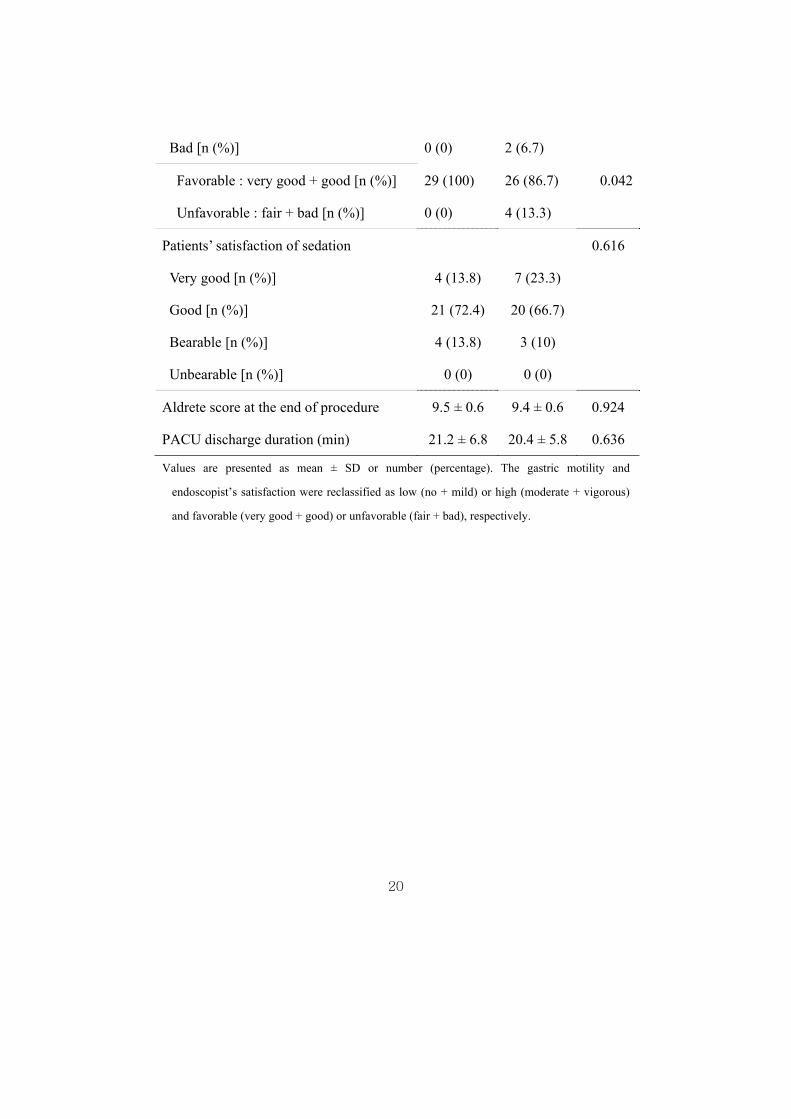
The Aldrete score at the end of the procedure was not different between the
groups (9.5 ± 0.6 in the DR group and 9.4 ± 0.6 in the PR group, P = 0.924)
and all patients left the PACU within 30 min (21.2 ± 6.8 min in the DR group
and 20.4 ± 5.8 min in the PR group, P = 0.636).
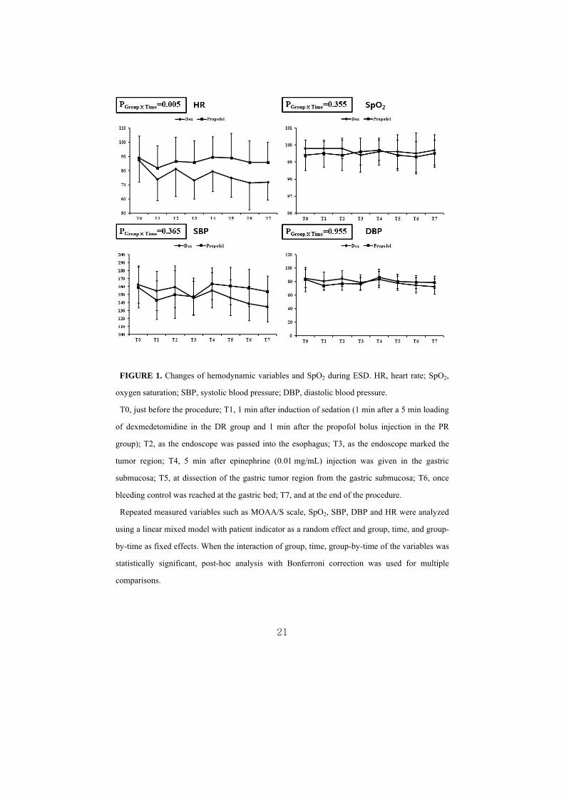
No differences in the MOAA/S scale score, SBP, DBP, or SpO2 were
observed, except HR was different between the groups (Figure 1). No cases of
oxygen desaturation or any adverse hemodynamic events were observed during
the ESD procedures in either group.
19
TABLE 7. Efficacy of procedural performance
DR group
(n=29)
PR group
(n=30)
p-value
Advancing scope into throat 0.010
Very easy [n (%)] 7 (24.1) 17 (56.7)
Easy [n (%)] 14 (48.3) 12 (40)
Slight difficult [n (%)] 1 (3.4) 1 (3.3)
Difficult [n (%)] 7 (24.1) 0 (0)
Gastric motility 0.101
No [n (%)] 21 (72.4) 16 (53.3)
Mild [n (%)] 7 (24.1) 6 (20)
Moderate [n (%)] 1 (3.4) 7(23.3)
Vigorous [n (%)] 0 (0) 1 (3.3)
Low : no + mild [n (%)] 28 (96.6) 22 (73.3) 0.013
High : moderate + vigorous [n (%)] 1 (3.4) 8 (26.7)
Butylscopolamine use
Frequency [n (%)] 4 (13.8) 10 (33.3) 0.078
Endoscopist’s general satisfaction 0.216
Very good [n (%)] 21 (72.4) 17 (56.7)
Good [n (%)] 8 (27.6) 9 (30)
Fair [n (%)] 0 (0) 2 (6.7)
20
Bad [n (%)] 0 (0) 2 (6.7)
Favorable : very good + good [n (%)] 29 (100) 26 (86.7) 0.042
Unfavorable : fair + bad [n (%)] 0 (0) 4 (13.3)
Patients’ satisfaction of sedation 0.616
Very good [n (%)] 4 (13.8) 7 (23.3)
Good [n (%)] 21 (72.4) 20 (66.7)
Bearable [n (%)] 4 (13.8) 3 (10)
Unbearable [n (%)] 0 (0) 0 (0)
Aldrete score at the end of procedure 9.5 ± 0.6 9.4 ± 0.6 0.924
PACU discharge duration (min) 21.2 ± 6.8 20.4 ± 5.8 0.636
Values are presented as mean ± SD or number (percentage). The gastric motility and
endoscopist’s satisfaction were reclassified as low (no + mild) or high (moderate + vigorous)
and favorable (very good + good) or unfavorable (fair + bad), respectively.
21
FIGURE 1. Changes of hemodynamic variables and SpO2 during ESD. HR, heart rate; SpO2,
oxygen saturation; SBP, systolic blood pressure; DBP, diastolic blood pressure.
T0, just before the procedure; T1, 1 min after induction of sedation (1 min after a 5 min loading
of dexmedetomidine in the DR group and 1 min after the propofol bolus injection in the PR
group); T2, as the endoscope was passed into the esophagus; T3, as the endoscope marked the
tumor region; T4, 5 min after epinephrine (0.01 mg/mL) injection was given in the gastric
submucosa; T5, at dissection of the gastric tumor region from the gastric submucosa; T6, once
bleeding control was reached at the gastric bed; T7, and at the end of the procedure.
Repeated measured variables such as MOAA/S scale, SpO2, SBP, DBP and HR were analyzed
using a linear mixed model with patient indicator as a random effect and group, time, and group-
by-time as fixed effects. When the interaction of group, time, group-by-time of the variables was
statistically significant, post-hoc analysis with Bonferroni correction was used for multiple
comparisons.

22
IV. DISCUSSION
We found that minimal sedation using dexmedetomidine-remifentanil was
safe for patients, and that endoscopists were satisfied with the procedural
efficacy perhaps due to lower gastric motility.
This study has some clinical implications regarding the sedating protocol for
ESD. First, our results suggest the importance of analgesics and optimal
sedation level to avoid patient anxiety. ESD was safely performed under
MOAA/S sedation levels of 4–5 if adequate analgesic was provided. As shown
in Figure 1, no patient needed management due to hemodynamic instability or
adverse respiratory events despite the decreased HR in the DR group. We
believe that continuous infusion of remifentanil enabled patients to tolerate this
procedure well in an orientated and anxiety-free state. The analgesic
requirement for a painful procedure was evident in a previous colonoscopy trial,
which was terminated early before enrolling the planned number of patients
because of the higher rate of supplemental fentanyl required and adverse
hemodynamic events in the group of patients administered dexmedetomidine
alone14. In fact, the sedation level for endoscopic procedures is controversial.
International sedation guidelines for gastrointestinal endoscopic procedures15, 16
recommend sedating patients to improve procedural performance. However,
23
the adequate level of sedation for patients has not been well defined (conscious
sedation vs. deep sedation). Takimoto et al.9 compared the efficacy and safety
of conscious sedation for ESD targeting a Ramsay sedation score (RSS) of 2–3
among propofol, dexmedetomidine, and midazolam. They found that
dexmedetomidine provided comparable hemodynamic stability and improved
oxygen saturation as well as no major surgical complications compared to
propofol or midazolam, whereas two patients who received propofol or
midazolam developed gastric perforation. An RSS of 2–3 represent a level of
sedation that is similar to, but slightly more extending than, the MOAA/S of 4–
5 used in the present study (MOAA/S 4 = responding to normal verbal tone;
RSS 3 = responding to commands). Sasaki et al.14 reported hypoxemia in 15.9–
17.8% of patients and hypotension in 19.3–34.4% of patients, suggesting a
deeper sedation level and a higher rate of complications. In the present study,
minimal sedation, regardless of the group, allowed the patients to achieve an
Aldrete score of 9.5 at the end of the procedure and to leave the PACU within
30 min. This may also be an economic benefit of minimizing sedation.
Second, regarding procedural performance, endoscopists felt that the
endoscope could be more easily advanced into the throat (in 7 of 29 patients in
the DR group vs. 17 of 30 patients in the PR group, P = 0.01). The underlying
causes of this difference are unclear but might be explained, in part, by the
24
different effect of propofol and dexmedetomidine on the pharyngeal function.
Kiriyama et al.17 assessed the effects of a bolus of 0.5 mg/kg propofol injected
before ESD compared to no bolus of propofol, found that the propofol bolus
decreased pharyngeal muscle tone and obtunded the scope-stimulated
pharyngeal reflex in 77% of patients compared to 21% of patients with no
bolus. Therefore, in the present study, the intact pharyngeal function in the DR
group may have made it more difficult for the endoscopists to advance the
scope into the throat, because our sedation protocols included a bolus 0.5
mg/kg propofol or dexmedetomidine known to preserve the pharyngeal tone.
Inhibiting gastric motility is crucial to successfully perform ESD, and this is
the first report of endoscopist-evaluated gastric motility during ESD, in relation
to two different sedation protocols (Table 6). The endoscopists graded gastric
motility as low (no and mild among four grades) in 96.6% of the DR group and
in 73.3% of the PR group (P = 0.013). This result was also noted as less of a
requirement for butylscopolamine to suppress gastric motility. The effects of
dexmedetomidine on gastric motility seemed to differ according to subject and
dosage. In a previous study, infusion with a 1.0 µg/kg loading dose for 20 min
followed by infusion of 0.7 µg/h/kg inhibited gastric emptying in healthy
volunteers, as measured by paracetamol absorption compared to 0.1 mg/kg
morphine or placebo10. In contrast, Memis et al.18 found no difference in gastric
25
emptying time between propofol (2 mg/h/kg) and dexmedetomidine (0.2
µg/h/kg) for 5 h in critically ill patients. This discrepancy may have resulted
from the different doses of drugs and measuring methods (direct visualization
vs. indirect paracetamol absorption test) used the two studies.
Dexmedetomidine itself does not alter gastric motility in rats but markedly
enhances the inhibitory effect of morphine on gastric motility19. We are
uncertain of the interactive effect of dexmedetomidine and remifentanil on
gastric motility. As another evaluation of performance efficacy, the
endoscopists were able to perform 94.4% of the complete resections of 36 en
bloc resections (DR group) and 100% of the complete resections of 32 en bloc
resections (PR group), suggesting that both sedation protocols were effective
and safe for ESD.
Our study had some limitations. We analyzed a small number of patients,
which limited the statistical power of our results. Gastric motility did not differ
between the two groups (P = 0.101) when measured using the four grades (no,
mild, moderate, and vigorous); however, there was a significant difference
when just two grades of low (no/mild) and high (moderate/vigorous) were
applied (P = 0.013). This same issue was also observed with the statistical
analysis of endoscopist satisfaction. We did not find any statistical difference
when the ratings were based on four grades (very good, good, fair, and bad).
26
However, when satisfaction was divided into favorable (very good/good) and
unfavorable (fair/bad), endoscopists were in favor of the DR group treatment
(favorable, 100% in DR group vs. 86.7% in PR group, P = 0.042). Although we
intended this to be a prospective endoscopist-blinded study, we are unsure
whether each endoscopist was aware of the type of sedative drugs because of
the difference in the pharmacologic properties between dexmedetomidine and
propofol even though we covered the patient’s venous access site with a drape.
Therefore, we could not conclusively eliminate any bias of personal preference
when they answered the questionnaires. Finally, our study design did not
include a psychometric test for patients or comprehensive questionnaires to
assess patient and endoscopist satisfaction as suggested by Vargo20.
In conclusion, use of dexmedetomidine and remifentanil targeting minimal
sedation resulted in safe and effective ESD procedures, perhaps by suppressing
gastric motility. However, further studies with a greater number of subjects
may be required.
27
REFERENCES
1. Sasaki T, Tanabe S, Ishido K, Azuma M, Katada C, Higuchi K, et al.
Recommended sedation and intraprocedural monitoring for gastric endoscopic
submucosal dissection. Digestive endoscopy 2013;25 Suppl 1:79-85.
2. Kiriyama S, Gotoda T, Sano H, Oda I, Nishimoto F, Hirashima T, et al.
Safe and effective sedation in endoscopic submucosal dissection for early
gastric cancer: a randomized comparison between propofol continuous infusion
and intermittent midazolam injection. Journal of gastroenterology
2010;45(8):831-7.
3. Wehrmann T, Kokabpick S, Lembcke B, Caspary WF, Seifert H.
Efficacy and safety of intravenous propofol sedation during routine ERCP: a
prospective, controlled study. Gastrointestinal endoscopy 1999;49(6):677-83.
4. Sethi S, Wadhwa V, Thaker A, Chuttani R, Pleskow DK, Barnett SR,
et al. Propofol versus traditional sedative agents for advanced endoscopic
procedures: A meta-analysis. Digestive endoscopy 2013.
5. Park CH, Min JH, Yoo YC, Kim H, Joh DH, Jo JH, et al. Sedation
methods can determine performance of endoscopic submucosal dissection in
patients with gastric neoplasia. Surgical endoscopy 2013;27(8):2760-2767.
6. Cote GA, Hovis RM, Ansstas MA, Waldbaum L, Azar RR, Early DS,
et al. Incidence of Sedation-Related Complications With Propofol Use During
Advanced Endoscopic Procedures. Clin Gastroenterol H 2010;8(2):137-42.
7. Shin S, Lee SK, Min KT, Kim HJ, Park CH, Yoo YC. Sedation for
interventional gastrointestinal endoscopic procedures: are we overlooking the
"pain"? Surgical endoscopy 2014;28(1):100-7.
8. Dere K, Sucullu I, Budak ET, Yeyen S, Filiz AI, Ozkan S, et al. A
comparison of dexmedetomidine versus midazolam for sedation, pain and
hemodynamic control, during colonoscopy under conscious sedation. European
journal of anaesthesiology 2010;27(7):648-52.
28
9. Takimoto K, Ueda T, Shimamoto F, Kojima Y, Fujinaga Y, Kashiwa A,
et al. Sedation with dexmedetomidine hydrochloride during endoscopic
submucosal dissection of gastric cancer. Digestive endoscopy : official journal
of the Japan Gastroenterological Endoscopy Society 2011;23(2):176-81.
10. Iirola T, Vilo S, Aantaa R, Wendelin-Saarenhovi M, Neuvonen PJ,
Scheinin M, et al. Dexmedetomidine inhibits gastric emptying and oro-caecal
transit in healthy volunteers. British journal of anaesthesia 2011;106(4):522-7.
11. Chassard D, Lansiaux S, Duflo F, Mion F, Bleyzac N, Debon R, et al.
Effects of subhypnotic doses of propofol on gastric emptying in volunteers.
Anesthesiology 2002;97(1):96-101.
12. Hiki N, Kaminishi M, Yasuda K, Uedo N, Honjo H, Matsuhashi N, et
al. Antiperistaltic effect and safety of L-menthol sprayed on the gastric mucosa
for upper GI endoscopy: a phase III, multicenter, randomized, double-blind,
placebo-controlled study. Gastrointestinal endoscopy 2011;73(5):932-41.
13. Eberl S, Preckel B, Bergman JJ, Hollmann MW. Safety and
effectiveness using dexmedetomidine versus propofol TCI sedation during
oesophagus interventions: a randomized trial. BMC gastroenterology
2013;13:176.
14. Sasaki T, Tanabe S, Azuma M, Sato A, Naruke A, Ishido K, et al.
Propofol sedation with bispectral index monitoring is useful for endoscopic
submucosal dissection: a randomized prospective phase II clinical trial.
Endoscopy 2012;44(6):584-9.
15. Heneghan S, Myers J, Fanelli R, Richardson W, Society of American
Gastrointestinal Endoscopic S. Society of American Gastrointestinal
Endoscopic Surgeons (SAGES) guidelines for office endoscopic services.
Surgical endoscopy 2009;23(5):1125-9.
16. Riphaus A, Wehrmann T, Weber B, Arnold J, Beilenhoff U, Bitter H,
et al. [S3-guidelines--sedation in gastrointestinal endoscopy]. Zeitschrift fur
Gastroenterologie 2008;46(11):1298-330.
17. Kiriyama S, Naitoh H, Fukuchi M, Fukasawa T, Saito K, Tabe Y, et al.
29
Evaluation of Pharyngeal Function between No Bolus and Bolus Propofol
Induced Sedation for Advanced Upper Endoscopy. Diagnostic and therapeutic
endoscopy 2014;2014:248097.
18. Memis D, Dokmeci D, Karamanlioglu B, Turan A, Ture M. A
comparison of the effect on gastric emptying of propofol or dexmedetomidine
in critically ill patients: preliminary study. European journal of anaesthesiology
2006;23(8):700-4.
19. Asai T, Mapleson WW, Power I. Interactive effect of morphine and
dexmedetomidine on gastric emptying and gastrointestinal transit in the rat.
British journal of anaesthesia 1998;80(1):63-7.
20. Vargo J, Howard K, Petrillo J, Scott J, Revicki DA. Development and
validation of the patient and clinician sedation satisfaction index for
colonoscopy and upper endoscopy. Clinical gastroenterology and hepatology :
the official clinical practice journal of the American Gastroenterological
Association 2009;7(2):156-62.
30
ABSTRACT (IN KOREAN)
내시경적 점막하절제술을 시행받는 환자에서 덱스메데토미딘-
레미펜타닐(dexmedetomidine-remifentanil)과 프로포폴-레미
펜타닐(propofol-remifentanil)의 진정효과 및 안전성 비교
<지도교수 민경태>
연세대학교 대학원 의학과
김 남 오
내시경적 점막하절제술은 충분한 진정과 진통이 필요한 시술인데,
이를 위해 프로포폴과 레미펜타닐의 사용이 많이 추천되고 있다. 덱
스메데토미딘은 진정작용과 진통작용을 모두 가지고 있는 약제로 위
장관 운동 억제 작용이 있으며, 이는 내시경적 점막하 절제술을 시행
하는데 있어서 매우 중요한 요소가 될 수 있다. 본 연구에서는 내시
경적 점막하절제술 동안 덱스메데토미딘-레미펜타닐과 프로포폴-레
미펜타닐 약제의 유효성과 안전성을 비교하였다.
내시경적 점막하절제술이 예정되어 있던 총 59명의 환자가 무작위
로 덱스메데토미딘-레미펜타닐 군과 프로포폴-레미펜타닐 군으로 나
뉘었다.
환자의 진정작용을 위하여 덱스메데토미딘 혹은 프로포폴을 투여하
31
여 Modified Observer’s Assessment of Alertness/Sedation 척도의
4-5 레벨을 유지하였다. 두 군 모두 진통작용을 위하여 레미펜타닐
이 6 μg/hr/kg 의 용량으로 연속적으로 투여되었다. 내시경 기구를
환자의 식도로 삽입 시 수월한 정도, 위 연동운동 정도, 시술자와 환
자의 만족도 등이 조사되었다. 혈역학적 변수들과 저산소증 발생 여
부를 조사하여 환자의 안정성을 평가하였다.
조사한 두 군간의 환자의 특성 및 종양의 특성 차이는 없었다. 심
박수가 덱스메데토미딘-레미펜타닐 군에서 낮았던 것 이외에 혈역학
적 변수나 산소포화도는 두 군간에 차이를 보이지 않았다. 저산소증
은 두 군에서 발생하지 않았다. 내시경 기구를 환자의 식도로 넘길
때 덱스메데토미딘-레미펜타닐 군보다 프로포폴-레미펜타닐 군이 보
다 수월하였으며(내시경 집도 의사를 대상으로 한 설문조사 결과 “매
우 수월” 7 vs. 17, P=0.01), 위 연동운동은 덱스메데토미딘-레미펜타
닐 군에서 더 억제되었다(내시경 집도 의사를 대상으로 한 설문조사
결과 “연동운동 적음” 28 vs. 22, P=0.013). 보다 많은 내시경 집도
의사들이 덱스메데토미딘-레미펜타닐 군에서 시술이 용이하다고 느
꼈다(내시경 집도 의사를 대상으로 한 설문조사 결과 “시술에 호의
적” 29 vs. 26, P=0.042). 두 군간 환자 만족도는 차이가 없었다.
결론적으로 본 연구를 통하여 내시경적 점막하절제술의 시행시
덱스메데토미딘-레미펜타닐의 사용과 프로포폴-레미펜타닐의 사용은
유효성과 안정성에서 차이를 보이지 않았다. 그러나 위 연동운동
억제효과로 인하여 덱스메데토미딘이 보다 선호된 것으로 생각해볼
수 있다.
---------------------------------------------------
핵심되는 말: 덱스메데토미딘, 유효성, 안전성, 위 연동운동,
내시경적 점막하절제술