A CLINICAL STUDY ON INCISIONAL HERNIArepository-tnmgrmu.ac.in/8616/1/220101014anbu.pdf · A...
128
A CLINICAL STUDY ON INCISIONAL HERNIA Dissertation submitted to THE TAMIL NADU DR. MGR. MEDICAL UNIVERSITY CHENNAI – 600 032 In partial fulfilment of the regulations For the awards of the degree of M.S. DEGREE BRANCH – I GENERAL SURGERY GOVERNMENT MOHAN KUMARAMANGALAM MEDICAL COLLEGE, SALEM APRIL 2014
Transcript of A CLINICAL STUDY ON INCISIONAL HERNIArepository-tnmgrmu.ac.in/8616/1/220101014anbu.pdf · A...

A CLINICAL STUDY ON INCISIONAL HERNIA
Dissertation submitted to
THE TAMIL NADU DR. MGR. MEDICAL UNIVERSITY
CHENNAI – 600 032
In partial fulfilment of the regulations
For the awards of the degree of
M.S. DEGREE BRANCH – I
GENERAL SURGERY
GOVERNMENT MOHAN KUMARAMANGALAM
MEDICAL COLLEGE, SALEM
APRIL 2014


ACKNOWLEDGEMENT
I am extremely thankful to Prof. Dr.A. KARTHIKEYAN, M.D.,
Dean, Govt. Mohan Kumaramangalam Medical College and Hospital, for
allowing me to utilize the hospital facilities for doing this work.
I am also thankful to Prof. Dr. T. SWAMINATHAN, M.S,
Medical Superintendent, Govt. Mohan Kumaramangalam Medical
College and Hospital, for his whole hearted support and encouragement
for the completion of this dissertation.
I express deep thanks to Prof. Dr. R. KATTABOMMAN. M.S.,
Head of the Department of Surgery, for his esteemed guidance and
valuable suggestions from the very beginning of this study.
I express my deep sense of gratitude and indebtedness to my unit
chief Prof. Dr. C. RAJASEKARAN, M.S for giving me inspiration,
valuable guidance and his unstinting help in completing the course and
preparing this dissertation.
I thank all surgical unit chiefs Prof. Dr. P. RAMALINGAM, M.S,
Prof. Dr. K.SANTHI, M.S, Prof. Dr. A.NIRMALA, M.S.,
Prof. Dr. V.LAKSHMI NARAYANI, M.S for their advice and kind help.

It is my privileged duty to profusely thank my assistant professors
Dr.R.SWAMINATHAN,M.S. Dr.N. JEEVA, M.S. Dr. V.SIVAKUMAR, M.S,
who helped and guided me in many aspects of this study.
I sincerely thank The Professor and Assistant Professors in the
Department of General Surgery for their encouragement and guidance in
various aspects of this study.
I take this opportunity to thank all my Post Graduate colleagues
and friends who helped me a lot in completing this dissertation
successfully.
I cordially thank my parents and AnuSelvaraj(sister), Suresh,
Jayaprakash, Sivaskumar, Firdhose who have always been there with me
whenever I needed their help and cooperation.
I am deeply obliged to my patients, without whose help the present
study would not have been possible.


ABSTRACT
Incisional hernia is a hernia arising from previous surgical or
wound site. Incisional hernia constitutes 2% – 12 % of all abdominal
surgical incisions. There are many surgical techniques are available but
no one is best always.
AIM & OBJECTIVE
To evaluate the incidence of incisional hernia and to compare the
various surgical techniques to repair the incisional hernia.
MATERIALS & METHODS
This prospective study of 60 consecutive cases of incisional hernia
admitted in Government Mohan Kumaramangalam Medical College
Hospital, Salem was done in the period from July 2011 to June 2013. The
cases were evaluated through proper history taking, clinical examination,
operative procedure and post operative follow-ups.
OBSERVATION
In our study, commonest age group having incisional hernia is
between 30 – 50 years (46.6%). Females are common (61.7%), the most

common previous surgery to develop the incisional hernia is
Laparotomy(58.3%) and common surgical incision is Median
Vertical(45.1%). Most of the patients (27.3%) had wound dehiscence.
47% of patient had incisional hernia within one year of previous surgery.
In this study, I found 10% of recurrence rate, 25% of per operative
bleeding and 25% of chronic pain for onlay mesh repair. Sublay,
Component Separation Techinique and Laparoscopic technique had no
recurrence; Chronic pain was noticed 10% patients in Component
Separation and Laparoscopic techniques while only 5% patients in Sublay
repair.Hospital stay is less required in sublay repairs and laparoscopic
repairs. Sublay technique had less post operative complication.
CONCLUSION
Open Sublay mesh repair and Laparoscopic mesh repair are the
best method in our study compared to others. Middle aged female patients
who underwent laparotomy with wound dehiscence having midline
vertical incision had incisional hernia commonly.
KEY WORDS
Incisional Hernia, Mesh repair, Onlay, Sublay, Component
Separation, Laparoscopy,

CONTENTS
S. No. TITLE PAGE
NO.
1. INTRODUCTION 1
2. REVIEW OF LITERATURE 3
3. AIMS AND OBJECTIVES 43
4. MATERIALS AND METHODS 44
5. OBSERVATION AND RESULTS 52
6. DISCUSSION 72
7. CONCLUSION 78
8. ANNEXURES
BIBLIOGRAPHY
PROFORMA
PATIENT CONSENT FORM
MASTER CHART

LIST OF FIGURES
Sl. NO.
PICTURE PAGE
NO.
1. Layers of Anterior Abdominal Wall 6
2. The Arrangement of Muscles in Anterior Abdominal
Wall 7
3. Rectus Abdominus Muscles and contents of Rectus
Sheath 11
4. Schematic Transverse sections through ventral
abdominal wall 12
5. The Blood supply of ventral abdominal wall 14
6. The Schematic view-Direction of Blood supply and its
anastomosis 15
7. Venous and Lymphatic Drainage of Anterior
Abdominal Wall 16
8. Nerve Supply of Anterior Abdominal Wall 17
9. Neurovascular Course in Anterior Abdominal Wall 18
10. Incisional Hernia 22

11. The Algoritham for Abdonimal wall reconstruction 27
12. The various mesh position 32
13. Sublay mesh repair 34
14. Diagrammatic Representation of Pascal’s Principle
Hydrostatic Pressure 34
15. Diagram Represents Laparoscopic Port Position 35
16. The Component Separation Technique 38
17. The Component Separation Technique - Flap
advancement in abdominal wall 39
18. Schematic transverse section of abdominal wall after
completing component separation technique. 39
19. The Flap Reconstruction 41

LIST OF TABLES
Sl.
No TABLES
Page
No.
1. Age incidence of incisional hernia 52
2 Sex incidence of incisional hernia 53
3 Types of surgery causing incisional hernia 54
4 Types of incision causing incisional hernia 56
5 Previous surgical complications causing incisional
hernia
58
6 Time period for the onset of incisional hernia 60
7 Various incisional hernia repair 62
8 The Complications Of Per-Operative Period 64
9 The Complications Of Post-Operative Period 66
10 Hospital Stay In Various Technique 68
11 Follow Up 70

LIST OF CHART
Sl. NO
CHART PAGE
NO.
1. Age Incidence of Incisional Hernia 52
2 Sex Incidence of Incisional Hernia 53
3 Types of Surgery Causing Incisional Hernia 55
4 Types of Incision Causing Incisional Hernia 57
5 Previous surgical complications causing
incisional hernia
59
6 Time period for the onset of incisional hernia 61
7 Various incisional hernia repair 63
8 The complications of per-operative period 65
9 The complications of post-operative period 67
10 Hospital stay in various technique 69
11 Follow up 71

LIST OF ABBREVIATIONS USED
Yrs-years
No of-number of
CT-computerized tomography
MRI-magnetic resonant imaging
DOA-date of admission
DOD-date of discharge
DOS-date of surgery
DM-diabetes mellitus
RR-respiratory rate
BP-blood pressure
P/R-per rectal
P/V-per vaginal
Hb-haemoglobin
ECG-electrocardiogram
USG-ultra sonogram
SA-spinal anesthesia
GA-general anesthesia

1
INTRODUCTION
“No disease of the human body belonging to the province of the
Surgeon requires in its treatment a better combination of accurate
anatomical knowledge with surgical skill than Hernia in all it’s
varieties”-Sir AstelyPastonCopper(1804).
Abdominal wall hernias are very popular in general surgery. Many
people are affected by this problem. Some of them are symptomatic and
others are asymptomatic. It may be congenital problem or acquired.
Hernia presentations are commonly inguinal, ventral and incisional
hernia. Most of the patients undergoing surgery for this problem because
of incarceration or obstruction may occurs at any time.
“Hernia is derived from Latin word – meaning as ‘Rupture1’. In
Greek work HERNIAS/ KELE meaning is bud / off shoot2.”
“Hernia is defined as abnormal protrusion of whole or part of
viscus through the walls that contains it.”
Hernia continuously enlarges in size because of increased intra
abdominal pressure which is the greatest part of the hernia formation. The
Pascal’s principle and the law of LaPlace can explain it.

2
Incisional hernia is defined as hernia developing in the previous
surgical scar or trauma. Various techniques are available for incisional
hernia repair. Each technique has its own merits and demerits.
Recurrence is common depends on technical error, patient’s error and due
to materials.
The key concept for hernia repair is tension free repair. Now a days
mesh is used for fascial defect closure without tension. The anatomical
repair, Ramirez’s component separation is technique is available for
infected wound, large hernia and multiple defects with tension free repair.
In this study, I have analyzed various techniques available to repair
the incisional hernia and I have found out the incidence of various
etiological factors to cause the incisional hernia among the 60 patients
admitted in our hospital during the period of July 2011 to June 2013.

3
HISTORY
“History of hernia repair is the history of surgery” - stated by
Nyhus3.
Manuscripts referring hernia were found in Egyptian and
Mesopotamian cultures4.
Around 1550 BC, famous PapyrusEbers quoted hernia and
treatment as2,4
‘Then you shall say concerning it “This is a swelling of the
coverings of his abdomen, an illness which I will treat”. It is the
heat of his bladder in front of his belly, which creates it. Falling
to the ground, it returns likewise. You should heat (“shemen”) it
to imprison it in his belly. You treat it like the “sahemen”
treatment’
“Old masters” of Greek and roman antiquity- elaborated origin,
pathology, symptoms and treatment of hernia.
Hippocratic Corpus stated hernia resulted from drinking water
from large river or experiencing traumatic events to belly and
described hernia as ‘tear in the abdomen’2,6.

4
Erasistratus of Keos – considered as “the father of physiology”
performed hernia surgery in Egypt5.
Paulus of Aegina, opened hernia sac and reduced into belly by
imagination with probe and applying cauterization to skin.
In 1903, for large hernia and recurrent hernia Halsted used
fascia graft.
In 1950, Francis Usher used polypropylene as prosthesis7.
Preperitoneal mesh repair is introduced by Stoppa in 19732.
Wantz’s introduced unilateral preperitoneal mesh repair.
In classic 1973 Paper, Rives stated, “The problem which we
must solve is not just a simple problem of technique”9.
In 1982, Ger performed first laparoscopic hernia by simply
closing the peritoneal opening with staples.
Ramirez introduced the component separation technique in
199010.
Laparoscopic technique was introduced for incisional or ventral
hernia since 19938.

5
ANATOMY11, 12, 13
Anterior abdominal wall has nine layers
• Skin
• Subcutaneous tissue -Camper's fascia
• Superficial fascia - Scarpa's fascia
• External oblique muscle
• Internal oblique muscle
• Transverse abdominus muscle
• Transversalis fascia
• Preperitoneal fat and areolar tissue
• Peritoneum

Fig : 1 Layers of Anterior Abdominal Wall
6

Fig : 2 The Arra
SKIN &SUBCUTA
It is the seco
fascia :
• Fascia cam
• Fascia sca
Fascia camp
Second layer of su
fibrous tissue .It co
angement of Muscles in Anterior Abbdominal Wall
7
ANEOUS TISSUE
ond layer and deep to the skin which coonsist of the two
mper
arpa
er is the superficial layer which consi
ubcutaneous tissue is scapra's fascia
ntinues as fascia lata in the thigh.
ists of bulky fat.
which is dense

8
EXTERNAL OBLIQUE MUSCLE
Origin : Lower seven ribs
Insertion : Posterior most fibres inserted into iliac crest,
At mid clavicular level fibres inserted into linea alba, Lower most
fibres gives the form inguinal or Poupart's ligament between anterior
superior iliac spine and pubic tubercle.
It is the longest, thick, flat abdominal wall muscle .Its direction is
downwards and medially, as "Hand in Pocket "
INTERNAL OBLIQUE MUSCLE
Origin : Iliopsoas fascia, anterior 2/3 of the iliac crest an
lumbodorsal fascia.
Insertion : Upper most fibres are inserted into lower 5 ribs
and its cartilage.
Central fibres develop aponeurosis at the semilunar line. Above the
semicircular line ( just below the umbilicus ) it acts as part of anterior and
posterior rectus sheath. It forms a part of Anterior rectus sheath below the
semicircular line . Lower fibres give to develop part of cremaster muscle
around the spermatic cord.

9
Its direction is just opposite to External oblique apponeurosis from
inferolateral to superomedial direction.
TRANSVERSE ABDOMINUS MUSCLE
It is the smallest muscle
Origin: Lower six costal cartilages
The spine of the lumbar vertebra , the iliac crest and the iliopsoas
fascia .
Insertion : It continuously forms part of the anterior and
posterior rectus sheath above the semicircular line.
Below the semicircular line it forms a part of anterior rectus sheath.
In lower abdomen it joins with internal oblique muscle to form conjoint
tendon which is part of inguinal canal boundaries.
HASSELBACH'S TRIANGLE
It is the site of Indirect inguinal hernia formation and is bounded
Medially by lateral margin of rectus muscle.
Superolaterally by the inferior epigastric artery
Inferiorly by the inguinal ligament
Floor is formed by transversalis fascia

10
TRANSVERSALIS FASCIA14, 15, 16
It is otherwise called groin's achilles tendon which is
endoabdominal fascia. It prevents the formation of hernia by covering the
trianglesof medial part of groin. It connects between transverse
abdominus and iliopubic tract.
MYOPECTINEAL ORIFICE OF FRUCHAUD15, 16
It is common site for direct and indirect inguinal hernia, femoral
hernia and interstitial hernia of groin.
Boundaries:
Medially - Central border of rectus muscle sheath
Lateral - Iliopsoas muscle
Superiorly - Conjoint tendon
Inferiorly - Superior pubic ramus

RECTUS ABDOM
Origin :
Insertion :
Fig : 3 Rectus Ab
11
MINUS MUSCLE
Anterior surface of 5, 6, 7th costal cart
xiphoid process.
tilage and
Pubic crest and pubic symphysis. It h
insertions which attached to anterior re
There is no attachment to the posterio
Each rectus adjacent to each other wh
by linea alba.
has 3-5 tendinous
ectus sheath.
or rectus sheath.
hich is separated
bdominus Muscles and Contents of RRectus Sheath.

PYRAMIDALIS
Pyramidalis presents in the lower abdomen with the Anterior rectus
sheath. It originates from pubic tubercle and pubic symphysis ends in
linea alba in midline, a region between umbilicus and pubic symphysis.
Muscle site is variable in nature. Pyramidalis is supplied by inferior
epigastric artery and deep circumflex iliac artery. Its branches cross each
other and anastomosis takes place in midline which give trouble bleeding
during lower midline surgical incision .It has nerve supply from terminal
branch of subcostal nerve.
RECTUS SHEATH
The rectus muscle is covered by anterior and posterior rectus
sheath above the semicircular line.Below the semicircular line, Rectus is
covered by anterior rectus only.
Fig : 4 Schematic Transverse sections through ventral abdominal wall 12

13
Below the semicircular line, Three aponeurotic layers going to
develop the anterior rectus sheath, above Internal and external oblique
develop the anterior sheath , internal and transverse abdominus develop
in posterior sheath.
LINEA ALBA
In midline, band of dense fibres called as linea alba, which joins
the two rectus muscle to close each other.
PREPERITONEAL SPACE AND PERITONEUM
Space between fascia transversalis and the parietal peritoneum is
called as Prepritoneal space.
Retzius - It is defined as prevesical space, just posterior to the
pubis
Bogros - Space just posterior to posterior lamina transversalis
fascia.
CLOSE RELATED STRUCTURES:
1. Medial umbilical ligament
2. Median umbilical ligament
3. Falciform ligament of liver

4. Infeerior epigastric artery and vein
5. Rouund ligament
6.Vas deferens and pampiform plexus
BLOOD SUPPLYY
The anterola
artery and last 6
superior and inferio
ateral abdominal wall has blood supp
intercostal artery, deep circumflex
or epigastric artery.
ply from lumbar
iliac artery and
wall Fig : 5 TThe Blood supply of ventral abdominal
The lumbar
iliohypogastric, in
transverse abdomin
r and intercostal artery together w
ntercostal nerve pass between intern
nus muscle. Finally at midline, it sup
14
with ilioinguinal,
nal oblique and
pplies the rectus

muscle by the perfo
superior epigastric
forator arteries and communicates with
arteries.
h the inferior and
SUPERIOR EPIGGASTRIC ARTERY:
It is a branc
surface of the rec
muscle and anastom
ch of Internal mammary artery and r
ctus abdominus muscle. It descends
mosis with epigastric artery.
reaches posterior
through rectus
INFERIOR EPIGGASTRIC ARTERY:
It is a bran
inguinal ligament.
into rectus muscle a
nch of External iliac artery and is j
It courses through the preperitoneal s
at level of semilunar line of Douglas.
ust proximal to
space and enters
Fig : 6 The Scchematic view-Direction of Blood supply an
15
nd its anastomosis

The deep circumflex artery arises from external iliac artery just
before or near the inferior epigastric artery and supplies the anterior
abdominal wall musculature.
VENOUS DRAINAGE
Upper abdomen drains into superior vena cava internal memory,
intercostal long thoracic veins. Below the umblicus, the abdomen drains
into inferior vena cava through superficial epigastric, circumflex illiac
and pudendal veins via saphenous vein.
16
Fig : 7 Venous and Lymphatic Drainage of Anterior Abdominal wall

LYMPHATIC SU
Above umbil
the axillary group o
level drains into the
NERVE SUPPLY
• Anterior abd
nerve.
• Motor suppli
• Ilioinguinal
innervation t
F
17
UPPLY
licus level, anterior abdominal wall lym
of lymph nodes. Abdominal wall belo
e superficial inguinal nodes.
mphatics reaches
ow the umbilicus
dominal wall is innervated or supplied by 7-12 thoracic
ies is provided by 7th and 8th nerve
and iliohypogastric nerve supplie
to hypogastrium and lower abdominal w
es the sensory
wall.
ig : 8 Nerve supply of Anterior Abdominaal wall

Fig : 9 Ne
The Neurova
peculiar one. The N
muscle and transv
course between the
"Hernia is d
viscus through the w
Abdominal h
hernia.
eurovascular Course in Anterior Abdominnal wall
ascular innervations in the anterior ab
Neurovascular bundle passes between
verse abdominus muscle. There is n
internal and external oblique muscles.
bdominal wall is
internal oblique
no neurovascular
.
18
HERNIA
defined as abnormal protrusion of vi
wall that contain it."
iscus or part of
hernia in adults is classified into inguuinal and ventral

19
CLASSIFICATION OF VENTRAL HERNIA:17
1. Congenital hernia
- Umbilical hernia - infant
-Gastrochisis
-Omphalocele
2. Acquired hernia
- In midline
- Diastasis recti
-Epigastric
- Umbilical hernia in adult
- Supravesical - anteior, posterior and lateral
- In paramedian
-Spigelian
-Interparietal - preperitoneal , interstitial and
superficial .

20
3. Incisional hernia
- Midline incisional hernia
- Paramedian
- Transverse
- Port site hernia
- Special operative site hernia
- Boundry hernia
- Incisional hernia with loss of abdominal domains
4. Traumatic
- Penetrating
- Blunt Injury
- Destructive
Hernia occurs when there is loss or weakness of the function of
muscle tendon and fascia. Here basic mechanism, pathology in fascia
causing defect and failure of wound healing, over all cellular and
extracellular molecular defect is common.

21
WOUND HEALING18, 19, 20, 21
Wound healing is a complex process. Several cells and cellular
matrix are involved. The quality and quantity of connective tissue
determines the stability of wound or scar site. The type 1 and type 3
collagen are involved. Type 1 collagen have tensile property which act
against resistance. Type 3 collagen have less strength compared to type 1.
In later ,a type 3 in converted into type 1 collagen. The lack or decreased
synthesis of type 1 causes incisional hernia. Due to dysfunction of
collagen and decreased ratio of type 1 and 3 , incisional hernia is
commonly occurred.
SURGICAL WOUND HEALING
Process of wound healing:
- Lag phase
- Proliferative phase
- Remodelling phase

During laporotomy and hernia repair, there is delay or defective in
repair mechanism which causes incisional hernia. Fascial or wound
dehiscence is caused by early mechanical wound failure.
Wound infection, steroids, wound ischaemia affect the healing
pathway .
INCISIONAL HERNIA
Hernia arising from previous surgical scar or trauma wound is
called incisional hernia. Here, primary mechanism is failure or defective
wound healing.
Fig : 10 Incisional Hernia 22

23
AGE AND GENDER 22
Above 45 years, wound healing is delayed and defective cellular
matrix. Older people have less migration of fibroblast and defective
synthesis of collagen23, 24 as well as prevalence of concomitant diseases.
Most of the studies suggest, men are commonly affected with
incisional hernia compared to female25, 26.
CAUSES :
LOCAL DISTURBANCES OF WOUND HEALING 27-30
Wound infection, seroma formation, foreign bodies causes increase
risk of incisional hernia formation. It produces cytokines and proteases
which causes decreased production of fibroblast and wound stability.
Tension suture causes necrosis that produces defective wound healing.
CONCOMITTENT DISEASE 28, 31, 32
Poor nutritional status, diabetes mellitus, obesity, anemia,
underlying malignancy are identified as independent risk to form the
incisional hernia.

24
ENVIRONMENTAL INFLUENCE33-35
Smoking is one of the most common causes that develop hernia
both primary and secondary. It is 4 fold increased risk for incisional
hernia. ACE inhibitors, steroids, NSAIDS, chemotherapeutic agents
affect the wound healing.
HEREDITARY CONNECTIVE TISSUE DISORDER36-39
Ehler- Danlos syndrome, cutis laxa , type 3 pre collagen disorder,
osteogenesis imperfecta are associated with increased incidence of
incisional hernia.
DEFECTIVE TECHNIQUE
Incisional hernia also occurs due to defective technique to close the
abdomen and the choosing of incorrect materials cause to develop
incisional hernia.
Midline incisional hernia is common compared to transverse
incision because transverse suture is mechanically more stable because it
encircles tissue collagen bundles. In vertical incision, we split the
collagen bundles.40

25
Closure under tension causes increased incisional hernia formation.
In the first post operative years the rate is 50 %, then 75 % in second post
operative year and 90% in third year.41
Midline incision has highest rate of 10.5% compared to 7.5% in
transverse incision and paramedian incision has 2.5%.42
Non absorbable monofilament has less incidence compared to
absorbable suture materials. Jenkins realised that the suture length to
incision ratio is 4:1 for abdominal closure.43
Incisional hernia is the iatrogenic hernia.
If there are multiple ways of treating a problem, there is no one
good way”
REPAIR PRINCIPLE44-48
Indication for repair is the presence of incisional hernia because
hernia causes multiple problems. Actual size of the hernia is defined as “
The size of the parietal wall defect to be repaired, which is often
significantly larger than the palpable clinical defect”.

26
THE GOALS OF INCISIONAL HERNIA REPAIR
• Provision of dynamic muscular support
• Prevention of visceral eventration
• Incorporation of the remaining abdominal wall
• In tension-free manner, to restore the abdominal wall
continuity.
In olden days, primary closure was done for incisional hernia but it
has highest recurrence rates. Currently tension free repair by using
prosthesis as mesh is the gold standard treatment. Main problem in mesh
repair are infection, extrusion and fistula formation. Recently we are
using myocutaneous flap cover, biological mesh to treat the incisional
hernia.

Fig : 11 The Algoritham for Abdonimal wall reconstruction (Onlay)
PRIMARY SUTURE REPAIR49-53
Simple suture repair was the gold standard technique until 1990s.
Even in small defect (<5cm), high recurrence rate (25% to 63%) was
reported in many studies.
27

28
Here, non absorbable monofilament suture material is used with
suture to wound ratio of 4:1.54
Burger and his colleagues reported recurrence rate for suture repair
as 63% compared to 23% in mesh repair.
MESH REPAIR
Increased usage of mesh for hernia repair because of high
recurrence rate of incisional hernia after suture technique. Mesh allows
tension free repair and to restore abdominal wall structural integrity.
Advantages :
Tension free repair
Absence of donor site morbidity
Strength of the repair
Easy availability
Disadvantages
Infection
Mesh migration
Mesh reaction
Bowel erosion, if contact

29
The average rate of early and late mesh infection is 7% which
depends on mesh type used.55, 56
Staphylococcus aureus and Staphylococcus epididymis are the
commonest organisms. 56
Law reported infected mesh causes weakening and predisposes to
higher recurrence and also we must remove it if uncontrollable.
CUMBERLAND’S CHARACTERISTICS OF IDEAL MESH16, 57
1. Chemically inert
2. Not modified physically by tissue fluid
3. Should not be carcinogenic
4. Should not cause any allergic or hypersensitivity reaction
5. Pliable and movable
6. Resistant to mechanical strain
7. Should allow the tissue growth in it
8. Easily sterilized by conventional methods

30
IDEAL MESH PORTFOLIO7
1. Should not have adhesion potential
2. Good tissue integration
3. Should be cost effective
4. Good memory
5. Less shinkage
6. Should not affect patient’s normal activity
7. Should not promote infection, fistula formation and seroma.
Mesh that fulfil the Cumberland’s characteristics
- Dacron
- Monofilament or polyfilamentpolypropelene
- Polyster mesh ( MERSILENE)
- Expanded polytetrafluoro ethylene (ePTFE)
- polyglactin 910 mesh (vicryl )
- Polyglycolic acid mesh (dexon)

31
TYPES OF MESH
• Synthetic Mesh
o Non Absorable
Polyprophylene (Prolene)
Polythylene (Marlex)
Polyester (Dacron)
PTFE (Teflon/ Gordex)
o Absorbable
Vicryl
o Combined
Vipro
• Synthetic With Absorbable Barrier (Dual Mesh)
Parietex
Proceed etc
• Biological Mesh
Surgisis (Porcine Submucosa)
Alloderm (Cadaceric Human Dermis)
CHOOSING OF MESH
• Site of the Herina
• Size of the Herina : Small Vs Large
• Type of repair : Open Vs Lap
• Site of placement of Mesh : Inlay / Onlay / OPOM
• Surgeon’s Preference
• Cost factor

Abdominal w
tension strength ac
cross-section of
Abdominal wall pre
wall tensile strength is calculated by
ccording to LaPlace’s formula ( P=2
the abdomen. According to Pas
essure itself to protect the mesh positio
y the product of
2T/r) and one of
cal’s Principle,
on.
In coverage
anterior abdominal
approximately 32N
is important which
VARIOUS MESH
o Onlay me
o Inlay mes
o Sublay m
o Underlay
F32
, maximum tensile strength require
l wall is 16N/cm.56 All mesh have
N/cm postoperatively interface between
determines the durability and strength
H REPAIR
esh repair
sh repair
mesh repair
mesh repair
Fig : 12 The various mesh position
ed to close the
tensile strength
n mesh and tissue
of mesh repair.

33
ONLAY MESH REPAIR
Most of the surgeons are using this technique nowadays because it
is technically easy. Chevrel and Rath identified a recurrence rate of
18.4% without the use of mesh compared with 5.5% with the use of
polypropeleneonlay mesh and 0.97% with the use of fibrin glue in
addition to onlay mesh. Here major drawback is recurrence and wound
related complications.
INLAY MESH REPAIR
It involves excision of the hernial sac and healthy facial margin
identification. It provides tension free repair and avoids the wide
undermining. Here the recurrence rate is high about 10-20%49, 53
SUBLAY MESH REPAIR
In sublay mesh repair, mesh is placed between visceral peritoneum
and anterior abdominal wall. Here, the hernia sac is preserved , which
avoids mesh being in direct contact with the bowel. The rule of this
procedure is atleast 4 cm of mesh must be placed in contact with fascia.
According to Pascal’s principle,it distributes the pressure in all areas. The
pressure induced apposition aggravated the fibrous growth at mesh-
fascial interface.

After implan
procedure recurrenc
Fig : 14 Di
34
Fig : 13 Sublay Mesh Repair
ntation , the prolene mesh shrink upto
ce rate is less than 10%.60, 61
iagrammatic representation of Pascal’s
Hydrostatic Pressure
30%58, 59. In this
Principle

INTRAPERITONEAL/ UNDERLAY MESH REPAIR
It secures the mesh placement and promotes tissue ingrowth
between mesh and tissue. In this technique, various fixation available
from approximation to full thickness fixation in the lateral border.
Recurrence rate is less than 5%.62
Fig : 15 DIAGRAM REPRESENTS LAPAROSCOPIC PORT POSITION
ADVANTAGES: 63-65
- Lower recurrence rate 2%-5%
- Shorter hospital stay
- Decreased infection rate
- Wound complication is reduced 35

36
DISADVANTAGES: 66
- There is no restoration of dynamic abdominal wall function
- Scar tissue (old surgical scar) is retained.
BIOPROSTHESIS
Major problem in prosthesis mesh is infection which causes
recurrence. Bioprosthesis materials are derived from human and animal
tissue which heals by using regenerative methods than scar tissue
formation. It provides mechanical integrity because it has collagen base
extracellular matrix.
ADVANTAGES :
- Tolerance of cutaneous exposure
- Resistence to infection
- Mechanical stability
DISADVANTAGES
- High cost
- Lack of long term follow up studies

37
COMPONENT SEPARATION TECHNIQUES
This technique is used to cover large defect which is introduced by
Ramirez. It has two methods.
1. Classic component technique
2. Perforators preservation technique
Classic Component Technique
Incision made around the wound scar mobilization of extensive
skin flap is done away from the rectus muscles upto anterior axillary line.
Then 1cm lateral to rectus, external oblique apponeurosis is incised from
the costal margin to inguinal ligament. Sliding myocutaneous flap is
created which consisting transverse abdominis and internal oblique
muscle. After releasing the lateral rectus attachment, rectus muscle is
mobilized medially. The posterior rectus sheath is incised longitudinally,
if any additional mobilization is needed. Rectus is approximated medially
in tension free manner. The major drawback of the procedure is skin
necrosis and seroma formation.

Perforators Preser
In this techn
separation. After m
external oblique m
towards head end
sponge are used to
external oblique ap
this procedure to av
The Compon
Fig : 16 The
38
rvation Technique
nique, separate inguinal incision is m
making incision, the balloon dissector i
muscle or apponeurosis. It advance
d. The headlamp illumination narrow
o complete separation. By using utlra
pponeurosis is incised. All perforators
void flap necrosis.
made for fascial
is passed deep to
ed and inflated
w retractor and
asonic shear, the
are preserved in
nent Separation technique
Component Separation Technique
a. Elevation of su b. Dividing of Ex apponeurosis c. External obliqu dividing of pos
ubcutaneous Flap
xternal Oblique
ue falp elevation and sterior rectus sheath

Fig : 17 The Compone
Fig : 18 Schematic tr
FLAP RECONST
Hernia which
be treated by local a
nt Separation Technique - Flap advancement i
in abdominal wall
ransverse section of abdominal wall after comp
separation technique.
pleting component
39
RUCTION
h cannot be corrected by advancement
and distal flap.
t technique, may

40
Flaps used for abdomen defect
Fasciocutaneous flap46, 67-70
• Thorocoepigastric flap
• Iliolumbarbipedicled flap
• Groin flap
Used for upper 1/3rd wall defect
Middle 1/3rd wall defect
Lower 1/3rd wall defect
Local muscular flap67, 68
• Rectus muscle flap71, 72
Based on superior epigastric artery and
deep inferior epigastric artery.
Used for midline defect.
Recurrence: 30%; Wound
complication: 25%73
• External oblique flap74
Based on posterior intercostal arteries.
Used for para-median defects.
Recurrence: 3%
Distal muscular flap
(It is either free flaps or pedicle
flaps)
• Tensor fascia lata75, 76
Based on lateral femoral circumflux
artery.
It is not provide dynamic
reconstruction.
Donor site morbitity: 15% - 20%
Distal necrosis: 20% - 25%
Recurrence rate: 9% - 42%

• Antero lateral thigh flap67,
77, 78
Used for lower abdominal wall defect.
Based on lateral femoral circumflux
artery.
Along with Tensor fascia lata, there is
no recurrence.
• Rectus femoris muscle
flap79, 80
Used for lower 2/3rd abdominal wall
defect.
Based on lateral femoral circumflux
artery.
• Latissmusdorsi flap81, 82 Used for upper abdomen defect.
Based on thorocodorsal pedicle.
• Gracillis muscle flap83
Used for lower 1/3rd abdominal wall
defect.
Based on medial circumflux artery.
Fig : 19 The Flap Reconstruction
41

42
TISSUE EXPANSION84, 85
It provides autologous, well vascularized and innervated tissue for
abdominal wall reconstruction. It is used for congenital defects and large
hernias.
It is placed in subcutaneous plane or inter muscular plane. If we
place in the avascular plane between external oblique and internal oblique
muscle, external oblique apponeurosis allows external expansion and
internally internal oblique apponeurosis expanded.

43
AIMS AND OBJECTIVES
1. To study the age incidence of incisional hernia.
2. To study the sex incidence of incisional hernia
3. To study the etiological factors.
4. To study the time of occurrence of incisional hernia.
5. To find the incidence of incisional hernia following various
abdominal incisions.
6. To study to compare various technique of repair
- Onlay technique
- Sublay technique
- Component separation technique
- Laproscopy repair technique
7. To study about management of incisional hernia with loss of
abdominal domains

44
METHODS AND MATERIALS
TITLE
A clinical study on incisional hernia
OBJECTIVE OF THE STUDY
To evaluate the incidence of incisional hernia based on age, sex ,
incision causing hernia, time of occurance from previous surgery.To
compare the various surgical technique such as onlay, inlay, laproscopy
mesh and componenet separation study.
SOURCES OF DATA
Patient admitted in our hospital with features of incisional hernia
including my criteria are undergoing surgery for incisional hernia from
July 2011 to June 2013.
METHODS OF COLLECTION OF DATA
It was collected by thorough history taking, clinical examination,
investigations and collection of per operative and post operative data
regarding procedure and complications and follow up.

45
STUDY DESIGN
Prospective study
STUDY PERIOD
July 2011 to June 2013
PLACE OF STUDY
Govt Mohan Kumaramangalam Medical College Hospital, Salem
SAMPLE SIZE
60 cases
ETHICAL CLEARANCE
Obtained from institutional ethical committee
INCLUSION CRITERIA
All cases presenting with features of incisional hernia in general
surgery ward.
EXCLUSION CRITERIA
- Patient with associated blunt injury abdomen
- Patient with associated portal hypertension with ascites

46
- Patient associated with intra-abdominal malignancy and
malignant ascites
- Patient with features suggestive of complications
STUDY METHODOLOGY
In this study, from patients who met the inclusion and exclusion
criteria, written informal consent was obtained.
In proforma, history, clinical examination such as symptoms and
signs are noted.
All patients underwent the following investigations
• Blood glucose and urea
• Serum creatinine
• Complete blood count
• Urine routine examination
• Xray chest PAview
• Xray abdomen erect or lateral decubitus view if needed
• Ultrasonogram abdomen and pelvis
• CT/MRI( if needed)

47
20 patients were subjected to onlay, 20 patients were sublay mesh
repair; 10 patients were laproscopic mesh repair and 10 patients were
component separation technique by randomization.
SURGICAL TECHNIQUE
Onlay Mesh Repair
Patients are placed in supine position. The incision is made around
the scar tissue. Then deepening the incision is done. The scar tissue is
excised then cutaneous flap is raised around the sac. In small sac, the sac
is reduced. If sac is larger, excess sac is removed. Then healthy margin of
anterior rectus sheath is approximated after suturing the sac. Complete
haemostasis is achieved. Then defect size is measured and rectus
approximated. Then prolene mesh is kept over the anterior rectus sheath.
Lateral extension of the mesh is 5cm from the defect site and fixed
with anterior rectus by using 2-0 prolene material. 18G or 16G sized
suction drain is placed in the subcutaneous plane to prevent the seroma
formation and promotes granulation. Finally wound is closed layer by
layer. Adequate dressing is applied.

48
Sublay Mesh Repair
Subcutaneous flap raised as in onlay mesh repair. After raising the
subcutaneous flap, incision is made in virgin area over the anterior rectus
sheath. The rectus muscle is separated and preperitoneal space is reached.
Blunt dissection is made out around the sac. The myocutaneous flap is
raised minimum 5cm away from the sac. Sac is reduced. If sac is larger in
size, excess sac is removed. Peritoneum is closed by using 2-0 vicryl
material. Mesh size is measured to place in preperitoneal space. Mesh
lateral margin which is 5cm distal to the defect site and fixed with
anterior myocutaneous flap. Suction drain is kept in preperitoneal plane
and subcutaneous plane. Wound is closed layer by layer. Adequate
dressing is applied.
Component Separation Technique
Eliptical incision is made in the the scar site. Peritoneal cavity is
entered. Decapping of the scar and excision of scar is done. Subcutaneous
flap is raised from the rectus sheath and the external oblique apponeurosis
about 5cm away or lateral to the rectus border. Incision is made over
external oblique apponeurosis 1cm lateral to rectus border from the
inguinal ligament to costal margin. External oblique apponeurosis
myofasial flap is raised from the internal oblique apponeurosis upto mid

49
axillary line. This is avascular plane. Posterior rectus sheath is incised if
needed. Same procedure is followed in opposite side also. Finally anterior
rectus sheath is medialised on both sides. Complete haemostasis is
achieved. After keeping the suction drain in place, wound is closed layer
by layer.
By this method, if we do in bilateral rectus complex mobility, we
can achieve to cover 20cm defect in umbilical region(mid abdomen),
12cm defect in upper abdomen and 10cm defect in lower abdomen defect.
Laparoscopic Mesh Repair
In laparoscopic method, three port we used. The port sites are
depends on hernial , site. For midline hernia, we used three ports in left
hypochondrium, left lumbar region and left iliac fossa. All are 5mm or
10mm port. We used 30 degree telescope after creating
pneumoperitoneum Adhesiolysis is done. Content is reduced and defect
is identified. Dual mesh was placed under the parietal peritoneum and
tagged with anterior abdominal wall using 2-0 prolene materials in the
lateral margin of the mesh. After the mesh was fixed, pneumoperitoneum
was released. All ports were removed and wound was closed layer by
layer.

50
POST OPERATIVE EVENTS
Patient was shifted to ward and adequate analgesics and antibiotics
were given. Oral fluids were started after 8 hours of surgery. Diet was
given if patient was tolerated. If patients improved clinically, patient were
discharged and reviewed after one week. Patients were advised to come
for regular follow up.
ASSESSMENT TOOLS
PER OPERATIVE COMPLICATION
Bleeding and peritoneal tear (significant), bowel injury and bladder
injury were taken as assessment factors.
POST OPERATIVE COMPLICATION
Wound Infection And Wound Dehiscence:
Wound infection causes premature busting of the wound along the
suture line. It requires resuturing of the wound and adequate antibiotic
covering.

51
Seroma:
Collection of the serus discharge in the wound called as Seroma
which is avoided by keeping the suction drain in the flap surface. If
persistent seroma is occurred, then we must let it out from the wound.
Pelvic Collection:
It is due to inadequate heamostasis and peritoneal lavage.
Pain
Pain is an important assessment tool. Post operative pain was
managed with analgesics.
Mesh infection
Suppurative infection of the wound with exposure of the mesh with
discharge.
Recurrence
Bulge or swelling was appeared during cough or straining in the
post operative follow up.
Hospital stay
Patient admission and discharge times are noted to calculate the
hospital stay.

OB
AGE INCIDENCE
In our study
yrs presented with
Ta
C
51 – 60y11
18%
> 60y13
22%
Age grou
<30 yrs
31 – 40y
41 – 50y
51 – 60y
> 60yrs
Total
BSERVATION AND RESULTSS
52
E
of 60 cases, Peoples of age group fro
h features of incisional hernia. Mean ag
om 24 yrs to 75
ge was 48.13 yrs.
able : 1 Age Incidence of incisional h
hart: 1 Age Incidence of incisional h
< 30 yrs8
14%
31 – 40yrs14
23%41 – 50yrs
1423%
yrs
yrs
%
No. of cases
ups No. of cases Pe
s 8 13
rs 14 23
rs 14 23
rs 11 18
s 13 21
60 10
hernia
< 30 yrs
31 – 40yrs
41 – 50yrs
51 – 60yrs
> 60yrs
hernia
ersentage
.3%
.3%
.3%
8.3%
.7%
00%

SEX INCIDENCE
In our study
males. Hence abou
of 60 patients, 37 patients were fema
ut 62% of incisional hernia occurred in
ales and 23 were
females.
Sex
Male
Female
Total
T
C
Incidence in
53
No. of cases Per
23 3
37 6
60 1
centage
8.3%
61.7%
100%
able : 2 Sex Incidence of incisional hernia
hart : 2 Sex Incidence of incisional h
n females - 62% , Incidence in ma
Male23
38%
Female37
62%
No. of cases
Male
Female
hernia
ales - 38%

PREVIOUS SURGERY
In our study of 60 patients with Incisional hernia, 35 patients had
previous history of Emergency laparotomy and 13 had previous history of
LSCS and few others have history of hysterectomy(8),
cholecystectomy(3), and sterilisation(1).
Hence patients with previous laparotomy history had more
incidence of Incisional hernia(58%), followed by LSCS(22%).
Table : 3 Types of Surgery causing incisional hernia
54
Previous surgery No of case Percentage
Emergency Laparotomy 35 58.3%
LSCS 13 21.7%
Hysterectomy 8 13.3%
Cholecystectomy 3 5%
Sterilisation 1 1.7%

PREVIOUS SURGERY
Chart: 3 Types of Surgery causing incisional hernia
Incidence of incisional hernia following
Emergency Laparotomy - 58%
LSCS - 22%
Others - 20%
55
38
35
13
1
Cholecystectomy
Hysterectomy
Emergency Laparotomy
LSCS
Sterilisation

VARIOUS INCISIONS
In our study of 60 cases, patients with median vertical
incision(50%) had more incidence of incisional hernia compared to
Pfannensteil incision(26.7%).
Table: 4 Types of incision causing incisional hernia
56
Various Incisions No. of cases Percentage
Median Vertical 30 50
Pfannensteil 16 26.7
ParaMedian 6 10
Port Site 2 3.3
Sub Costal 3 5
Transverse 4 6.7
Lumbar 1 1.7

VARIOUS INCISIONS
0
10
20
30
40
50
30
16
Chart
57
t: 4 Types of incision causing incision
Incidence of incisional h
Median vertical
Pfannensteil in
No. of cases
62 3 4
1
No. of cases
Percentage
nal hernia
hernia following
l incision – 50%
ncision – 26.7%

PREVIOUS SURGICAL COMPLICATIONS
In our study of 60 patients, about 21 patients experienced wound
dehiscence (for which resuturing done previously), 19 patients had wound
infection,14 had cough, few others urinary syndromes and constipation
and about 12 didn't experience any complications after previous surgery.
This reveals that patients with wound dehiscence and secondary
suturing (35%) had more incidence incisional hernia compared to wound
infection (32%).
Table: 5 Previous surgical complications causing
incisional hernia
58
Previous Surgical
Complications No. of cases Percentage
NIL 12 20%
COMPLICATION 48 80%
Cough 14 23.3%
Wound Dehiscence 21 35%
Wound Infection 19 31.7%
Urinary Syndrome 6 10%
Constipation 5 8.3%

PREVIOOUS SURGICAL COMPLICATTIONS
0
5
10
15
20
25
121
Chart: 5 Previou
59
us surgical complications causing inc
Incidence of incisional h
Wound dehicence and re
Wound
14
2119
65
No. of cases
No. of cases
cisional hernia
hernia following
esuturing – 35%
infection - 32%

DURATION OF OCCURRENCE
AFTER PREVIOUS SURGERY
In our study of 60 cases, incisional hernia occurred within 1 yr of
previous surgery in 47% of cases and in about 28% cases it occurred in 1
- 2 year duration of previous surgery.
Table: 6 Time period for the onset of incisional hernia
60
Years <=1 year 1-2 year 2-3 year >3 year
No. of cases 28 17 12 3
Percentage 46.7% 28.3% 20% 5%

DU
A
URATION OF OCCURRENCE
AFTER PREVIOUS SURGERY
E
0%10%20%30%40%50%60%70%80%90%
100%
Chart: 6 T
61
Time period for the onset of incisiona
Duration of occurrence after p
With
Within 1
Afte
No. of cases
<=1 year28
1-2 year17
2-3 year12
>3 year3
>3 year
2-3 year
1-2 year
<=1 year
al hernia
revious surgery
hin 1 year - 47%
1 - 2 year - 28%
er 2 years - 25%

PROCEDURE DONE
In our study of 60 cases with incisional hernia, we proceeded with
20 cases onlay repair, 20 sublay repair, 10 component separation repair
and 10 laparoscopic repair.
Table: 7 Various incisional hernia repair
62
Procedure No. of cases Percentage
Onlay 20 33%
Sublay 20 33%
Component 10 17%
Laparoscopy 10 17%

PROCEDURE DONE
Component10
17%
Laparosco10
17%
Cha
compon
63
art: 7 Various incisional hernia repai
Procedures Done For In
onlay and sublay rep
nent separation and laparoscopic rep
Onlay20
33%
Sublay20
33%
pyNo. of cases
Onlay
Sublay
Component
Laparoscopy
ir
ncisional Hernia
pair - 33% cases
pair - 17% cases

PER-OPERATIVE COMPLICATIONS
In our study of 60 cases, while comparing onlay / sublay /
component separation technique / laparoscopic repair in the treatment of
incisional hernia,
• 25% of onlay repair patients had per operative bleeding,
• 15% of sublay repair patients had peritoneal tear,
• 10% of component separation repair patients had per
operative bleeding,
• 20% of laparoscopic repair patients had per operative
bleeding
Table: 8 The complications of Per-operative period
64
Per-op
Complications
Onlay
(20 cases)
Sublay
(20 cases)
Component
(10 cases)
Laparoscopy
(10 cases)
cases % cases % Cases % Cases %
Bleeding 5 25% 2 10% 1 10% 2 20%
Peritoneal
Tear
- - 3 15% - - - -
Bowel injury - - - - - - - -
Bladder injury - - - - - - - -

PER-OPERATIVE COMPLICATIONS
Chart: 8 The complications of Per-operative period
Bleeding is the most common complication that occurred in
25% of onlay repair, 10% of sublay repair, 10% of component
separation technique and 20% of laparoscopic repair.
Peritoneal tear occurred in 15% of sublay repairs.
65
0 1 2 3 4 5 6
Bleeding
Peritoneal Tear
Bowel injury
Bladder injury
Laparoscopy
Component
Sublay
Onlay

66
POST OPERATIVE COMPLICATIONS
In our study, 35% of onlay repair patients experienced post
operative pain and seroma collections, 15% of sublay repair patients
experienced post operative pain and seroma, 20% of component repair
patients experienced post operative pain, wound infection and seroma,
30% of laparoscopic repair patients experienced post operative pain.
Few others experienced wound dehiscence, wound infection, post
operative fever and mesh infection.
Post-op complications
Onlay (20 cases)
Sublay (20 cases)
Component (10 cases)
Laparoscopy (10 cases)
cases % Case
s % cases % cases %
Pain 7 35% 3 15% 2 20% 3 30%
Pelvic collection - - - - - - 2 20%
Mesh infection 1 5% - - - - - -
Wound infection 3 15% 2 10% 2 20% - -
Wound dehiscence
2 10% - - 1 10% - -
Seroma 7 35% 3 15% 2 20% 1 10%
Fever 3 15% 1 5% 1 10% 2 20%
Table : 9 The complications of Post-operative period

POST OPERATIVE COMPLICATIONS
Cahrt : 9 The complications of Post-operative period
67
7
0
1
3
2
7
3
3
0
0
2
0
3
1
2
0
0
2
1
2
1
3
2
0
0
0
1
2
0% 20% 40% 60% 80% 100%
Pain
Pelvic collection
Mesh infection
Wound infection
Wound dehiscence
Seroma
Fever
Onlay
Sublay
Component
Laparoscopy

HOSPITAL STAY
In our study, about 70% of onlay cases required 1 - 2 week of
hospital stay, 65% of sublay cases required just 1 week of hospital stay,
80% component separation repair required 1 - 2 week of hospital stay,
30% of laparoscopic repair required 1 - 2 week of hospital stay.
Table : 10 Hospital Stay in various technique
68
Hospital
Stay
Onlay Sublay Component Laparoscopy
cases % cases % cases % cases %
<7 days 1 5% 13 65% 1 10% 7 70%
7-14 days 14 70% 7 35% 8 80% 3 30%
>14 days 5 25% - - 1 10% - -
Mean 11 8 10 8

HOSPITAL STAY
0
2
4
6
8
10
12
14
<7 days
1
13
1
7-14 days >14 days
14
5
7
0
8
1
7
3
0
Onlay
Sublay
Component
Laparoscopy
Chart : 10 Hospital Stay in various techniique
Most of the
week of hospital
hospital stay most
sublay repairs and laparoscopic rep
stay and other techniques required
tly.
pair required <1
d 1 - 2 week of
69

FOLLOW UP
ON FOLLOWING UP IN OUR STUDY,
• 25% of onlay cases had chronic pain and 10% had recurrence of
incisional hernia,
• 5% sublaycaes, component separation repair cases and
laparoscopic repair cases had chronic pain.
Table : 11 Follow up
70
Follow-up
Onlay 20 cases
Sublay 20 cases
Component 10 cases
Laparoscopy10 cases
cases % cases % cases % case
s %
Chronic pain 5 25% 1 5% 1 5% 1 5%
Recurrence 2 10% - - -

FOLLOW UP
Chart : 11 Follow up
71
Onlay, 5
Onlay, 2
Sublay, 1
Sublay, 0
Component, 1
Component, 0
Laparoscopy, 1
Laparoscopy, 0
0 1 2 3 4 5 6
Chronic pain
Recurrence
Laparoscopy
Component
Sublay
Onlay

72
DISCUSSION
I have studied about 60 cases of incisional hernia. 20 cases were
operated using onlay mesh repair and 20 cases using sublay mesh repair.
10 cases were operated by the component separation technique and
remaining 10 cases by Laparoscopic technique. The collected information
was analyzed.
In the present study, the youngest case was 24 years and oldest
being 75 years. The mean age of the patients presenting with incisional
hernia was 48.13 years. Here most of the cases with incisional hernia had
reported in 4th and 5th decade of life.
Carlson et al identified the patients with incisional hernia were
between 25 to 90 years and mean age of 60.3 years. In our study, most of
them are 4th and 5th decade of life because predominance of female
patient who underwent surgery for childbirth.
In this study, male to female sex ratio was 2:3. 38.3% are male and
61.7% are female patients with incisional hernia. Regnad et al found
male to female ratio is 1:5 in his study. Female patient had higher
incidence of incisional hernia because of repeated pregnancy causing

73
laxity of abdominal wall and obesity which caused infection post
operatively.
In our study, Emergency Laparotomy wound causing incisional
hernia is 58.3% which is the most common previous surgical procedure
compare to others such Cholecystectomy(5%), Hysterectomy(13.3%),
LSCS(21.7%) and sterilization(1.7%).
Out of 60 patients, most of them operated for emergency surgery
such as duodenal perforation, intestinal obstruction and others.
Patience with median vertical incision (50%) had more incidence
of incisional hernia compared to others. In present study, I found
incisional hernia with pfannensteil incision (26.7%), para median
incision(10%), port site incision (3.5%), sub costal incision(5%),
transverse incision(6.7%), lumbar incision(1.7%).
Most of the study reported midline vertical incision is most
common incision causing incisional hernia compared to others. Report of
our study also support to this point.
Most of the patients with port site hernia underwent the
laparoscopic procedure. Because all of them had small sized hernia which
is best suited for laparoscopic procedure.

74
Among 60 patients in our study about 35% of patients experienced
wound dehiscence and 31.7% patients had wound infection in previous
surgery which was most common complication to develop incisional
hernia.
Buchnall TE et al reported that previous surgery had been
complicated by post operative wound infection in 48.8% developed
hernia. Larson et al reported 35.9% and Bose had 53.6%.
The incidence of cough in post operative complication in previous
surgery was 23.3% among the 60 patients in our study. Incidence of
wound related complications were most common compare to others such
as cough, urinary symptoms, constipation.
Most of them developed incisional hernia within one year of their
post operative period(46.7%). Only 5% patients developed the incisional
hernia after three years from previous surgery. Many studies support this
report.
In this study, 25% patients had bleeding per operative during onlay
mesh repair, 10% patients in sublay, 10% patients in component
separation technique, 20% in laparoscopy procedure during adhesiolysis.

75
One patient had bleeding in onlay and component separation
technique which was managed by blood transfusion and supportive IV
fluids.
Three patients had peritoneal tear during sublay mesh repair due to
previous surgical adhesion. The peritoneal tear was sutured using 2-0
vicryl material. No one had bladder injury or bowel injury in our study.
The post operative wound infection was 3 patients(15%) in onlay
mesh repair, 2 patients(10%) in sublay mesh repair, 2 patients(20%) in
component separation technique. All were managed by antibiotics based
on wound culture and sensitivity. Wound dehiscence occurred to two
patients in onlay mesh repair, one patient in component separation
technique, which was managed by resuturing. The post operative periods
were uneventful.
In onlay mesh repair, one patient was affected by mesh infection.
Infection was controlled with antibiotics. Post operatively patient was
treated successfully and discharged well.
Generally pain is the significant problem in hernia surgery. Pain
developed to 7 cases in onlay mesh repair, 3 cases in sublay mesh repair,
2 cases in component separation technique. According to this study, pain

76
is mostly occurred in onlay mesh repair. In laparoscopic procedure, pelvic
collection occurred in two patients.
Post operative period of this study seroma formation occurred 35%
patients in onlay, 30% in sublay, 20% in component separation technique,
10% in laparoscopic surgery.
In our study, in onlay mesh repair patients had prolonged hospital
stay because of seroma formation, wound dehiscence, wound infection
and pain.70% of patients underwent onlay repair, discharged in the
second week of post operative period. Mean period of onlay mesh repair
11.6 days.
Component separation technique patients had discharged, 1 patient
in 1st week, 8 in 2nd week of post operative period. One patient had
prolonged serous discharge and wound gapping. So the discharge was
delayed upto 3rd week.
Patients underwent laparoscopic procedure discharged in 1st week
of post operative period because of pelvic collection and pain. Sublay
mesh repair patients discharged mostly in the end of 1st week (65%).
In the follow up period, one patients had recurrence who
underwent onlay mesh repair(5%). Molloy RG et al reported 8% of

77
recurrence in 45 months of follow up period in onlay mesh repair. In our
study, recurrence rate was lesser. Vries experienced 23% in onlay mesh
repair.
Overall, Onlay mesh repair had recurrence, wound infection,
seroma formation and prolonged hospital stay. In early period of
laparoscopic surgery, pelvic collection and pain are common which
caused prolonged hospital stay. Component separation technique had
chronic pain, prolonged hospital stay and wound infection.
Sublay mesh repair and laparoscopic repair had less and acceptable
post operative complication, less hospital stay and no recurrence.

78
CONCLUSION
Open Sublay mesh repair and laparoscopic repair were the best
method in our study compared to others. Middle aged female patients
who underwent laparotomy with wound dehiscence having midline
vertical incision had incisional hernia commonly.
PITFALL
• A large scale study is needed to reveal the actual situation
regarding various etiology and to identify ideal procedure.
• We need more experience in laparoscopic surgery when
comparing with other procedures.

i
BIBLIOGRAPHY
1. Patrick J Javid, David C Brooks; Hernias; Michael J Zinner,
Staley W Ashley; “Maingot’s abdominal operations, 11th
edition” McGraw Hill, USA,2007, pg 103-104
2. I Katedra Chirugrgii Ogolnej CM UJ, Krakow “The history of
treatment of groin hernia” Folia Med Cracov, 2008; 49(1-2);
57-74;
3. Lorelei J Grunwaldt, Steven D Schwaitzberg, David W Rattner
“Is Laparoscopic Inguinal Hernia Repair an Operation of the
Past?” jamcollsurg.2004.10.033
4. R. Van Hee “History of inguinal hernia repair.” Jumalul de
Chirurgie, Iasi, 2011, Vol. 7, Nr. 3[ISSN 1584 – 9341]
5. John E. Skandalakis, Gene I, Colbom, Thomas A. Weidman,
Roger S. Foster, Jr., Andrew N. Kingsnorth, Lee J. Skandalakis
“Shandalakis’ Surgical Anatomy” Paschalidis medical
publication, Greece, 2004.
6. Guy J. Maddern “Hernia repair open vs laparoscopic” Churchill
livingstone, New York, 1997.

ii
7. S. Bringman, J. Conze, D. Cuccurullo, J. Deprest, K. Junge
“Hernia Repair: The Search For Ideal Meshes” Springer, Hernia
(2010) 14:81-87
8. Saiz AA,, Willis IH, Paul DK, Sivina M. “Laparoscopic ventral
hernia repair: A community hospital experience” Am surg
1996;62;336-8.
9. Rives J. Lardennois B, Pire J-CI, et al. “Les grandes
eventrations. Importance du ‘volet abdominal’ et des troubles
redspiratoires qui lui sont secondaires.” Chirurgie
1973 :99 : 547- 63.
10. Ramirez O, Ruas E, Dellon L. “Components separation method
for closure of abdominal wall defects: an anatomical and
clinical study” Plast Reconstr Surg 1990:86:519-26
11. Susan Standirg, GRAY’S Anatomy, “The Anatomical Basis of
Clinical Practice” 39th Ed, 2008; section10 SYSTEMIC
OVERVIEW Embryogenesis
12. Richard H. Turnage, MD et.al. SECTION X – Abdomen
CHAPTER 43 – “Abdominal Wall, Umbilicus, Peritoneum,

iii
Mesenteries, Omentum, and Retroperitoneum”: Sabiston
Textbook of Surgery, 18th ed.2007
13. John E. Skandalakis, et.al. Skandalakis' Surgical Anatomy
14. Souba, Wiley W; Fink, Mitchell P. Robert J. Fitzgibbons Jr.
“ACS Surgery: Principles & Practice, 6th edition” Chapter 27
Open Hernia Repair pg 1-25
15. Parveen Bhatia “Indian association of gastrointestinal endo-
surgeons, 2nd edition” Chapter 13, understanding of endoscopic
anatomy of inguinal region.
16. Arthur I Gilbert, Michael F.Graham “Inguinal hernia:
anatomy and management”
http://surgery.medscape.com/medscape/surgery
17. Nyhus and Condon’s “Hernia” edited by Fitzgibbons R.J. and
Greenburg A.G., 5th ed, Philadelphia: Lippincott Williams &
Wilkins;2002.
18. Birk DE, Mayne R: Localization of collagen types I, III and V
during tendon development. Changes in collagen types I and III
are correlated with changes in fibril diameter. Eur J Cell Biol
1997; 72: 352–61.

iv
19. Junge K, Klinge U, Klosterhalfen B, Mertens PR, Rosch R,
Schachtrupp A et al.: Influence of mesh materials on collagen
deposition in a rat model. J Invest Surg 2002; 15: 319–28.
20. Stadelmann WK, Digenis AG, Tobin GR: Physiology and
healing dynamics of chronic cutaneous wounds. Am J Surg
1998; 176 (2A Suppl.): 26S–38S.
21. Junge K, Klinge U, Rosch R, Mertens PR, Kirch J,
Klosterhalfen B et al.: Decreased collagen type I/III ratio in
patients with recurring hernia after implantation of alloplastic
prostheses. Langenbecks Arch Surg 2004; 389: 17–22.
22. Hoer J, Lawong G, Klinge U, Schumpelick V: Factors
influencing the development of incisional hernia. A
retrospective study of 2,983 laparotomy patients over a period
of 10 years. Chirurg 2002; 73: 474–80.
23. Ballas CB, Davidson JM: Delayed wound healing in aged rats is
associated with increased collagen gel remodeling and
contraction by skin fibroblasts, not with differences in apoptotic
or myofibroblast cell populations.Wound Repair Regen 2001; 9:
223–37.

v
24. Gottrup F: Healing of incisional wounds in stomach and
duodenum. The influence of aging. Acta Chir Scand 1981; 147:
363–9.
25. Eypasch E, Paul A: Bauchwandhernien: Epidemiologie,
Ökonomie und chirurgische Technik – ein Überblick. Zentralbl
Chir 1997; 122: 855–8.
26. Pollock AV, Greenall MJ, Evans M: Single-layer mass closure
of major laparotomies by continuous suturing. J R Soc Med
1979; 72: 889–93.
27. da Silva AL, Petroianu A: Incisional hernias: factors influencing
development. South Med J 1991; 84: 1500, 1504.
28. Santora TA, Roslyn JJ: Incisional hernia. Surg Clin North Am
1993; 73: 557–70.
29. Yahchouchy-Chouillard E, Aura T, Picone O, Etienne JC,
Fingerhut A: Incisional hernias. Related risk factors. Dig Surg
2003; 20: 3–9.
30. Bucknall TE: The effect of local infection upon wound healing:
an experimental study. Br J Surg 1980; 67: 851–5.

vi
31. Hesselink V et al.: An evaluation of risk factors in incisional
hernia recurrence. Surg Gyn Obstet 1993; 176: 228–34.
32. Derzie AJ, Silvestri F, Liriano E, Benotti P:Wound closure
technique and acute wound complications in gastric surgery for
morbid obesity: a prospective randomized trial. J Am Coll Surg
2000; 191: 238–43.
33. Sorensen LT, Hemmingsen UB, Kirkeby LT, Kallehave F,
Jorgensen LN: Smoking is a risk factor for incisional
hernia.Arch Surg 2005; 140: 119–23.
34. Sorensen LT, Friis E, Jorgensen T, Vennits B, Andersen BR,
Rasmussen GI et al.: Smoking is a risk factor for recurrence of
groin hernia.World J Surg 2002; 26: 397–400.
35. Junge K, Klinge U, Klosterhalfen B, Rosch R, Stumpf M,
Schumpelick V: Review of wound healing with reference to an
unrepairable abdominal hernia. Eur J Surg 2002; 168: 67–73.
36. Girotto JA, Malaisrie SC, Bulkely G, Manson PN: Recurrent
ventral herniation in Ehlers-Danlos syndrome. Plast Reconstr
Surg 2000; 106: 1520–6.

vii
37. Liem MS, van der GY, Beemer FA, van Vroonhoven TJ:
Increased risk for inguinal hernia in patients with Ehlers-Danlos
syndrome. Surgery 1997; 122: 114–5.
38. Rowe DW, Shapiro JR, Poirier M, Schlesinger S: Diminished
type I collagen synthesis and reduced alpha 1(I) collagen
messenger RNA in cultured fibroblasts from patients with
dominantly inherited (type I) osteogenesis imperfecta.J Clin
Invest 1985; 76: 604–11.
39. Uden A, Lindhagen T: Inguinal hernia in patients with
congenital dislocation of the hip. A sign of general connective
tissue disorder. Acta Orthop Scand 1988; 59: 667–8.
40. Korenkov M, et al. “Biomechanical and morphological types of
the linea alba and its possible role in the pathogenesis of
midline incisional hernia”. Eur J Surg 2001; 167(12):909–14.
41. Millikan KW. “Incisional hernia repair”. Surg Clin North Am
2003; 83:1223–34.
42. Carlson MA, Ludwig KA, Condon RE. “Ventral hernia and
othercomplications of 1,000 midline laparotomies”. South Med
J 1995; 88(4):450–

viii
43. Cassar K, Munro A. “Surgical treatment of incisional hernia”.
Br J Surg 2002;89:534–45
44. Wantz GE. Incisional hernia: the problem and the cure.
JAmColl Surg 1999;188(4):433–47.
45. Core GB, Grotting JC. Reoperative surgery of the abdominal
wall. In: Grotting JC, editor. Aesthetic and reconstructive
plastic surgery. St Louis (MO): Quality Medical Publishing,
Incorporated; 1995. p. 1327–75.
46. Rohrich RJ, Lowe JB, Baty JD, et al. An algorithm for
abdominal wall reconstruction.Plast Reconstr Surg
2000;105(1):202–16.
47. DiBello JN Jr, Moore JH Jr. Sliding myofascial flap of rectus
abdominus muscles for the closure of recurrent ventral hernias.
Plast Reconstr Surg 1996;98(3):464–9
48. Nguyen V, Shestak KC. Separation of anatomic components
method of abdominal wall reconstruction: clinical outcome
analysis and an update of surgical modifications using the
technique. Clin Plast Surg 2006;33:247–57.

ix
49. Luijendijk RW, Hop WC, Van den Tol MP, et al. A comparison
of suture repair with mesh repair for incisional hernia. N Engl J
Med 2000;343(6):392–8.
50. Burger JW, Luijendijk RW, Hop WC, et al. Long term follow
up of a randomized controlled trial of suture versus mesh repair
of incisonal hernia. Ann Surg 2004;240(4):578–83 [discussion:
583–5].
51. Anthony T, Bergen PC, Kim LT. Factors affecting recurrence
following incisional herniorrhaphy.World J Surg
2000;24(1):95–101.
52. Read RC, Yoder G. Recent trends in management of incisional
herniation. Arch Surg 1989;124:485–8.
53. Millikan KW. Incisional hernia repair. Surg Clin North Am
2003;83:1223-34.
54. Korenkov M, Paul A, Sauerland S, et al. Classification and
surgical treatment: results of an experts’ meeting. Langenbecks
Arch Surg 2001;386:65–73.

x
55. Leber GE, Garb JL, Alexander AJ, et al. Long term
complications associated with prosthetic repair of incisional
hernias. Arch Surg 1998;133(4):378–82.
56. Grevious MA, Cohen M, Jean-Pierre F, et al. The use of
prosthetics in abdominal wall reconstruction. Clin Plast Surg
2006;33:181–97.
57. Satyaprasad C. Burjonrappa, Samuel Cemaj, Robert J.
Fitzgbbons Jr, Charles J. Yeo “Shackelford’s Surgery of the
Alimentary Tract, 6th Edition” Elsevier, Philadelphia, 2007,
chapter 45.
58. Klinge U, Klosterhalfen B, Conze J, et al. Modified mesh for
hernia repair that is adapted to the physiology of the abdominal
wall. Eur J Surg 1998;164(12):951–60.
59. Klinge U, Conze B, Klosterhalfen B, et al. Changes in
abdominal wall mechanics after mesh implantation:
experimental changes in mesh stability. Langenbecks Arch Chir
1996;381(6):323–32.
60. Temudom T, Siadati M, Sarr MG. Repair of complex giant or
recurrent ventral hernias by using tension-free intraparietal

xi
prosthetic mesh (Stoppa technique): lessons learned from our
initial experience (fifty patients). Surgery 1996;120:738–43.
61. Gillion JF, Begin GF, Marecos C, et al. Expanded
polytetrafluoroethylene patches used in the intraperitoneal or
extraperitoneal position for repair on incisional hernias of the
anterolateral abdominal wall. Am J Surg 1997;174:16–9.
62. Millikan KW, Baptista M, Amin B, et al. Intraperitoneal
underlay ventral hernia repair utilizing bilayer ePTFE and
polypropylene mesh. Am Surg 2003;69:258–63.
63. Carbajo MA, Martin del Olmo JC, Blanco JI, et al.
Laparoscopic treatment versus open surgery in the solution of
major incisional and abdominal wall hernias with mesh. Surg
Endosc 1999;13:250–2.
64. Ramshaw BJ, Schwab J, Mason EM, et al. Comparison of
laparoscopic and open ventral herniorrhaphy. Am Surg
1999;65:827–31.
65. Park A, Burch DW, Lovrics P. Laparoscopic and open
incisional hernioplasty. Surg Endosc 1997;11:32–5.

xii
66. Voeller GR, Ramshaw B, Park AE, et al. Incisional hernia. J
Am Coll Surg 1999;189(6):635–7.
67. Lowe JB. Updated algorithm for abdominal wall reconstruction.
Clin Plast Surg 2006;33:225–40.
68. Mathes SJ, Nahai F. Clinical application of muscle and
musculocutaneous flaps. St Louis(MO): Mosby; 1982.
69. Mathes SJ, Nahai F. Reconstructive surgery: principles,
anatomy, and technique. New York: Churchill Livingstone;
1997.
70. Ohtsuka H, Ochi K, Seike H. Reconstruction of a large lateral
abdominal wall defect with an iliolumbar bipedicled flap. Br J
Plast Surg 1984;37(3):327–9.
71. Parkash S, Palepu J. Rectus abdominus myocutaneous flap:
clinical experience with ipsilateral and contralateral flaps. Br J
Surg 1983;70(2):68–70.
72. Mathes SJ, Bostwick J III. A rectus abdominus myocutaneous
flap to reconstruct abdominal wall defects. Br J Plast Surg
1977;30(4):282–3.

xiii
73. DeFranzo AJ, Kingman GJ, Sterchi JM, et al. Rectus turnover
flaps for the reconstruction of large midline abdominal wall
defects. Ann Plast Surg 1996;37(1):18–23.
74. Spear SL, Walker RK. The external oblique flap for
reconstruction of the rectus sheath. Plast Reconstr Surg
1992;90(4):608–13.
75. Disa JJ, Goldberg NH, Carlton JM, et al. Restoring abdominal
wall integrity in contaminated tissue deficient wounds using
autologous fascia grafts. Plast Reconstr Surg 1988; 101(4):
979–86.
76. Williams JK, Carlson GW, deChalain T, et al. Role of tensor
fascia lata in abdominal wall reconstruction. Plast Reconstr
Surg 1998;101(3):802–5.
77. Kimata Y, UchiyamaK, Sekido M, et al. Anterolateral thigh flap
for abdominal wall reconstruction.Plast Reconstr Surg
1999;103(4):1191–7.
78. Sasaki K, Nozaki M, Nakazawa H, et al. Reconstruction of a
large abdominal wall defect using combined free tensor fascia

xiv
latae musculocutaneous flap and anterolateral thigh flap. Plast
Reconstr Surg 1998;102(6):2244–52.
79. Caulfield WH, Curtsinger L, Powell G. Donor leg morbidity
after pedicled rectus femoris muscle flap transfer for abdominal
and pelvic reconstruction. Ann Plast Surg 1994;32(4): 377–982.
80. McCraw JB, Dibbell DG, Carraway JH. Clinical definition of
independent myocutaneous vascular territories. Plast Recostr
1977;60(3):341–52.
81. Ninkovic M, Kronberger P, Harpf C. Free innervated latissimus
dorsi muscle for reconstruction of full-thickness abdominal wall
defects. Plast Reconstr Surg 1998;101(4):971–8.
82. Bostwick J 3rd, Vasconez LO, Nahai F, et al. Sixty latissimus
dorsi flaps. Plast Reconstr Surg 1979;63(1):31–41.
83. Venugopalan S. Repair of midline abdominal incisional hernia
by gracilis muscle transfer. Br J Plast Surg 1980;33(1):43–5.
84. Jacobsen WM, Petty PM, Bite U, et al. Massive abdominal wall
hernia reconstruction with expanded external/internal oblique
transversalis fascia. Plast Reconstr Surg 1997;100(2):326–35.

xv
85. Hobar PC, Rohrich RJ, Byrd HS. Abdominal-wall
reconstruction with expanded musculofascial tissue in a
posttraumatic defect. Plast Reconstr Surg 1994; 94(2): 379–83.

xvi
PROFORMA
Name : DOA :
Age : DOS :
Sex : DOD :
Occupation : IP.NO :
Address :
DIAGNOSIS : PROCEDURE
Clinical Presentation:
Diffuse bulge only
Pain
Obstructive symptoms
Chronic cough / constipation / Dysuria
Any other relevant complaints.
PAST HISTORY
Trauma to anterior abdominal wall - bull gore / stab injury
Complication of previous surgery
No. of surgery
Details of operation - emergency / elective
Type of incision Wound
Infection Disruption of wound Post operative abdominal
distention
Post operative cough/Swelling / Hiccough
Presence and duration of drains
H/o DM/malnutrition /anaemia / hypoproteinemia / Jaundice
/malignancy/others

xvii
PERSONAL HISTORY
Smoking/Alcohol use
Menstrual history in women.
PHYSICAL EXAMINATION
GENERAL
Clinical survey – obese / not
Anaemia, jaundice Edema
Lymph nodes status.
VITALS
Temperature RR
Pulse BP
SYSTEMIC EXAMINATION
- Ex of RS / Abdomen / Other system
LOCAL EXAMINATION
Inspection – swellin- position and extent, size, shape,
-Visible peristalsis,
-Skin over swelling,
-Signs of inflammation,
- Scars of previous surgery,
-Impulse on Coughing Reducibility

xviii
PALPATION
- Position & extent
- Warmth
- Consistency – rough granular elastic
- tense and tender(Strangulated hernia)
- Reducibility
Defects in anterior abdominal wall – position, size
Percussion – Resonant / dull
Auscultation – Bowel Sound heard or not
ESTIMATION OF THE TONE OF ABDOMINAL MUSCLES
P/R & P/V EXAMINATION.
Investigations:
Blood Hb
Blood – Urea, Sugar, Serum - Creatinine
Urine – Albumin., Sugar, Deposit
ECG
X-Ray chest
USG – Abdomen and Pelvis – to rule out any associated pathology CT / MRI

xix
TYPE OF SURGERY PERFORMED –
Emergency Or Elective
Anaesthesia SA/Epidural/GA
Post operative progress
Advice given - Diet
Treatment to be carried out
Change of occupation if any
Follow up - C ondition
Bowel habits
Abdominal pain
Recurrence
Any complication

xx
PATIENT CONSENT FORM
STUDY TITLE : A CLINICAL STUDY ON INCISIONAL HERNIA
STUDY CENTRE : Department of General surgery, GMKMCH Salem
PARTICIPANT NAME : AGE : SEX:
I.D. NO :
I confirm that I have understood the purpose of surgical procedure for the
above study. I have the opportunity to ask the question and all my questions and
doubts have been answered to my satisfaction.
I have been explained about the possible complications that may occur during
surgical and post surgical procedure. I understand that my participation in the study is
voluntary and that I am free to withdraw at any time without giving any reason.
I understand that investigator, regulatory authorities and the ethics committee
will not need my permission to look at my health records both in respect to the current
study and any further research that may be conducted in relation to it, even if I
withdraw from the study. I understand that my identity will not be revealed in any
information released to third parties or published, unless as required under the law.
I agree not to restrict the use of any data or results that arise from the study.
I hereby consent to participate in this study for various surgical procedures
and their outcomes.
Time : Date : signature / thumb impression of patient Place : Patient’name: Signature of the investigator: ______________________ Name of the investigator : ______________________
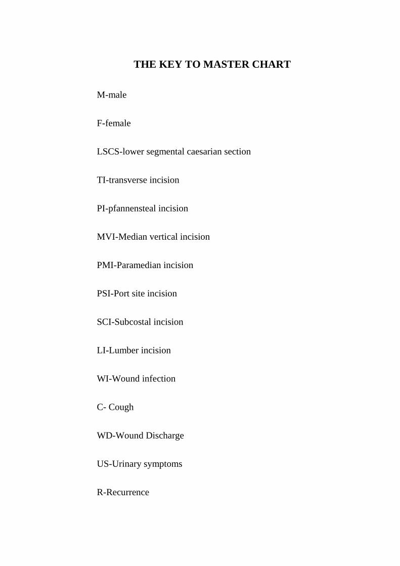
THE KEY TO MASTER CHART
M-male
F-female
LSCS-lower segmental caesarian section
TI-transverse incision
PI-pfannensteal incision
MVI-Median vertical incision
PMI-Paramedian incision
PSI-Port site incision
SCI-Subcostal incision
LI-Lumber incision
WI-Wound infection
C- Cough
WD-Wound Discharge
US-Urinary symptoms
R-Recurrence

Cs-constipation
m-Month
y-years
ON-Onlay mesh repair
SUB-Sublay mesh repair
COMP-component separation technique
LAP-laparoscopic technique
P-pain
B-bleeding
F-fever
PC-pelvic collection
MI-mesh infection
FN-flap necrosis
NC-no compliant