CLINICAL PRACTICE GUIDELINE Clinical Practice Guideline for the
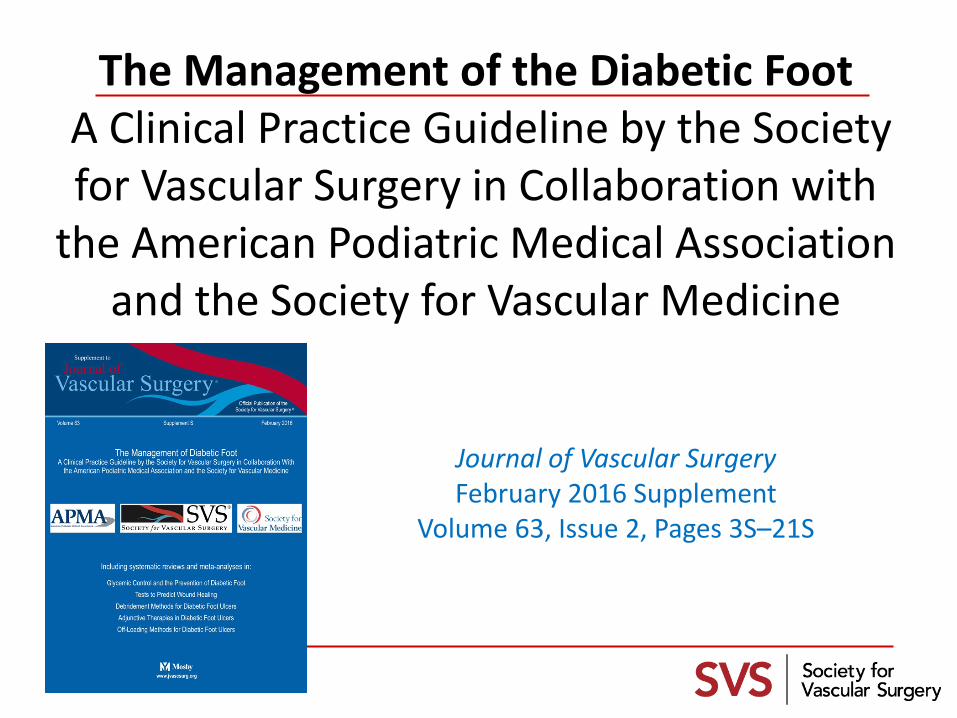
The Management of the Diabetic FootA Clinical Practice Guideline by the Society for Vascular Surgery in Collaboration with
the American Podiatric Medical Association and the Society for Vascular Medicine
Journal of Vascular SurgeryFebruary 2016 Supplement
Volume 63, Issue 2, Pages 3S–21S

Prevention of Diabetic Foot Ulceration
Off-loading DFUs
Wound Care for DFUs
Diagnosis of Diabetic Foot Osteomyelitis (DFO)
Peripheral Arterial Disease (PAD) and the DFU
5 Areas of Focus - DFU

Level of Evidence: A (high quality), B (moderate quality), and C (low quality)
Grade 2: Benefits and Risks Are More Closely Matched
Grade 1: Benefit Clearly Outweighs Risk
Grade 1 (strong) or Grade 2 (weak)
Grade the Strength of Recommendations & to Rate the Quality of Evidence (confidence in the estimates)
Grades of RecommendationAssessment, Development, and Evaluation (GRADE)

5 key questions were deemed to be in need of a full systematic review and meta-analysis. The evidence in several other areas
was summarized by consensus of committee members.
5 systematic reviews addressed the effect of glycemic control on
preventing DFU, the evidence supporting different off-loading methods, adjunctive therapies,
débridement, and tests to predict wound healing.
Numerous randomized controlled trials were identified in every
systematic review; however, most of these trials were small. Therefore, searches were
expanded to include nonrandomized trials as well.
5 Systematic Reviews and Meta-Analysis

A Systematic Review and Meta-Analysis of Glycemic Control for the Prevention of Diabetic Foot Syndrome
Journal of Vascular Surgery February 2016 SupplementVolume 63, Issue 2, Pages 22S–28S

A Systematic Review and Meta-Analysis of Tests to Predict Wound Healing in Diabetic Foot
Journal of Vascular Surgery February 2016 SupplementVolume 63, Issue 2, Pages 29S–36S

A Systematic Review and Meta-Analysis of DébridementMethods for Chronic Diabetic Foot Ulcers
Journal of Vascular Surgery February 2016 SupplementVolume 63, Issue 2, Pages 37S–45S

A Systematic Review and Meta-Analysis of Adjunctive Therapies in Diabetic Foot Ulcers
Journal of Vascular Surgery February 2016 SupplementVolume 63, Issue 2, Pages 46S–58S
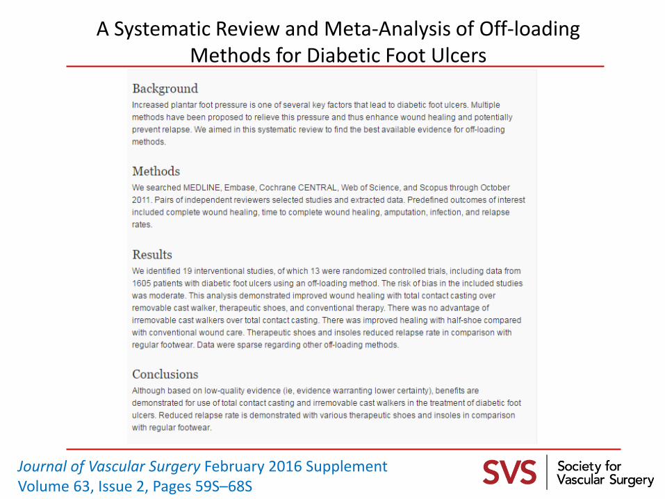
A Systematic Review and Meta-Analysis of Off-loading Methods for Diabetic Foot Ulcers
Journal of Vascular Surgery February 2016 SupplementVolume 63, Issue 2, Pages 59S–68S

Recommendation 1: We recommend that patients with diabetes undergo annual interval foot inspections by physicians (MD, DO, DPM) or advanced practice providers with training in foot care (Grade 1C)
Prevention of Diabetic Foot Ulceration

Category
0
1
2
3
Suggested Frequency for Follow-up Evaluation
Risk Profile
Normal
Peripheral Neuropathy
Neuropathy with Deformity and/or PAD
Previous Ulcer or Amputation
Evaluation Frequency
Annual
Semiannual
Quarterly
Monthly or Quarterly

Recommendation 2: We recommend that foot examination include testing for peripheral neuropathy using the Semmes-Weinstein test (Grade 1B)
Prevention of Diabetic Foot Ulceration

Recommendation 3: We recommend education of the patients and their families about preventive foot care (Grade 1C)
Prevention of Diabetic Foot Ulceration
Recommendation 4: a. We suggest against the routine use of specialized therapeutic footwear in average-risk diabetic patients (Grade 2C) b. We recommend using custom therapeutic footwear in high-risk diabetic patients, including those with significant neuropathy, foot deformities, or previous amputation(Grade 1B)
Prevention of Diabetic Foot Ulceration

Recommendation 5: We suggest adequate glycemic control (hemoglobin A1c < 7% with strategies to minimize hypoglycemia) to reduce the incidence of diabetic foot ulcers (DFUs) and infections, with subsequent risk of amputation (Grade 2B)
Prevention of Diabetic Foot Ulceration

Recommendation 6: We recommend against prophylactic arterial revascularization to prevent DFU (Grade 1C)
Prevention of Diabetic Foot Ulceration

Recommendation 1: In patients with plantar DFU, we recommend offloading with a total contact cast (TCC) or irremovable fixed ankle walking boot (Grade 1B)
Off-loading DFUs

Recommendation 2: In patients with DFU requiring frequent dressing changes, we suggest off-loading using a removablecast walker as an alternative to TCC and irremovable fixed ankle walking boot (Grade 2C). We suggest against using postoperative shoes or standard or customary footwear for off-loading plantar DFUs (Grade 2C)
Off-loading DFUs
Recommendation 3: In patients with nonplantar wounds, we recommend using any modality that relieves pressure at the site of the ulcer, such as a surgical sandal or heel relief shoe (Grade 1C)
Off-loading DFUs

Recommendation 4: In high-risk patients with healed DFU (including those with a prior history of DFU, partial foot amputation, or Charcot foot), we recommend wearing specific therapeutic footwear with pressure-relieving insoles to aid in prevention of new or recurrent foot ulcers (Grade 1C)
Off-loading DFUs

Recommendation 1: In patients with a diabetic foot infection (DFI) with an open wound, we suggest doing a probe to bone (PTB) test to aid in diagnosis (Grade 2C)
Diagnosis of Diabetic Foot Osteomyelitis (DFO)

Recommendation 2: In all patients presenting with a new DFI, we suggest that serial plain radiographs of the affected foot be obtained to identify bone abnormalities (deformity, destruction) as well as soft tissue gas and radiopaque foreign bodies (Grade 2C)
Diagnosis of DFO
Recommendation 3: For those patients who require additional (i.e., more sensitive or specific) imaging, particularly when soft tissue abscess is suspected or the diagnosis of osteomyelitis remains uncertain, we recommend using magnetic resonance imaging (MRI) as the study of choice. MRI is a valuable tool for diagnosis of osteomyelitis if the PTB test is inconclusive of if the plain film is not useful (Grade 1B)
Diagnosis of DFO

Recommendation 4: In patients with suspected DFO for whom MRI is contraindicated or unavailable, we suggest a leukocyte or antigranulocyte scan, preferably combined with a bone scan as the best alternative (Grade 2B)
Diagnosis of DFO
Recommendation 5: In patients at high risk for DFO, we recommend that the diagnosis is most definitively established by the combined findings on bone culture and histology (Grade 1C). When bone is débrided to treat osteomyelitis, we recommend sending a sample for culture and histology (Grade 1C)
Diagnosis of DFO
Recommendation 6: For patients not undergoing bone débridement, we suggest that clinicians consider obtaining a diagnostic bone biopsy when faced with diagnostic uncertainty, inadequate culture information, or failure of response to empirical treatment (Grade 2C)
Diagnosis of DFO

Recommendation 1: We recommend frequent evaluation at 1- to 4-week intervals with measurements of diabetic foot wounds to monitor reduction of wound size and healing progress (Grade 1C)
Recommendation 1.1: We recommend evaluation for infection on initial presentation of all diabetic foot wounds, with initial sharp débridement of all infected diabetic ulcers, and urgent surgical intervention for foot infections involving abscess, gas, or necrotizing fasciitis (Grade 1B)
Recommendation 1.2: We suggest that treatment of DFIs should follow the most current guidelines published by the Infectious Diseases Society of America (IDSA) (Ungraded)
Wound Care for DFUs

Recommendation 2: We recommend use of dressing products that maintain a moist wound bed, control exudate, and avoid maceration of surrounding intact skin for diabetic foot wounds (Grade 1B)
Wound Care for DFUs

Recommendation 3: We recommend sharp débridementof all devitalized tissue and surrounding callus material from diabetic foot ulcerations at 1- to 4-week intervals (Grade 1B)
Wound Care for DFUs

Recommendation 4: Considering lack of evidence for superiority of any given débridement technique, we suggest initial sharp débridement with subsequent choice of débridementmethod based on clinical context, availability of expertise and supplies, patient tolerance and preference, and cost-effectiveness (Grade 2C)
Wound Care for DFUs

Recommendation 5: For DFUs that fail to demonstrate improvement (>50% wound area reduction) after a minimum of 4 weeks of standard wound therapy, we recommend adjunctive wound therapy options. These include negative pressure therapy, biologics (platelet-derived growth factor [PDGF], living cellular therapy, extracellular matrix products, amnionic membrane products), and hyperbaric oxygen therapy. Choice of adjuvant therapy is based on clinical findings, availability of therapy, and cost-effectiveness; there is no recommendation on ordering of therapy choice. Re-evaluation of vascular status, infection control, and off-loading is recommended to ensure optimization before initiation of adjunctive wound therapy (Grade 1B)
Wound Care for DFUs

Recommendation 6: We suggest the use of negative pressure wound therapy for chronic diabetic foot wounds that do not demonstrate expected healing progression with standard or advanced wound dressings after 4 to 8 weeks of therapy (Grade 2B)
Wound Care for DFUs

Recommendation 7: We suggest consideration of the use of PDGF(becaplermin) for the treatment of DFUs that are recalcitrant to standard therapy (Grade 2B)
Wound Care for DFUs

Recommendation 8: We suggest consideration of living cellular therapy using a bilayered keratinocyte/fibroblast construct or a fibroblast-seeded matrix for treatment of DFUs when recalcitrant to standard therapy (Grade 2B)
Wound Care for DFUs

Recommendation 9: We suggest consideration of the use of extracellular matrix products employing acellular human dermis or porcine small intestinal submucosal tissue as an adjunctive therapy for DFUs when recalcitrant to standard therapy (Grade 2C)
Wound Care for DFUs

Recommendation 10: In patients with DFU who have adequate perfusion that fails to respond two 4 to 6 weeks of conservative management, we suggest hyperbaric oxygen therapy (Grade 2B)
Wound Care for DFUs

Recommendation 1.1: We suggest that patients with diabetes have ankle-brachial index (ABI) measurements performed when they reach 50 years of age (Grade 2C).
Peripheral Arterial Disease (PAD) and the DFU

Recommendation 1.2: We suggest that patients with diabetes who have a prior history of DFU, prior abnormal vascular examination, prior intervention for peripheral vascular disease, or known atherosclerotic cardiovascular disease (eg, coronary, cerebral, or renal) have an annular vascular examination of the lower extremities and feet including ABI and toe pressures (Grade 2C)
Peripheral Arterial Disease (PAD) and the DFU

Recommendation 2: We recommend that patients with DFU have pedal perfusion assessed by ABI, ankle and pedal Doppler arterial waveforms, and either toe systolic pressure or transcutaneous oxygen pressure (TcPO2) annually (Grade 1B)
PAD and the DFU

Recommendation 3: In patients with DFU with PAD, we recommend revascularization by either surgical bypass or endovascular therapy (Grade 1B)
PAD and the DFU

Recommendation 3 (continued): Prediction of patients most likely to require and to benefit from revascularization can be based on the Society for Vascular Surgery (SVS) Wound, Ischemia, and foot Infection (WIfI) lower extremity threatened limb classification.
PAD and the DFU

Recommendation 3 (continued): A combination of clinical judgment and careful interpretation of objective assessments of perfusion along with consideration of the wound and infection extent is required to select patients appropriately for revascularization.
PAD and the DFU

Recommendation 3 (continued): In functional patients with long-segment occlusive disease and a good autologous conduit, bypass is likely to be preferable.
PAD and the DFU

Recommendation 3 (continued): In the setting of tissue loss and diabetes, prosthetic bypass is inferior to bypass with vein conduit.
PAD and the DFU

Recommendation 3 (continued): The choice of intervention depends on the degree of ischemia, the extent of arterial disease, the extent of the wound, the presence or absence of infection, and the available expertise.
PAD and the DFU
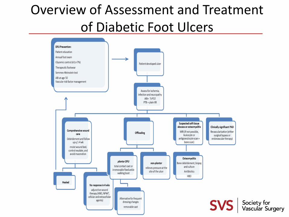
Overview of Assessment and Treatment of Diabetic Foot Ulcers