A case series of ovarian ectopic pregnancy at a rural ...
4
Indian Journal of Obstetrics and Gynecology Research 2020;7(4):600–603 Content available at: https://www.ipinnovative.com/open-access-journals Indian Journal of Obstetrics and Gynecology Research Journal homepage: www.ijogr.org Case Report A case series of ovarian ectopic pregnancy at a rural tertiary care hospital Kratika Kamath 1 , Gomathy E 1, * 1 Dept. of Obstetrics and Gynecology, Sri Devaraj Urs Medical College, Sri Devaraj Urs Academy of Higher Education and Research (SDUAHER), Kolar, Karnataka, India ARTICLE INFO Article history: Received 22-07-2020 Accepted 04-08-2020 Available online 07-12-2020 Keywords: Ovarian ectopic pregnancy Intrauterine contraceptive device use Adnexal mass ABSTRACT Ovarian ectopic pregnancy (OEP), is one of the variants of the non-tubal ectopic pregnancy, which varies one in 7,000–16,000 deliveries, and its prevalence is 1-3% among ectopic pregnancy. OEP is a complication which occurs when implantation and embryo development happens outside of the uterus. Usually, 91% of OEP terminates with rupture before the end of 1 st trimester and it can lead to pregnancy complications such as hemorrhage and hypovolemic shock. Clinical presentation of OEP is lower abdominal or pelvic pain, or both. In addition to this, other symptoms include nausea, vomiting, and constipation. In this, we present 3 cases of OEP who reported to our hospital as adnexal mass for evaluation, who came with acute abdomen and was subsequently confirmed at surgery and proven on histopathology or USG detected adnexal mass with hemoperitoneum with suspicion of ectopic pregnancy. Judicious use of ultrasound in an appropriate clinical setting can thus prevent mishaps and enable better management of such conditions. © This is an open access article distributed under the terms of the Creative Commons Attribution License (https://creativecommons.org/licenses/by/4.0/) which permits unrestricted use, distribution, and reproduction in any medium, provided the original author and source are credited. 1. Introduction Ovarian ectopic pregnancy (OEP), is one of the variants of the non-tubal ectopic pregnancy, which varies one in 7,000–16,000 deliveries, and its prevalence is 1-3% among ectopic pregnancy. The incidence of OEP increases following the use of ovulation-inducing agents and also increased usage of assisted reproductive techniques and intrauterine device. 1,2 OEP is a complication which occurs when implantation and embryo development happens outside of the uterus. When such ectopic pregnancy occurs in overy, it is known as an OEP. The Spiegelberg criterion is used for diagnosing ovarian pregnancies. Usually, 91% of OEP terminates with rupture before the end of 1 st trimester, 5.3% end in second trimester, and 3.7% end in the third trimester and it can lead to pregnancy complications such as hemorrhage and hypovolemic shock. 3 Clinical presentation of OEP is lower * Corresponding author. E-mail address: [email protected] (Gomathy E). abdominal or pelvic pain, or both. Pain being the common symptom and varies according the severity. In addition to this, other symptoms include nausea, vomiting, and constipation. 4 The risk factors of OEP are history of intrauterine contraceptive device use (IUCD), pelvic inflammatory disease (PID), sexually transmitted infections (STIs), use of assisted reproductive technologies, prior pelvic surgery, endometriosis, previous ectopic pregnancy, advanced maternal age and multi-parity. 5 The pre-operative diagnosis of OEP is challenging, despite the enhanced modern sonographic techniques and is most often made at the time of surgery. In this, we report here a series of 3 cases of ovarian ectopic pregnancy who reported to hospital as adnexal mass for evaluation, who came with acute abdomen and was subsequently confirmed at surgery and proven on histopathology or USG detected adnexal mass with hemoperitoneum with suspicion of ectopic pregnancy. Judicious use of ultrasound in an appropriate clinical setting https://doi.org/10.18231/j.ijogr.2020.127 2394-2746/© 2020 Innovative Publication, All rights reserved. 600
Transcript of A case series of ovarian ectopic pregnancy at a rural ...

Indian Journal of Obstetrics and Gynecology Research 2020;7(4):600–603
Content available at: https://www.ipinnovative.com/open-access-journals
Indian Journal of Obstetrics and Gynecology Research
Journal homepage: www.ijogr.org
Case Report
A case series of ovarian ectopic pregnancy at a rural tertiary care hospital
Kratika Kamath1, Gomathy E1,*1Dept. of Obstetrics and Gynecology, Sri Devaraj Urs Medical College, Sri Devaraj Urs Academy of Higher Education andResearch (SDUAHER), Kolar, Karnataka, India
A R T I C L E I N F O
Article history:Received 22-07-2020Accepted 04-08-2020Available online 07-12-2020
Keywords:Ovarian ectopic pregnancyIntrauterine contraceptive device useAdnexal mass
A B S T R A C T
Ovarian ectopic pregnancy (OEP), is one of the variants of the non-tubal ectopic pregnancy, which variesone in 7,000–16,000 deliveries, and its prevalence is 1-3% among ectopic pregnancy.OEP is a complication which occurs when implantation and embryo development happens outside of theuterus. Usually, 91% of OEP terminates with rupture before the end of 1st trimester and it can lead topregnancy complications such as hemorrhage and hypovolemic shock. Clinical presentation of OEP islower abdominal or pelvic pain, or both. In addition to this, other symptoms include nausea, vomiting, andconstipation.In this, we present 3 cases of OEP who reported to our hospital as adnexal mass for evaluation, whocame with acute abdomen and was subsequently confirmed at surgery and proven on histopathology orUSG detected adnexal mass with hemoperitoneum with suspicion of ectopic pregnancy. Judicious use ofultrasound in an appropriate clinical setting can thus prevent mishaps and enable better management ofsuch conditions.
© This is an open access article distributed under the terms of the Creative Commons AttributionLicense (https://creativecommons.org/licenses/by/4.0/) which permits unrestricted use, distribution, andreproduction in any medium, provided the original author and source are credited.
1. Introduction
Ovarian ectopic pregnancy (OEP), is one of the variantsof the non-tubal ectopic pregnancy, which varies onein 7,000–16,000 deliveries, and its prevalence is 1-3%among ectopic pregnancy. The incidence of OEP increasesfollowing the use of ovulation-inducing agents and alsoincreased usage of assisted reproductive techniques andintrauterine device.1,2
OEP is a complication which occurs when implantationand embryo development happens outside of the uterus.When such ectopic pregnancy occurs in overy, it is knownas an OEP. The Spiegelberg criterion is used for diagnosingovarian pregnancies. Usually, 91% of OEP terminates withrupture before the end of 1st trimester, 5.3% end in secondtrimester, and 3.7% end in the third trimester and it canlead to pregnancy complications such as hemorrhage andhypovolemic shock.3 Clinical presentation of OEP is lower
* Corresponding author.E-mail address: [email protected] (Gomathy E).
abdominal or pelvic pain, or both. Pain being the commonsymptom and varies according the severity. In additionto this, other symptoms include nausea, vomiting, andconstipation.4
The risk factors of OEP are history of intrauterinecontraceptive device use (IUCD), pelvic inflammatorydisease (PID), sexually transmitted infections (STIs), useof assisted reproductive technologies, prior pelvic surgery,endometriosis, previous ectopic pregnancy, advancedmaternal age and multi-parity.5 The pre-operative diagnosisof OEP is challenging, despite the enhanced modernsonographic techniques and is most often made at the timeof surgery.
In this, we report here a series of 3 cases of ovarianectopic pregnancy who reported to hospital as adnexalmass for evaluation, who came with acute abdomenand was subsequently confirmed at surgery and provenon histopathology or USG detected adnexal mass withhemoperitoneum with suspicion of ectopic pregnancy.Judicious use of ultrasound in an appropriate clinical setting
https://doi.org/10.18231/j.ijogr.2020.1272394-2746/© 2020 Innovative Publication, All rights reserved. 600

Kamath and Gomathy E / Indian Journal of Obstetrics and Gynecology Research 2020;7(4):600–603 601
can thus prevent mishaps and enable better management ofsuch conditions.
2. Case 1
A 28-year-old with history of previous LSCS camewith complains of sudden onset pain abdomen, with noaggravating or relieving factors, associated with 2 episodesof vomiting to casualty. Clinically her vitals were stable.Her systemic examination was within normal limits. Shehad missed her regular periods for one month, and her urinepregnancy test was positive.
Ultrasound of her abdomen showed uterus of about10-12 weeks size with endometrium was about 8 mmthick (Figures 1 and 2). No intrauterine or extrauterinegestational sac was seen.5,6 For better visualization of theuterus, and adnexal TVS scan was performed. A normal leftovary was demonstrated. To our surprise, a large irregularheterogenous lesion of about 410cc was noted in rightadnexa, extending up to the POD. The right ovary was notvisualized separately from the lesion. Thus, in this clinicalcontext, diagnosis of ruptured OEP was offered.
The patient was planned for exploratory laparotomy,where right ruptured ectopic pregnancy was observed,and right salpingoophorectomy was done. Samples weresent to histopathological analysis, which demonstrated thepresence of trophoblastic tissue with chorionic villi in theovarian tissue and confirmed this to be a case of rightruptured ovarian ectopic pregnancy (Figure 3).
Fig. 1: (a):Intraoperative finding in an exploratory laparotomy.(b): USG grey scale images of pelvis show uterus are normal,endometrium measured ~ 8 mm. No evidence of sac/ decidualreaction in the endometrium. Right adnexa showed large irregularheterogenous lesion, extending to pouch of doughlas
3. Case 2
A 29-year-old, with Copper T insitu came with complainsof pain abdomen since 1 day, sudden in onset withhistory 4 episodes of vomiting. Clinically her vitalswere stable. Her systemic examination was within normallimits. Her last menstrual cycle was 1 month back, withpositive urine pregnancy test. On ultrasound abdomen,her uterus had copper T insitu, with collection in PODof about 170ml, likely clots, with right ovary visualized
Fig. 2: USG grey scale images of pelvis show uterus is normal,endometrium measured ~ 8 mm. No evidence of sac/ decidualreaction in the endometrium. Right adnexa showed large irregularheterogenous lesion ~ 10.1 x 9.3 x 8.2 (410 cc) with internalanechoic cystic areasextending to pouch of doughlas (POD).
Fig. 3: Histopathological examination of ovarian tissue (a):Demonstrated the presence of trophoblastic tissue; (b): Showschorionic villiand confirmed this to be a case of right rupturedovarian ectopic pregnancy
within the clot with adjacent focal hyperechoic collection-representing-sentinel clot sign. On exploratory laparotomyand proceed, intraoperatively, right ovary was found tohave a bleeding point? Ruptured ectopic pregnancy wassuspected. Moderate hemoperitoneum was found. Copper Twas removed at the end of the procedure.
4. Case 3
A 20 year old, came with complains of pain abdomensince 1 day, sudden in onset with history 3episodes ofvomiting. She also gave history of 1 and half monthsof amenorrhea and urine pregnancy test was found to bepositive. On ultrasound abdomen, it was observed thatround heterogenous lesion with central anechoic gestational
602 Kamath and Gomathy E / Indian Journal of Obstetrics and Gynecology Research 2020;7(4):600–603
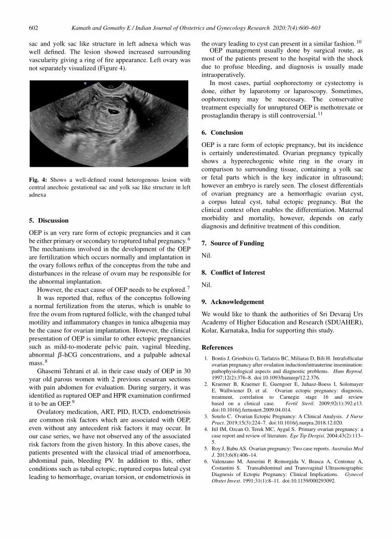
sac and yolk sac like structure in left adnexa which waswell defined. The lesion showed increased surroundingvascularity giving a ring of fire appearance. Left ovary wasnot separately visualized (Figure 4).
Fig. 4: Shows a well-defined round heterogenous lesion withcentral anechoic gestational sac and yolk sac like structure in leftadnexa
5. Discussion
OEP is an very rare form of ectopic pregnancies and it canbe either primary or secondary to ruptured tubal pregnancy.6
The mechanisms involved in the development of the OEPare fertilization which occurs normally and implantation inthe ovary follows reflux of the conceptus from the tube anddisturbances in the release of ovum may be responsible forthe abnormal implantation.
However, the exact cause of OEP needs to be explored.7
It was reported that, reflux of the conceptus followinga normal fertilization from the uterus, which is unable tofree the ovum from ruptured follicle, with the changed tubalmotility and inflammatory changes in tunica albugenia maybe the cause for ovarian implantation. However, the clinicalpresentation of OEP is similar to other ectopic pregnanciessuch as mild-to-moderate pelvic pain, vaginal bleeding,abnormal β -hCG concentrations, and a palpable adnexalmass.8
Ghasemi Tehrani et al. in their case study of OEP in 30year old parous women with 2 previous cesarean sectionswith pain abdomen for evaluation. During surgery, it wasidentified as ruptured OEP and HPR examination confirmedit to be an OEP.9
Ovulatory medication, ART, PID, IUCD, endometriosisare common risk factors which are associated with OEP,even without any antecedent risk factors it may occur. Inour case series, we have not observed any of the associatedrisk factors from the given history. In this above cases, thepatients presented with the classical triad of amenorrhoea,abdominal pain, bleeding PV. In addition to this, otherconditions such as tubal ectopic, ruptured corpus luteal cystleading to hemorrhage, ovarian torsion, or endometriosis in
the ovary leading to cyst can present in a similar fashion.10
OEP management usually done by surgical route, asmost of the patients present to the hospital with the shockdue to profuse bleeding, and diagnosis is usually madeintraoperatively.
In most cases, partial oophorectomy or cystectomy isdone, either by laparotomy or laparoscopy. Sometimes,oophorectomy may be necessary. The conservativetreatment especially for unruptured OEP is methotrexate orprostaglandin therapy is still controversial.11
6. Conclusion
OEP is a rare form of ectopic pregnancy, but its incidenceis certainly underestimated. Ovarian pregnancy typicallyshows a hyperechogenic white ring in the ovary incomparison to surrounding tissue, containing a yolk sacor fetal parts which is the key indicator in ultrasound;however an embryo is rarely seen. The closest differentialsof ovarian pregnancy are a hemorrhagic ovarian cyst,a corpus luteal cyst, tubal ectopic pregnancy. But theclinical context often enables the differentiation. Maternalmorbidity and mortality, however, depends on earlydiagnosis and definitive treatment of this condition.
7. Source of Funding
Nil.
8. Conflict of Interest
Nil.
9. Acknowledgement
We would like to thank the authorities of Sri Devaraj UrsAcademy of Higher Education and Research (SDUAHER),Kolar, Karnataka, India for supporting this study.
References1. Bontis J, Grimbizis G, Tarlatzis BC, Miliaras D, Bili H. Intrafollicular
ovarian pregnancy after ovulation induction/intrauterine insemination:pathophysiological aspects and diagnostic problems. Hum Reprod.1997;12(2):376–8. doi:10.1093/humrep/12.2.376.
2. Kraemer B, Kraemer E, Guengoer E, Juhasz-Boess I, SolomayerE, Wallwiener D, et al. Ovarian ectopic pregnancy: diagnosis,treatment, correlation to Carnegie stage 16 and reviewbased on a clinical case. Fertil Steril. 2009;92(1):392.e13.doi:10.1016/j.fertnstert.2009.04.014.
3. Sotelo C. Ovarian Ectopic Pregnancy: A Clinical Analysis. J NursePract. 2019;15(3):224–7. doi:10.1016/j.nurpra.2018.12.020.
4. Itil IM, Ozcan O, Terek MC, Aygul S. Primary ovarian pregnancy: acase report and review of literature. Ege Tip Dergisi. 2004;43(2):113–5.
5. Roy J, Babu AS. Ovarian pregnancy: Two case reports. Australas MedJ. 2013;6(8):406–14.
6. Valenzano M, Anserini P, Remorgida V, Brasca A, Centonze A,Costantini S. Transabdominal and Transvaginal UltrasonographicDiagnosis of Ectopic Pregnancy: Clinical Implications. GynecolObstet Invest. 1991;31(1):8–11. doi:10.1159/000293092.

Kamath and Gomathy E / Indian Journal of Obstetrics and Gynecology Research 2020;7(4):600–603 603
7. Zhu Q, Li C, Zhao WH, Yuan JJ, Yan M, Qin GJ, et al. Risk factorsand clinical features of ovarian pregnancy: a case–control study. BMJOpen. 2014;4(12). doi:10.1136/bmjopen-2014-006447.
8. Kadau JV. Sonographic Detection of Ovarian EctopicPregnancy. J Diagnostic Med Sonography. 2016;32(5):299–3.doi:10.1177/8756479316663163.
9. Middleton WD, Kurtz AB, Hertzberg BS. Ultrasound: The Requisites.St Louis: Mosby; 2004.
10. Tehrani HG, Hamoush Z, Ghasemi M, Hashemi L. Ovarian ectopicpregnancy: A rare case. Iran J Reprod Med. 2014;12(4):281–4.
11. Siddiqua SF, Abbasi S, Rijvi S, Hasan AS. Ovarian Pregnancy- ARare Case Report. Anwer Khan Modern Med Coll J. 2019;10(2):183–5. doi:10.3329/akmmcj.v10i2.44134.
Author biography
Kratika Kamath, Post Graduate Student
Gomathy E, Professor
Cite this article: Kamath K, Gomathy E. A case series of ovarianectopic pregnancy at a rural tertiary care hospital. Indian J ObstetGynecol Res 2020;7(4):600-603.