A Case report of Nasal r hinosporidiosis presenting as an ...
6
44 p-ISSN:2231-6140 * Corresponding Author: Dr Krishna Potdukhe E-mail: krishnapotdukhe92@ A Case report of Nasal mass. Dr Kalpesh Patel 1 , Dr Krishna 1 Associate Professor, 2 Resident Civil Hospital, Ahmedabad. 3 Resident doctor, Department of Abstract: Rhinosporidiosis is a r lesions of the mucous mem nasopharynx, conjunctiva an subcontinent, but remains qui male patient, native of Banask The patient presented with a epistaxis for 2 months. Diagn was successfully operated wit of nasal mass in our institute. nasal polyps, angiofibroma an region to consider rhinospo presenting with nasal swelling Key words: Nasal obstruction Introduction: Rhinosporidiosis is a mucocutaneous disease caused which affects mainly the naso other mucosal surfaces. [1] It oc reported in the Americas, Eu with the highest incidences Lanka. [1] Infection occurs afte in stagnant water ponds, lakes In the nasal mucosa, the dis friable, polypoidal, hyperpl confused with malignant lesio The clinical diagnosis and histopathological confirm delineate the extent of the lesi out other common clinical diff BJKines-NJBAS Volume-10(1), June 0, e-ISSN:2395-7859 @gmail.com l rhinosporidiosis presenting as an a Potdukhe 2* , Dr Saketh Rao 3 doctor, Department of Otorhinolaryngology, B. Radiology, B. J. Medical College, Civil Hospital rare chronic granulomatous disease, character mbrane. It commonly affects the mucous and palate. The disease is more prevale ite rare. We hereby present a case descriptio kantha district, diagnosed with polypoid nas history of nasal obstruction since 10 month nosis was confirmed by histopathological e th endoscopic resection of mass. This was a Nasal rhinosporidiosis lesions commonly mi nd angiomatous polyp, it is therefore crucial f oridiosis as a differential diagnosis when gs. n, Nasopharynx, Rhinosporidosis. an infectious, granulomatous, d by Rhinosporidium Seeberi, o-oropharynx and occasionally ccurs worldwide and has been urope, East Africa, and Asia, registered in India and Sri er inoculation of spores found s, and rivers, and even in dust. sease manifests as a reddish, lastic mass, which can be ons, especially when ulcerated. [2] s of rhinosporidiosis, affecting the nose and mation is essential. Radiological evaluation i ion, the status of underlying structures and m ferentials. Case report: A 19-year old male e 2018 2018 Case Report oropharyngeal . J. Medical College, l, Ahmedabad. rised by polypoidal membrane of the ent in the Indian on of a 19 year old sal rhinosporidiosis. hs and intermittent examination and he very unusual cause imic other ordinary for clinicians in our assessing patients d throat, is difficult is often required to maybe useful to rule presented to our
Transcript of A Case report of Nasal r hinosporidiosis presenting as an ...

44 p-ISSN:2231-6140,
* Corresponding Author: Dr Krishna Potdukhe E-mail: [email protected]
A Case report of Nasal rmass.
Dr Kalpesh Patel1, Dr Krishna Potdukhe1Associate Professor, 2Resident doctor, Department of Otorhinolaryngology, B. J. Medical College, Civil Hospital, Ahmedabad.
3Resident doctor, Department of Radiology, B. J. Medical College, Civil Hospital, Ahmedabad.
Abstract:
Rhinosporidiosis is a rare chronic granulomatouslesions of the mucous membrane. It commonly affects the mucous membrane of the nasopharynx, conjunctiva and palate. The disease is more prevalent in the Indian subcontinent, but remains quite rare. We hereby present a male patient, native of Banaskantha district,The patient presented with a history of nasal obstruction since 10 months and intermittent epistaxis for 2 months. Diagnosis waswas successfully operated with endoscopic resection of mass. This was a very unusual cause of nasal mass in our institute. Nasal rhinosporidiosis lesions commonly mimic other ordinary nasal polyps, angiofibroma and angiomatous polyp, it is therefore crucial for clinicians in our region to consider rhinosporidiosis as a differential diagnosis when assessing patients presenting with nasal swellings.
Key words: Nasal obstruction
Introduction:
Rhinosporidiosis is an infectious, granulomatous, mucocutaneous disease caused by Rhinosporidiumwhich affects mainly the nasoother mucosal surfaces.[1] It occurs worldwide and has been reported in the Americas, Europe, East Africa, and Asia, with the highest incidences registered in India and Sri Lanka.[1] Infection occurs after inoculation of spores found in stagnant water ponds, lakes, and rivers, and In the nasal mucosa, the disease manifests as afriable, polypoidal, hyperplastic massconfused with malignant lesions, especially when ulcerated.
The clinical diagnosis of rhinosporidiosis, affecting the nose and thand histopathological confirmation is essential. Radiological evaluation is often required to delineate the extent of the lesion, the status of underlying structures and maybe useful to rule out other common clinical differentials.
BJKines-NJBAS Volume-10(1), June
6140, e-ISSN:2395-7859
A Case report of Nasal rhinosporidiosis presenting as an
, Dr Krishna Potdukhe2*, Dr Saketh Rao3
Resident doctor, Department of Otorhinolaryngology, B. J. Medical College,
Resident doctor, Department of Radiology, B. J. Medical College, Civil Hospital, Ahmedabad.
Rhinosporidiosis is a rare chronic granulomatous disease, characterised by polypoidallesions of the mucous membrane. It commonly affects the mucous membrane of the nasopharynx, conjunctiva and palate. The disease is more prevalent in the Indian subcontinent, but remains quite rare. We hereby present a case description of a 19 year old male patient, native of Banaskantha district, diagnosed with polypoid nasal rThe patient presented with a history of nasal obstruction since 10 months and intermittent epistaxis for 2 months. Diagnosis was confirmed by histopathological examination and he was successfully operated with endoscopic resection of mass. This was a very unusual cause of nasal mass in our institute. Nasal rhinosporidiosis lesions commonly mimic other ordinary
and angiomatous polyp, it is therefore crucial for clinicians in our region to consider rhinosporidiosis as a differential diagnosis when assessing patients presenting with nasal swellings.
asal obstruction, Nasopharynx, Rhinosporidosis.
Rhinosporidiosis is an infectious, granulomatous, mucocutaneous disease caused by Rhinosporidium Seeberi, which affects mainly the naso-oropharynx and occasionally
It occurs worldwide and has been reported in the Americas, Europe, East Africa, and Asia, with the highest incidences registered in India and Sri
Infection occurs after inoculation of spores found in stagnant water ponds, lakes, and rivers, and even in dust. In the nasal mucosa, the disease manifests as a reddish,
polypoidal, hyperplastic mass, which can be confused with malignant lesions, especially when ulcerated.[2]
The clinical diagnosis of rhinosporidiosis, affecting the nose and thand histopathological confirmation is essential. Radiological evaluation is often required to delineate the extent of the lesion, the status of underlying structures and maybe useful to rule out other common clinical differentials.
Case report:
A 19-year old male presented to our
10(1), June 2018 2018
Case Report
oropharyngeal
Resident doctor, Department of Otorhinolaryngology, B. J. Medical College,
Resident doctor, Department of Radiology, B. J. Medical College, Civil Hospital, Ahmedabad.
disease, characterised by polypoidal lesions of the mucous membrane. It commonly affects the mucous membrane of the nasopharynx, conjunctiva and palate. The disease is more prevalent in the Indian
case description of a 19 year old diagnosed with polypoid nasal rhinosporidiosis.
The patient presented with a history of nasal obstruction since 10 months and intermittent confirmed by histopathological examination and he
was successfully operated with endoscopic resection of mass. This was a very unusual cause of nasal mass in our institute. Nasal rhinosporidiosis lesions commonly mimic other ordinary
and angiomatous polyp, it is therefore crucial for clinicians in our region to consider rhinosporidiosis as a differential diagnosis when assessing patients
The clinical diagnosis of rhinosporidiosis, affecting the nose and throat, is difficult and histopathological confirmation is essential. Radiological evaluation is often required to delineate the extent of the lesion, the status of underlying structures and maybe useful to rule
year old male presented to our
45 p-ISSN:2231-6140,
hospital with long standing history of leftgave history of occasional epistaxis, intermittent postbody sensation in oral cavity with occasional cough since 2 months. He reported no history of significant constitutional symptoms. The patiensimilar illness in the family.
On general examination, we observed that the young man had good nutritional status, was not pale or dyspnoeic and had poor general hygiene.
Intraoral examination revealed a pinkish rthe left lateral aspect of oropharynx, behind the faucial pillar, measuring asize. The lesion was firm and bled on touch on palpation with the tongue depressor. examination there was a polypThe mass was erythematous, nonendoscope couldn’t be passed beyond the lesion.enlargement of local-regional lymph nodes was observed. Other systemic examination was unremarkable.
The following differential diagnoses were considered: Vascular polyp, Nasopharyngeal carcinoma and M
Work-up of the patient comprised of biochemical and haematological profiles as well as radiological work up. Biochemical and haematological parameters were essentially normal.
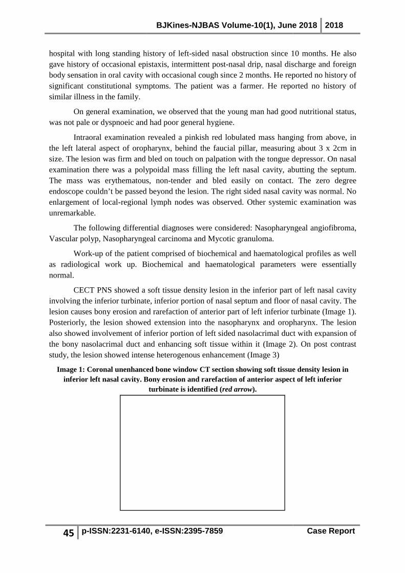
CECT PNS showed a soft tissue density lesion in the inferior part of left nasalinvolving the inferior turbinate, inferior portion of nasal septum and floor of nasal cavitylesion causes bony erosion and rarefaction of anterior partPosteriorly, the lesion showed extension into the naalso showed involvement of inferior portion of left sided nasolacrimal duct with expansion of the bony nasolacrimal duct and enhancing soft tissue within it (study, the lesion showed intense h
Image 1: Coronal unenhanced bone window CT section showing soft tissue density lesion in inferior left nasal cavity. Bony erosion and rarefaction of anterior aspect of left inferior
BJKines-NJBAS Volume-10(1), June
6140, e-ISSN:2395-7859
hospital with long standing history of left-sided nasal obstruction since 10 months. He also gave history of occasional epistaxis, intermittent post-nasal drip, nasal discharge and foreign body sensation in oral cavity with occasional cough since 2 months. He reported no history of significant constitutional symptoms. The patient was a farmer. He reported no history of
On general examination, we observed that the young man had good nutritional status, was not pale or dyspnoeic and had poor general hygiene.
ntraoral examination revealed a pinkish red lobulated mass hanging from above, in the left lateral aspect of oropharynx, behind the faucial pillar, measuring a
The lesion was firm and bled on touch on palpation with the tongue depressor. polypoidal mass filling the left nasal cavity, abutting the septum.
The mass was erythematous, non-tender and bled easily on contact. The zero degree endoscope couldn’t be passed beyond the lesion. The right sided nasal cavity was normal. No
regional lymph nodes was observed. Other systemic examination was
The following differential diagnoses were considered: Nasopharyngeal angiofibroma, Nasopharyngeal carcinoma and Mycotic granuloma.
up of the patient comprised of biochemical and haematological profiles as well as radiological work up. Biochemical and haematological parameters were essentially
CECT PNS showed a soft tissue density lesion in the inferior part of left nasalinvolving the inferior turbinate, inferior portion of nasal septum and floor of nasal cavitylesion causes bony erosion and rarefaction of anterior part of left inferior turbinate (Posteriorly, the lesion showed extension into the nasopharynx and oropharynx. The lesion also showed involvement of inferior portion of left sided nasolacrimal duct with expansion of
and enhancing soft tissue within it (Image 2). On post contrast study, the lesion showed intense heterogenous enhancement (Image 3)
1: Coronal unenhanced bone window CT section showing soft tissue density lesion in inferior left nasal cavity. Bony erosion and rarefaction of anterior aspect of left inferior
turbinate is identified (red arrow).
10(1), June 2018 2018
Case Report
10 months. He also nasal drip, nasal discharge and foreign
body sensation in oral cavity with occasional cough since 2 months. He reported no history of t was a farmer. He reported no history of
On general examination, we observed that the young man had good nutritional status,
ed lobulated mass hanging from above, in the left lateral aspect of oropharynx, behind the faucial pillar, measuring about 3 x 2cm in
The lesion was firm and bled on touch on palpation with the tongue depressor. On nasal abutting the septum.
tender and bled easily on contact. The zero degree The right sided nasal cavity was normal. No
regional lymph nodes was observed. Other systemic examination was
asopharyngeal angiofibroma,
up of the patient comprised of biochemical and haematological profiles as well as radiological work up. Biochemical and haematological parameters were essentially
CECT PNS showed a soft tissue density lesion in the inferior part of left nasal cavity involving the inferior turbinate, inferior portion of nasal septum and floor of nasal cavity. The
of left inferior turbinate (Image 1). sopharynx and oropharynx. The lesion
also showed involvement of inferior portion of left sided nasolacrimal duct with expansion of 2). On post contrast
1: Coronal unenhanced bone window CT section showing soft tissue density lesion in inferior left nasal cavity. Bony erosion and rarefaction of anterior aspect of left inferior

46 p-ISSN:2231-6140,
Image 2: Axial unenhanced bone window CT cavity extending posteriorly into nasopharynx on left side (
bony portion of left sided nasolacrimal duct with soft tissue wi
Image 3: Axial contrast enhanced soft window CT enhancing (red arrow) in left nasal cavity extending into nasopharynx on left side. Also note
enhancing lesion in expanded left bony nasolacrimal du
Patient underwent endoscopic excision of mass coupled with electric cauterisation of the base. Anterior nasal packing was done which was removed after 2 days. Postperiod was uneventful; the patient did remarkably well and was discharged dapsone for a 6 month period. Followrecurrence.
Gross examination of the specimen depicted an intact elongated, polypoidal soft friable greyish mass measuring about 9 x 4 x 1cm (Histopathological examination of the excised tissue showed granulation tissue containing plasma cells, lymphocytes and collection of histiocytes and neutrophils. Globular cysts containing spores (sporangia) in different stages of developmpredominantly in the stroma of the polypoidal tissue. These findings were pathognomonic of rhinosporidiosis. Henceforth, a final diagnosis of rhinosporidiosis was established.
BJKines-NJBAS Volume-10(1), June
6140, e-ISSN:2395-7859
Image 2: Axial unenhanced bone window CT scan showing soft tissue density lesion in left nasal cavity extending posteriorly into nasopharynx on left side (yellow arrow). Expansion of inferior
bony portion of left sided nasolacrimal duct with soft tissue within it (
Image 3: Axial contrast enhanced soft window CT scan showing intense heterogeneously ) in left nasal cavity extending into nasopharynx on left side. Also note
enhancing lesion in expanded left bony nasolacrimal duct.
Patient underwent endoscopic excision of mass coupled with electric cauterisation of the base. Anterior nasal packing was done which was removed after 2 days. Post
he patient did remarkably well and was discharged dapsone for a 6 month period. Follow-up visit six months later revealed no signs of
Gross examination of the specimen depicted an intact elongated, polypoidal soft friable greyish mass measuring about 9 x 4 x 1cm (Image 4). Cut surfHistopathological examination of the excised tissue showed granulation tissue containing plasma cells, lymphocytes and collection of histiocytes and neutrophils. Globular cysts containing spores (sporangia) in different stages of development were identified predominantly in the stroma of the polypoidal tissue. These findings were pathognomonic of rhinosporidiosis. Henceforth, a final diagnosis of rhinosporidiosis was established.
10(1), June 2018 2018
Case Report
showing soft tissue density lesion in left nasal ). Expansion of inferior
thin it (red arrow).
showing intense heterogeneously ) in left nasal cavity extending into nasopharynx on left side. Also note
ct.
Patient underwent endoscopic excision of mass coupled with electric cauterisation of the base. Anterior nasal packing was done which was removed after 2 days. Post-operative
he patient did remarkably well and was discharged with tablet up visit six months later revealed no signs of
Gross examination of the specimen depicted an intact elongated, polypoidal soft 4). Cut surface was whitish.
Histopathological examination of the excised tissue showed granulation tissue containing plasma cells, lymphocytes and collection of histiocytes and neutrophils. Globular cysts
ent were identified predominantly in the stroma of the polypoidal tissue. These findings were pathognomonic of rhinosporidiosis. Henceforth, a final diagnosis of rhinosporidiosis was established.

BJKines-NJBAS Volume-10(1), June 2018 2018
47 p-ISSN:2231-6140, e-ISSN:2395-7859 Case Report
Image 4: Excised polypoidal specimen (Soft friable greyish with irregular surface.)
Discussion:
Rhinosporidiosis has existed since ancient times. Initial account of this disease entity was made over a century ago in Latin America.[3] Though it appears to occur universally, rhinosporidiosis remains largely endemic in Indian subcontinent. The mode of transmission to humans is not clearly understood, however most researchers believe that direct contact with spores through dust, soil or prolonged exposure to stagnant water are among the major risk factors for acquisition of the infection.[3]Many patients may give similar history which points towards the diagnosis. We believe that in our case, the patient being a farmer by occupation, his frequent bathing in ponds and stagnant waters and overall lack of personal hygiene, were high risk factors contributing to contraction of such an infection.
Rhinosporidiosis is a non-contagious chronic granulomatous infection seen mainly in adult males aged between 20 and 40 years.[4] Usually the patient presents with a history of gradual nasal obstruction, occasional epistaxis, nasal itching, sneezing, and at times post-nasal dripping.[1] Clinically, nasal rhinosporidiosis is characterised by development of single pedunculated or sessile polyp, multiple sessile polypoidal lesions or a combination of both [3]. Our case had a single polypoidal lesion. The classical clinical finding in RS is the presence of
Image 5 Multiple varying sized mature and immature sporangia
[H&E, 4X]
Image 6 Sporangia containing microspores [H&E, 10X]

BJKines-NJBAS Volume-10(1), June 2018 2018
48 p-ISSN:2231-6140, e-ISSN:2395-7859 Case Report
a reddish mass with greyish-whitish dots on the surface (representing sporangia); however this was not seen in our case. The lesion often bleeds on touch and has a soft friable consistency. Location of the lesion plays an important role in narrowing down the differential diagnosis. Contrary to ordinary polyps which often arise from the middle turbinate, rhinosporidiosis frequently involves mucosal lining of the naso-pharynx, anterior nares and inferior nasal cavity. [5]Nasal polyps originating from these locations should always be treated with high index of suspicion. In the nasal cavity, the most common site involved was the inferior nasal cavity, comprising nasal floor, inferior turbinate, and inferior meatus.[2] According to Banjara et al., the most common sites of involvement in rhinosporidiosis in order of frequency are nasal cavity, nasopharynx, lacrimal sac, and conjunctiva.[1]
Radiological features, though nonspecific may provide a clue and may give rise to suspicion of rhinosporidiosis infection. The most common imaging appearance of nasal RS is that of a polypoidal lesion centred in the inferior nasal cavity, (as opposed to the nasopharynx and sphenopalatine foramen in JNAF) involving the nasal floor, inferior turbinate, inferior meatus and inferior part of septum, as was the case in our patient. Involvement of surrounding bones is common in rhinosporidiosis, as seen in our case. Bone involvement is seen as irregularity, rarefaction, partial or complete erosion of inferior turbinate, thinning of medial maxillary wall, and septal erosion. Maxillary sinus extension is uncommon and this feature may help in differentiating it from other nasal masses such as antro-choanal polyps and inverted papillomas. nasolacrimal duct involvement is common due to frequent presence of rhinosporidiosis in inferior meatus and is diagnosed when nasolacrimal duct is dilated with extension of soft tissue density into it, as was seen in our case.[1]
Apart from history and clinical and radiological findings, histopathology is mandatory for definitive diagnosis of rhinosporidiosis. Definitive diagnosis of rhinosporidiosis depends upon identification of the pathogen in its diverse stages on biopsied or resected tissues.[4]
Histopathological sections show multiple sporangia in various stages of maturity, enclosed in a thin chitinous wall. The sporangia are 50-1000 µm in diameter, containing numerous endospores of diameter 5-10 µm. Overlying epithelium is usually hyperplastic, with or without areas of ulceration and loose fibrovascular stroma infiltrated with lymphocytes, macrophages, plasma cells and even polymorphonuclear leucocytes. Rupture of sporangia can cause giant cell reaction.[5]
Surgical excision remains the mainstay of treatment for rhinosporidiosis lesions.[3] Wide, complete and meticulous excision of the polyp followed by thorough electro-cautery of the lesion's base is recommended. It is hypothesized that cauterisation of the lesion's base may abate recurrence resulting from spillage of endospores on the adjacent mucosa.[3]
Besides surgery, a variety of adjuvant medical therapies have been tried in the treatment of rhinosporidiosis. These include drugs like griseofluvin, amphoterecin B and dapsone (4, 4-diaminodiphenyl sulphone). Dapsone is a promising drug that is used as an adjunct to surgery as it arrests the maturation of sporangia and promotes fibrosis. However, by far there has been no tangible success with medical therapy. [2]
Conclusion:

BJKines-NJBAS Volume-10(1), June 2018 2018
49 p-ISSN:2231-6140, e-ISSN:2395-7859 Case Report
Nasal rhinosporidiosis seldom remains a diagnostic consideration in our region. However, with emerging reports of sporadic cases, it is consequently imperative for clinicians in our setting to consider rhinosporidiosis as a differential diagnosis when evaluating patients presenting with nasal growth.
References
1. Prabhu S. M., Irodi A., Khiangte H. L., Rupa V., Naina P. (2013). Imaging features of rhinosporidiosis on contrast CT. The Indian Journal of Radiology & Imaging, 23(3), 212–218.
2. Rath R., Baig S. A., Debata T. (2015). Rhinosporidiosis presenting as an oropharyngeal mass: A clinical predicament? Journal of Natural Science, Biology, and Medicine, 6(1), 241–245.
3. Saha J., Basu A. J., Sen I., Sinha R., Bhandari A. K., Mondal S. (2011). Atypical Presentations of Rhinosporidiosis: A Clinical Dilemma? Indian Journal of Otolaryngology and Head & Neck Surgery, 63(3), 243–246.
4. Kumari R, Nath AK, Rajalakshmi R, Adityan B, Thappa DM. Disseminated cutaneous rhinosporidiosis: Varied morphological appearances on the skin. Indian J Dermatol Venereol Leprol 2009; 75:68-71.
5. Sinha A, Phukan JP, Bandyopadhyay G, et al. Clinicopathological study of rhinosporidiosis with special reference to cytodiagnosis. Journal of Cytology / Indian Academy of Cytologists. 2012; 29(4):246-249. doi:10.4103/0970-9371.103943.