A case of primary adenosquamous carcinoma of the liver · Magnetic resonance imaging study showed a...
4
Korean J Hepatobiliary Pancreat Surg 2013;17:38-41 Case Report A case of primary adenosquamous carcinoma of the liver Gu Hyum Kang 1 , Byung Seok Lee 1 , and Dae Young Kang 2 Departments of 1 Internal Medicine and 2 Pathology, Chungnam National University School of Medicine, Daejeon, Korea Adenosquamous carcinoma (ASC) is a rare tumor of the liver, which carries a poor prognosis. It has been reported mostly in the form of case studies. Recently, we experienced a case of primary ASC of the liver that has been pre- operatively considered as cholangiocelluar carcinoma of the liver. Microscopically, the tumor was composed of ad- enocarcinoma, squamous cell carcinoma, as well as transitional area, including the transformation of adenocarcinoma to squamous cell carcinoma. The patient remained well with no evidence of post-resection complication or recurrence for 15 month. (Korean J Hepatobiliary Pancreat Surg 2013;17:38-41) Key Words: Cholangiocelluar carcinoma; Adenosquamous carcinoma; Liver Received: June 5, 2012; Revised: August 27, 2012; Accepted: October 15, 2012 Corresponding author: Byung Seok Lee Department of Internal Medicine, Chungnam National University Hospital, 640, Daesa-dang, Jung-gu, Daejeon 301-721, Korea Tel: +82-42-280-8049, Fax: +82-42-257-5753, E-mail: [email protected] Copyright Ⓒ 2013 by The Korean Association of Hepato-Biliary-Pancreatic Surgery Korean Journal of Hepato-Biliary-Pancreatic Surgery ∙ ISSN: 1738-6349 INTRODUCTION Adenosquamous carcinoma (ASC) is a tumor composed of both adenocarcinoma and squamous cell carcinoma components, especially adenocarcinoma containing sig- nificant amounts of unequivocal squamous carcinomatous elements, for example keratin and/or intercelluar bridges. Primary ASC is considered as a subtype of cholangio- celluar carcinoma (CCC). The incidence of ASC is rather common in the gall bladder, pancreas, stomach, thyroid, and large intestine, but hepatic ASC is very rare. Primary ASC of the liver has been reported mostly in a form of case studies in South Korea. ASC tends to present more aggressive clinicopathologic features and has a worse prognosis than CCC. 1,2 From five hepatobiliary ASC for the past 10 years in our institute, one primary hepatic ASC was observed. We describe a case of primary hepatic ASC, preoperatively diagnosed as CCC, along with inves- tigating the literature of primary hepatic ASC and at- tempting to explore it clinicopathologically. CASE A 73-year-old man complained of fever and chill, and was hospitalized for urinary tract infection at the local clinic. During hospitalization, hepatic mass was observed from ultrasonography (US) and was transferred to Chung- nam National University Hospital for further investigation of the liver mass. He had a history of endoscopic retro- grade cholangiopancreatography (ERCP) and endoscopic sphincterotomy with stone extraction, due to cholangitis accompanying bile duct stone 6 years ago. He had neither specific family history nor history of smoking, as well as drinking. He was currently working in the field of agricul- ture. On admission, he was chronic ill looking with icteric sclera. He had a temperature of 36.2 o C and physical ex- amination was unremarkable. Initial laboratory evaluation were as follows: WBC 7,880/mm 3 , Hb 13.1 g/dl, platelet 258,000/mm 3 , PT (pro- thrombin time) 12.5 sec (INR: 1.1), aPTT (partial throm- boplastin time) 23.6 sec, total protein 7.5 mg/dl, Albumin 3.5 mg/dl, AST 13 IU/L, ALT 8 IU/L, ALP 151 IU/L, GGT (gamma glutamyl transferase) 89 IU/L, total bilir- ubin 1.4 mg/dl. HBsAg and HBsAb were both negative and HCV Ab was negative. CA 19-9 (carbohydrate anti- gen 19-9) and CEA (carcinoembryonic antigen) were 1,600 U/ml and 7.2 ng/ml. Abdominal computed tomog- raphy (CT) from the local clinic showed low-density mass with irregular margin on the left lobe. Hepatic mass showed rim enhancement on early phase with slow central
Transcript of A case of primary adenosquamous carcinoma of the liver · Magnetic resonance imaging study showed a...

Korean J Hepatobiliary Pancreat Surg 2013;17:38-41 Case Report
A case of primary adenosquamous carcinoma of the liver
Gu Hyum Kang1, Byung Seok Lee1, and Dae Young Kang2
Departments of 1Internal Medicine and 2Pathology, Chungnam National University School of Medicine, Daejeon, Korea
Adenosquamous carcinoma (ASC) is a rare tumor of the liver, which carries a poor prognosis. It has been reported mostly in the form of case studies. Recently, we experienced a case of primary ASC of the liver that has been pre-operatively considered as cholangiocelluar carcinoma of the liver. Microscopically, the tumor was composed of ad-enocarcinoma, squamous cell carcinoma, as well as transitional area, including the transformation of adenocarcinoma to squamous cell carcinoma. The patient remained well with no evidence of post-resection complication or recurrence for 15 month. (Korean J Hepatobiliary Pancreat Surg 2013;17:38-41)
Key Words: Cholangiocelluar carcinoma; Adenosquamous carcinoma; Liver
Received: June 5, 2012; Revised: August 27, 2012; Accepted: October 15, 2012Corresponding author: Byung Seok LeeDepartment of Internal Medicine, Chungnam National University Hospital, 640, Daesa-dang, Jung-gu, Daejeon 301-721, KoreaTel: +82-42-280-8049, Fax: +82-42-257-5753, E-mail: [email protected]
Copyright Ⓒ 2013 by The Korean Association of Hepato-Biliary-Pancreatic SurgeryKorean Journal of Hepato-Biliary-Pancreatic Surgery ∙ ISSN: 1738-6349
INTRODUCTION
Adenosquamous carcinoma (ASC) is a tumor composed of both adenocarcinoma and squamous cell carcinoma components, especially adenocarcinoma containing sig-nificant amounts of unequivocal squamous carcinomatous elements, for example keratin and/or intercelluar bridges. Primary ASC is considered as a subtype of cholangio-celluar carcinoma (CCC). The incidence of ASC is rather common in the gall bladder, pancreas, stomach, thyroid, and large intestine, but hepatic ASC is very rare. Primary ASC of the liver has been reported mostly in a form of case studies in South Korea. ASC tends to present more aggressive clinicopathologic features and has a worse prognosis than CCC.1,2 From five hepatobiliary ASC for the past 10 years in our institute, one primary hepatic ASC was observed. We describe a case of primary hepatic ASC, preoperatively diagnosed as CCC, along with inves-tigating the literature of primary hepatic ASC and at-tempting to explore it clinicopathologically.
CASE
A 73-year-old man complained of fever and chill, and was hospitalized for urinary tract infection at the local
clinic. During hospitalization, hepatic mass was observed from ultrasonography (US) and was transferred to Chung-nam National University Hospital for further investigation of the liver mass. He had a history of endoscopic retro-grade cholangiopancreatography (ERCP) and endoscopic sphincterotomy with stone extraction, due to cholangitis accompanying bile duct stone 6 years ago. He had neither specific family history nor history of smoking, as well as drinking. He was currently working in the field of agricul-ture. On admission, he was chronic ill looking with icteric sclera. He had a temperature of 36.2oC and physical ex-amination was unremarkable.
Initial laboratory evaluation were as follows: WBC 7,880/mm3, Hb 13.1 g/dl, platelet 258,000/mm3, PT (pro-thrombin time) 12.5 sec (INR: 1.1), aPTT (partial throm-boplastin time) 23.6 sec, total protein 7.5 mg/dl, Albumin 3.5 mg/dl, AST 13 IU/L, ALT 8 IU/L, ALP 151 IU/L, GGT (gamma glutamyl transferase) 89 IU/L, total bilir-ubin 1.4 mg/dl. HBsAg and HBsAb were both negative and HCV Ab was negative. CA 19-9 (carbohydrate anti-gen 19-9) and CEA (carcinoembryonic antigen) were 1,600 U/ml and 7.2 ng/ml. Abdominal computed tomog-raphy (CT) from the local clinic showed low-density mass with irregular margin on the left lobe. Hepatic mass showed rim enhancement on early phase with slow central
Gu Hyum Kang, et al. Adenosquamous carcinoma of the liver 39
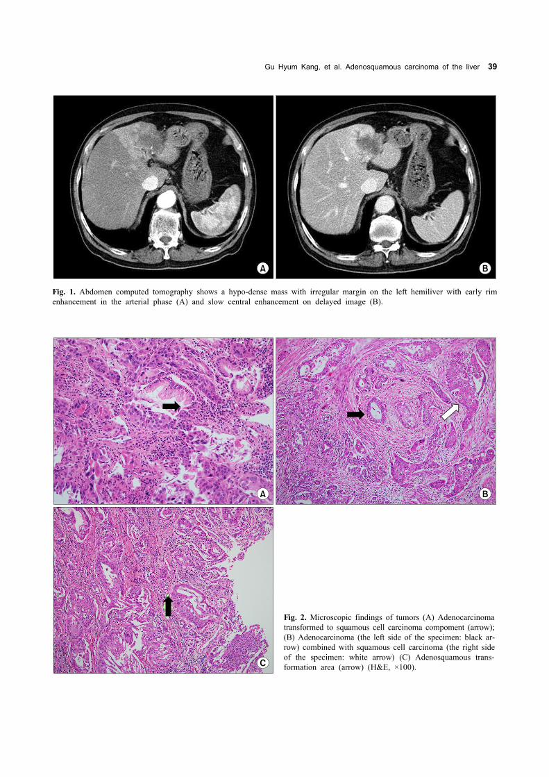
Fig. 1. Abdomen computed tomography shows a hypo-dense mass with irregular margin on the left hemiliver with early rim enhancement in the arterial phase (A) and slow central enhancement on delayed image (B).
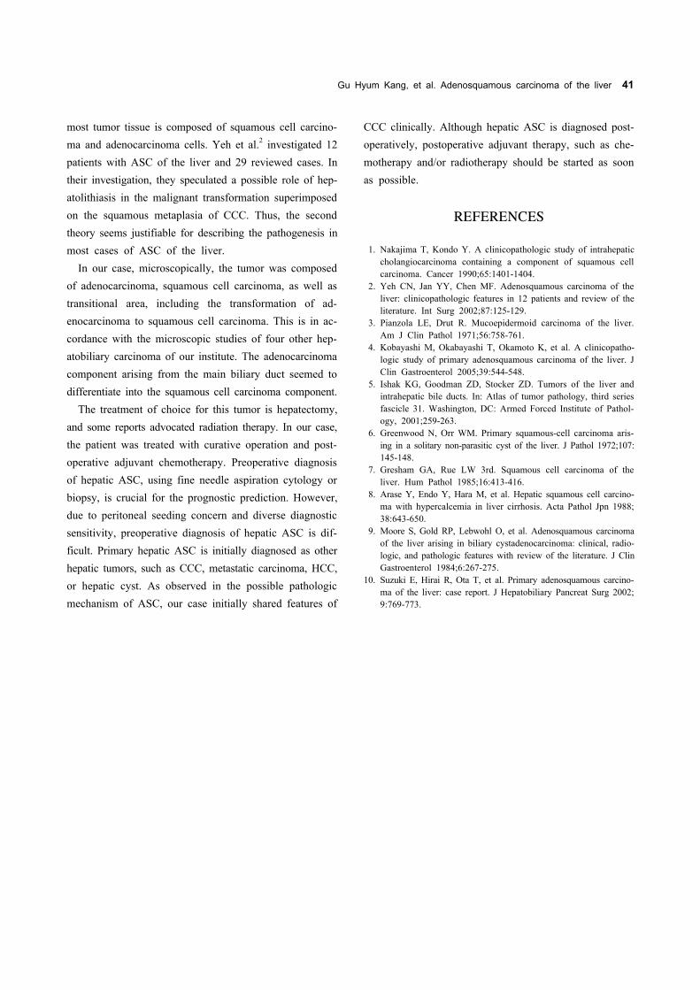
Fig. 2. Microscopic findings of tumors (A) Adenocarcinoma transformed to squamous cell carcinoma compoment (arrow); (B) Adenocarcinoma (the left side of the specimen: black ar-row) combined with squamous cell carcinoma (the right side of the specimen: white arrow) (C) Adenosquamous trans-formation area (arrow) (H&E, ×100).
40 Korean J Hepatobiliary Pancreat Surg Vol. 17, No. 1, February 2013
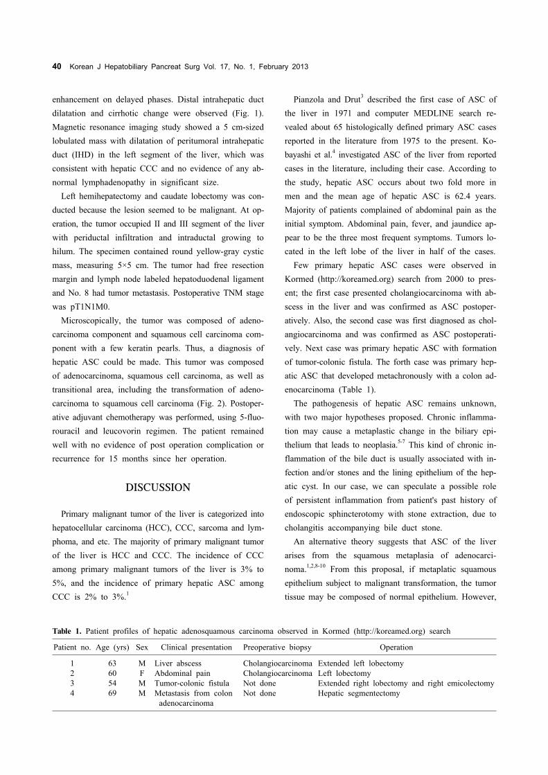
Table 1. Patient profiles of hepatic adenosquamous carcinoma observed in Kormed (http://koreamed.org) search
Patient no. Age (yrs) Sex Clinical presentation Preoperative biopsy Operation
1234
63605469
MFMM
Liver abscessAbdominal painTumor-colonic fistulaMetastasis from colon
adenocarcinoma
CholangiocarcinomaCholangiocarcinomaNot doneNot done
Extended left lobectomyLeft lobectomyExtended right lobectomy and right emicolectomyHepatic segmentectomy
enhancement on delayed phases. Distal intrahepatic duct dilatation and cirrhotic change were observed (Fig. 1). Magnetic resonance imaging study showed a 5 cm-sized lobulated mass with dilatation of peritumoral intrahepatic duct (IHD) in the left segment of the liver, which was consistent with hepatic CCC and no evidence of any ab-normal lymphadenopathy in significant size.
Left hemihepatectomy and caudate lobectomy was con-ducted because the lesion seemed to be malignant. At op-eration, the tumor occupied II and III segment of the liver with periductal infiltration and intraductal growing to hilum. The specimen contained round yellow-gray cystic mass, measuring 5×5 cm. The tumor had free resection margin and lymph node labeled hepatoduodenal ligament and No. 8 had tumor metastasis. Postoperative TNM stage was pT1N1M0.
Microscopically, the tumor was composed of adeno-carcinoma component and squamous cell carcinoma com-ponent with a few keratin pearls. Thus, a diagnosis of hepatic ASC could be made. This tumor was composed of adenocarcinoma, squamous cell carcinoma, as well as transitional area, including the transformation of adeno-carcinoma to squamous cell carcinoma (Fig. 2). Postoper-ative adjuvant chemotherapy was performed, using 5-fluo-rouracil and leucovorin regimen. The patient remained well with no evidence of post operation complication or recurrence for 15 months since her operation.
DISCUSSION
Primary malignant tumor of the liver is categorized into hepatocellular carcinoma (HCC), CCC, sarcoma and lym-phoma, and etc. The majority of primary malignant tumor of the liver is HCC and CCC. The incidence of CCC among primary malignant tumors of the liver is 3% to 5%, and the incidence of primary hepatic ASC among CCC is 2% to 3%.1
Pianzola and Drut3 described the first case of ASC of the liver in 1971 and computer MEDLINE search re-vealed about 65 histologically defined primary ASC cases reported in the literature from 1975 to the present. Ko-bayashi et al.4 investigated ASC of the liver from reported cases in the literature, including their case. According to the study, hepatic ASC occurs about two fold more in men and the mean age of hepatic ASC is 62.4 years. Majority of patients complained of abdominal pain as the initial symptom. Abdominal pain, fever, and jaundice ap-pear to be the three most frequent symptoms. Tumors lo-cated in the left lobe of the liver in half of the cases.
Few primary hepatic ASC cases were observed in Kormed (http://koreamed.org) search from 2000 to pres-ent; the first case presented cholangiocarcinoma with ab-scess in the liver and was confirmed as ASC postoper-atively. Also, the second case was first diagnosed as chol-angiocarcinoma and was confirmed as ASC postoperati-vely. Next case was primary hepatic ASC with formation of tumor-colonic fistula. The forth case was primary hep-atic ASC that developed metachronously with a colon ad-enocarcinoma (Table 1).
The pathogenesis of hepatic ASC remains unknown, with two major hypotheses proposed. Chronic inflamma-tion may cause a metaplastic change in the biliary epi-thelium that leads to neoplasia.5-7 This kind of chronic in-flammation of the bile duct is usually associated with in-fection and/or stones and the lining epithelium of the hep-atic cyst. In our case, we can speculate a possible role of persistent inflammation from patient's past history of endoscopic sphincterotomy with stone extraction, due to cholangitis accompanying bile duct stone.
An alternative theory suggests that ASC of the liver arises from the squamous metaplasia of adenocarci-noma.1,2,8-10 From this proposal, if metaplatic squamous epithelium subject to malignant transformation, the tumor tissue may be composed of normal epithelium. However,
Gu Hyum Kang, et al. Adenosquamous carcinoma of the liver 41
most tumor tissue is composed of squamous cell carcino-ma and adenocarcinoma cells. Yeh et al.2 investigated 12 patients with ASC of the liver and 29 reviewed cases. In their investigation, they speculated a possible role of hep-atolithiasis in the malignant transformation superimposed on the squamous metaplasia of CCC. Thus, the second theory seems justifiable for describing the pathogenesis in most cases of ASC of the liver.
In our case, microscopically, the tumor was composed of adenocarcinoma, squamous cell carcinoma, as well as transitional area, including the transformation of ad-enocarcinoma to squamous cell carcinoma. This is in ac-cordance with the microscopic studies of four other hep-atobiliary carcinoma of our institute. The adenocarcinoma component arising from the main biliary duct seemed to differentiate into the squamous cell carcinoma component.
The treatment of choice for this tumor is hepatectomy, and some reports advocated radiation therapy. In our case, the patient was treated with curative operation and post-operative adjuvant chemotherapy. Preoperative diagnosis of hepatic ASC, using fine needle aspiration cytology or biopsy, is crucial for the prognostic prediction. However, due to peritoneal seeding concern and diverse diagnostic sensitivity, preoperative diagnosis of hepatic ASC is dif-ficult. Primary hepatic ASC is initially diagnosed as other hepatic tumors, such as CCC, metastatic carcinoma, HCC, or hepatic cyst. As observed in the possible pathologic mechanism of ASC, our case initially shared features of
CCC clinically. Although hepatic ASC is diagnosed post-operatively, postoperative adjuvant therapy, such as che-motherapy and/or radiotherapy should be started as soon as possible.
REFERENCES
1. Nakajima T, Kondo Y. A clinicopathologic study of intrahepatic cholangiocarcinoma containing a component of squamous cell carcinoma. Cancer 1990;65:1401-1404.
2. Yeh CN, Jan YY, Chen MF. Adenosquamous carcinoma of the liver: clinicopathologic features in 12 patients and review of the literature. Int Surg 2002;87:125-129.
3. Pianzola LE, Drut R. Mucoepidermoid carcinoma of the liver. Am J Clin Pathol 1971;56:758-761.
4. Kobayashi M, Okabayashi T, Okamoto K, et al. A clinicopatho-logic study of primary adenosquamous carcinoma of the liver. J Clin Gastroenterol 2005;39:544-548.
5. Ishak KG, Goodman ZD, Stocker ZD. Tumors of the liver and intrahepatic bile ducts. In: Atlas of tumor pathology, third series fascicle 31. Washington, DC: Armed Forced Institute of Pathol-ogy, 2001;259-263.
6. Greenwood N, Orr WM. Primary squamous-cell carcinoma aris-ing in a solitary non-parasitic cyst of the liver. J Pathol 1972;107: 145-148.
7. Gresham GA, Rue LW 3rd. Squamous cell carcinoma of the liver. Hum Pathol 1985;16:413-416.
8. Arase Y, Endo Y, Hara M, et al. Hepatic squamous cell carcino-ma with hypercalcemia in liver cirrhosis. Acta Pathol Jpn 1988; 38:643-650.
9. Moore S, Gold RP, Lebwohl O, et al. Adenosquamous carcinoma of the liver arising in biliary cystadenocarcinoma: clinical, radio-logic, and pathologic features with review of the literature. J Clin Gastroenterol 1984;6:267-275.
10. Suzuki E, Hirai R, Ota T, et al. Primary adenosquamous carcino-ma of the liver: case report. J Hepatobiliary Pancreat Surg 2002; 9:769-773.