A Case of Abortive Staphylococcal Scalded Skin Syndrome · Brief Report 624 Ann Dermatol Received...
3
Brief Report 624 Ann Dermatol Received August 14, 2017, Revised October 10, 2017, Accepted for publication October 19, 2017 Corresponding author: Chun Wook Park, Department of Dermatology, Kangnam Sacred Heart Hospital, Hallym University College of Medicine, 1 Singil-ro, Yeongdeungpo-gu, Seoul 07441, Korea. Tel: 82-2-829-5221, Fax: 82-2-832-3237, E-mail: [email protected] ORCID: https://orcid.org/0000-0003-4512-8668 Hye One Kim, Department of Dermatology, Kangnam Sacred Heart Hospital, Hallym University College of Medicine, 1 Singil-ro, Yeongdeungpo-gu, Seoul 07441, Korea. Tel: 82-2-829-5221, Fax: 82-2-832-3237, E-mail: [email protected] ORCID: https://orcid.org/0000-0001-5846-0008 *These authors contributed equally and should be considered co-first authors. This is an Open Access article distributed under the terms of the Creative Commons Attribution Non-Commercial License (http://creativecommons.org/ licenses/by-nc/4.0) which permits unrestricted non-commercial use, distribution, and reproduction in any medium, provided the original work is properly cited. Copyright © The Korean Dermatological Association and The Korean Society for Investigative Dermatology Fig. 1. Diffuse erythroderma involves the neck, trunk, and extremities with sandpaper-like coarseness (A), and perioral crust and fissure (B) were observed. Several vesicobullae and erosive lesions were observed in the lower abdomen, without Nikolsky sign (C). The desquamation worsened and the erythema gradually faded on the 8th day after admission (D∼F) (We received the patient’s consent form about publishing all photographic materials). https://doi.org/10.5021/ad.2018.30.5.624 A Case of Abortive Staphylococcal Scalded Skin Syndrome Bo Young Chung*, Jee Hee Son*, Min Je Jung, Yong Won Choi, Yong Se Cho, Hye One Kim, Chun Wook Park Department of Dermatology, Kangnam Sacred Heart Hospital, Hallym University College of Medicine, Seoul, Korea Dear Editor: A 5-year-old boy presented with diffuse erythematous patches and fever, started three days earlier. Ten days ear- lier, second-generation cephalosporin and mupirocin oint- ment was used for a mosquito bite site on left elbow at an- other hospital. Three days ago, oral steroids were pre- scribed in another hospital to treat a trunk rash and perio- ral crust, but they did not work. At the time of arrival, the
Transcript of A Case of Abortive Staphylococcal Scalded Skin Syndrome · Brief Report 624 Ann Dermatol Received...

Brief Report
624 Ann Dermatol
Received August 14, 2017, Revised October 10, 2017, Accepted for publication October 19, 2017
Corresponding author: Chun Wook Park, Department of Dermatology, Kangnam Sacred Heart Hospital, Hallym University College of Medicine, 1 Singil-ro, Yeongdeungpo-gu, Seoul 07441, Korea. Tel: 82-2-829-5221, Fax: 82-2-832-3237, E-mail: [email protected]: https://orcid.org/0000-0003-4512-8668
Hye One Kim, Department of Dermatology, Kangnam Sacred Heart Hospital, Hallym University College of Medicine, 1 Singil-ro, Yeongdeungpo-gu, Seoul 07441, Korea. Tel: 82-2-829-5221, Fax: 82-2-832-3237, E-mail: [email protected]: https://orcid.org/0000-0001-5846-0008
*These authors contributed equally and should be considered co-first authors.
This is an Open Access article distributed under the terms of the Creative Commons Attribution Non-Commercial License (http://creativecommons.org/licenses/by-nc/4.0) which permits unrestricted non-commercial use, distribution, and reproduction in any medium, provided the original work is properly cited.
Copyright © The Korean Dermatological Association and The Korean Society for Investigative Dermatology
Fig. 1. Diffuse erythroderma involves the neck, trunk, and extremities with sandpaper-like coarseness (A), and perioral crust and fissure (B) were observed. Several vesicobullae and erosive lesions were observed in the lower abdomen, without Nikolsky sign (C). The desquamation worsened and the erythema gradually faded on the 8th day after admission (D∼F) (We received the patient’s consent form about publishing all photographic materials).
https://doi.org/10.5021/ad.2018.30.5.624
A Case of Abortive Staphylococcal Scalded Skin Syndrome
Bo Young Chung*, Jee Hee Son*, Min Je Jung, Yong Won Choi, Yong Se Cho, Hye One Kim, Chun Wook Park
Department of Dermatology, Kangnam Sacred Heart Hospital, Hallym University College of Medicine, Seoul, Korea
Dear Editor:A 5-year-old boy presented with diffuse erythematous patches and fever, started three days earlier. Ten days ear-lier, second-generation cephalosporin and mupirocin oint-
ment was used for a mosquito bite site on left elbow at an-other hospital. Three days ago, oral steroids were pre-scribed in another hospital to treat a trunk rash and perio-ral crust, but they did not work. At the time of arrival, the
Brief Report
Vol. 30, No. 5, 2018 625
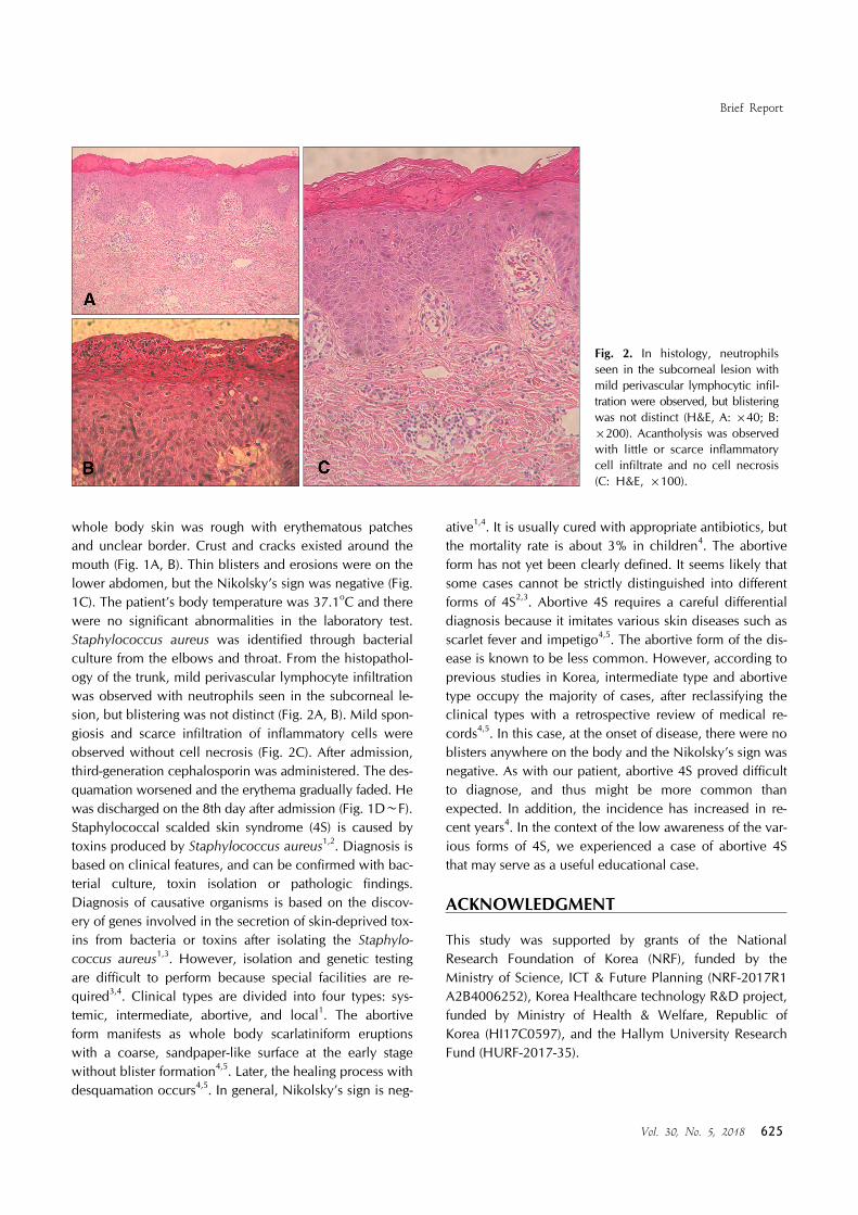
Fig. 2. In histology, neutrophils seen in the subcorneal lesion with mild perivascular lymphocytic infil-tration were observed, but blistering was not distinct (H&E, A: ×40; B: ×200). Acantholysis was observed with little or scarce inflammatory cell infiltrate and no cell necrosis (C: H&E, ×100).
whole body skin was rough with erythematous patches and unclear border. Crust and cracks existed around the mouth (Fig. 1A, B). Thin blisters and erosions were on the lower abdomen, but the Nikolsky’s sign was negative (Fig. 1C). The patient’s body temperature was 37.1oC and there were no significant abnormalities in the laboratory test. Staphylococcus aureus was identified through bacterial culture from the elbows and throat. From the histopathol-ogy of the trunk, mild perivascular lymphocyte infiltration was observed with neutrophils seen in the subcorneal le-sion, but blistering was not distinct (Fig. 2A, B). Mild spon-giosis and scarce infiltration of inflammatory cells were observed without cell necrosis (Fig. 2C). After admission, third-generation cephalosporin was administered. The des-quamation worsened and the erythema gradually faded. He was discharged on the 8th day after admission (Fig. 1D∼F).Staphylococcal scalded skin syndrome (4S) is caused by toxins produced by Staphylococcus aureus1,2. Diagnosis is based on clinical features, and can be confirmed with bac-terial culture, toxin isolation or pathologic findings. Diagnosis of causative organisms is based on the discov-ery of genes involved in the secretion of skin-deprived tox-ins from bacteria or toxins after isolating the Staphylo-coccus aureus1,3. However, isolation and genetic testing are difficult to perform because special facilities are re-quired3,4. Clinical types are divided into four types: sys-temic, intermediate, abortive, and local1. The abortive form manifests as whole body scarlatiniform eruptions with a coarse, sandpaper-like surface at the early stage without blister formation4,5. Later, the healing process with desquamation occurs4,5. In general, Nikolsky’s sign is neg-
ative1,4. It is usually cured with appropriate antibiotics, but the mortality rate is about 3% in children4. The abortive form has not yet been clearly defined. It seems likely that some cases cannot be strictly distinguished into different forms of 4S2,3. Abortive 4S requires a careful differential diagnosis because it imitates various skin diseases such as scarlet fever and impetigo4,5. The abortive form of the dis-ease is known to be less common. However, according to previous studies in Korea, intermediate type and abortive type occupy the majority of cases, after reclassifying the clinical types with a retrospective review of medical re-cords4,5. In this case, at the onset of disease, there were no blisters anywhere on the body and the Nikolsky’s sign was negative. As with our patient, abortive 4S proved difficult to diagnose, and thus might be more common than expected. In addition, the incidence has increased in re-cent years4. In the context of the low awareness of the var-ious forms of 4S, we experienced a case of abortive 4S that may serve as a useful educational case.
ACKNOWLEDGMENT
This study was supported by grants of the National Research Foundation of Korea (NRF), funded by the Ministry of Science, ICT & Future Planning (NRF-2017R1 A2B4006252), Korea Healthcare technology R&D project, funded by Ministry of Health & Welfare, Republic of Korea (HI17C0597), and the Hallym University Research Fund (HURF-2017-35).

Brief Report
626 Ann Dermatol
Received October 13, 2017, Accepted for publication November 14, 2017
Corresponding author: Chun Wook Park, Department of Dermatology, Kangnam Sacred Heart Hospital, Hallym University College of Medicine, 1 Singil-ro, Yeongdeungpo-gu, Seoul 07441, Korea. Tel: 82-2-829-5221, Fax: 82-2-832-3237, E-mail: [email protected]: https://orcid.org/0000-0003-4512-8668
Hye One Kim, Department of Dermatology, Kangnam Sacred Heart Hospital, Hallym University College of Medicine, 1 Singil-ro, Yeongdeungpo-gu, Seoul 07441, Korea. Tel: 82-2-829-5221, Fax: 82-2-832-3237, E-mail: [email protected] ORCID: https://orcid.org/0000-0001-5846-0008
*These authors contributed equally and should be considered co-first authors.
This is an Open Access article distributed under the terms of the Creative Commons Attribution Non-Commercial License (http://creativecommons.org/licenses/by-nc/4.0) which permits unrestricted non-commercial use, distribution, and reproduction in any medium, provided the original work is properly cited.
Copyright © The Korean Dermatological Association and The Korean Society for Investigative Dermatology
CONFLICTS OF INTEREST
The authors have nothing to disclose.
REFERENCES
1. Park AY, Yeon EK, Lee HK, Shin MY. A clinical review of staphylococcal scalded skin syndrome for the last 10 years.
Soonchunhyang Med Sci 2012;18:32-37.
2. Patel GK, Finlay AY. Staphylococcal scalded skin syndrome:
diagnosis and management. Am J Clin Dermatol 2003; 4:165-175.
3. Ladhani S, Evans RW. Staphylococcal scalded skin syndrome.
Arch Dis Child 1998;78:85-88.4. Park JW, Hwang DK, Yu HJ. Staphylococcal scalded skin
syndrome, review of 20 cases. Korean J Dermatol 2002;40:
1051-1057. 5. Kang JD, Park SD. Reclassification of staphylococcal scalded
skin syndrome by clinical analysis of 25 cases. Korean J
Dermatol 2004;42:398-405.
https://doi.org/10.5021/ad.2018.30.5.626
Influence of Weight Loss on Severity of Atopic Dermatitis in a 20-Year-Old Female with Atopic Dermatitis
Jee Hee Son*, Bo Young Chung*, Min Je Jung, Yong Won Choi, Hye One Kim, Chun Wook Park
Department of Dermatology, Kangnam Sacred Heart Hospital, Hallym University College of Medicine, Seoul, Korea
Dear Editor:A 20-year-old female patient had atopic dermatitis (AD) from her second year. She had been treated with antihist-amine, and with topical steroid and calcineurin inhibitor, for about one year. Four months ago, her symptoms got worse. At first visit, her body mass index (BMI) was 26.81 kg/cm2 (73 kg, 165 cm), waist circumstance was 82 cm, Eczema Area and Severity Index (EASI) score was 16.8, and visual analogue scale (VAS) for pruritus was ‘7’. Physical examination revealed diffuse erythematous patch-
es and maculopapules with slight crust and scales over her whole body, especially her face, back, and posterior as-pects of her thighs (Fig. 1A∼E). Symptoms did not im-prove after cyclosporine (100 mg/d) was given for three weeks. Maintaining existing therapy, she was referred to Family Medicine to manage her obesity. Dietary control and exercise were prescribed. Aerobic exercise of moder-ate intensity (METs 6∼7) was recommended 150 mi-nutes/week. She recorded the quantity, type of exercise, and total daily calorie intake in a diary. One tablet of anti-