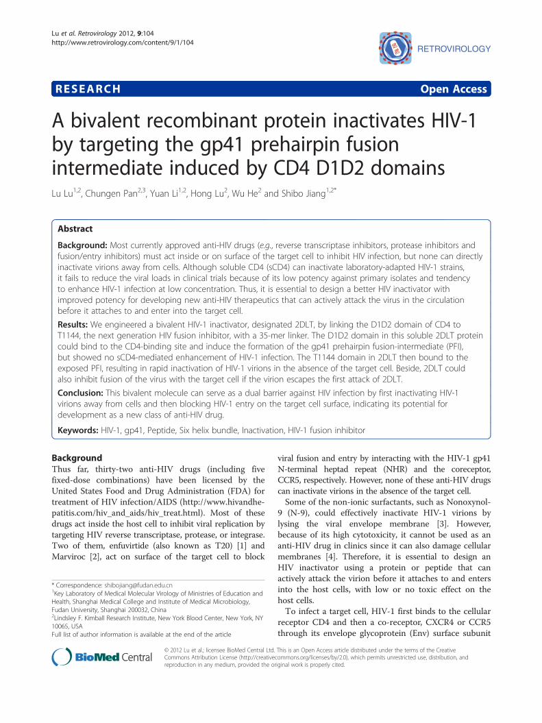
A bivalent recombinant protein inactivates HIV-1 by targeting the
14
RESEARCH Open Access A bivalent recombinant protein inactivates HIV-1 by targeting the gp41 prehairpin fusion intermediate induced by CD4 D1D2 domains Lu Lu 1,2 , Chungen Pan 2,3 , Yuan Li 1,2 , Hong Lu 2 , Wu He 2 and Shibo Jiang 1,2* Abstract Background: Most currently approved anti-HIV drugs (e.g., reverse transcriptase inhibitors, protease inhibitors and fusion/entry inhibitors) must act inside or on surface of the target cell to inhibit HIV infection, but none can directly inactivate virions away from cells. Although soluble CD4 (sCD4) can inactivate laboratory-adapted HIV-1 strains, it fails to reduce the viral loads in clinical trials because of its low potency against primary isolates and tendency to enhance HIV-1 infection at low concentration. Thus, it is essential to design a better HIV inactivator with improved potency for developing new anti-HIV therapeutics that can actively attack the virus in the circulation before it attaches to and enter into the target cell. Results: We engineered a bivalent HIV-1 inactivator, designated 2DLT, by linking the D1D2 domain of CD4 to T1144, the next generation HIV fusion inhibitor, with a 35-mer linker. The D1D2 domain in this soluble 2DLT protein could bind to the CD4-binding site and induce the formation of the gp41 prehairpin fusion-intermediate (PFI), but showed no sCD4-mediated enhancement of HIV-1 infection. The T1144 domain in 2DLT then bound to the exposed PFI, resulting in rapid inactivation of HIV-1 virions in the absence of the target cell. Beside, 2DLT could also inhibit fusion of the virus with the target cell if the virion escapes the first attack of 2DLT. Conclusion: This bivalent molecule can serve as a dual barrier against HIV infection by first inactivating HIV-1 virions away from cells and then blocking HIV-1 entry on the target cell surface, indicating its potential for development as a new class of anti-HIV drug. Keywords: HIV-1, gp41, Peptide, Six helix bundle, Inactivation, HIV-1 fusion inhibitor Background Thus far, thirty-two anti-HIV drugs (including five fixed-dose combinations) have been licensed by the United States Food and Drug Administration (FDA) for treatment of HIV infection/AIDS (http://www.hivandhe- patitis.com/hiv_and_aids/hiv_treat.html). Most of these drugs act inside the host cell to inhibit viral replication by targeting HIV reverse transcriptase, protease, or integrase. Two of them, enfuvirtide (also known as T20) [1] and Marviroc [2], act on surface of the target cell to block viral fusion and entry by interacting with the HIV-1 gp41 N-terminal heptad repeat (NHR) and the coreceptor, CCR5, respectively. However, none of these anti-HIV drugs can inactivate virions in the absence of the target cell. Some of the non-ionic surfactants, such as Nonoxynol- 9 (N-9), could effectively inactivate HIV-1 virions by lysing the viral envelope membrane [3]. However, because of its high cytotoxicity, it cannot be used as an anti-HIV drug in clinics since it can also damage cellular membranes [4]. Therefore, it is essential to design an HIV inactivator using a protein or peptide that can actively attack the virion before it attaches to and enters into the host cells, with low or no toxic effect on the host cells. To infect a target cell, HIV-1 first binds to the cellular receptor CD4 and then a co-receptor, CXCR4 or CCR5 through its envelope glycoprotein (Env) surface subunit * Correspondence: [email protected] 1 Key Laboratory of Medical Molecular Virology of Ministries of Education and Health, Shanghai Medical College and Institute of Medical Microbiology, Fudan University, Shanghai 200032, China 2 Lindsley F. Kimball Research Institute, New York Blood Center, New York, NY 10065, USA Full list of author information is available at the end of the article © 2012 Lu et al.; licensee BioMed Central Ltd. This is an Open Access article distributed under the terms of the Creative Commons Attribution License (http://creativecommons.org/licenses/by/2.0), which permits unrestricted use, distribution, and reproduction in any medium, provided the original work is properly cited. Lu et al. Retrovirology 2012, 9:104 http://www.retrovirology.com/content/9/1/104
Transcript of A bivalent recombinant protein inactivates HIV-1 by targeting the
Lu et al. Retrovirology 2012, 9:104http://www.retrovirology.com/content/9/1/104
RESEARCH Open Access
A bivalent recombinant protein inactivates HIV-1by targeting the gp41 prehairpin fusionintermediate induced by CD4 D1D2 domainsLu Lu1,2, Chungen Pan2,3, Yuan Li1,2, Hong Lu2, Wu He2 and Shibo Jiang1,2*
Abstract
Background: Most currently approved anti-HIV drugs (e.g., reverse transcriptase inhibitors, protease inhibitors andfusion/entry inhibitors) must act inside or on surface of the target cell to inhibit HIV infection, but none can directlyinactivate virions away from cells. Although soluble CD4 (sCD4) can inactivate laboratory-adapted HIV-1 strains,it fails to reduce the viral loads in clinical trials because of its low potency against primary isolates and tendencyto enhance HIV-1 infection at low concentration. Thus, it is essential to design a better HIV inactivator withimproved potency for developing new anti-HIV therapeutics that can actively attack the virus in the circulationbefore it attaches to and enter into the target cell.
Results: We engineered a bivalent HIV-1 inactivator, designated 2DLT, by linking the D1D2 domain of CD4 toT1144, the next generation HIV fusion inhibitor, with a 35-mer linker. The D1D2 domain in this soluble 2DLT proteincould bind to the CD4-binding site and induce the formation of the gp41 prehairpin fusion-intermediate (PFI),but showed no sCD4-mediated enhancement of HIV-1 infection. The T1144 domain in 2DLT then bound to theexposed PFI, resulting in rapid inactivation of HIV-1 virions in the absence of the target cell. Beside, 2DLT couldalso inhibit fusion of the virus with the target cell if the virion escapes the first attack of 2DLT.
Conclusion: This bivalent molecule can serve as a dual barrier against HIV infection by first inactivating HIV-1virions away from cells and then blocking HIV-1 entry on the target cell surface, indicating its potential fordevelopment as a new class of anti-HIV drug.
Keywords: HIV-1, gp41, Peptide, Six helix bundle, Inactivation, HIV-1 fusion inhibitor
BackgroundThus far, thirty-two anti-HIV drugs (including fivefixed-dose combinations) have been licensed by theUnited States Food and Drug Administration (FDA) fortreatment of HIV infection/AIDS (http://www.hivandhe-patitis.com/hiv_and_aids/hiv_treat.html). Most of thesedrugs act inside the host cell to inhibit viral replication bytargeting HIV reverse transcriptase, protease, or integrase.Two of them, enfuvirtide (also known as T20) [1] andMarviroc [2], act on surface of the target cell to block
* Correspondence: [email protected] Laboratory of Medical Molecular Virology of Ministries of Education andHealth, Shanghai Medical College and Institute of Medical Microbiology,Fudan University, Shanghai 200032, China2Lindsley F. Kimball Research Institute, New York Blood Center, New York, NY10065, USAFull list of author information is available at the end of the article
© 2012 Lu et al.; licensee BioMed Central Ltd.Commons Attribution License (http://creativecreproduction in any medium, provided the or
viral fusion and entry by interacting with the HIV-1 gp41N-terminal heptad repeat (NHR) and the coreceptor,CCR5, respectively. However, none of these anti-HIV drugscan inactivate virions in the absence of the target cell.Some of the non-ionic surfactants, such as Nonoxynol-
9 (N-9), could effectively inactivate HIV-1 virions bylysing the viral envelope membrane [3]. However,because of its high cytotoxicity, it cannot be used as ananti-HIV drug in clinics since it can also damage cellularmembranes [4]. Therefore, it is essential to design anHIV inactivator using a protein or peptide that canactively attack the virion before it attaches to and entersinto the host cells, with low or no toxic effect on thehost cells.To infect a target cell, HIV-1 first binds to the cellular
receptor CD4 and then a co-receptor, CXCR4 or CCR5through its envelope glycoprotein (Env) surface subunit
This is an Open Access article distributed under the terms of the Creativeommons.org/licenses/by/2.0), which permits unrestricted use, distribution, andiginal work is properly cited.
Lu et al. Retrovirology 2012, 9:104 Page 2 of 14http://www.retrovirology.com/content/9/1/104
gp120. Subsequently, the HIV-1 Env transmembranesubunit gp41 changes its conformation from native toprehairpin fusion intermediate (PFI) state by insertingthe fusion peptide (FP) into the target cell membrane.Three molecules of the gp41 N-terminal heptad repeats(NHR) interact with each other to form a N-trimer, aPFI conformation, which is then associated with thegp41 C-terminal heptad repeats (CHR) to form ahairpin-like six-helix bundle (6-HB) (Figure 1A), bring-ing the viral and cellular membranes into close proxim-ity necessary for fusion [5-7]. Mutations in the multiplehighly conserved tyrosine and dileucine motifs in gp41cytoplasmic domain lead to a loss of HIV-1 Env-mediated membrane fusion [8], and several specificamino acid changes in gp120 V3 region and gp41 areassociated together with CXCR4 and/or CCR5 usage [9],suggesting that both gp120 and gp41 play importantroles in HIV-1 entry and are attractive targets for devel-oping an HIV-1 inactivator and/or entry inhibitors [10].Monomeric soluble CD4 (sCD4) that can specifically
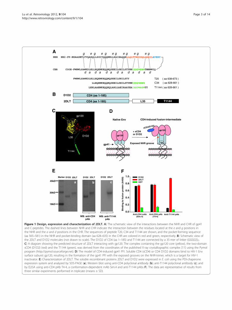
binds to the HIV-1 gp120 and then inactivate the virionwas one of the first anti-HIV-1 agents tested in clinicaltrial. Unfortunately, it failed to reduce the viral loads inHIV-1-infected individuals [12,13]. However, sCD4 andCD4-mimetics could efficiently induce the formation ofthe gp41 PFI with the exposed grooves on the NHR-trimer [14], which is the target of peptidic HIV fusioninhibitors, such as SJ-2176 [15], T20 [16], C34 [17,18]and T1144 [19,20]. These results suggest that a moleculecontaining a CD4 or CD4-mimetic and a gp41 PFI-binding domain (such as T1144) can inactivate HIV-1more efficiently than sCD4 or CD4-mimetic since T1144can bind to the exposed gp41 grooves induced by bind-ing of sCD4 or CD4-mimetic to gp120 to speed the virusinactivation. Based on this hypothesis, we engineered abivalent protein, designated 2DLT, in which the D1D2domains of CD4 were linked to T1144 by a 35-mer flex-ible linker to allow the free movement of the two func-tional domains in the bivalent molecule (Figure 1B). TheD1D2 fragment in this bivalent protein is expected tobind specifically with gp120 on the surface of HIV vir-ions or HIV-infected cells (Figure 1C) and trigger forma-tion of the gp41 PFI with the exposed hydrophobicgrooves (Figure 1D), while the T1144 domain can bindto the exposed grooves on the gp41 NHR-trimer, result-ing in rapid inactivation of the cell-free HIV-1 before itsattachment to the target cell. Indeed, the 2DLT proteincould effectively bind to both gp120 and gp41, blockgp41 6-HB formation, inactivate cell-free HIV-1 and in-hibit HIV-1 Env-mediated cell-cell fusion, but withoutthe sCD4-mediated enhancing effects on HIV-1 infec-tion. Therefore, this engineered bivalent molecule hassubstantial potential for development as an anti-HIVtherapeutic for treatment of patients who fail to respond
to the current anti-HIV drugs and as a topical microbi-cide for preventing sexual transmission of HIV.
ResultsConstruction, expression and characterization of thebivalent fusion protein 2DLTThe expression plasmids pD1D2-PDI and p2DLT-PDIwere constructed by linking the DNA fragment encodingD1D2 with those coding the 35-mer linker (GGGGS)7and T1144 sequentially by three-step overlapping PCRusing the corresponding primer pairs. The nucleotidesequences of the vectors were confirmed by DNA se-quencing. The recombinant bivalent protein 2DLT andthe control protein D1D2 (Figure 1B) were expressed inE. coli. To avoid the formation of inclusion bodies, weused the protein disulfide isomerase (PDI) chaperone-expression system since we and others have shown thatPDI, as a fusion partner, could significantly increase thesoluble expression of recombinant proteins in the cyto-plasm of E. coli [21,22], and we successfully obtainedsoluble D1D2 and 2DLT proteins. After purification, weanalyzed these proteins with SDS-PAGE and Westernblot. On the gels, two proteins migrated near theexpected position of the proteins (Figure 1Ea, D1D2:~23KD; 2DLT: ~29KD). Both displayed specific inter-action with anti-CD4 polyclonal antibodies (pAb)(Figure 1Eb). While the anti-T1144 pAb bound with2DLT at the predicted size, no band was revealed inthe D1D2 lane (Figure 1Ec). Similarly, anti-CD4 pAb(T4-4) could react with both D1D2 and 2DLT, whileanti-T1144 pAb was able to recognize 2DLT only in theELISA. Notably, both D1D2 and 2DLT could react withthe CD4-specific and conformation-dependent mono-clonal antibody (mAb) Sim.4 (Figure 1F), suggestingthat the soluble D1D2 and 2DLT may have a correctlyfolded conformation.
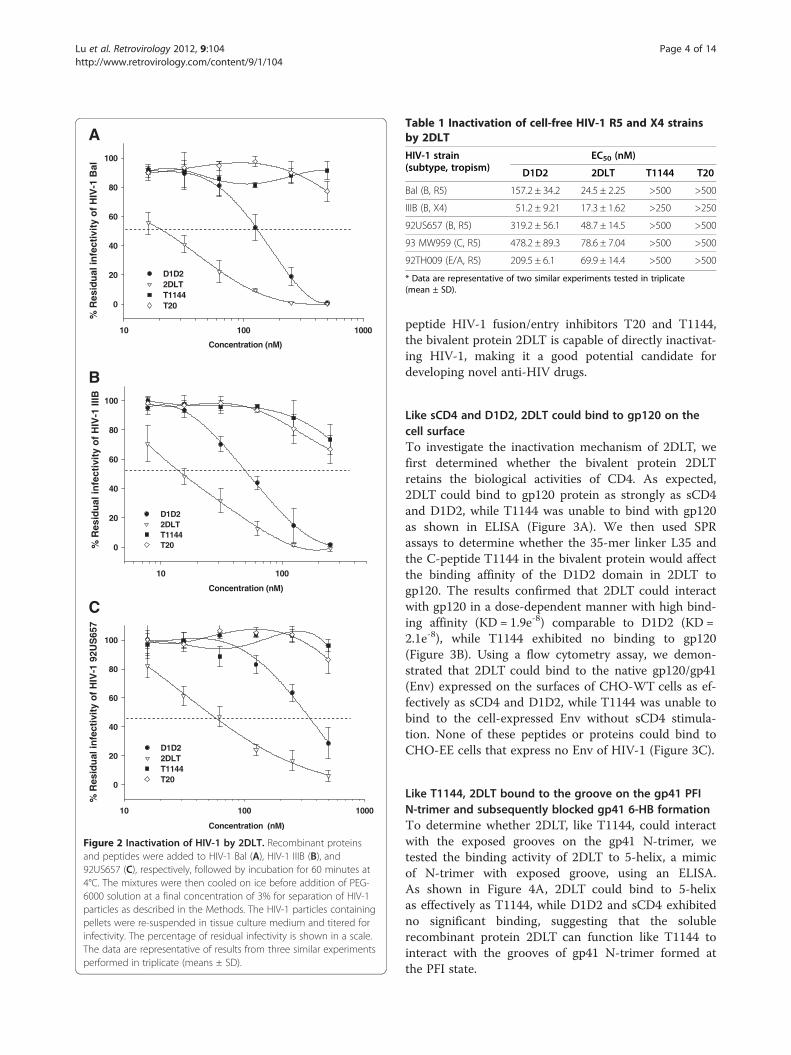
2DLT inactivated cell-free HIV-1 virionsTo determine whether 2DLT could inactivate HIV-1virions, we used PEG-6000 method, as previouslydescribed [23-26], to separate HIV-1 from the recombin-ant proteins 2DLT and D1D2, as well as the control pep-tides T1144 and T20, and then measured the residualinfectivity of the treated HIV-1 particles. As shown inFigure 2 and Table 1, 2DLT inactivated cell-free HIV-1Bal (laboratory-adapted R5 virus), HIV-1 IIIB (labora-tory-adapted X4 virus), and primary R5 isolates with dif-ferent subtypes (92US657, 93 MW959, and 92TH009) ina dose-dependent manner with 50% effective concentra-tion (EC50) ranging from 17.3 to 78.6 nM, which isabout 2- to 6-fold more potent than D1D2 against thecorresponding viral strains. In contrast, T20 and T1144at a concentration up to 500 nM did not show signifi-cant effects on virus inactivation. Different from the
B D1D2
2DLT
CD4 (aa 1-185)
CD4 (aa 1-185) L35 T1144
0.0
0.2
0.4
0.6
0.8
1.0 D1D22DLTGST
Anti-CD4 mAb (Sim.4)
Anti-CD4 pAb (T4-4)
Anti-T1144 pAb
Bin
din
g t
o f
usi
on
pro
tein
(A
450)
E F
WB: anti-CD4 pAb
WB: anti-T1144 pAb
18kD
30kD
45kD
Marker D1D2 2DLT
a b c
D1D2 2DLT D1D2 2DLT
C
gp120
V3
D
D1D2
T1144
L35
gp120
D
CD4-induced fusion-intermediate Native Env
Exposed NHR groove
+ sCD4or D1D2
gp41
gp120
gp120sCD4/D1D2
A
C34 ( aa 628-661 )
T20 ( aa 638-673 )
NHR NH2-FP-MGAASMTLTVQARQLLSGIVQQQNNLLRAIEAQQHLLQLTVWGIKQLQARILAVERYg e g e g e g e g e g e g
CHR COOH-FWNWLSAWKDLELLEQENKEQQNQSEEILSHILSTYNNIERDWEMWTMNNWIQd a d a d a d a d a d a d
LLEQENKEQQNQSEEILSHILSTYNNIERDWEMW
FWNWLSAWKDLELLEQENKEQQNQSEEILSHILSTY
T1144 ( aa 628-661 )LERLAAENKEQQEQLARLLAEIRAAYEAIARDWAEWTT
Figure 1 Design, expression and characterization of 2DLT. A) The schematic view of the interactions between the NHR and CHR of gp41and C-peptides. The dashed lines between NHR and CHR indicate the interaction between the residues located at the e and g positions inthe NHR and the a and d positions in the CHR. The sequences of peptide T20, C34 and T1144 are shown, and the pocket-forming sequence(aa 565–581) in the NHR and pocket-binding domain (aa 628–635) in the CHR are colored in red and green, respectively. B) Schematic view ofthe 2DLT and D1D2 molecules (not drawn to scale). The D1D2 of CD4 (aa 1–185) and T1144 are connected by a 35-mer of linker (GGGGS)7.C) A diagram showing the predicted structure of 2DLT interacting with gp120. The complex containing the gp120 core (yellow), the two-domainsCD4 (D1D2) (red) and the T1144 (green), was derived from the coordinates of the published X-ray crystallographic complex [11] using the Pymolprogram (http://pymol.sourceforge.net). D) The model of CD4-induced gp41 PFI. Soluble CD4 (sCD4) or CD4 D1D2 domains bind to HIV-1 Envsurface subunit gp120, resulting in the formation of the gp41 PFI with the exposed grooves on the NHR-trimer, which is a target for HIV-1inactivator. E) Characterization of 2DLT. The soluble recombinant proteins 2DLT and D1D2 were expressed in E. coli using the PDI-chaperoneexpression system and analyzed by SDS-PAGE (a); Western blot using anti-CD4 polyclonal antibody (b); anti-T1144 polyclonal antibody (c); andby ELISA using anti-CD4 pAb T4-4, a conformation-dependent mAb Sim.4 and anti-T1144 pAbs F). The data are representative of results fromthree similar experiments performed in triplicate (means ± SD).
Lu et al. Retrovirology 2012, 9:104 Page 3 of 14http://www.retrovirology.com/content/9/1/104
000100101
0
20
40
60
80
100
D1D22DLTT1144T20
Concentration (nM)
% R
esid
ual
infe
ctiv
ity
of
HIV
-1 9
2US
657
C
00101
0
20
40
60
80
100
D1D22DLTT1144T20
Concentration (nM)
% R
esid
ual
infe
ctiv
ity
of
HIV
-1 II
IB
B
000100101
0
20
40
60
80
100
D1D22DLTT1144T20
Concentration (nM)
% R
esid
ual
infe
ctiv
ity
of
HIV
-1 B
al
A
Figure 2 Inactivation of HIV-1 by 2DLT. Recombinant proteinsand peptides were added to HIV-1 Bal (A), HIV-1 IIIB (B), and92US657 (C), respectively, followed by incubation for 60 minutes at4°C. The mixtures were then cooled on ice before addition of PEG-6000 solution at a final concentration of 3% for separation of HIV-1particles as described in the Methods. The HIV-1 particles containingpellets were re-suspended in tissue culture medium and titered forinfectivity. The percentage of residual infectivity is shown in a scale.The data are representative of results from three similar experimentsperformed in triplicate (means ± SD).
Table 1 Inactivation of cell-free HIV-1 R5 and X4 strainsby 2DLT
HIV-1 strain(subtype, tropism)
EC50 (nM)
D1D2 2DLT T1144 T20
Bal (B, R5) 157.2 ± 34.2 24.5 ± 2.25 >500 >500
IIIB (B, X4) 51.2 ± 9.21 17.3 ± 1.62 >250 >250
92US657 (B, R5) 319.2 ± 56.1 48.7 ± 14.5 >500 >500
93 MW959 (C, R5) 478.2 ± 89.3 78.6 ± 7.04 >500 >500
92TH009 (E/A, R5) 209.5 ± 6.1 69.9 ± 14.4 >500 >500
* Data are representative of two similar experiments tested in triplicate(mean ± SD).
Lu et al. Retrovirology 2012, 9:104 Page 4 of 14http://www.retrovirology.com/content/9/1/104
peptide HIV-1 fusion/entry inhibitors T20 and T1144,the bivalent protein 2DLT is capable of directly inactivat-ing HIV-1, making it a good potential candidate fordeveloping novel anti-HIV drugs.
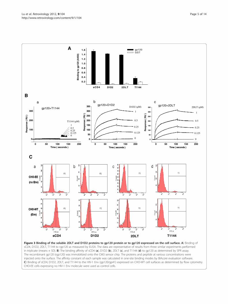
Like sCD4 and D1D2, 2DLT could bind to gp120 on thecell surfaceTo investigate the inactivation mechanism of 2DLT, wefirst determined whether the bivalent protein 2DLTretains the biological activities of CD4. As expected,2DLT could bind to gp120 protein as strongly as sCD4and D1D2, while T1144 was unable to bind with gp120as shown in ELISA (Figure 3A). We then used SPRassays to determine whether the 35-mer linker L35 andthe C-peptide T1144 in the bivalent protein would affectthe binding affinity of the D1D2 domain in 2DLT togp120. The results confirmed that 2DLT could interactwith gp120 in a dose-dependent manner with high bind-ing affinity (KD = 1.9e-8) comparable to D1D2 (KD =2.1e-8), while T1144 exhibited no binding to gp120(Figure 3B). Using a flow cytometry assay, we demon-strated that 2DLT could bind to the native gp120/gp41(Env) expressed on the surfaces of CHO-WT cells as ef-fectively as sCD4 and D1D2, while T1144 was unable tobind to the cell-expressed Env without sCD4 stimula-tion. None of these peptides or proteins could bind toCHO-EE cells that express no Env of HIV-1 (Figure 3C).
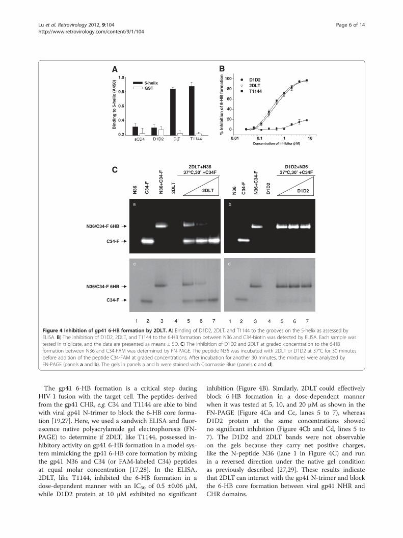
Like T1144, 2DLT bound to the groove on the gp41 PFIN-trimer and subsequently blocked gp41 6-HB formationTo determine whether 2DLT, like T1144, could interactwith the exposed grooves on the gp41 N-trimer, wetested the binding activity of 2DLT to 5-helix, a mimicof N-trimer with exposed groove, using an ELISA.As shown in Figure 4A, 2DLT could bind to 5-helixas effectively as T1144, while D1D2 and sCD4 exhibitedno significant binding, suggesting that the solublerecombinant protein 2DLT can function like T1144 tointeract with the grooves of gp41 N-trimer formed atthe PFI state.
sCD4
Bin
din
g t
o g
p12
0 (A
450)
0 .2
0.4
0.6
0.8
1.0
1.2
1.4
1.6 gp120GST
2DLT 4411T2D1D
A
C
T11442DLTD1D2sCD4
Time ( seconds )0 50 100 150 200 0 50 100 150 200 0 50 100 150 200
Res
po
nse
( R
U )
0
100
200
300gp120+T1144
10.50.250.1250
T1144 (µM)
Time ( seconds )R
esp
on
se (
RU
)
0
50
100
150
200
250
300
350gp120+2DLT
1
0.5
0.25
0.125
0
2DLT (µM)
Time ( seconds )
Res
po
nse
( R
U )
0
50
100
150
200
250
300
350 gp120+D1D2
1
0.5
0.25
0.125
0
D1D2 (µM)
a b c
B
Figure 3 Binding of the soluble 2DLT and D1D2 proteins to gp120 protein or to gp120 expressed on the cell surface. A) Binding ofsCD4, D1D2, 2DLT, T1144 to rgp120 as measured by ELISA. The data are representative of results from three similar experiments performedin triplicate (means ± SD). B) The binding affinity of sCD4 (a), D1D2 (b), 2DLT (c), and T1144 (d) to gp120 as determined by SPR assay.The recombinant gp120 (rgp120) was immobilized onto the CM3 sensor chip. The proteins and peptide at various concentrations wereinjected onto the surface. The affinity constant of each sample was calculated in one-site binding modes by BIAcore evaluation software.C) Binding of sCD4, D1D2, 2DLT, and T1144 to the HIV-1 Env (gp120/gp41) expressed on CHO-WT cell surfaces as determined by flow cytometry.CHO-EE cells expressing no HIV-1 Env molecule were used as control cells.
Lu et al. Retrovirology 2012, 9:104 Page 5 of 14http://www.retrovirology.com/content/9/1/104
C
N36/C34-F 6HB
C34-F
N36/C34-F 6HB
C34-F
b
d
N36
C34
-F
N36
+C34
-F
D1D
2
D1D2
D1D2+N3637oC,30’ +C34F
1 2 3 4 5 6 7
a
c
N36
C34
-F
N36
+C34
-F
2DL
T
2DLT
2DLT+N3637oC,30’ +C34F
1 2 3 4 5 6 7
Concentration of inhibitor ( M)
0.01 0.1 1 10
% In
hib
itio
n o
f 6-
HB
fo
rmat
ion
0
20
40
60
80
100 D1D22DLTT1144
B
0.2
0.4
0.6
0.8
1.05-helixGST
sCD4 D1D2 DLT T1144
Bin
din
g t
o 5
-hel
ix (
A45
0)
A
Figure 4 Inhibition of gp41 6-HB formation by 2DLT. A) Binding of D1D2, 2DLT, and T1144 to the grooves on the 5-helix as assessed byELISA. B) The inhibition of D1D2, 2DLT, and T1144 to the 6-HB formation between N36 and C34-biotin was detected by ELISA. Each sample wastested in triplicate, and the data are presented as means ± SD. C) The inhibition of D1D2 and 2DLT at graded concentration to the 6-HBformation between N36 and C34-FAM was determined by FN-PAGE. The peptide N36 was incubated with 2DLT or D1D2 at 37°C for 30 minutesbefore addition of the peptide C34-FAM at graded concentrations. After incubation for another 30 minutes, the mixtures were analyzed byFN-PAGE (panels a and b). The gels in panels a and b were stained with Coomassie Blue (panels c and d).
Lu et al. Retrovirology 2012, 9:104 Page 6 of 14http://www.retrovirology.com/content/9/1/104
The gp41 6-HB formation is a critical step duringHIV-1 fusion with the target cell. The peptides derivedfrom the gp41 CHR, e.g. C34 and T1144 are able to bindwith viral gp41 N-trimer to block the 6-HB core forma-tion [19,27]. Here, we used a sandwich ELISA and fluor-escence native polyacrylamide gel electrophoresis (FN-PAGE) to determine if 2DLT, like T1144, possessed in-hibitory activity on gp41 6-HB formation in a model sys-tem mimicking the gp41 6-HB core formation by mixingthe gp41 N36 and C34 (or FAM-labeled C34) peptidesat equal molar concentration [17,28]. In the ELISA,2DLT, like T1144, inhibited the 6-HB formation in adose-dependent manner with an IC50 of 0.5 ±0.06 μM,while D1D2 protein at 10 μM exhibited no significant
inhibition (Figure 4B). Similarly, 2DLT could effectivelyblock 6-HB formation in a dose-dependent mannerwhen it was tested at 5, 10, and 20 μM as shown in theFN-PAGE (Figure 4Ca and Cc, lanes 5 to 7), whereasD1D2 protein at the same concentrations showedno significant inhibition (Figure 4Cb and Cd, lines 5 to7). The D1D2 and 2DLT bands were not observableon the gels because they carry net positive charges,like the N-peptide N36 (lane 1 in Figure 4C) and runin a reversed direction under the native gel conditionas previously described [27,29]. These results indicatethat 2DLT can interact with the gp41 N-trimer and blockthe 6-HB core formation between viral gp41 NHR andCHR domains.

Lu et al. Retrovirology 2012, 9:104 Page 7 of 14http://www.retrovirology.com/content/9/1/104
2DLT could disrupt the function of the CD4-induced gp41PFI, and caused no significant enhancement of HIV-1infection in CD4-/CCR5+ cellsRecently, Haim et al. have demonstrated that binding ofsCD4 or CD4-mimetics to HIV-1 gp120 could induce ashort-lived PFI N-trimer, resulting in increased binding
Interval between the proteins pulse and addition of C34-biotin at 25 OC ( min )
0 10 20 30 40
C34
-bio
tin
bin
din
g (
RL
U )
0
1e+5
2e+5
3e+5
4e+5
5e+5
D1D22DLTT1144
Interval between the proteins pulse and addition of C34-biotin at 4 OC ( min )
0 20 40 60
C34
-bio
tin
bin
din
g (
RL
U )
0
1e+5
2e+5
3e+5
4e+5D1D22DLTT1144
b
aA
Concentrationof inhibitor (nM )
1 10 100 1000
% A
ctiv
ated
infe
ctio
n o
f H
IV-1
(B
al)
in t
he
CD
4-/C
CR
5+ c
ells
0
200
400
600
800
1000
1200
1400
1600D1D2 2DLTT1144
B
Figure 5 2DLT-mediated rapid decay of the CD4-induced gp41PFI and lack of D1D2-induced enhancement of HIV-1 infectionin CD4-/CCR5+ cells. A) Decay of the gp41 PFI at 25°C (a) and 4°C(b) after pulse activation with D1D2 (2.5 μM), 2DLT (2.5 μM) orT1144 (2.5 μM) was measured by a cell-based ELISA, as described inthe Methods. B) The infectivity of HIV-1 Bal (100 TCID50/well) inCf2Th-CCR5 (CD4-/CCR5+) cells in the presence of D1D2, 2DLT orT1144 was determined by ELISA for p24 antigen production asdescribed in the Materials and Methods.
of the C34-Ig protein to the groove on the N-trimer,using a cell-based ELISA as described in Methods. Usinga similar approach, we examined whether 2DLT may acton this intermediate to rapidly inactive viruses by theinteraction of T1144 domain on it and NHR of gp41 onEnv. Indeed, binding of C34 to HIV-1 Env was rapidlyenhanced after D1D2 pulse, and then graduallydecreased as the time of D1D2 pulse was prolonged at25°C (Figure 5Aa) or 4°C (Figure 5Ab), which are coinci-dent with the report by Haim et al. There was no C34binding immediately after T1144 pulse. Strikingly, thebinding ability of C34 to the pulsed Env was lost morerapidly in the 2DLT group than the D1D2 group at 25°C(Figure 5Aa) and 4°C (Figure 5Ab). These results suggestthat because of the presence of T1144 in the bivalentmolecule, 2DLT can decay the N-trimer-exposed PFI ata faster rate than D1D2. Therefore, we believe that2DLT may have a mechanism of action different fromD1D2 molecule in inactivating the CD4-induced gp41PFI. The D1D2 domain in 2DLT may destabilize thegp41 PFI triggered by its binding to gp120, while itsT1144 domain may interact with the exposed N-trimerin the viral gp41 to form heterogonous 6-HB, resultingin the stabilization of the gp41 PFI. It has been reportedthat sCD4 can modestly enhance the infectivity of someHIV-1 strains at suboptimal concentrations in CD4-
CCR5+ cells because sCD4 may efficiently replace CD4on cell surface to drive infection of the CD4-negative andCCR5-positive cells [30]. Here, we investigated whether2DLT could enhance infectivity of CCR5-using HIV-1strain Bal in Cf2Th-CCR5 (CD4-/CCR5+) cells, usingD1D2 and T1144 as controls. As shown in Figure 5B,D1D2 at the concentrations from 5 to 200 nM signifi-cantly enhanced the infection of the virus in the CD4-/CCR5+ cells, while 2DLT and T1144 at the same concen-trations showed no enhancement of HIV-1 infection, sug-gesting that 2DLT exhibits no enhancing effect on HIV-1infectivity in CD4-/CCR5+ cells.
2DLT inhibited HIV-1 infection and HIV-1-mediated cell-cell fusionSubsequently, we determined the inhibitory activities of2DLT against HIV-1 IIIB-mediated cell-cell fusion andagainst infection by laboratory-adapted HIV-1 IIIB(X4) and Bal (R5) strains in MT-2 and TZM-b1 cells, re-spectively, as well as several primary HIV-1 isolates inPBMCs using D1D2, T1144 and T20 as controls. LikeT1144 and T20, 2DLT also exhibited potent inhibitoryactivities against HIV-1 IIIB-mediated cell-cell fusion(IC50 = 19.03 nM) and infection by HIV-1 strains IIIB(IC50 = 5.64 nM) and Bal (IC50 = 10.78 nM), which areabout 5- to 15-fold more potent than D1D2 (Figure 6).Similarly, 2DLT could also significantly inhibit infectionby the primary HIV-1 isolates tested at a low nanomolar
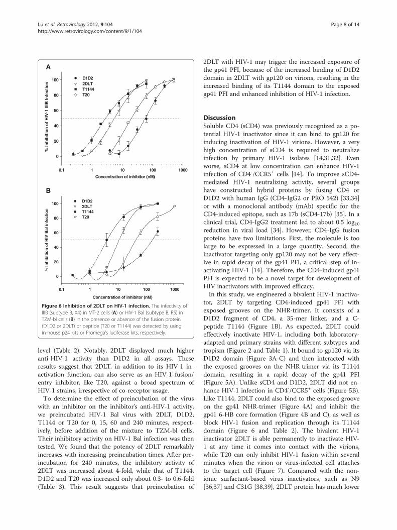
0.1 1 10 100 1000
0
20
40
60
80
100 D1D22DLTT1144T20
% In
hib
itio
n o
f H
IV-1
IIIB
Infe
ctio
n
Concentration of inhibitor (nM)
Concentration of inhibitor (nM)
0.1 1 10 100 1000
% In
hib
itio
n o
f H
IV B
al in
fect
ion
0
20
40
60
80
100D1D22DLTT1144T20
A
B
Figure 6 Inhibition of 2DLT on HIV-1 infection. The infectivity ofIIIB (subtype B, X4) in MT-2 cells (A) or HIV-1 Bal (subtype B, R5) inTZM-bl cells (B) in the presence or absence of the fusion protein(D1D2 or 2DLT) or peptide (T20 or T1144) was detected by usingin-house p24 kits or Promega’s luciferase kits, respectively.
Lu et al. Retrovirology 2012, 9:104 Page 8 of 14http://www.retrovirology.com/content/9/1/104
level (Table 2). Notably, 2DLT displayed much higheranti-HIV-1 activity than D1D2 in all assays. Theseresults suggest that 2DLT, in addition to its HIV-1 in-activation function, can also serve as an HIV-1 fusion/entry inhibitor, like T20, against a broad spectrum ofHIV-1 strains, irrespective of co-receptor usage.To determine the effect of preincubation of the virus
with an inhibitor on the inhibitor’s anti-HIV-1 activity,we preincubated HIV-1 Bal virus with 2DLT, D1D2,T1144 or T20 for 0, 15, 60 and 240 minutes, respect-ively, before addition of the mixture to TZM-bl cells.Their inhibitory activity on HIV-1 Bal infection was thentested. We found that the potency of 2DLT remarkablyincreases with increasing preincubation times. After pre-incubation for 240 minutes, the inhibitory activity of2DLT was increased about 4-fold, while that of T1144,D1D2 and T20 was increased only about 0.3- to 0.6-fold(Table 3). This result suggests that preincubation of
2DLT with HIV-1 may trigger the increased exposure ofthe gp41 PFI, because of the increased binding of D1D2domain in 2DLT with gp120 on virions, resulting in theincreased binding of its T1144 domain to the exposedgp41 PFI and enhanced inhibition of HIV-1 infection.
DiscussionSoluble CD4 (sCD4) was previously recognized as a po-tential HIV-1 inactivator since it can bind to gp120 forinducing inactivation of HIV-1 virions. However, a veryhigh concentration of sCD4 is required to neutralizeinfection by primary HIV-1 isolates [14,31,32]. Evenworse, sCD4 at low concentration can enhance HIV-1infection of CD4-/CCR5+ cells [14]. To improve sCD4-mediated HIV-1 neutralizing activity, several groupshave constructed hybrid proteins by fusing CD4 orD1D2 with human IgG (CD4-IgG2 or PRO 542) [33,34]or with a monoclonal antibody (mAb) specific for theCD4-induced epitope, such as 17b (sCD4-17b) [35]. In aclinical trial, CD4-IgG2 treatment led to about 0.5 log10reduction in viral load [34]. However, CD4-IgG fusionproteins have two limitations. First, the molecule is toolarge to be expressed in a large quantity. Second, theinactivator targeting only gp120 may not be very effect-ive in rapid decay of the gp41 PFI, a critical step of in-activating HIV-1 [14]. Therefore, the CD4-induced gp41PFI is expected to be a novel target for development ofHIV inactivators with improved efficacy.In this study, we engineered a bivalent HIV-1 inactiva-
tor, 2DLT by targeting CD4-induced gp41 PFI withexposed grooves on the NHR-trimer. It consists of aD1D2 fragment of CD4, a 35-mer linker, and a C-peptide T1144 (Figure 1B). As expected, 2DLT couldeffectively inactivate HIV-1, including both laboratory-adapted and primary strains with different subtypes andtropism (Figure 2 and Table 1). It bound to gp120 via itsD1D2 domain (Figure 3A-C) and then interacted withthe exposed grooves on the NHR-trimer via its T1144domain, resulting in a rapid decay of the gp41 PFI(Figure 5A). Unlike sCD4 and D1D2, 2DLT did not en-hance HIV-1 infection in CD4-/CCR5+ cells (Figure 5B).Like T1144, 2DLT could also bind to the exposed grooveon the gp41 NHR-trimer (Figure 4A) and inhibit thegp41 6-HB core formation (Figure 4B and C), as well asblock HIV-1 fusion and replication through its T1144domain (Figure 6 and Table 2). The bivalent HIV-1inactivator 2DLT is able permanently to inactivate HIV-1 at any time it comes into contact with the virions,while T20 can only inhibit HIV-1 fusion within severalminutes when the virion or virus-infected cell attachesto the target cell (Figure 7). Compared with the non-ionic surfactant-based virus inactivators, such as N9[36,37] and C31G [38,39], 2DLT protein has much lower
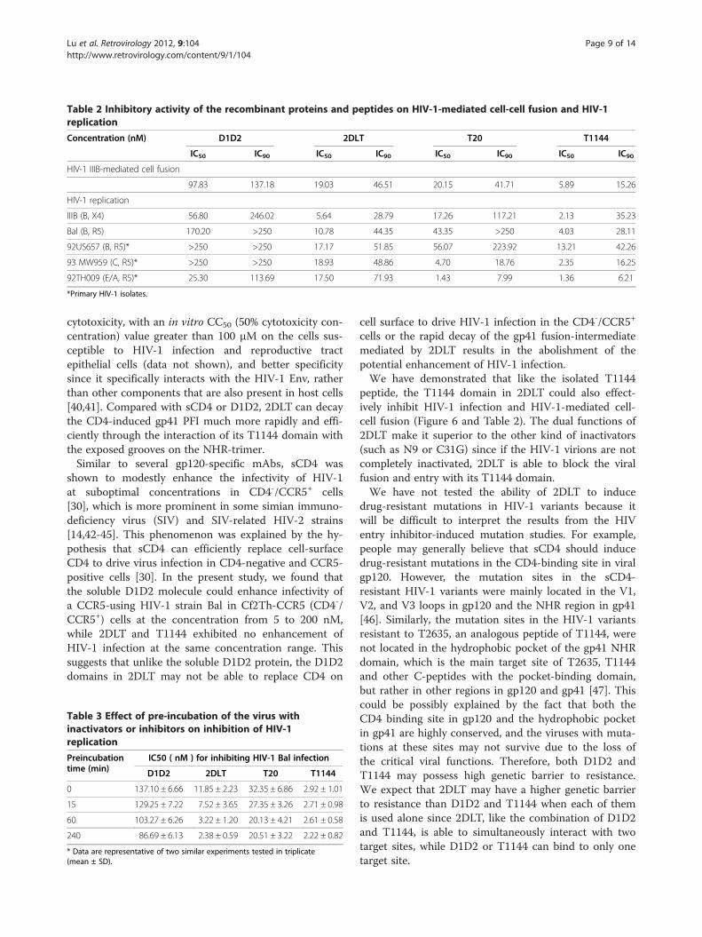
Table 2 Inhibitory activity of the recombinant proteins and peptides on HIV-1-mediated cell-cell fusion and HIV-1replication
Concentration (nM) D1D2 2DLT T20 T1144
IC50 IC90 IC50 IC90 IC50 IC90 IC50 IC90
HIV-1 IIIB-mediated cell fusion
97.83 137.18 19.03 46.51 20.15 41.71 5.89 15.26
HIV-1 replication
IIIB (B, X4) 56.80 246.02 5.64 28.79 17.26 117.21 2.13 35.23
Bal (B, R5) 170.20 >250 10.78 44.35 43.35 >250 4.03 28.11
92US657 (B, R5)* >250 >250 17.17 51.85 56.07 223.92 13.21 42.26
93 MW959 (C, R5)* >250 >250 18.93 48.86 4.70 18.76 2.35 16.25
92TH009 (E/A, R5)* 25.30 113.69 17.50 71.93 1.43 7.99 1.36 6.21
*Primary HIV-1 isolates.
Lu et al. Retrovirology 2012, 9:104 Page 9 of 14http://www.retrovirology.com/content/9/1/104
cytotoxicity, with an in vitro CC50 (50% cytotoxicity con-centration) value greater than 100 μM on the cells sus-ceptible to HIV-1 infection and reproductive tractepithelial cells (data not shown), and better specificitysince it specifically interacts with the HIV-1 Env, ratherthan other components that are also present in host cells[40,41]. Compared with sCD4 or D1D2, 2DLT can decaythe CD4-induced gp41 PFI much more rapidly and effi-ciently through the interaction of its T1144 domain withthe exposed grooves on the NHR-trimer.Similar to several gp120-specific mAbs, sCD4 was
shown to modestly enhance the infectivity of HIV-1at suboptimal concentrations in CD4-/CCR5+ cells[30], which is more prominent in some simian immuno-deficiency virus (SIV) and SIV-related HIV-2 strains[14,42-45]. This phenomenon was explained by the hy-pothesis that sCD4 can efficiently replace cell-surfaceCD4 to drive virus infection in CD4-negative and CCR5-positive cells [30]. In the present study, we found thatthe soluble D1D2 molecule could enhance infectivity ofa CCR5-using HIV-1 strain Bal in Cf2Th-CCR5 (CD4-/CCR5+) cells at the concentration from 5 to 200 nM,while 2DLT and T1144 exhibited no enhancement ofHIV-1 infection at the same concentration range. Thissuggests that unlike the soluble D1D2 protein, the D1D2domains in 2DLT may not be able to replace CD4 on
Table 3 Effect of pre-incubation of the virus withinactivators or inhibitors on inhibition of HIV-1replication
Preincubationtime (min)
IC50 ( nM ) for inhibiting HIV-1 Bal infection
D1D2 2DLT T20 T1144
0 137.10 ± 6.66 11.85 ± 2.23 32.35 ± 6.86 2.92 ± 1.01
15 129.25 ± 7.22 7.52 ± 3.65 27.35 ± 3.26 2.71 ± 0.98
60 103.27 ± 6.26 3.22 ± 1.20 20.13 ± 4.21 2.61 ± 0.58
240 86.69 ± 6.13 2.38 ± 0.59 20.51 ± 3.22 2.22 ± 0.82
* Data are representative of two similar experiments tested in triplicate(mean ± SD).
cell surface to drive HIV-1 infection in the CD4-/CCR5+
cells or the rapid decay of the gp41 fusion-intermediatemediated by 2DLT results in the abolishment of thepotential enhancement of HIV-1 infection.We have demonstrated that like the isolated T1144
peptide, the T1144 domain in 2DLT could also effect-ively inhibit HIV-1 infection and HIV-1-mediated cell-cell fusion (Figure 6 and Table 2). The dual functions of2DLT make it superior to the other kind of inactivators(such as N9 or C31G) since if the HIV-1 virions are notcompletely inactivated, 2DLT is able to block the viralfusion and entry with its T1144 domain.We have not tested the ability of 2DLT to induce
drug-resistant mutations in HIV-1 variants because itwill be difficult to interpret the results from the HIVentry inhibitor-induced mutation studies. For example,people may generally believe that sCD4 should inducedrug-resistant mutations in the CD4-binding site in viralgp120. However, the mutation sites in the sCD4-resistant HIV-1 variants were mainly located in the V1,V2, and V3 loops in gp120 and the NHR region in gp41[46]. Similarly, the mutation sites in the HIV-1 variantsresistant to T2635, an analogous peptide of T1144, werenot located in the hydrophobic pocket of the gp41 NHRdomain, which is the main target site of T2635, T1144and other C-peptides with the pocket-binding domain,but rather in other regions in gp120 and gp41 [47]. Thiscould be possibly explained by the fact that both theCD4 binding site in gp120 and the hydrophobic pocketin gp41 are highly conserved, and the viruses with muta-tions at these sites may not survive due to the loss ofthe critical viral functions. Therefore, both D1D2 andT1144 may possess high genetic barrier to resistance.We expect that 2DLT may have a higher genetic barrierto resistance than D1D2 and T1144 when each of themis used alone since 2DLT, like the combination of D1D2and T1144, is able to simultaneously interact with twotarget sites, while D1D2 or T1144 can bind to only onetarget site.
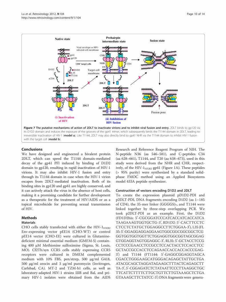
Target cell membrane
(i) Inactivation of HIV
Native state
CD4+ target cells
2DLT
Viral envelope or HIV-infected cell membrane
Prehairpin intermediate state
CD4
fusion state
X
CHR
NHR
FP
gp41gp120
(ii) Inhibition of HIV fusion
D1D2L35T1144
2DLT
2DLT
2DLT
X
D1D2
sCD4
Fusion pore
Figure 7 The putative mechanisms of action of 2DLT to inactivate virions and to inhibit viral fusion and entry. 2DLT binds to gp120 viaits D1D2 domain and induces the exposure of the grooves of the gp41 trimer, which subsequently binds the T1144 domain in 2DLT, leading toirreversible inactivation of HIV-1 (model a). Like T1144, 2DLT may also directly bind to gp41 NHR via the T1144 domain to inhibit HIV-1 fusionwith the target cell (model b).
Lu et al. Retrovirology 2012, 9:104 Page 10 of 14http://www.retrovirology.com/content/9/1/104
ConclusionsWe have designed and engineered a bivalent protein2DLT, which can speed the T1144 domain-mediateddecay of the gp41 PFI induced by binding of D1D2domain to gp120, resulting in rapid inactivation of HIV-1virions. It may also inhibit HIV-1 fusion and entrythrough its T1144 domain in case when the HIV-1 virionescapes from 2DLT-mediated inactivation. Both of itsbinding sites in gp120 and gp41 are highly conserved, andit can actively attack the virus in the absence of host cells,making it a promising candidate for further developmentas a therapeutic for the treatment of HIV/AIDS or as atopical microbicide for preventing sexual transmissionof HIV.
MethodsMaterialsCHO cells stably transfected with either the HIV-1HXB2
Env-expressing vector pEE14 (CHO-WT) or controlpEE14 vector (CHO-EE) were cultured in Glutamine-deficient minimal essential medium (GMEM-S) contain-ing 400 μM Methionine sulfoximine (Sigma, St. Louis,MO). Cf2Th/syn CCR5 cells stably expressing CCR5receptors were cultured in DMEM complementedmedium with 10% FBS, pen/strep, 500 μg/ml G418,500 μg/ml zeocin and 3 μg/ml puromycin (Invitrogen,Carlsbad, CA). MT-2 and TZM-b1 cells, as well aslaboratory-adapted HIV-1 strains IIIB and Bal, and pri-mary HIV-1 isolates were obtained from the AIDS
Research and Reference Reagent Program of NIH. TheN-peptide N36 (aa 546–581), and C-peptides C34(aa 628–661), T1144, and T20 (aa 638–673), used in thisstudy were derived from the NHR and CHR, respect-ively, of the HIV-1HXB2 gp41 (Figure 1A). These peptides(> 95% purity) were synthesized by a standard solid-phase FMOC method using an Applied Biosystemsmodel 433A peptide synthesizer.
Construction of vectors encoding D1D2 and 2DLTTo create the expression plasmid pD1D2-PDI andp2DLT-PDI, DNA fragments encoding D1D2 (aa 1–185of CD4), the 35-mer linker (GGGGS)7, and T1144 werelinked together by three-step overlapping PCR. Wetook p2DLT-PDI as an example. First, the D1D2(FD1D2his: 50-CGCGGATCCCATCACCATCACCATCATAAGAAAGTGGTGCTG-30, RD1D2: 50-CACTTCCTCCTCCTCTATGCTGGAGGCCTTCTGGAA-30), L35 (FL35: 50-GGAGGAGGAGGAAGTGGCGGCGGCGGCTCGGGTGGTGGTGGTTCTGGAGGTGGCGGTAGCGGAGGTGGAGGTAGTGGAGGC-30, RL35: 50-GCTACCTCCGCCTCCCGAACCTCCGCCTCCACTACCTCCACCTCCGCTACCGCCACCTCCAGAACCACCACCACCCGAG-30) and T1144 (FT1144: 50-GAGGCGGAGGTAGCACGACCTGGGAAGCATGGGACAGAGCTATTGCTGAATACGCAGCTAGGATAGAAGCTTTACTCAGAGCTTTA-30, 50-CGGAGATCTCTATAATTCCCTTAAGGCTGCTTCATTCTTTTCTTGCTGTTCTTGTAAAGCTCTGAGTAAAGCTTCTATCC-30) DNA fragments were genera-
Lu et al. Retrovirology 2012, 9:104 Page 11 of 14http://www.retrovirology.com/content/9/1/104
ted by overlapping PCR using the corresponding primerpairs. Second, the DNA fragments coding for L35 andT1144 were linked by overlapping PCR with the primersFL35 and RT1144. Third, the two DNA fragmentsencoding D1D2 and L35-T1144 were linked by overlap-ping PCR with the DNA fragment D1D2 and the primersFD1D2his and RT1144. Finally, the amplified DNA frag-ment coding for 2DLT was digested by BamHI and EcoR Iand inserted into the expression vector pGEX-6p-1 togenerate the p2DLT plasmid. In order to prevent the for-mation of the inclusion bodies in E. coli, we inserted aprotein disulfide isomerase (PDI) [21,22] DNA sequence(aa 18–508) with PreScission protease cutting site (calledppase site) in the N terminus into the EcoR I and Xho Isites located at the C terminus of His-2DLT gene inthe plasmid p2DLT to extend the GST-his-2DLT read-ing frame, resulting in the generation of chimeric GST-his-2DLT-ppase-PDI. This plasmid is called p2DLT-PDI.The sequences were confirmed by DNA sequencing.
Protein expression and purificationTo express D1D2 and 2DLT fusion proteins, Escherichiacoli strain Rosetta 2 (DE3) pLysS (Novagen) was trans-formed with pD1D2-PDI and p2DLT-PDI, respectively,cultured at 37°C to OD600 = 0.4, then induced at 16-22°C for 8–12 h with 0.4 mM IPTG. The cells wereharvested and lysed by sonication in the presence ofprotease inhibitor mixture (Roche). After centrifugation,supernatants containing the fusion protein were collected.The protein was purified with Glutathione-Sepharose 4Baffinity columns and cleaved with PreScissionTM Protease(GE Healthcare). These fusion proteins were purifiedby His•BindW Purification Kit (Novagen) and fast proteinliquid chromatography (FPLC), and then analyzed bySDS-PAGE.
SDS-PAGE and Western blot analysisPurified fusion proteins were analyzed by SDS-PAGE aspreviously described [48]. Briefly, D1D2 or 2DLT wasmixed with 4X SDS sample buffer (Novagen, Gibbstown,NJ) and boiled for 5 minutes or kept at roomtemperature (RT) before loading onto a 10-20% Tricine-Glycine gel (Invitrogen, Carlsbad, CA). The electro-phoresis was conducted in SDS-PAGE running bufferwith 125 V constant voltage at 4°C. In Western blot, theanti-human CD4 and anti-T1144 polyclonal antibodieswere used.
Enzyme-linked immunosorbent assay (ELISA)D1D2 and 2DLT fusion proteins were characterized byELISA as previously described [49]. Briefly, they werecoated onto a 96-well polystyrene plate (Costar, CorningInc., Corning, NY) (10 μg/ml in 0.1 M Tris–HCl,pH 8.8), which was blocked with 2% non-fat milk in
PBS. The polyclonal antibodies against CD4, conformation-dependent monoclonal antibody against CD4 (Sim.4) andanti-T1144 polyclonal antibody, respectively, were addedto the plate. After incubation at 37°C for 60 min, horse-radish peroxidase (HRP)-labeled antibodies (ZYMEDLaboratories, S. San Francisco, CA) and the substrateTMB (Sigma) were added, sequentially. The bindingof D1D2 and 2DLT to gp120 or gp41 NHR, and theirinhibitory activity on gp41 6-HB formation were deter-mined by ELISA as previously described [50].
Flow cytometryThe binding of D1D2 and 2DLT to gp120/gp41expressed on the cell surface was detected by flow cyto-metry as previously described [49,51]. Briefly, the cul-tured CHO-WT (with Env) and CHO-EE (with no Env)cells were detached from plate and washed with washbuffer (PBS containing 5% GBS) three times and incu-bated with the testing protein for 1 h at 4°C. After threewashes, anti-CD4 or anti-T1144 polyclonal antibody wasadded for 1 h at 4°C. After three washes, FITC-conjugated anti-rabbit or mouse antibody was addedand incubated for 1 h at 4°C. After three washes, thecells were examined by flow cytometry and the fluores-cence intensity was recorded by FACSCalibur (BectonDickinson).
Surface plasmon resonance (SPR) assayThe binding affinity of D1D2 and 2DLT to gp120 wasmeasured by SPR using the BIAcore3000 system (Phar-macia, Piscataway, NJ), following the Manual of the Bio-molecular Interaction Analysis (BIA) Technology asdescribed previously [52]. Briefly, gp120 (100 μg/ml) wasimmobilized onto the CM3 sensor chip by amine coup-ling, and the unreacted sites were blocked with ethanola-mine. The dissociation reaction was done by washingwith running buffer (10 mM HEPES pH7.4 containing0.15 M NaCl, 3.4 mM EDTA and 0.005% v/v surfactant)for at least 2 min.
Fluorescence native polyacrylamide gel electrophoresis(FN-PAGE)FN-PAGE for detecting 6-HB formation as described be-fore [27]. Briefly, a testing peptide or protein (100 μM)was pre-incubated with N36 (100 μM) at 37°C for30 min, followed by addition of C34-FAM (100 μM) at37°C for 30 minutes. The mixtures were added intoTris-glycine native sample buffer (Invitrogen, Carlsbad,CA). The samples (20 μl) were then loaded onto Tris-glycine gels (18%; Invitrogen, Carlsbad, CA), which wererun under 120 V constant voltage at room tempera-ture for 1 h. The gels were stained and visualized withthe FluorChem 8800 Imaging System (Alpha InnotechCorp., San Leandro, CA) using a transillumination UV
Lu et al. Retrovirology 2012, 9:104 Page 12 of 14http://www.retrovirology.com/content/9/1/104
light source with excitation wavelength at 520 nm andthen with Coomassie Blue.
Inhibition of HIV-1 infectionInhibitory activities of D1D2 and 2DLT on HIV-1 infec-tion were determined as previously described [53,54].For inhibition of HIV-1 IIIB (subtype B, X4) infection,100 TCID50 of the virus was added to 1 × 10⁴/ml MT-2cells in RPMI medium 1640 containing 10% FBS inthe presence or absence of the test peptide or proteinovernight. The culture supernatants were removed, andfresh media were added. On the fourth day post-infec-tion, culture supernatants were collected for detectionof p24 antigen by ELISA. For inhibition of infectionby the HIV-1 strain Bal (subtype B, R5), TZM-bl cells(1 × 10⁵/ml) were pre-cultured overnight and infectedwith Bal at 100 TCID50 in the presence or absence ofthe test peptide or protein overnight. The cells were har-vested and lysed on the fourth day post-infection withlysing reagent. The luciferase activity was analyzed usinga luciferase kit (Promega, Madison, WI) and a lumin-ometer (Ultra 386, Tecan, Durham, NC) according tothe manufacturer’s instructions. For testing the effect ofpre-incubation times on the inhibitory activity of 2DLT,the TZM-b1 assay was performed after pre-incubationof the HIV-1 Bal virus with the inhibitors for 0, 15, 60and 240 min. For inhibition of primary HIV-1 isolate in-fection, peripheral blood mononuclear cells (PBMCs)were isolated from the blood of healthy donors. ThePHA-stimulated cells were infected with a primary HIV-1 isolate at a multiplicity of infection (MOI) of 0.01 inthe absence or presence of peptide or protein at gradedconcentrations. The supernatants were collected onthe 7th day post-infection and tested for p24 antigenby ELISA as previously described [54,55]. The per-cent inhibition of p24 production or luciferase activitywas calculated.
Inactivation of HIV-1 virionsThe virus inactivation by D1D2 and 2DLT was deter-mined as previously described [25,56,57]. Briefly, 100 μlof the protein or peptide at graded concentration wereadded to 100 μl of an HIV-1 strain (200 TCID50/ml), fol-lowed by incubation at 4°C for 1 h. Then, PEG-6000 wasadded to the treated virus at final concentration of 3%,4°C for 1 h. The mixture was centrifuged on a microfugeat 15,000 rpm for 30 min. The supernatants wereremoved and the pellet was washed with 3% PEG in PBScontaining 10 mg/ml BSA. The viral pellet was thenresuspended in 100 μl of PBS, before addition of 100 μlMT-2 or TZM-bl cells (1 × 10⁵/ml). After cultur at 37°Cfor 3 days, p24 production in MT-2 cell culture or luci-ferase activity in TZM-bl cell culture was tested as previ-ously described [58-60].
Cell-based ELISATo measure the effects of D1D2 and 2DLT on inductionof short-lived PFI of HIV-1 Env, we used a cell-basedELISA as previously described [14]. Briefly, CHO-WTcells steadily expressing HIV-1 Env were seeded in 96-well plates (5 × 10⁴/well). Cells were then harvested andwashed twice with blocking buffer (35 mg/ml BSA,10 mg/ml non-fat dry milk, 1.8 mM CaCl2, 1 mMMgCl2, 25 mM Tris, pH 7.5 and 140 mM NaCl). Forpulse activation experiments, the cells were incubatedwith D1D2 (2.5 μM) or 2DLT (2.5 μM) suspended inblocking buffer for three minutes, washed three timeswith blocking buffer and incubated with the C34-biotin(2 μM). To study the temperature dependence of NHRgroove exposure, the D1D2- or 2DLT-pulsed cells wereincubated at the requisite temperature for differentlengths of time. The cells were subsequently returned toroom temperature for incubation with C34-biotin. Cellswere then washed four times with blocking bufferand four times with washing buffer (140 mM NaCl,1.8 mM CaCl2, 1 mM MgCl2 and 20 mM Tris, pH 7.5).A horseradish peroxidase-conjugated streptavidin (ZYMEDLaboratories, S. San Francisco, CA) was then incubatedwith the samples for 45 minutes at room temperature.Cells were washed 5 times with blocking buffer and5 times with washing buffer. HRP enzyme activity wasdetermined after the addition of 33 μl per well of a 1:1mixture of Western Lightning oxidizing and luminalreagents (Perkin Elmer Life Sciences) supplemented with150 mM NaCl. Light emission was measured.
HIV-1 infectivity in CD4-/CCR5+ cells treated by D1D2or 2DLTThe effects of D1D2 and 2DLT on HIV-1 infection ofCD4-/CCR5+ target cells were evaluated as previouslydescribed [14]. Briefly, HIV-1 Bal (100 TCID50/well wascultured with Cf2Th-CCR5 cells (1 × 10⁶ cells per well)at room temperature for 1 h, then was resuspended,followed by addition of D1D2 or 2DLT at different con-centrations. After a short centrifugation, the mixturewas incubated for 8–12 h at room temperature. Theviral infectivity was measured three days later.
Inhibition of HIV-1-mediated cell-cell fusionHIV-1 mediated cell-cell fusion was measured with adye transfer assay as previously described [27,29]. Inbrief, the HIV-1IIIB chronically infected H9 (H9/HIV-1IIIB)cells were labeled with Calcein-Am (Molecular Probes,Inc., Eugene, OR). After washes, the fluorescence-labeledH9/HIV-1IIIB cells were incubated with MT-2 cells at37°C for 2 h in the absence or presence of an inhibitorat a graded concentration. The percentage of fused cellswas counted under a fluorescence microscope (Zeiss,
Lu et al. Retrovirology 2012, 9:104 Page 13 of 14http://www.retrovirology.com/content/9/1/104
Germany), and the 50% inhibitory concentration ofeach drug was calculated with the Calcusyn softwareprogram [27,29].
AbbreviationsCHR: C-terminal heptad repeat; NHR: N-terminal heptad repeat; HR: Heptadrepeat; PFI: Prehairpin fusion-intermediate; Native-PAGE: Nativepolyacrylamide gel electrophoresis; 6-HB: Six-helix bundle; sCD4: SolubleCD4; CC50: 50% cytotoxicity concentrations.
Competing interestsThe authors declare that they have no competing interests.
Authors’ contributionsSJ conceived the idea and designed research. LL, CP, YL, HL, and WHperformed research. LL, and SJ analyzed the data and wrote the paper. Allauthors read and approved the final manuscript.
AcknowledgementsMT-2, TZM-b1, CHO-WT, CHO-EE, and Cf2Th/syn CCR5 cells, the HIV-1IIIBchronically infected H9 cells, and HIV-1 strains were obtained from the AIDSResearch and Reference Reagent Program, NIH, USA. This work wassupported by grants from the National Natural Science Foundation of China(#81173098 to SJ and #81102476 to LL), 973 Programme of China(#2012CB519001 to SJ) and “Chen Guang” Project of SMEC and SEDF(11CG03) to LL.
Author details1Key Laboratory of Medical Molecular Virology of Ministries of Education andHealth, Shanghai Medical College and Institute of Medical Microbiology,Fudan University, Shanghai 200032, China. 2Lindsley F. Kimball ResearchInstitute, New York Blood Center, New York, NY 10065, USA. 3The Institute ofHuman Virology, Key Laboratory of Tropical Disease Control of MOE,Zhongshan School of Medicine, Sun Yat-Sen University, Guangzhou 510080,China.
Received: 3 September 2012 Accepted: 11 November 2012Published: 7 December 2012
References1. Lalezari JP, Henry K, O’Hearn M, Montaner JS, Piliero PJ, Trottier B,
Walmsley S, Cohen C, Kuritzkes DR, Eron JJ Jr, Chung J, DeMasi R,Donatacci L, Drobnes C, Delehanty J, Salgo M: Enfuvirtide, an HIV-1 fusioninhibitor, for drug-resistant HIV infection in north and south America.N Engl J Med 2003, 348:2175–2185.
2. Dorr P, Westby M, Dobbs S, Griffin P, Irvine B, Macartney M, Mori J,Rickett G, Smith-Burchnell C, Napier C, Webster R, Armour D, Price D,Stammen B, Wood A, Perros M: Maraviroc (UK-427,857), a potent, orallybioavailable, and selective small-molecule inhibitor of chemokinereceptor CCR5 with broad-spectrum anti-human immunodeficiency virustype 1 activity. Antimicrob Agents Chemother 2005, 49:4721–4732.
3. Polsky B, Baron PA, Gold JW, Smith JL, Jensen RH, Armstrong D: In vitroinactivation of HIV-1 by contraceptive sponge containing nonoxynol-9.Lancet 1988, 1:1456.
4. Phillips DM, Taylor CL, Zacharopoulos VR, Maguire RA: Nonoxynol-9 causesrapid exfoliation of sheets of rectal epithelium. Contraception 2000,62:149–154.
5. Chan DC, Kim PS: HIV entry and its inhibition. Cell 1998, 93:681–684.6. Jiang S, Debnath AK: Development of HIV entry inhibitors taregeted to
the coiled coil regions of gp41. Biochem Biophys Res Commun 2000,269:641–646.
7. Melikyan GB: Common principles and intermediates of viralprotein-mediated fusion: the HIV-1 paradigm. Retrovirology 2008, 5:111.
8. Bhakta SJ, Shang L, Prince JL, Claiborne DT, Hunter E: Mutagenesis oftyrosine and di-leucine motifs in the HIV-1 envelope cytoplasmicdomain results in a loss of Env-mediated fusion and infectivity.Retrovirology 2011, 8:37.
9. Dimonte S, Mercurio F, Svicher V, D’Arrigo R, Perno CF, Ceccherini-Silberstein F:Selected amino acid mutations in HIV-1 B subtype gp41 are associatedwith specific gp120v(3) signatures in the regulation of co-receptor usage.Retrovirology 2011, 8:33.
10. D’Souza MP, Cairns JS, Plaeger SF: Current evidence and future directionsfor targeting HIV entry: therapeutic and prophylactic strategies.JAMA 2000, 284:215–222.
11. Huang CC, Lam SN, Acharya P, Tang M, Xiang SH, Hussan SS, Stanfield RL,Robinson J, Sodroski J, Wilson IA, Wyatt R, Bewley CA, Kwong PD:Structures of the CCR5 N terminus and of a tyrosine-sulfated antibodywith HIV-1 gp120 and CD4. Science 2007, 317:1930–1934.
12. Daar ES, Li XL, Moudgil T, Ho DD: High concentrations of recombinantsoluble CD4 are required to neutralize primary humanimmunodeficiency virus type 1 isolates. Proc Natl Acad Sci U S A 1990,87:6574–6578.
13. Schooley RT, Merigan TC, Gaut P, Hirsch MS, Holodniy M, Flynn T, Liu S,Byington RE, Henochowicz S, Gubish E: Recombinant soluble CD4 therapyin patients with the acquired immunodeficiency syndrome (AIDS) andAIDS-related complex. A phase I-II escalating dosage trial. Ann Intern Med1990, 112:247–253.
14. Haim H, Si Z, Madani N, Wang L, Courter JR, Princiotto A, Kassa A, DeGraceM, Gee-Estrada K, Mefford M, Gabuzda D, Smith AB III, Sodroski J: SolubleCD4 and CD4-mimetic compounds inhibit HIV-1 infection by inductionof a short-lived activated state. PLoS Pathog 2009, 5:e1000360.
15. Jiang S, Lin K, Strick N, Neurath AR: HIV-1 inhibition by a peptide.Nature 1993, 365:113.
16. Wild CT, Shugars DC, Greenwell TK, McDanal CB, Matthews TJ: Peptidescorresponding to a predictive alpha-helical domain of humanimmunodeficiency virus type 1 gp41 are potent inhibitors of virusinfection. Proc Natl Acad Sci USA 1994, 91:9770–9774.
17. Lu M, Blacklow SC, Kim PS: A trimeric structural domain of the HIV-1transmembrane glycoprotein. Nat Struct Biol 1995, 2:1075–1082.
18. Lu M, Kim PS: A trimeric structural subdomain of the HIV-1transmembrane glycoprotein. J Biomol Struct Dyn 1997, 15:465–471.
19. Delmedico M, Bray B, Cammack N, Davison D, Dwyer J, Frick L, Tvermoes N,Wring S, Zhang H, Greenberg M: Next generation HIV peptide fusioninhibitor candidates achieve potent, durable suppression of virusreplication in vitro and improved pharmacokinetic properties. 13thConference on Retroviruses and Opportunistic Infections 2006, Abstract No. 48.
20. Dwyer JJ, Wilson KL, Davison DK, Freel SA, Seedorff JE, Wring SA, TvermoesNA, Matthews TJ, Greenberg ML, Delmedico MK: Design of helical,oligomeric HIV-1 fusion inhibitor peptides with potent activityagainst enfuvirtide-resistant virus. Proc Natl Acad Sci USA 2007,104:12772–12777.
21. Liu Y, Zhao TJ, Yan YB, Zhou HM: Increase of soluble expression inEscherichia coli cytoplasm by a protein disulfide isomerase gene fusionsystem. Protein Expr Purif 2005, 44:155–161.
22. Lu L, Zhu Y, Diao J, Wang Z, Chen YH: V3 CTL epitope density in a singlerecombinant molecule antigen differentially affects the number andactivity of primary and memory CD8+ T cells. Vaccine 2008, 26:845–852.
23. Kutner RH, Zhang XY, Reiser J: Production, concentration and titration ofpseudotyped HIV-1-based lentiviral vectors. Nat Protoc 2009, 4:495–505.
24. Ibegbu CC, Kennedy MS, Maddon PJ, Deen KC, Hicks D, Sweet RW,McDougal JS: Structural features of CD4 required for binding to HIV.J Immunol 1989, 142:2250–2256.
25. Neurath AR, Strick N, Li YY: Anti-HIV-1 activity of anionic polymers: acomparative study of candidate microbicides. BMC Infect Dis 2002, 2:27.
26. Neurath AR, Strick N, Jiang S, Li YY, Debnath AK: Anti-HIV-1 activity ofcellulose acetate phthalate: synergy with soluble CD4 and induction of“dead-end” gp41 six-helix bundles. BMC Infect Dis 2002, 2:6.
27. Liu S, Lu H, Xu Y, Wu S, Jiang S: Different from the HIV fusion inhibitorC34, the anti-HIV drug Fuzeon (T-20) inhibits HIV-1 entry by targetingmultiple sites in gp41 and gp120. J Biol Chem 2005, 280:11259–11273.
28. Liu S, Zhao Q, Jiang S: Determination of the HIV-1 gp41 postfusionconformation modeled by synthetic peptides: applicable foridentification of the HIV-1 fusion inhibitors. Peptide 2003, 24:1303–1313.
29. Li L, He L, Tan S, Guo X, Lu H, Qi Z, Pan C, An X, Jiang S, Liu S: 3-Hydroxyphthalic anhydride-modified chicken ovalbumin exhibits potentand broad anti-HIV-1 activity: a potential microbicide for preventingsexual transmission of HIV-1. Antimicrob Agents Chemother 2010,54:1700–1711.
30. Sullivan N, Sun Y, Binley J, Lee J, Barbas CF 3, Parren PW, Burton DR,Sodroski J: Determinants of human immunodeficiency virus type 1envelope glycoprotein activation by soluble CD4 and monoclonalantibodies. J Virol 1998, 72:6332–6338.
Lu et al. Retrovirology 2012, 9:104 Page 14 of 14http://www.retrovirology.com/content/9/1/104
31. Chertova E, Bess JJ Jr, Crise BJ, Sowder RC II, Schaden TM, Hilburn JM, HoxieJA, Benveniste RE, Lifson JD, Henderson LE, Arthur LO: Envelopeglycoprotein incorporation, not shedding of surface envelopeglycoprotein (gp120/SU), Is the primary determinant of SU content ofpurified human immunodeficiency virus type 1 and simianimmunodeficiency virus. J Virol 2002, 76:5315–5325.
32. McDougal JS, Kennedy MS, Orloff SL, Nicholson JK, Spira TJ: Mechanismsof human immunodeficiency virus Type 1 (HIV-1) neutralization:irreversible inactivation of infectivity by anti-HIV-1 antibody. J Virol1996, 70:5236–5245.
33. Allaway GP, Davis-Bruno KL, Beaudry GA, Garcia EB, Wong EL, Ryder AM,Hasel KW, Gauduin MC, Koup RA, McDougal JS: Expression andcharacterization of CD4-IgG2, a novel heterotetramer that neutralizesprimary HIV type 1 isolates. AIDS Res Hum Retroviruses 1995, 11:533–539.
34. Jacobson JM, Israel RJ, Lowy I, Ostrow NA, Vassilatos LS, Barish M, Tran DN,Sullivan BM, Ketas TJ, O’Neill TJ, Nagashima KA, Huang W, Petropoulos CJ,Moore JP, Maddon PJ, Olson WC: Treatment of advanced humanimmunodeficiency virus type 1 disease with the viral entry inhibitor PRO542. Antimicrob Agents Chemother 2004, 48:423–429.
35. Dey B, Del Castillo CS, Berger EA: Neutralization of humanimmunodeficiency virus type 1 by sCD4-17b, a single-chain chimericprotein, based on sequential interaction of gp120 with CD4 andcoreceptor. J Virol 2003, 77:2859–2865.
36. Balzarini J, Velazquez S, San-Felix A, Karlsson A, Perez-Perez MJ,Camarasa MJ, De CE: Human immunodeficiency virus type 1-specific[20,50-bis-O-(tert- butyldimethylsilyl)-beta-D-ribofuranosyl]-3'-spiro-5"-(4"-amino-1",2"- oxathiole-2",2"-dioxide)-purine analogues show a resistancespectrum that is different from that of the human immunodeficiencyvirus type 1-specific non-nucleoside analogues. Mol Pharmacol 1993,43:109–114.
37. Feldblum PJ, Weir SS, Cates W: The protective effect of condoms andnonoxynol-9 against HIV infection: a response to Wittkowski andcolleagues. Am J Public Health 1999, 89:108–110.
38. Feldblum PJ, Adeiga A, Bakare R, Wevill S, Lendvay A, Obadaki F,Olayemi MO, Wang L, Nanda K, Rountree W: SAVVY vaginal gel (C31G) forprevention of HIV infection: a randomized controlled trial in Nigeria.PLoS One 2008, 3:e1474.
39. Krebs FC, Miller SR, Malamud D, Howett MK, Wigdahl B: Inactivation ofhuman immunodeficiency virus type 1 by nonoxynol-9, C31G, or analkyl sulfate, sodium dodecyl sulfate. Antiviral Res 1999, 43:157–173.
40. Catalone BJ, Miller SR, Ferguson ML, Malamud D, Kish-Catalone T,Thakkar NJ, Krebs FC, Howett MK, Wigdahl B: Toxicity, inflammation, andanti-human immunodeficiency virus type 1 activity following exposureto chemical moieties of C31G. Biomed Pharmacother 2005, 59:430–437.
41. Cone RA, Hoen T, Wong X, Abusuwwa R, Anderson DJ, Moench TR: Vaginalmicrobicides: detecting toxicities in vivo that paradoxically increasepathogen transmission. BMC Infect Dis 2006, 6:90.
42. Schenten D, Marcon L, Karlsson GB, Parolin C, Kodama T, Gerard N,Sodroski J: Effects of soluble CD4 on simian immunodeficiency virusinfection of CD4-positive and CD4-negative cells. J Virol 1999,73:5373–5380.
43. Clapham PR, McKnight A, Weiss RA: Human immunodeficiency virus type2 infection and fusion of CD4-negative human cell lines: induction andenhancement by soluble CD4. J Virol 1992, 66:3531–3537.
44. Allan JS, Strauss J, Buck DW: Enhancement of SIV infection with solublereceptor molecules. Science 1990, 247:1084–1088.
45. Moore JP, McKeating JA, Huang YX, Ashkenazi A, Ho DD: Virions of primaryhuman immunodeficiency virus type 1 isolates resistant to soluble CD4(sCD4) neutralization differ in sCD4 binding and glycoprotein gp120retention from sCD4-sensitive isolates. J Virol 1992, 66:235–243.
46. Bouma P, Leavitt M, Zhang PF, Sidorov IA, Dimitrov DS, Quinnan GV Jr:Multiple interactions across the surface of the gp120 core structuredetermine the global neutralization resistance phenotype of humanimmunodeficiency virus type 1. J Virol 2003, 77:8061–8071.
47. Eggink D, Bontjer I, Langedijk JP, Berkhout B, Sanders RW: Resistance ofhuman immunodeficiency virus type 1 to a third-generation fusioninhibitor requires multiple mutations in gp41 and is accompanied by adramatic loss of gp41 function. J Virol 2011, 85:10785–10797.
48. Papanikolopoulou K, Forge V, Goeltz P, Mitraki A: Formation of HighlyStable Chimeric Trimers by Fusion of an Adenovirus Fiber Shaft
Fragment with the Foldon Domain of Bacteriophage T4 Fibritin.J Biol Chem 2004, 279:8991–8998.
49. Jiang S, Lin K, Lu M: A conformation-specific monoclonal antibodyreacting with fusion-active gp41 from the HIV-1 envelope glycoprotein.J Virol 1998, 72:10213–10217.
50. Huang JH, Liu ZQ, Liu S, Jiang S, Chen YH: Identification of the HIV-1 gp41core-binding motif - HXXNPF. FEBS Lett 2006, 580:4807–4814.
51. Zhu Y, Lu L, Xu L, Yang H, Jiang S, Chen YH: Identification of a gp41core-binding molecule with homologous sequence of humanTNNI3K-like protein as a novel human immunodeficiency virus type 1entry inhibitor. J Virol 2010, 84:9359–9368.
52. Lu L, Zhu Y, Huang J, Chen X, Yang H, Jiang S, Chen YH: Surface Exposureof the HIV-1 Env Cytoplasmic Tail LLP2 Domain during the MembraneFusion Process: INTERACTION WITH gp41 FUSION CORE. J Biol Chem2008, 283:16723–16731.
53. Jiang S, Lu H, Liu S, Zhao Q, He Y, Debnath AK: N-substituted pyrrolederivatives as novel human immunodeficiency virus type 1 entryinhibitors that interfere with the gp41 six-helix bundle formation andblock virus fusion. Antimicrob Agents Chemother 2004, 48:4349–4359.
54. Lu H, Zhao Q, Wallace G, Liu S, He Y, Shattock R, Neurath AR, Jiang BS:Cellulose acetate 1,2-benzenedicarboxylate inhibits infection by cell-freeand cell-associated primary HIV-1 isolates. AIDS Res Hum Retroviruses 2006,22:411–418.
55. Rimsky LT, Shugars DC, Matthews TJ: Determinants of humanimmunodeficiency virus type 1 resistance to gp41-derived inhibitorypeptides. J Virol 1998, 72:986–993.
56. Neurath AR, Strick N, Li YY, Debnath AK: Cellulose acetate phthalate, acommon pharmaceutical excipient, inactivates HIV-1 and blocks thecoreceptor binding site on the virus envelope glycoprotein gp120.BMC Infect Dis 2001, 1:17.
57. Bilello J, Belay S, Weislow O: Development of novel approaches to theevaluation of virucidal agents: separation of treated virions from testcompounds. AIDS 2001, 15(Suppl. 1):S39.
58. Li L, Qiao P, Yang J, Lu L, Tan S, Lu H, Zhang X, Chen X, Wu S, Jiang S, Liu S:Maleic anhydride-modified chicken ovalbumin as an effective andinexpensive anti-HIV microbicide candidate for prevention of HIV sexualtransmission. Retrovirology 2010, 7:37.
59. Lu L, Tong P, Yu X, Pan C, Zou P, Chen YH, Jiang S: HIV-1 variants with asingle-point mutation in the gp41 pocket region exhibiting differentsusceptibility to HIV fusion inhibitors with pocket- or membrane-bindingdomain. Biochim Biophys Acta 2012, 1818:2950–2957.
60. Yu X, Lu L, Cai L, Tong P, Tan S, Zou P, Meng F, Chen YH, Jiang S:Mutations of Gln64 in the HIV-1 gp41 N-terminal heptad repeat renderviruses resistant to peptide HIV fusion inhibitors targeting the gp41pocket. J Virol 2012, 86:589–593.
doi:10.1186/1742-4690-9-104Cite this article as: Lu et al.: A bivalent recombinant protein inactivatesHIV-1 by targeting the gp41 prehairpin fusion intermediate induced byCD4 D1D2 domains. Retrovirology 2012 9:104.
Submit your next manuscript to BioMed Centraland take full advantage of:
• Convenient online submission
• Thorough peer review
• No space constraints or color figure charges
• Immediate publication on acceptance
• Inclusion in PubMed, CAS, Scopus and Google Scholar
• Research which is freely available for redistribution
Submit your manuscript at www.biomedcentral.com/submit