digitallibrary.amnh.org › bitstream › handle › 2246 › 6555 ›...
44
Copyright © American Museum of Natural History 2014 ISSN 0003-0082 AMERICAN MUSEUM NOVITATES Number 3816, 44 pp. October 22, 2014 Cranial osteology of Haplocheirus sollers Choiniere et al., 2010 (eropoda: Alvarezsauroidea) JONAH N. CHOINIERE, 1,2,3 JAMES M. CLARK, 2 MARK A. NORELL, 3 AND XING XU 4 ABSTRACT e basalmost alvarezsauroid Haplocheirus sollers is known from a single specimen col- lected in Upper Jurassic (Oxfordian) beds of the Shishugou Formation in northwestern China. Haplocheirus provides important data about the plesiomorphic morphology of the theropod group Alvarezsauroidea, whose derived members possess numerous skeletal autapomorphies. We present here a detailed description of the cranial anatomy of Haplocheirus. ese data are important for understanding cranial evolution in Alvarezsauroidea because other basal mem- bers of the clade lack cranial material entirely and because derived parvicursorine alvarezsau- roids have cranial features shared exclusively with members of Avialae that have been interpreted as synapomorphies in some analyses. We discuss the implications of this anatomy for cranial evolution within Alvarezsauroidea and at the base of Maniraptora. INTRODUCTION Alvarezsauroidea is a clade of theropod dinosaurs whose derived members possess remarkably birdlike features, including a lightly built, kinetic skull, several vertebral modi- fications, a keeled sternum, a fused carpometacarpus, a fully retroverted pubis and ischium 1 Evolutionary Studies Institute, University of the Witwatersrand; DST/NRF Centre of Excellence in Palaeo- sciences, University of the Witwatersrand. 2 Department of Biological Sciences, George Washington University. 3 Division of Paleontology, American Museum of Natural History. 4 Key Laboratory of Vertebrate Evolution and Human Origins, Institute for Vertebrate Paleontology and Paleoanthropology, Chinese Academy of Sciences.
Transcript of digitallibrary.amnh.org › bitstream › handle › 2246 › 6555 ›...

Copyright © American Museum of Natural History 2014 ISSN 0003-0082
A M ERI C A N M USE U M N OV ITATES
Number 3816, 44 pp. October 22, 2014
Cranial osteology of Haplocheirus sollers Choiniere et al., 2010 (Theropoda: Alvarezsauroidea)
JonAh n. Choiniere,1,2,3 JAmes m. ClArk,2 mArk A. norell,3 And Xing Xu4
AbstrACt
The basalmost alvarezsauroid Haplocheirus sollers is known from a single specimen col-lected in upper Jurassic (oxfordian) beds of the shishugou Formation in northwestern China. Haplocheirus provides important data about the plesiomorphic morphology of the theropod group Alvarezsauroidea, whose derived members possess numerous skeletal autapomorphies. We present here a detailed description of the cranial anatomy of Haplocheirus. These data are important for understanding cranial evolution in Alvarezsauroidea because other basal mem-bers of the clade lack cranial material entirely and because derived parvicursorine alvarezsau-roids have cranial features shared exclusively with members of Avialae that have been interpreted as synapomorphies in some analyses. We discuss the implications of this anatomy for cranial evolution within Alvarezsauroidea and at the base of maniraptora.
introduCtion
Alvarezsauroidea is a clade of theropod dinosaurs whose derived members possess remarkably birdlike features, including a lightly built, kinetic skull, several vertebral modi-fications, a keeled sternum, a fused carpometacarpus, a fully retroverted pubis and ischium 1 evolutionary studies institute, university of the Witwatersrand; dst/nrF Centre of excellence in Palaeo-
sciences, university of the Witwatersrand. 2 department of biological sciences, george Washington university.3 division of Paleontology, American museum of natural history.4 key laboratory of Vertebrate evolution and human origins, institute for Vertebrate Paleontology and
Paleoanthropology, Chinese Academy of sciences.
2 AmeriCAn museum noVitAtes no. 3816
that do not contact at the body midline, and a gracile hind limb. Furthermore, derived alvarezsauroids possess highly specialized forelimbs consisting of a short, robust humerus with large muscle attachments, an ulna with an extensive olecranon process, and a single functional claw on the manus that is hypertrophied relative to the other digits (bonaparte, 1991; Perle et al., 1993; novas, 1996; Chiappe et al., 1998; suzuki et al., 2002; longrich and Currie, 2008; Xu et al., 2010; Xu et al., 2011). there is strong direct (as opposed to phylo-genetic) evidence of a feathered body covering in one member of this group (schweitzer et al., 1999).
The recognition of Alvaresauroidea as a monophyletic clade of maniraptoran theropods is relatively recent, with members of the group first being described in 1991 (bonaparte). The most complete skeletal material of alvarezsauroids is from late Cretaceous deposits in mon-golia, first described in 1993 (Perle et al., 1993; Perle et al., 1994; Chiappe et al., 1998). recently, there has been an explosion of alvarezsauroid discoveries in Asia (Xu et al., 2010; nesbitt et al., 2011; Xu et al., 2011; hone et al., 2012; Xu et al., 2013), europe (naish and dyke, 2004; kessler et al., 2005), north America (longrich and Currie, 2008), and south America (martinelli and Vera, 2007; Agnolin et al., 2012).
several birdlike features of derived alvarezsauroids initially led to phylogenetic hypotheses that placed these taxa either within, or sister to, the derived theropod group Avialae (Perle et al., 1993; Chiappe et al., 1998). This phylogenetic result was contentious (Chiappe et al., 1997), and subsequent discovery of more plesiomorphic forms from south America (novas, 1996; 1997) led to a new hypothesis for Alvarezsauroidea as a basal coelurosaurian lineage (sereno, 2001; novas and Pol, 2002). The position of the clade is still an unresolved issue in theropod systematics (Zhou, 1995; Chiappe, 1996; Chiappe et al., 1997; sereno, 2001; novas and Pol, 2002; suzuki et al., 2002; lee and Worthy, 2011; spencer and Wilberg, 2013). one of the reasons for the phylo-genetic uncertainty is the scarcity of fossil material recovered for basal alvarezsauroid taxa. until recently, these were primarily known from isolated limb bones and scant vertebral material but no cranial material, whereas derived forms are known from more complete skeletons. Addition-ally, regardless of the position of Alvarezsauroidea within Coelurosauria, until recently a 70 mil-lion year ghost lineage (norell, 1993) was implied for the clade (Choiniere et al., 2010b), indicating the potential for a great deal of evolution away from plesiomorphic conditions.
The discovery of the new, basal alvarezsauroid Haplocheirus sollers from the lowest upper Jurassic shishugou Formation in Xinjiang, People’s republic of China (Choiniere et al., 2010b), provided a first look at the morphology of a plesiomorphic and stratigraphically old member of the clade. importantly, the holotype (iVPP V14988) of Haplocheirus preserves a nearly complete, uncrushed skull. Cranial material was previously known only from a few derived parvicursorine alvarezsauroids, including: two skulls of Shuvuuia (Chiappe et al., 1998), partial cranial material of Mononykus (Perle et al., 1993; Perle et al., 1994), and a partial skull of Ceratonykus (Alifanov and barsbold, 2009). here we present the detailed description of the cranial anatomy of Haplocheirus and discuss the implications of this mate-rial for cranial evolution and feeding ecology in the earliest alvarezsaurs.

2014 Choiniere et Al.: HAPLOCHEIRUS SOLLERS 3
systemAtiC PAleontology
dinosauria owen, 1842
Theropoda marsh, 1881
tetanurae gauthier, 1986
Coelurosauria sensu gauthier, 1986
Alvarezsauroidea bonaparte, 1991
Haplocheirus Choiniere et al., 2010
H. sollers Choiniere et al., 2010
holotype: iVPP V14988, a nearly complete skeleton lacking the dorsal parts of the ilium and the caudal vertebrae distal to caudal 13. An articulated skeleton of a crocodyliform is preserved surrounding its cervical vertebrae.
stratigraphic and geographic distribution: “middle beds” of the shishugou Forma-tion, Xinjiang, China (fig. 1). The section of the shishugou Formation at Wucaiwan in which the specimen was found (fig. 2) is under- and overlain by radiometrically dated volcanic tuffs (eberth et al., 2001). They bracket the age of the fossils to between 159.7+/-0.3 and 162.2+/-0.2 ma (previ-ously reported as between 158.7 ± 0.3 and 161.2 ± 0.2 mya [Clark et al., 2006], but recalibration of the Fish Canyon sanidine [kuiper et al., 2008)] adds 0.6% to our previous dates Clark et al., 2006), which corresponds to the oxfordian stage (gradstein et al., 2012). unlike the recently described shishugou theropods Guanlong (Xu et al., 2006) and Limusaurus (Xu et al., 2009), which were discovered in mud mires (eberth et al., 2010), the holotype of Haplocheirus was discovered in a fine-grained red to brown mudstone, with no evidence of miring.
Figure 1. A. map showing location of shishugou Formation and Wucaiwan locality in Xinjiang, People’s republic of China. B. View of type locality of Haplocheirus sollers at Wucaiwan. View is looking toward the sW.

4 AmeriCAn museum noVitAtes no. 3816

2014 Choiniere et Al.: HAPLOCHEIRUS SOLLERS 5
revised diagnosis: differs from all other theropods in: ventral edge of the distal end of the paroccipital processes twisted posteriorly; metacarpal iii one-half the length of meta-carpal ii. differs from all other alvarezsauroids in the following derived cranial features: dorsally expanded distal end of the jugal process of the maxilla; heterodont dentary tooth row with enlarged tooth in the 4th alveolus; alveolar margin of anterior end of dentary dor-sally convex; maxillary and dentary teeth with serrations on distal carinae. Additional research on the holotype skull indicates that a second mandibular fenestra, considered by Choiniere et al. (2010b) as an autapomorphy of Haplocheirus, is a preservational artifact.
desCriPtion
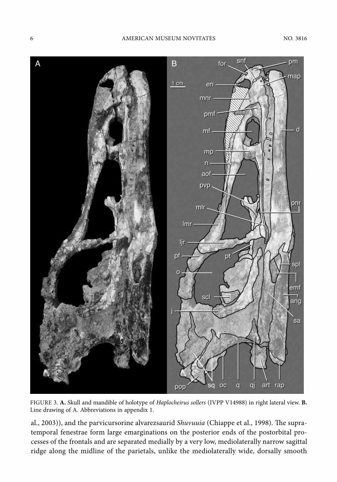
general overview and openings: The skull and mandible are nearly complete and uncrushed, although many of the skull bones are in very poor condition (figs. 3–12). The skull exhibits no mediolateral crushing and is only mildly distorted, the most significant aspect of which is the slight dorsoventral displacement of the posterior bones on the right half. The skull roof is poorly preserved, with numerous breaks and missing cortical bone in the nasals, fron-tals, and parietals. The right parietal, squamosal, frontal, and postorbital are absent. The ante-rior end of the right nasal is missing. many of the maxillary teeth are missing on the left side, and the right maxillary and most of the dentary teeth are obscured by matrix.
The rostrum is long and low, as in ornithomimosaurs (makovicky et al., 2004), Shuvuuia (Chiappe et al., 1998; Chiappe et al., 2002) and some troodontids (makovicky and norell, 2004). The orbital region and posterior ends of the skull are expanded from the narrow ros-trum both mediolaterally and dorsoventrally. The antorbital fossa is large and anteroventrally pointed, extending almost to the anteriormost tip of the maxilla and dorsally onto the ven-trolateral surface of the nasals. The internal margin of the antorbital fenestra is bordered dorsally by the maxilla and the lacrimal, but the dorsal margin of the antorbital fossa is rimmed by the nasal and the lacrimal. A small maxillary fenestra is anteriorly rounded and squared posteriorly, and it is separated from the antorbital fenestra by an anteroposteriorly narrow maxillary pila (interfenestral bar). it is offset posteriorly from the anterior margin of the antorbital fossa and is located approximately at midlevel in the antorbital fossa unlike the dorsally displaced maxillary fenestrae of many dromaeosaurids (turner et al., 2012). A dorsoventrally tall, slitlike promaxillary foramen is located under the anterior margin of the antorbital fossa, and is hidden in lateral view by the lateral lamina of the nasal ramus of the maxilla at the anteroventral margin of the fossa. The orbits face anterolaterally. The maxilla would have participated in the posterior margin of the external naris, although it probably only contributed a small portion. The ovoid external naris is anteroposteriorly long and dorsoventrally low, and its long axis is oriented nearly horizontally. This suite of features of the external naris is common to the basal tyrannosauroids Guanlong (iVPP V14531, V14532) and Proceratosaurus (rauhut et al., 2010), ornithomimosaurs (e.g., Gallimimus (igm 100/1133), troodontids (e.g., Sinovenator [iVPP V12632] and Byronosaurus (makovicky et
Figure 2. Composite stratigraphic section of the shishugou Formation at the Wucaiwan locality. stratigraphic position of holotype of Haplocheirus sollers and other theropod genera from this formation indicated by arrows.
6 AmeriCAn museum noVitAtes no. 3816
1 cm1 cm
forfor
jj
sclscl
oo
ljrljr
lmrlmr
mnrmnr
enen
raprapqqococpoppop
splspl
angang
emfemf
sasa
pvppvp
pnrpnrmlrmlr
pmpm
mapmap
dd
pmfpmf
mfmf
mpmp
aofaofnn
artart
pfpf ptpt
sq qjqj
snfsnfA B
Figure 3. A. skull and mandible of holotype of Haplocheirus sollers (iVPP V14988) in right lateral view. B. line drawing of A. Abbreviations in appendix 1.
al., 2003)), and the parvicursorine alvarezsaurid Shuvuuia (Chiappe et al., 1998). The supra-temporal fenestrae form large emarginations on the posterior ends of the postorbital pro-cesses of the frontals and are separated medially by a very low, mediolaterally narrow sagittal ridge along the midline of the parietals, unlike the mediolaterally wide, dorsally smooth

2014 Choiniere et Al.: HAPLOCHEIRUS SOLLERS 7
pmpm
poppop sqsq
epiepi
popo
oo
pfpf
laclac
pvppvp
mnrmnr
nmsnms
nn
nmpnmp
enen
artartqq
pmgpmg
dd
qjqj
qprqpr
jjcppcpp
emfemf
pnrpnr
mlrmlr
aofaof
mpmp
idpidp
mfmf
forfor
1 cm1 cm
dgdg
prapra
bpp
bspbsp
ptpt
lmrlmr
qfoqfo
A B
Figure 4. A. skull and mandible of holotype of Haplocheirus sollers (iVPP V14988) in left lateral view. B. line drawing of A. Abbreviations in appendix 1.
portions of the parietals that separate the supratemporal fenestrae in many basal theropods (rauhut, 2003), ornithomimosaurs (makovicky et al., 2004), and Shuvuuia (igm 100/977). The lateral margin of the supratemporal fenestra is straight and the medial margin is medi-ally convex. The infratemporal fenestra is dorsoventrally high and anteroposteriorly short,

8 AmeriCAn museum noVitAtes no. 3816
as in ornithomimosaurs (e.g., Garudimimus [kobayashi and barsbold, 2005a]), and the ther-izinosaurid Erlikosaurus (Clark et al., 1994). it is mesially constricted by the quadratojugal and squamosal approaching the postorbital bar.
skullPremaxilla: both premaxillary bodies are well preserved, but their nasal and maxillary
processes are distally broken (figs. 3–5, 8). The premaxillary body is square in lateral view, and only a small portion of it underlies the external naris, with the majority of the body located ante-rior to the anterior narial margin, as in Ornitholestes (Amnh FArb 619). in ventral view, the articulated premaxillae form a u-shaped junction. sutural marks on the anterior surface of the
nn
ococpoppopsppspp
stfstfpsppsp
lsplsp
ffjjoo
poppopbcbcpp
oo
laclac
pmpm
fnsfns
pfpf
1 cm1 cm
maxmax
laclac
jj
pfpf
A B
Figure 5. A. skull and mandible of holotype of Haplocheirus sollers (iVPP V14988) in dorsal view. B. line drawing of A. Abbreviations in appendix 1.

2014 Choiniere et Al.: HAPLOCHEIRUS SOLLERS 9
sclscl
ococpoppop
splspl
angang
dd
prapra
ampamp poppop
qcqc
nn
prapra
artart
hyhy
hyhy
jj
alvalv
1 cm1 cm
grgr
sasa
angang
mm
A B
Figure 6. A. skull and mandible of holotype of Haplocheirus sollers (iVPP V14988) in ventral view. B. line drawing of A. Abbreviations in appendix 1.
nasal ramus of the maxilla show that the maxillary process of the premaxilla would not have contacted the nasals on the posteroventral border of the naris. This differs from the condition in almost all ornithomimosaurs, in which the maxillary process extends posteriorly to contact the

10 AmeriCAn museum noVitAtes no. 3816
nasals, excluding the maxilla from participating in the external naris. often, as in many drom-aeosaurids, the maxillary ramus of the premaxilla extends between the nasomaxillary suture. The condition in Shuvuuia (igm 100/977, 100/1001) cannot be determined because the maxillary processes are either missing or broken in both skulls. The nasal processes, which form the inter-narial bar, are broken close to their bases above the anterior end of the external naris. The mor-phology of their bases suggests that they were dorsoventrally flat, as in troodontids (makovicky
pfpf
dd
sqsqpoppop CN X, CN XII CN X, CN XII
ococ
btbt
bspbsp
bsrbsr
qfoqfo
qcqc
bppbpp
qjqj
artart
qcqc
qfoqfo
socsoc
poppop
pmpm
mm
pnppnpenen
jj
popolaclac
nn
artart
1 cm1 cm
strstr
A CC
B DD
Figure 7. A. skull and mandible of holotype of Haplocheirus sollers (iVPP V14988) in anterior view. B. skull and mandible of holotype of Haplocheirus sollers (iVPP V14988) in posterior view. C. line drawing of A. D. line drawing of b. Abbreviations in appendix 1.
2014 Choiniere et Al.: HAPLOCHEIRUS SOLLERS 11
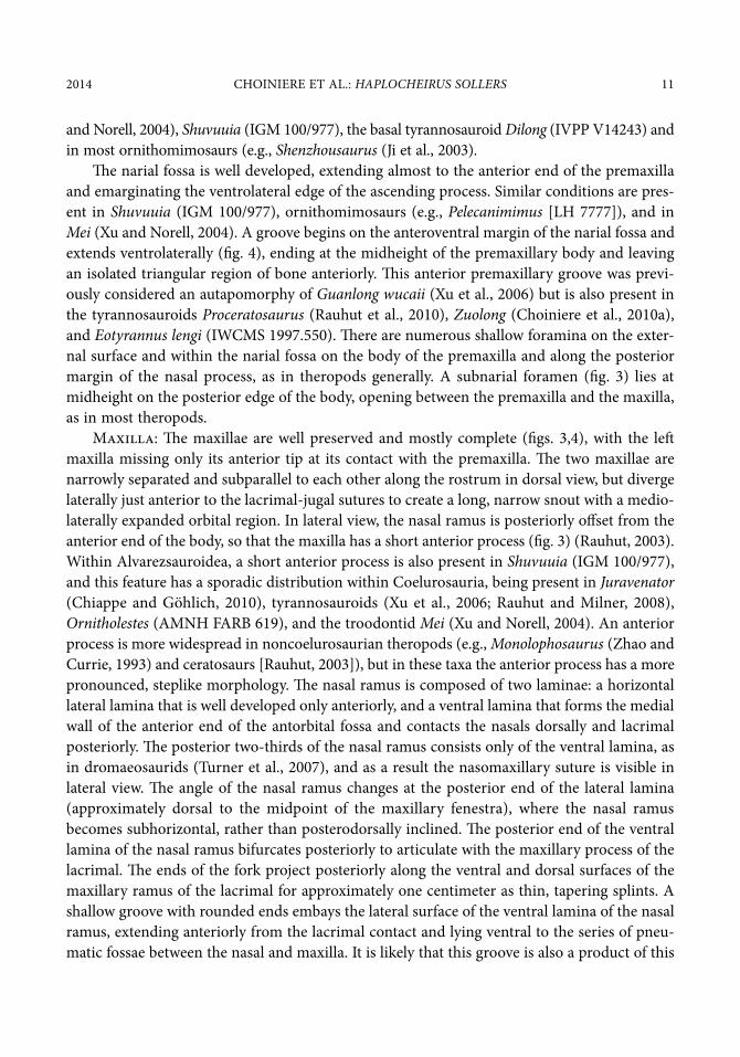
and norell, 2004), Shuvuuia (igm 100/977), the basal tyrannosauroid Dilong (iVPP V14243) and in most ornithomimosaurs (e.g., Shenzhousaurus (Ji et al., 2003).
The narial fossa is well developed, extending almost to the anterior end of the premaxilla and emarginating the ventrolateral edge of the ascending process. similar conditions are pres-ent in Shuvuuia (igm 100/977), ornithomimosaurs (e.g., Pelecanimimus [lh 7777]), and in Mei (Xu and norell, 2004). A groove begins on the anteroventral margin of the narial fossa and extends ventrolaterally (fig. 4), ending at the midheight of the premaxillary body and leaving an isolated triangular region of bone anteriorly. This anterior premaxillary groove was previ-ously considered an autapomorphy of Guanlong wucaii (Xu et al., 2006) but is also present in the tyrannosauroids Proceratosaurus (rauhut et al., 2010), Zuolong (Choiniere et al., 2010a), and Eotyrannus lengi (iWCms 1997.550). There are numerous shallow foramina on the exter-nal surface and within the narial fossa on the body of the premaxilla and along the posterior margin of the nasal process, as in theropods generally. A subnarial foramen (fig. 3) lies at midheight on the posterior edge of the body, opening between the premaxilla and the maxilla, as in most theropods.
maxilla: The maxillae are well preserved and mostly complete (figs. 3,4), with the left maxilla missing only its anterior tip at its contact with the premaxilla. The two maxillae are narrowly separated and subparallel to each other along the rostrum in dorsal view, but diverge laterally just anterior to the lacrimal-jugal sutures to create a long, narrow snout with a medio-laterally expanded orbital region. in lateral view, the nasal ramus is posteriorly offset from the anterior end of the body, so that the maxilla has a short anterior process (fig. 3) (rauhut, 2003). Within Alvarezsauroidea, a short anterior process is also present in Shuvuuia (igm 100/977), and this feature has a sporadic distribution within Coelurosauria, being present in Juravenator (Chiappe and göhlich, 2010), tyrannosauroids (Xu et al., 2006; rauhut and milner, 2008), Ornitholestes (Amnh FArb 619), and the troodontid Mei (Xu and norell, 2004). An anterior process is more widespread in noncoelurosaurian theropods (e.g., Monolophosaurus (Zhao and Currie, 1993) and ceratosaurs [rauhut, 2003]), but in these taxa the anterior process has a more pronounced, steplike morphology. The nasal ramus is composed of two laminae: a horizontal lateral lamina that is well developed only anteriorly, and a ventral lamina that forms the medial wall of the anterior end of the antorbital fossa and contacts the nasals dorsally and lacrimal posteriorly. The posterior two-thirds of the nasal ramus consists only of the ventral lamina, as in dromaeosaurids (turner et al., 2007), and as a result the nasomaxillary suture is visible in lateral view. The angle of the nasal ramus changes at the posterior end of the lateral lamina (approximately dorsal to the midpoint of the maxillary fenestra), where the nasal ramus becomes subhorizontal, rather than posterodorsally inclined. The posterior end of the ventral lamina of the nasal ramus bifurcates posteriorly to articulate with the maxillary process of the lacrimal. The ends of the fork project posteriorly along the ventral and dorsal surfaces of the maxillary ramus of the lacrimal for approximately one centimeter as thin, tapering splints. A shallow groove with rounded ends embays the lateral surface of the ventral lamina of the nasal ramus, extending anteriorly from the lacrimal contact and lying ventral to the series of pneu-matic fossae between the nasal and maxilla. it is likely that this groove is also a product of this

12 AmeriCAn museum noVitAtes no. 3816
1 cm1 cm nn
pvppvp mlrmlrmpmp
aofaofljrljr
nmsnms pnfpnf pnfpnflnslns
dd
pmgpmg
mfmf
forfor
enen
forfor
alvalvmlrmlr
aofaof mm
pnfpnf
lmrlmrnnsnns
nnlmnslmns
mfmf
mpmp
pnfpnfnmsnms mnrmnrpnfpnf
A
B
Figure 8. A. rostrum and anterior mandible of holotype of Haplocheirus sollers (iVPP V14988) in left lateral view. B. maxillary region of holotype of Haplocheirus sollers (iVPP V14988) in left ventrolateral view. Abbre-viations in appendix 1.

2014 Choiniere et Al.: HAPLOCHEIRUS SOLLERS 13
pneumatization. The medial surface of the maxillary nasal ramus is embayed by two shallow, vertically arranged depressions along its anterior junction with the maxillary pila. The more dorsal of the two is continuous with the anteromedial recess on the maxillary ramus of the lacrimal. The ventral depression is located slightly anterior to the dorsal depression. medial processes contact each other posterodorsal to the maxillary fenestra to form a short ventral floor for the nasal chamber.
The maxillary pila has a foramen centered on its posterior surface (the fenestra posterioris antri maxillaris) (Witmer, 1997b) that would have allowed communication between the maxil-lary fenestra and the antorbital fenestra (fig. 8). This foramen is present in the ornithomimo-saurs Gallimimus (igm 100/1133) and Pelecanimimus (lh 7777), the parvicursorine alvarezsauroid Shuvuuia (igm 100/977) and in most troodontids (makovicky and norell, 2004). A foramen in this position is also present in tyrannosaurids (Witmer, 1997b; brochu, 2003), but in these theropods it is developed as part of an inflated maxillary antrum. The lateral surface of the jugal ramus of the maxilla has a prominent horizontally oriented and laterally projecting lamina of bone extending from the anterior edge of the antorbital fossa to its pos-terior terminus that forms the ventral margin of the antorbital fossa. This lamina appears vari-ably in theropods (rauhut, 2003) and is present in two other small coelurosaurs from Wucaiwan, Zuolong (Choiniere et al., 2010a) and Aorun (Choiniere et al., 2014). on the anterior end of the lateral surface of the alveolar margin, immediately ventral to the lateral ridge, is a shallow sulcus (fig. 12b) containing several large, oval foramina. These are in the same position as the nutrient foramina of other theropods, but they are much larger than typical nutrient foramina and may represent further pneumatized pockets of the maxilla. The groove makes the tooth row very slightly medially inset from the lateral edge, an effect that is most pronounced in the middle of the tooth row.
on the medial surface of the jugal ramus, the palatal shelves converge at the anterior end of the antorbital fenestra, forming the secondary palate in conjunction with the premaxillae and likely the vomers, which are not preserved. evidence of a vomerine contribution to the secondary palate is the slight separation of the maxillary palatal shelves that are preserved in life position (although this may be an artifact of postmortem distortion as these bones are not suturally connected). in most maniraptorans, the vomer overlies the contacting maxillary shelves. The posterior end of the jugal ramus of the maxilla expands dorsally where it meets the descending ramus of the lacrimal, a morphology that is autapomorphic for Haplocheirus within Alvarezsauroidea. This expansion excludes the jugal from participation in the internal margin of the antorbital fenestra. The lateral component of the alveolar margin is missing and the interdental plates on the medial surface can be seen in lateral view. Though small, they are unfused, approximately square, and similar to the general morphology of most theropod inter-dental plates (however, see discussion in bever and norell, 2009).
nasal: The nasals are broken anteriorly, missing the premaxillary processes and the anterior termini of the maxillary processes (figs. 3–5). They are unfused and the midline contact between the adjoining nasals can be seen in both dorsal and ventral views. during preservation, the medial edges of the nasals were displaced slightly ventrally, so that the

14 AmeriCAn museum noVitAtes no. 3816
internasal suture is depressed ventrally, below the lateral margins. in life position, the nasals probably would have formed a shallow, dorsally convex arc along the dorsal surface of the rostrum in transverse cross section. The nasals form the posterior, posterodorsal, and pos-teroventral margins of the external nares. sutural marks on the anterior surface of the max-illa indicate that the maxillary process of the nasal failed to contact the maxillary process of the premaxilla along the ventral margin of the external naris. The dorsal surface of the nasal is smooth and expands slightly mediolaterally as it extends posteriorly. The dorsal surface of the anterior end of the left nasal bears three small, shallow foramina immediately posterior to the premaxillary contact (fig. 9A), typical of theropods generally. These foramina cannot be seen on the right side because the anterior end of the right nasal is not preserved. The ventrolateral surface of the nasal forms the dorsal margin of the antorbital fossa. it is under-cut by three deep, anteroposteriorly long, ovoid pneumatic fossae. These are similar to those of Sinraptor (Currie and Zhao, 1993), but they are more extensively developed along the lateral margin of the nasal in Haplocheirus and are ovoid rather than subcircular. The ven-tromedial surface of the right nasal shows that these fossae do not penetrate the lateral wall of the nasal.
The nasomaxillary suture follows the ventral edge of these pneumatic fossae and lies entirely within the antorbital fossa. The posterior end of the nasal is poorly preserved, but its dorsal suture with the anterior end of the frontal appears to have been transversely oriented. it overlaps the dorsal surface of the anterior end of the frontal in a lap joint that is perpendicu-lar to the long axis of the skull.
lacrimal: The right lacrimal is fractured but nearly complete (figs. 3, 4). The left lacrimal is missing the lateral margin of the angle, but the ventrally directed jugal ramus is mostly complete and the maxillary ramus is well preserved. The jugal ramus extends the full height of the anterior border of the orbit, forming its anterior margin and the posterior boundary of the antorbital fenestra. The presence of a foramen for the nasolacrimal duct leading through the jugal ramus cannot be confirmed because of the poor preservation of both sides. As in most maniraptorans and in ornithomimosaurs, the jugal ramus is only slightly expanded anteropos-teriorly as it extends ventrally. At the angle of the lacrimal, there is no evidence for a lacrimal foramen, as in Shuvuuia (igm 100/977, 100/99) and most other theropods, but this may be an artifact of preservation.
The maxillary ramus of the lacrimal is very long and tapers anteriorly as in Shuvuuia (igm 100/977) and in maniraptorans generally (e.g., Byronosaurus [makovicky et al., 2003]). it artic-ulates anteriorly with the forked posterior end of the nasal ramus of the maxilla and dorsally with the nasal along the posterior dorsal margin of the antorbital fossa. An embayment of the lateral surface of the maxillary ramus forms the posterodorsal corner of the antorbital fossa. The anterior end of the maxillary ramus of the right lacrimal extends along the medial surface of the rostrum, medial to the ventral lamina of the nasal process of the maxilla, and it extends anteriorly on the medial surface almost to the maxillary pila. The medial surface of the anterior end has a pocketlike ovoid foramen that extends posteriorly and slightly laterally from an ori-gin on the anterior tip. As in Shuvuuia, there is no well-developed lacrimal posterodorsal

2014 Choiniere et Al.: HAPLOCHEIRUS SOLLERS 15
process (Chiappe et al., 1998). Haplocheirus lacks the small “lacrimal horn” that projects pos-terolaterally in Shuvuuia (igm 100/977) and many other theropods.
Prefrontal: The left side of the skull preserves the prefrontal articulating with the pos-terior edge of the lacrimal angle (fig. 4), but the right lacrimal is badly damaged and is repre-sented only by a small area of broken bone adhering to the lacrimal angle (fig. 5). because the left lacrimal angle is poorly preserved, it is unclear whether the prefrontal was a completely separate ossification from the lacrimal or whether it was tightly sutured to the posterior margin of the lacrimal angle, as in Ornitholestes (Amnh FArb 619). unlike the hypertrophied pre-frontals of ornithomimosaurs (e.g., Gallimimus (igm 100/1133)) and Shuvuuia (igm 100/977), which are diamond shaped in dorsal view, the prefrontal of Haplocheirus is shaped like a nar-row triangle in dorsal view with its long axis oriented sagittally and the base oriented medio-laterally. A similar morphology is observed in the therizinosauroid Erlikosaurus (Clark et al., 1994). The prefrontal is mediolaterally widest at the lacrimal contact, and in dorsal view, it tapers to a posteriorly directed point. Also when viewed dorsally, the straight medial margin forms an extensive contact with the lateral edge of the frontal. The posterior end forms a por-tion of the anterodorsal orbital rim. There is a small fossa on the ventrolateral margin of the posterior end. The ventral surface has a slender ventral process that runs along the medial edge of the lacrimal, but the ventralmost terminus of this process is broken, so its full extent cannot be determined. The anterolateral surface has a distinct, anteriorly concave notch for articulation with the lacrimal angle, a feature also present in Ornitholestes (Amnh FArb 619) but is not developed in either ornithomimosaurs or Shuvuuia (igm 100/977, 100/99). The anterior end is broken, but it appears to have separated the posterior end of the lacrimal from the frontal margin. in ornithomimosaurs (e.g., Gallimimus [igm 100/1133] and Shuvuuia [igm 100/977]), the prefrontal separates the posterior half of the lacrimal from the frontal (sereno, 2001).
sclerotic ossicles: Well-preserved sclerotic rings are present in both orbits (figs. 3, 10; note breakage to specimen prior to final photography). The right sclerotic ring preserves the intact dorsal half, but it has been dislocated to the midline of the skull inside the orbit overlying the cultriform process and the pterygoid. The left sclerotic ring is mostly complete, but the dorsal half has been displaced ventrally and overlaps the ventral half. The sclerotic ring is composed of approximately 20 imbricating sclerotic ossicles. The ossicles are oval and medially flexed, so the outer surface of the ring is concave and the inner (eyeball side) of the ring is convex. The inner diameter of the ring is 33 mm, and the outer diameter is 39 mm. The inner diameter of the ring is approximately 76% of the diameter of the orbital fenestra.
Postorbital: The left postorbital is complete and preserved in articulation with the sur-rounding frontal, laterosphenoid, squamosal, and jugal (figs. 4, 10). The right postorbital is not preserved. The frontal ramus is missing its anteriormost tip. in lateral view, the frontal ramus forms a dorsally convex arc that makes up the majority of the posterior border of the orbit. it arches well above the level of the squamosal process, as is typical for maniraptorans, and is mediolaterally restricted to a thin bar of bone along the anterolateral edge of the frontal, unlike the mediolaterally wide frontal ramus of basal coelurosaurs like Zuolong (Choiniere et al., 2010a). Although it is broken, the anterior end of the frontal ramus probably did not contact

16 AmeriCAn museum noVitAtes no. 3816
the lacrimal (thus excluding the frontal from participation in the orbit) because these bones would have been separated by the prefrontal. The medial surface of the frontal ramus contacts the laterosphenoid at the level of the posterior end of the orbit. The contact surface for the laterosphenoid is medially concave and smaller in relative diameter than it is in more basal tetanuran theropods like Allosaurus (madsen, 1976). The dorsal surface extends dorsal to the level of the suture with the frontal, which may mark the anterolateral border of the supratem-poral fossa. This observation is tentative because of extensive breakage in this area and lack of preservation of the right postorbital.
The squamosal process is anteroposteriorly short, dorsoventrally low, and arches dorsally. The sutural boundary between it and the squamosal is unclear because of breakage, but there is possible fusion at the contact. The jugal ramus forms the anterodorsal half of the postorbital bar. it is triangular in cross section.
Jugal: The left jugal (figs. 4, 8, 10) is complete and well preserved. The right jugal is miss-ing its anterior end. The anterior jugal ends bluntly at the posteroventral corner of the antor-bital fossa and lacks an anterior spur that extends below the antorbital fenestra, a feature of many other maniraptorans (e.g., Erlikosaurus [Clark et al., 1994], Tsaagan [norell et al., 2006]). The jugal participates only in the posteroventralmost corner of the antorbital fossa, and is slightly emarginated on the anterolateral tip to form the posteroventral corner of the antorbital fossa. There is no jugal foramen at this corner as in some theropods (madsen, 1976; Currie and Zhao, 1993; Zhao and Currie, 1993; sereno et al., 1996; rauhut, 2003). The suborbital portion of the jugal is strongly laterally convex and dorsoventrally low, as in Shuvuuia (igm 100/977, 100/99), Erlikosaurus (Clark et al., 1994), and birds (baumel and Witmer, 1993). The maximal lateral extent of the jugal is at a level immediately anterior to the postorbital process. The jugals of Juravenator (Chiappe and göhlich, 2010) and ornithomimosaurs are also dorsoventrally low, but the lateral surface is generally flat in these taxa (e.g., Pelecanimimus [lh 7777]). The jugal expands dorsoventrally below the postorbital process to form a large triangular plate. The postorbital process extends two-thirds of the way up the anterior margin of the infratemporal fenestra and forms approximately half the postorbital bar before articulating with the overlap-ping postorbital.
The quadratojugal process is anteroposteriorly short and forked at the posterior end for reception of the jugal ramus of the quadratojugal. This morphology, typical of coelurosaurs, is unlike the condition in Shuvuuia (igm 100/977, 100/99), whose quadratojugal and jugal are fused and indistinguishable from one another. The dorsal and ventral processes of the fork are subequal in length and the articular facet for the jugal process of the quadratojugal forms an anteriorly tapering trough centered between these processes. two small pits are present on the ventral surface of the jugal lateral to the ectopterygoid contact. These pits do not appear to open into pneumatic foramina although they are clogged with matrix and their full depth can-not be determined.
Quadratojugal: The left quadratojugal is well preserved (fig. 4, 10), but the right qua-dratojugal is missing. it is a hook-shaped bone with a short, anteriorly tapering jugal process and a tall squamosal process. The angle between the squamosal process and the jugal process

2014 Choiniere et Al.: HAPLOCHEIRUS SOLLERS 17
is acute. The anteriorly expanded dorsal end of squamosal process trends anterodorsally, and together with the quadratojugal process of the squamosal this constricts the intfratemporal fenestra at midheight. The squamosal process overlaps the quadratojugal process of the squa-mosal laterally at the same level where it expands anteriorly to constrict the infratemporal fenestra. medially, the squamosal process contacts the lateral margin of the quadrate shaft. in posterior view, the posterior edge of the squamosal process wraps around the posterolateral edge of the quadrate. The jugal process is short relative to many other theropods. in most nonavian theropods (e.g., Ornitholestes [Amnh FArb 619], Guanlong [iVPP V14531]), the jugal process reaches anteriorly to at least the posterior margin of the postorbital process of the jugal. in Haplocheirus, the jugal process ends at approximately the midpoint of the infratem-poral fenestra, well posterior to the postorbital process.
Frontal: The frontals (fig. 5) are poorly preserved and the lateral edge of the right frontal above the orbit is missing. The frontals are not coossified, and the suture between them appears to have been simple, without complex interdigitation. The dorsal surface of the frontal is smooth. together, the frontals are hourglass shaped, with deeply concave orbital excavations on the lateral margins. The anterior end of the frontal terminates bluntly at the nasals in dorsal view, forming a transversely oriented suture, but in ventral view, a long, anteriorly tapering process of each frontal underlies the posterior ends of the nasals. The medial surfaces of these processes are poorly preserved, but they do not appear to have contacted each other along the midline of the skull. The ventral surface of each anterior process bears a low, anteroposteriorly oriented ridge. on the anterior end of the lateral surface, the frontal has a long, straight contact with the prefrontal. The poorly preserved orbital margin posterior to the prefrontal does not show any prominent rugosities or ornamentation.
Posterior to the lateral emargination for the orbits, the postorbital process is laterally exten-sive and anteroposteriorly short. The supratemporal fossa is well developed on the posterior edge of the postorbital process, forming a posterodorsally facing recessed area on almost the entire posterolateral margin. The anterior edge of this supratemporal emargination is anteriorly convex, not sinusoidal as it is in some paravians (turner et al., 2007). The posterior sutural boundary with the parietal is indistinct due to extensive breakage in this area. The cristae cranii are well developed on the ventral surface of the frontal, forming laterally concave, ventrally extending ridges. They are narrowly separated medially and diverge strongly from each other anteriorly and posteriorly.
Parietal: only the left half of the parietal is present (figs. 4, 5), but it is broken into small pieces that remain close to their original positions, so little can be described of its morphology. in ornithomimosaurs (makovicky et al., 2004), therizinosaurids, and alvarezsauroids (Chiappe et al., 1998) and other maniraptorans (e.g., dromaeosaurids [norell and makovicky, 2004]), the parietals are fused. This was likely the case in Haplocheirus based on its phylogenetic position and based on the overall morphology of the preserved area. The dorsal surface appears to have been smooth and probably formed a shallowly convex plate between the supratemporal fenes-trae, widely separating them along the skull midline. There is no sign of a sagittal crest or any other parietal ornamentation along the skull midline.

18 AmeriCAn museum noVitAtes no. 3816
squamosal: The left squamosal is complete but shattered and the right squamosal consists of only small fragments (figs. 3–5, 10, 12). unlike the reduced, triradiate squamosal of Shuvuuia (Chiappe et al., 1998), the squamosal of Haplocheirus is tetraradiate, with a postorbital process projected anteriorly, a quadratojugal process directed ventrally, a medial process that contacts the parietal and the laterosphenoid, and a paroccipital process extending over the quadrate head and contacting the dorsal margin of the paroccipital process of the exoccipital-opisthotic. The squa-mosal bounds the supratemporal fossa laterally and posteriorly. The postorbital process is short and the suture between it and the squamosal process of the postorbital is indistinct in lateral view. some of the obscurity is due to shattering in this area, but it is possible that this contact was fused. The quadratojugal process forms a sinuous posterior contact with the lateral margin of the quad-rate. it is angled 45° anteroventrally from the near vertical quadrate shaft, tapers distally and contacts the squamosal process of the quadratojugal ventrally. The paroccipital process is pen-dant, overhanging the posterior margin of the quadrate head in lateral view, but does not project laterally as a tab as it does in dromaeosaurids. The quadrate cotyle of the squamosal opens later-ally, exposing the head of the quadrate in lateral view, although some of the posterior portion of the quadrate head is covered laterally by the lateral expansion of the squamosal process. The posterior edge of the paroccipital process of the squamosal forms an extensive, horizontally ori-ented, flat contact with the dorsal margin of the paroccipital process itself. A low ridge of bone extends along the posterior boundary of the paroccipital process of the squamosal, defining this suture. The medial process that forms the posterior rim of the supratemporal fenestra is poorly preserved. A ventral extension of this process approaches the laterosphenoid along the medio-ventral wall of the supratemporal fossa; however, contact between the two elements cannot be confirmed. The dorsomedial aspect of the medial process is eroded away. it contacted the parietal at the posteromedial corner of the supratemporal fenestra. The dorsal surface of the body does not appear to have been emarginated by the supratemporal fossa.
Quadrate: both quadrates are preserved in their original articulations and positions. The shaft of the quadrate is anteriorly convex in lateral view and the mandibular condyles are situ-ated directly ventral to the quadrate head. The quadrate head is simple and distinct from the quadrate shaft, forming a dorsally convex structure that projects posterodorsally. There is no evidence of a second, medial quadrate head, as in derived avialans (baumel and Witmer, 1993), Shuvuuia (Chiappe et al., 1998), and oviraptorosaurs (maryanska and osmólska, 1997; balanoff et al., 2009). in posterior view, there is a thin, dorsoventrally oriented ridge of bone along the medial edge of the quadrate shaft, forming a pillarlike structure. lateral to this pillar, the ven-tral half of the posterior surface of the quadrate has a deep fossa extending medially from its lateral contact with the quadratojugal. This fossa is continuous with the small quadrate foramen that is present as a vertical slitlike opening between the quadrate and quadratojugal. The lateral side of the dorsal half of the posterior surface of the quadrate forms a large, laterally projecting flange above the quadrate foramen that has a sinuous contact with the squamosal in lateral view. The dorsal margin of this flange is convex.
The pterygoid ramus of the quadrate is anteriorly convex. The ventral margin has a notch close to the quadrate body, and as preserved, the basipterygoid process of the basisphenoid passes through this notch, although this is likely not life position.
2014 Choiniere et Al.: HAPLOCHEIRUS SOLLERS 19
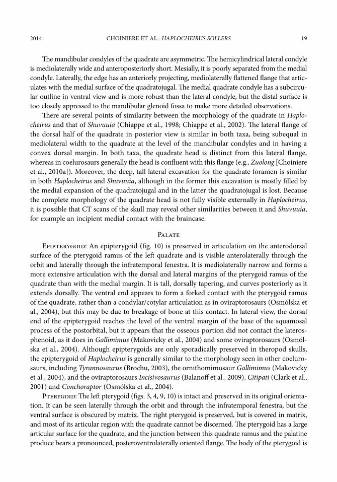
The mandibular condyles of the quadrate are asymmetric. The hemicylindrical lateral condyle is mediolaterally wide and anteroposteriorly short. mesially, it is poorly separated from the medial condyle. laterally, the edge has an anteriorly projecting, mediolaterally flattened flange that artic-ulates with the medial surface of the quadratojugal. The medial quadrate condyle has a subcircu-lar outline in ventral view and is more robust than the lateral condyle, but the distal surface is too closely appressed to the mandibular glenoid fossa to make more detailed observations.
There are several points of similarity between the morphology of the quadrate in Haplo-cheirus and that of Shuvuuia (Chiappe et al., 1998; Chiappe et al., 2002). The lateral flange of the dorsal half of the quadrate in posterior view is similar in both taxa, being subequal in mediolateral width to the quadrate at the level of the mandibular condyles and in having a convex dorsal margin. in both taxa, the quadrate head is distinct from this lateral flange, whereas in coelurosaurs generally the head is confluent with this flange (e.g., Zuolong [Choiniere et al., 2010a]). moreover, the deep, tall lateral excavation for the quadrate foramen is similar in both Haplocheirus and Shuvuuia, although in the former this excavation is mostly filled by the medial expansion of the quadratojugal and in the latter the quadratojugal is lost. because the complete morphology of the quadrate head is not fully visible externally in Haplocheirus, it is possible that Ct scans of the skull may reveal other similarities between it and Shuvuuia, for example an incipient medial contact with the braincase.
Palateepipterygoid: An epipterygoid (fig. 10) is preserved in articulation on the anterodorsal
surface of the pterygoid ramus of the left quadrate and is visible anterolaterally through the orbit and laterally through the infratemporal fenestra. it is mediolaterally narrow and forms a more extensive articulation with the dorsal and lateral margins of the pterygoid ramus of the quadrate than with the medial margin. it is tall, dorsally tapering, and curves posteriorly as it extends dorsally. The ventral end appears to form a forked contact with the pterygoid ramus of the quadrate, rather than a condylar/cotylar articulation as in oviraptorosaurs (osmólska et al., 2004), but this may be due to breakage of bone at this contact. in lateral view, the dorsal end of the epipterygoid reaches the level of the ventral margin of the base of the squamosal process of the postorbital, but it appears that the osseous portion did not contact the lateros-phenoid, as it does in Gallimimus (makovicky et al., 2004) and some oviraptorosaurs (osmól-ska et al., 2004). Although epipterygoids are only sporadically preserved in theropod skulls, the epipterygoid of Haplocheirus is generally similar to the morphology seen in other coeluro-saurs, including Tyrannosaurus (brochu, 2003), the ornithomimosaur Gallimimus (makovicky et al., 2004), and the oviraptorosaurs Incisivosaurus (balanoff et al., 2009), Citipati (Clark et al., 2001) and Conchoraptor (osmólska et al., 2004).
Pterygoid: The left pterygoid (figs. 3, 4, 9, 10) is intact and preserved in its original orienta-tion. it can be seen laterally through the orbit and through the infratemporal fenestra, but the ventral surface is obscured by matrix. The right pterygoid is preserved, but is covered in matrix, and most of its articular region with the quadrate cannot be discerned. The pterygoid has a large articular surface for the quadrate, and the junction between this quadrate ramus and the palatine produce bears a pronounced, posteroventrolaterally oriented flange. The body of the pterygoid is

20 AmeriCAn museum noVitAtes no. 3816
expanded near the articulation with the ectopterygoid. The palatine processes are thin, straplike elements. They taper slightly as they extend anteriorly, then expand again at their anterior tip where they contact the posterior end of the vomers and the vomeropterygoid process of the pala-tine. in lateral view, the palatine processes scribe a shallow, dorsally concave arc.
Palatine: The palatines are complete and well preserved (fig. 10). The tetraradiate palatine is located well anterior to the ectopterygoid as preserved, but its position may have shifted during preservation. on the medial edge, the contact between the palatine and the pterygoid is discontinuous in the midregion, resulting in a pronounced choana as in dromaeosaurids (norell and makovicky, 2004), ornithomimosaurs (rauhut, 2003), and the tyrannosaurid Das-pletosaurus (russell, 1970) (although not in Tyrannosaurus [brochu, 2003]). The maxillary process is long and anteriorly tapering, and it extends along the mediodorsal edge of the pos-teromedial margin of the maxillary palatal process. The jugal process forms a complicated quadruple junction at the posteroventromedial margin of the antorbital fossa, contacting the medial surfaces of the maxilla, the maxillary ramus of the lacrimal, and the maxillary process of the jugal. The portion of the body between the maxillary process and the vomeropterygoid process is inclined dorsomedially, and the portion of the body between the jugal process and the pterygoid process is subhorizontally oriented.
A dorsolaterally projecting ridge extends anteriorly from the junction with the lacrimal, jugal, and maxilla and grades into the dorsolateral surface of the palatine body lateral and ventral to the anterior pterygoid process. The ridge divides the posterior end of the palatine into medial and lateral sections. medial to this ridge, a deep fossa (fossa muscularis) (Witmer, 1997b) is present. Ventral and lateral to the anterior end of the ridge, an anteriorly opening, anteroposteriorly elongate pneumatic recess of the palatine (recessus pneumaticus palatinus) (Witmer, 1997b) is present. A similarly elongate fossa is present in Pelecanimimus (lh 7777) and Gallimimus (igm 100/1133). in avialans like Archaeopteryx (mayr et al., 2005), the palatine ridge is less pronounced and the anterior pneumatic fossa is enlarged and triangular relative to the condition in Haplocheirus, occupying the majority of the anterodorsal surface of the palatine. A pneumatic fossa is absent in most noncoelurosaurian theropods (Witmer, 1997b) including Allosaurus (madsen, 1976), but one is present in Sinraptor (Currie and Zhao, 1993), where it is large, circular, and perforated by two foramina. Witmer (1997b) reports that a fora-men is often developed in the position of the pneumatic recess in tyrannosaurids.
ectopterygoid: The left ectopterygoid (not figured) is located posterior to the palatine and preserved in articulation with the jugal and pterygoid, but most of the ventral surface is obscured by matrix. The jugal process of the right ectopterygoid is preserved in articulation with the jugal, but the contact with the pterygoid is not preserved. Although similar to other theropods in being hook shaped, it is relatively small and slender when compared to the ectopterygoids of dromaeosaurs (Currie, 1995). no other alvarezsauroid preserves an ectopter-ygoid (Chiappe et al., 2002). The ectopterygoid body is inflated near its contact with the ptery-goid, as in coelurosaurs generally (rauhut, 2003), but the extent of this inflation cannot be determined. A matrix-filled foramen on the dorsal surface of the ectopterygoid may represent a dorsal recess, as in some dromaeosaurids (ostrom, 1969; Currie, 1995), but it is possible it is a preservational artifact.

2014 Choiniere et Al.: HAPLOCHEIRUS SOLLERS 21
Vomer: A small fragment of the anterior end of the left vomer is preserved in contact with the medial edge of the anterior end of the maxillary palatal shelf.
braincaseexoccipital-opisthotic: The condylar portions of the exoccipitals (figs. 7, 11) are tightly
sutured to the occipital condyle, but the sutures can still be seen in posterior view. each exoccipi-tal contributes approximately one-quarter of the outer rim of the occipital condyle and forms the ventrolateral boundary of the foramen magnum, although they are separated along the ventral margin of this structure by the basioccipital. lateral to the condylar portion of the exoccipitals, a pair of horizontally arranged foramina open posteriorly. The circular lateral foramen is smaller than the medial foramen. The medial foramen is divided interiorly by a very thin lamina into two distinct openings that represent separate branches of the hypoglossal nerve (Cn Xii). A horizon-tal strut (fig. 7) projects from the condylar portion of the exoccipital to merge smoothly with the posterior surface of the paroccipital process, forming a triangular roof over these foramina. This roof forms the dorsal boundary of a shallow subcondylar recess (Witmer, 1997a), a presumably pneumatic feature common in theropods and within maniraptora (e.g., the basal therizinosauroid Falcarius [smith et al., 2011]). A second bony strut emerges from the lateral margin of the exoc-cipital to form the ventral margin of the paroccipital processes and forms the lateral boundary of the recess. The suture between the exoccipital and the basioccipital is visible within the subcon-dylar recess. it is oriented 45° to the plane of the paroccipital processes.
The paroccipital process is mediolaterally long, dorsoventrally narrow, and slightly pen-dant. The degree of ventral deflection of the pendant paroccipital process is similar to that of Erlikosaurus (Clark et al., 1994), but less than that of Incisivosaurus (balanoff et al., 2009) or other oviraptorosaurs (Clark et al., 2002). The ventral edge of the distal end of the process is twisted posteriorly, so that the ventral margin of the process is subhorizontal in distal view, but the dorsal margin retains its vertical orientation, a morphology autapomorphic for Haplochei-rus. This differs from the morphology present in some dromaeosaurids where the dorsal edge of the distal end is twisted anteriorly (norell and makovicky, 2004). The ventral rim of the base of the paroccipital process is situated below the midpoint of the occipital condyle. it is not known whether the paroccipital process is hollow as in some maniraptorans. The anterior surface of the opisthotic has a deep excavation for the caudal tympanic recess. A shallow dorsal tympanic recess is present on the anterodorsal surface of the opisthotic.
basioccipital: The basioccipital (figs. 7, 11) forms the majority of the occipital condyle. The size of the foramen magnum is difficult to determine because of poor preservation and matrix infill, although it is clear that it is relatively smaller than in parvicursorine alvarezsau-roids and avians where this opening is unusually large (Chiappe et al., 1998). The dorsal surface of the basioccipital is concave, emarginated by the foramen magnum of which it forms the median ventral margin. The basioccipital is mildly constricted anterior to its condylar portion, forming a very short “neck.” A poorly developed, shallow infracondylar fossa is developed on the ventral surface of the basioccipital neck. The lateral surfaces of the basal tubera are slightly eroded. Their medial margins are directly ventral to the lateral edge of the occipital condyle, and they are formed equally by the basioccipital and basisphenoid. The mediolaterally wide

22 AmeriCAn museum noVitAtes no. 3816
basal tubera are narrowly separated along the skull midline. The shallow subcondylar recess is more deeply excavated on the basioccipital than on the exoccipital, extending as a deep pocket onto the posterodorsal surfaces of the basal tubera. deep fossae are present at the ventromedial corners of the subcondylar recess, but they do not appear to fully penetrate the bone.
basisphenoid-Parasphenoid: The basisphenoid (fig. 11) is anteroposteriorly long and inclined approximately 45° from horizontal as it is in Shuvuuia (igm 100/99, 100/977) and in at least one small troodontid specimen (igm 100/1128). in the spinosauroid Baryonyx (nhmuk r9951) and in some oviraptorosaurs (e.g., Citipati [Clark et al., 2002]), the basisphe-noid is oriented nearly vertically, but more often it is horizontal in theropods. The basisphenoid forms the anterolateral portion of the basal tubera. The oval basisphenoid recess is deeply excavated and lacks the longitudinal midline ridge on its floor that is present in some mani-raptorans. due to the inclination of the basisphenoid, the basisphenoid recess is visible in posterior view. The left basipterygoid process can be seen through the infratemporal fenestra passing ventral to the pterygoid ramus of the quadrate and abutting the mediodorsal surface
1 cm1 cm
mpsmps
dd
mlrmlrmfmf
pmfpmfaomaom
mm
ljrljr
pnrpnr
vv
pvppvp
aofaof
nn
mpfmpf
Figure 9. maxillary and anterior palatal region of holotype of Haplocheirus sollers (iVPP V14988) in right dorsolateral view. Abbreviations in appendix 1.

2014 Choiniere et Al.: HAPLOCHEIRUS SOLLERS 23
1 cm1 cm popopsppsp
jjsclsclpfpf
artart
qjqjqprqprcpfcpf
cppcppqq ococ
epiepi
poppop
sppsppsqsq
ljrljr
prprpgpg
pnrpnr
vvptpt
pvppvp
mjcmjc
dd
mjrmjr
aofaof
A
B
Figure 10. A. Posterior end of skull and mandible of holotype of Haplocheirus sollers (iVPP V14988) in left lateral view, showing original preservation of sclerotic ring (subsequently damaged). B. Palate of holotype of Haplocheirus sollers (iVPP V14988) in right dorsolateral view. Abbreviations in appendix 1.

24 AmeriCAn museum noVitAtes no. 3816
of the surangular. it is long and distally tapering, projecting ventrolaterally as in Shuvuuia (Chiappe et al., 2002). The right basipterygoid process is obscured by matrix in anterior view.
The left lateral surface of the cultriform process of the parasphenoid can be seen inside the left orbit (figs. 4, 10). The cultriform process is long, low, and subhorizontally oriented. it tapers gradually as it extends anteriorly, unlike the proximally expanded and sharply anteriorly tapered cultriform process of troodontids, tyrannosaurids, and ornithomimosaurs (Chiappe et al., 1998; Currie and dong, 2001; Chiappe et al., 2002; makovicky et al., 2004; makovicky and norell, 2004; kobayashi and barsbold, 2005a). The ventral surface of the cultriform process is obscured by matrix. There is a large, oval, matrix-filled foramen or recess on the dorsolateral surface of the cultriform process. dufeau (2002) maintains that this opening represents the tuba auditiva in Shuvuuia, but this awaits confirmation with Ct scans in Haplocheirus.
laterosphenoid: only the capitate process of the left laterosphenoid (fig. 11) is well preserved. the capitate process is visible in dorsal view of the skull, extending mediolater-ally along the anterior border of the supratemporal fenestra ventral to the frontal. it is mediolaterally long, and underlies the postorbital process of the frontal, contacting the medial surface of the postorbital laterally with a very slender projection of bone. the base of the capitate process is ventrally flat. the posteromedial portion of the laterosphenoid is poorly preserved where it contacts the parietal. Ventral to this contact the trigeminal nerve foramina (Cn V) are obscured by matrix.
mandible
dentary: The dentary (figs. 3, 4, 6, 8–10, 12) is long, dorsoventrally low anteriorly and mildly dorsoventrally expanded posteriorly. The dentary symphyseal region is medially inturned slightly, making it u-shaped in ventral view. The dorsal surface of the anterior end expands from the alveolar margin to form a low, dorsally arcing eminence. A similar condition is pres-ent in the spinosaurid taxa Baryonyx (Charig and milner, 1997) and Suchomimus (sereno et al., 1998), and also in the primitive abelisaur Masiakasaurus (Carrano et al., 2002), although in the latter taxon the anterior dentary also shows a corresponding ventral expansion that is not present in Haplocheirus. This morphology contrasts with the dorsally convex anterior den-tary of some ornithomimosaurs (e.g., Garudimimus [kobayashi and barsbold, 2005a]), which is a function of a ventral deflection at the anterior end of the dentary rather than a dorsal development of the dorsal margin. large, widely separated mental foramina are present in positions ventral to each anterior dentary tooth on this expanded surface. Posterior to the anterior tip of the dentary the mental foramina descend progressively more posteriorly and ultimately join to form a deep, dorsolaterally located groove on the mid-to-posterior dentary. This groove deepens and moves dorsally as it extends posteriorly, ending posteriorly at a level just lateral to the tooth row on the dorsolateral margin of the dentary. Although the posterior ends of both dentaries are broken, the alveolar groove is continuous with a groove developed on the anterolateral surangular margin. This groove terminates at the posterior end of the mandibular fenestra, just anterior to the lateral surangular ridge. The groove is located more dorsally than the alveolar groove in troodontids (makovicky and norell, 2004), and is neither triangular nor dorsoventrally expanded as in that taxon.

2014 Choiniere et Al.: HAPLOCHEIRUS SOLLERS 25
Figure 11. braincase of holotype of Haplocheirus sollers (iVPP V14988). A. left lateral view and interpretive line drawing. B. Ventral view and interpretive line drawing. C. left anterodorsal view, through left orbit and interpretive line drawing. Abbreviations in appendix 1.
1 cm1 cm
jfjf
1 cm1 cm
bsrbsrbspbsp
forfor
sqsq X, CN XII X, CN XII
poppop
CN X, CN XIICN X, CN XII
nn
qcqc
jfjfforforocnocn
ococbsrbsrbtbt
poppop
pop pop qfoqfo
j j
popo
epiepipoppop
sppspp
lsplsppfpf
btbt
ococ
A
B
C
26 AmeriCAn museum noVitAtes no. 3816
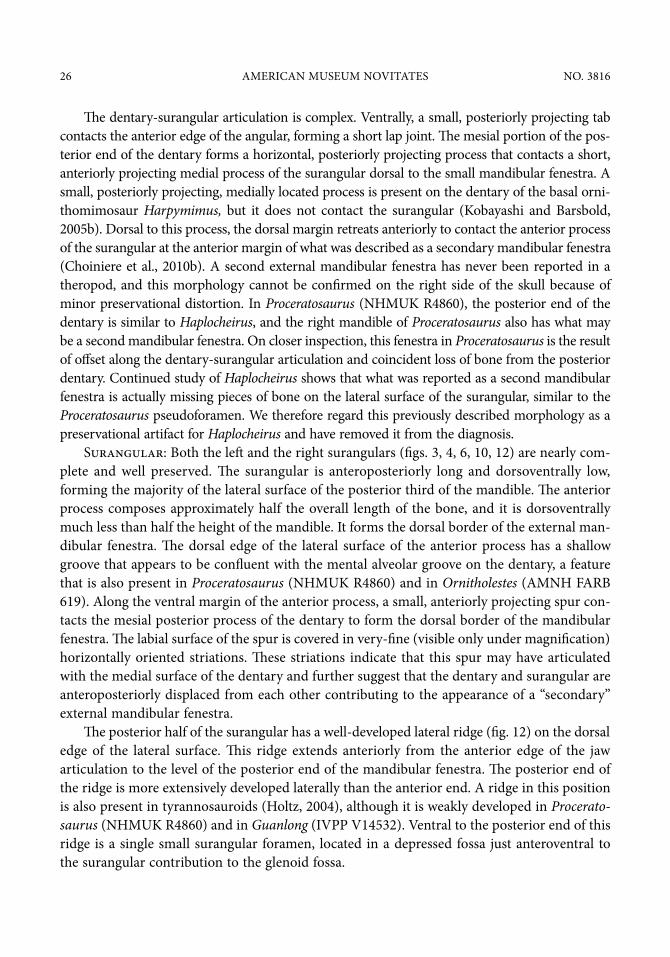
The dentary-surangular articulation is complex. Ventrally, a small, posteriorly projecting tab contacts the anterior edge of the angular, forming a short lap joint. The mesial portion of the pos-terior end of the dentary forms a horizontal, posteriorly projecting process that contacts a short, anteriorly projecting medial process of the surangular dorsal to the small mandibular fenestra. A small, posteriorly projecting, medially located process is present on the dentary of the basal orni-thomimosaur Harpymimus, but it does not contact the surangular (kobayashi and barsbold, 2005b). dorsal to this process, the dorsal margin retreats anteriorly to contact the anterior process of the surangular at the anterior margin of what was described as a secondary mandibular fenestra (Choiniere et al., 2010b). A second external mandibular fenestra has never been reported in a theropod, and this morphology cannot be confirmed on the right side of the skull because of minor preservational distortion. in Proceratosaurus (nhmuk r4860), the posterior end of the dentary is similar to Haplocheirus, and the right mandible of Proceratosaurus also has what may be a second mandibular fenestra. on closer inspection, this fenestra in Proceratosaurus is the result of offset along the dentary-surangular articulation and coincident loss of bone from the posterior dentary. Continued study of Haplocheirus shows that what was reported as a second mandibular fenestra is actually missing pieces of bone on the lateral surface of the surangular, similar to the Proceratosaurus pseudoforamen. We therefore regard this previously described morphology as a preservational artifact for Haplocheirus and have removed it from the diagnosis.
surangular: both the left and the right surangulars (figs. 3, 4, 6, 10, 12) are nearly com-plete and well preserved. The surangular is anteroposteriorly long and dorsoventrally low, forming the majority of the lateral surface of the posterior third of the mandible. The anterior process composes approximately half the overall length of the bone, and it is dorsoventrally much less than half the height of the mandible. it forms the dorsal border of the external man-dibular fenestra. The dorsal edge of the lateral surface of the anterior process has a shallow groove that appears to be confluent with the mental alveolar groove on the dentary, a feature that is also present in Proceratosaurus (nhmuk r4860) and in Ornitholestes (Amnh FArb 619). Along the ventral margin of the anterior process, a small, anteriorly projecting spur con-tacts the mesial posterior process of the dentary to form the dorsal border of the mandibular fenestra. The labial surface of the spur is covered in very-fine (visible only under magnification) horizontally oriented striations. These striations indicate that this spur may have articulated with the medial surface of the dentary and further suggest that the dentary and surangular are anteroposteriorly displaced from each other contributing to the appearance of a “secondary” external mandibular fenestra.
The posterior half of the surangular has a well-developed lateral ridge (fig. 12) on the dorsal edge of the lateral surface. This ridge extends anteriorly from the anterior edge of the jaw articulation to the level of the posterior end of the mandibular fenestra. The posterior end of the ridge is more extensively developed laterally than the anterior end. A ridge in this position is also present in tyrannosauroids (holtz, 2004), although it is weakly developed in Procerato-saurus (nhmuk r4860) and in Guanlong (iVPP V14532). Ventral to the posterior end of this ridge is a single small surangular foramen, located in a depressed fossa just anteroventral to the surangular contribution to the glenoid fossa.

2014 Choiniere et Al.: HAPLOCHEIRUS SOLLERS 27
Angular: both angulars (figs. 3, 4, 6, 12) are complete and preserved in articulation with the corresponding surangulars dorsally and dentaries anteriorly. The angular is long, extending the entire length of the external mandibular fenestra and covering the splenial laterally. The dor-soventral height of the angular below the external mandibular fenestra is approximately the same as the height of the surangular above the fenestra. The surangular-angular suture is directed posteroventrally and meets the ventral edge of the mandible well anterior to the glenoid. The medial surface is medially concave, forming the labial border of the anteroposteriorly long inter-nal mandibular fenestra. Ventrally, the angular forms an extensive medial contact with the prearticular.
splenial: The splenial (figs. 6, 12) is well preserved, but its medial surface is partly obscured by matrix. it is anteroposteriorly long and dorsoventrally low, with an anteroposteri-orly extensive, thin anterior end that extends almost to the dentary symphysis. The mylohyoid
1 cm1 cmdd
dd splsplmhfmhfsplspldd
dd
sasa
emfemfprapra
angang
hyhy
sarsar1 cm1 cm
A D
B
C
Figure 12. in situ dentition and mandibular articulation of holotype of Haplocheirus sollers (iVPP V14988). A. Anterior dentition of premaxilla, maxilla, and dentary in left lateral view. B. Posterior dentition of maxilla in left ventrolateral view. C. Anterior dentition of premaxilla, maxilla, and dentary in right lateral view. D. mandibular articular region in left posteroventrolateral view. Abbreviations in appendix 1.
28 AmeriCAn museum noVitAtes no. 3816
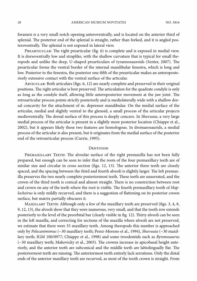
foramen is a very small notch opening anteroventrally, and is located on the anterior third of splenial. The posterior end of the splenial is straight, rather than forked, and it is angled pos-teroventrally. The splenial is not exposed in lateral view.
Prearticular: The right prearticular (fig. 6) is complete and is exposed in medial view. it is dorsoventrally low and straplike, with the shallow curvature that is typical for small the-ropods and unlike the deep, u-shaped prearticulars of tyrannosauroids (senter, 2007). The prearticular forms the ventral border of the internal mandibular fenestra, which is long and low. Posterior to the fenestra, the posterior one-fifth of the prearticular makes an anteroposte-riorly extensive contact with the ventral surface of the articular.
Articular: both articulars (figs. 6, 12) are nearly complete and preserved in their original positions. The right articular is best preserved. The articulation for the quadrate condyle is only as long as the condyle itself, allowing little anteroposterior movement at the jaw joint. The retroarticular process points strictly posteriorly and is mediolaterally wide with a shallow dor-sal concavity for the attachment of m. depressor mandibulae. on the medial surface of the articular, medial and slightly ventral to the glenoid, a small process of the articular projects medioventrally. The dorsal surface of this process is deeply concave. in Shuvuuia, a very large medial process of the articular is present in a slightly more posterior location (Chiappe et al., 2002), but it appears likely these two features are homologous. in dromaeosaurids, a medial process of the articular is also present, but it originates from the medial surface of the posterior end of the retroarticular process (Currie, 1995).
dentitionPremaxillary teeth: The alveolar surface of the right premaxilla has not been fully
prepared, but enough can be seen to infer that the roots of the four premaxillary teeth are of similar size and circular in cross section (figs. 12, 13). The anterior three teeth are closely spaced, and the spacing between the third and fourth alveoli is slightly larger. The left premax-illa preserves the two nearly complete posteriormost teeth. These teeth are unserrated, and the crown of the third tooth is conical and almost straight. There is no constriction between root and crown on any of the teeth where the root is visible. The fourth premaxillary tooth of Hap-locheirus is only mildly recurved, and there is a suggestion of flattening on its posterior crown surface, but matrix partially obscures it.
maxillary teeth: Although only a few of the maxillary teeth are preserved (figs. 3, 4, 8, 9, 12, 13), the alveoli show that they were numerous, very small, and that the tooth row extends posteriorly to the level of the preorbital bar (clearly visible in fig. 12). Thirty alveoli can be seen in the left maxilla, and correcting for sections of the maxilla where alveoli are not preserved, we estimate that there were 35 maxillary teeth. Among theropods this number is approached only by Pelecanimimus (~30 maxillary teeth; Perez-moreno et al., 1994), Shuvuuia (~30 maxil-lary teeth; igm 100/0977; Chiappe et al., 1998) and some troodontids such as Byronosaurus (~30 maxillary teeth; makovicky et al., 2003). The crowns increase in apicobasal height ante-riorly, and the anterior teeth are subconical and the middle teeth are labiolingually flat. The posteriormost teeth are missing. The anteriormost teeth entirely lack serrations. only the distal ends of the anterior maxillary teeth are recurved, as most of the tooth crown is straight. From

2014 Choiniere et Al.: HAPLOCHEIRUS SOLLERS 29
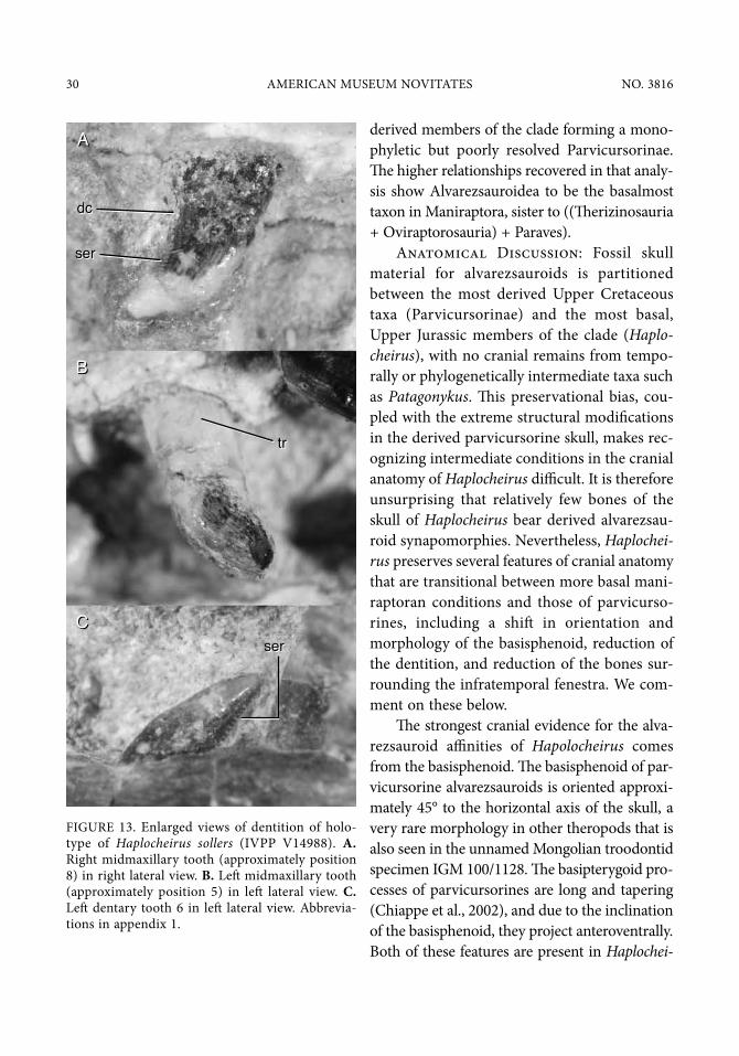
approximately the fifth tooth on, the middle teeth have serrations only on their distal carinae. in some of the middle teeth the serrations are developed along the entire distal carinae, but in others the serrations are developed only basally. marked heterodonty as described above is uncommon in theropods, but is known in the troodontid taxon Troodon (Currie, 1987) and in Ricardoestesia (Currie et al., 1990; longrich, 2008), a putative troodontid (hwang, 2005: exam-ining Paronychodon). it is difficult to get a precise count of serration density, because nearly all the teeth have some minor crown damage, but it appears to be in the range of 5–7 serrations per millimeter, which is typical for coelurosaurs (Choiniere et al., 2010a) and unlike the more sparsely serrated teeth of some troodontids (e.g., Saurornithoides (osborn, 1924; norell et al., 2009)), and therizinosauroids (Clark et al., 2004). The serrations extend directly posteriorly. As in some troodontids (norell et al., 2009), the serrations end well short of the tooth apices.
left maxillary tooth 3 is preserved within its alveolus. The crown is subconical and only slightly recurved, as in Pelecanimimus (lh 7777). This tooth has strong apicobasally oriented striations, as in spinosaurids (brusatte et al., 2007), although these striations are present only on one other maxillary tooth crown. only one midmaxillary tooth is preserved (on the right side of the skull) in its alveolus. This tooth is markedly smaller than the anterior teeth, about half the height of tooth 3. it also differs in shape, being apicobasally short and mesiodistally wide. in lateral view, the mesial margin of the tooth crown abruptly changes angle at midheight, deflecting strongly apicodistally, as in some troodontids (makovicky and norell, 2004). Poste-rior to the eighth maxillary tooth position, the maxillary alveoli progressively decrease in size.
interdental plates are well preserved between all the maxillary teeth and the broken pieces of osseous septae extend laterally from the lateral surface of the interdental plates between adjacent teeth. none of the preserved maxillary teeth show evidence of a constriction between the tooth crown and the root, as in Archaeopteryx, derived alvarezsauroids (Perle et al., 1993), ornithomi-mosaurs (e.g., Pelecanimimus (lh 7777)), and therizinosauroids (Clark et al., 2004).
dentary teeth: The anterior seven dentary teeth are larger than more posterior dentary teeth, unserrated, subconical, mildly recurved, and widely spaced (figs. 3, 4, 12, 13). The entire dentary tooth row is not preserved on either side, but we estimate 30–40 dentary teeth for Hap-locheirus based on the presence of alveolar foramina and alveolar notches in the dorsal dentary margin. The roots of these anterior teeth have circular cross sections. only three middentary teeth are preserved on the left side, and they are similar in size to the anterior teeth but more medio-laterally compressed. These middentary teeth bear serrations on the distal carinae. The serrations average approximately 5 per mm. The size of the alveoli in the dentary decreases posteriorly, indicating that tooth size probably diminished in correspondence to the maxillary teeth.
disCussion
We base our phylogenetic hypotheses and character distributions in the discussion below on the results of the large cladistic analysis of Coelurosauria by Choiniere et al. (2014: fig. 20A, and supplementary information therein). in that analysis (here reproduced in fig. 14), Alvarezsauroi-dea are monophyletic, with Haplocheirus as the basalmost member, the south American taxa Patagonykus and Alvarezsaurus forming a grade of intermediate alvarezsaurids, and with the most
30 AmeriCAn museum noVitAtes no. 3816
derived members of the clade forming a mono-phyletic but poorly resolved Parvicursorinae. The higher relationships recovered in that analy-sis show Alvarezsauroidea to be the basalmost taxon in maniraptora, sister to ((Therizinosauria + oviraptorosauria) + Paraves).
Anatomical discussion: Fossil skull material for alvarezsauroids is partitioned between the most derived upper Cretaceous taxa (Parvicursorinae) and the most basal, upper Jurassic members of the clade (Haplo-cheirus), with no cranial remains from tempo-rally or phylogenetically intermediate taxa such as Patagonykus. This preservational bias, cou-pled with the extreme structural modifications in the derived parvicursorine skull, makes rec-ognizing intermediate conditions in the cranial anatomy of Haplocheirus difficult. it is therefore unsurprising that relatively few bones of the skull of Haplocheirus bear derived alvarezsau-roid synapomorphies. nevertheless, Haplochei-rus preserves several features of cranial anatomy that are transitional between more basal mani-raptoran conditions and those of parvicurso-rines, including a shift in orientation and morphology of the basisphenoid, reduction of the dentition, and reduction of the bones sur-rounding the infratemporal fenestra. We com-ment on these below.
The strongest cranial evidence for the alva-rezsauroid affinities of Hapolocheirus comes from the basisphenoid. The basisphenoid of par-vicursorine alvarezsauroids is oriented approxi-mately 45° to the horizontal axis of the skull, a very rare morphology in other theropods that is also seen in the unnamed mongolian troodontid specimen igm 100/1128. The basipterygoid pro-cesses of parvicursorines are long and tapering (Chiappe et al., 2002), and due to the inclination of the basisphenoid, they project anteroventrally. both of these features are present in Haplochei-
trtr
ser ser
dcdc
serser
AA
BB
CC
Figure 13. enlarged views of dentition of holo-type of Haplocheirus sollers (iVPP V14988). A. right midmaxillary tooth (approximately position 8) in right lateral view. B. left midmaxillary tooth (approximately position 5) in left lateral view. C. left dentary tooth 6 in left lateral view. Abbrevia-tions in appendix 1.

2014 Choiniere et Al.: HAPLOCHEIRUS SOLLERS 31
rus, in more or less the same degree as in parvicursorines, and therefore this characteristic basisphenoid anatomy forms an Alvarezsauroid cranial synapo-morphy. Furthermore, Haplo-cheirus shows that this basic architecture of the braincase floor was already in place early in the evolution of Alvarezsau-roidea. Although reasons for this derived morphology are unclear, its presence in some small troodontids (e.g., igm 100/1128) suggests that similar evolutionary pressures (con-straints, adaptive scenarios, or otherwise) may have been act-ing in these lineages.
Parvicursorine alvarezsau-roids have dentitions that differ markedly from the plesiomor-phic coelurosaurian condition (Choiniere et al., 2010a) in the following ways: the number of maxillary and dentary teeth is greatly increased; both maxillary and dentary tooth crowns are homodont and set into an open groove (e.g., there are no inder-
Figure 14. Phylogenetic relation-ships of Haplocheirus sollers from Choiniere et al., 2014. tree-shape parameters discussed in text. tree is strict consensus of 80 mPts, length = 3045, Ci = 0.213, ri = 0.611. open circle denotes Coelurosauria; open triangle denotes maniraptora; open star denotes Paraves; closed triangle denotes Alvarezsauridae; closed circle denotes Parvicursori-nae. numbers below nodes are bremer support values.
32 AmeriCAn museum noVitAtes no. 3816
dental septae); the tooth crowns lack serrations on the carinae; the tooth crowns are only slightly recurved; and there is a constriction between the tooth crown and tooth root. Premaxillary teeth are unknown for parvicursorines, but from Haplocheirus it can be determined that early alva-rezsauroids retained the four premaxillary teeth that are plesiomorphic for coelurosaurs generally (rauhut, 2003; Choiniere et al., 2010a). Among other hyperdentate coelurosaur taxa, the troodon-tid Byronosaurus has four premaxillary teeth (makovicky et al., 2003) and the ornithomimosaur Pelecanimimus bears seven (Perez-moreno et al., 1994). Haplocheirus has at least 30 teeth in the maxilla (and likely many more) as well as in the dentary, and given similar tooth counts in Fal-carius (Zanno, 2010) it now appears that a moderate increase in the number of teeth is plesio-morphic for maniraptora. Within Alvarezsauroidea, the tooth count in Haplocheirus is apparently greater than even Shuvuuia (in which the posterior end of the tooth row is poorly preserved) (Chiappe et al., 1998). This perhaps indicates that the number of maxillary teeth was reduced during alvarezsauroid evolution.
interestingly, Haplocheirus bears interdental septae, whereas septae are lost in Shuvuuia (Chiappe et al., 1998). interdental septae are present in perinate specimens of the hyperdentate troodontid Byronosaurus, but they are markedly reduced in height relative to those of other maniraptorans (bever and norell, 2009). interdental septae are clearly lost in both the basal-most ornithomimosaur Nqwebasaurus and between some but not all the alveoli in the hyper-dentate basal ornithomimosaur Pelecanimimus (Perez-moreno et al., 1994; Choiniere et al., 2012). These observations suggest that an increase in tooth number and loss of separate alveoli are separate evolutionary processes. however, Haplocheirus has heterodont dental morphology (as do basal oviraptorosaurs: Xu et al., 2002a; balanoff et al., 2009; some troodontids: Currie, 1987; longrich, 2008; and therizinosaurs: Zanno, 2010), whereas the aforementioned hyper-dentate taxa are considerably more homodont. in developing Alligator mississipiensis speci-mens, interdental septae appear late in the ontogenetic sequence, and it is only after these septae are fully developed that the dentition becomes more heterodont (Westergaard and Fer-guson, 1990). This suggests that if theropod dental ontogeny is similar to that of extant toothed archosaurs, then the loss of interdental septae and increased homodonty in these theropods may be due to paedomorphic retention of earlier developmental stages in adults, a phenome-non currently hypothesized to be largely responsible for the evolution of skull shape in the theropod to bird transition (bhullar et al., 2012), or alternatively suggests that these taxa are represented by juvenile exemplars.
The distribution of derived tooth features in Haplocheirus is grossly similar to the pattern observed in basal members of the maniraptoran clades Therizinosauroidea and oviraptorosau-ria, where initial tooth modification begins with anterior dentition (Xu et al., 2002a; Zanno, 2010; Pu et al., 2013). This suggests a conserved maniraptoran tendency toward modification of premaxillary and anterior maxillary and dentary teeth rather than middle-to-posterior max-illary or dentary teeth, although anterior tooth modifications are apparently independently derived in Alvarezsauroidea, Therizinosauroidea, and oviraptorosauria.
specifically, the anterior teeth of Haplocheirus resemble the maxillary and dentary teeth of parvicursorines in lacking serrations and having only mild recurvature (Perle et al., 1993; Perle et al., 1994; Chiappe et al., 1998), whereas the middle and posterior maxillary and dentary teeth of

2014 Choiniere et Al.: HAPLOCHEIRUS SOLLERS 33
Haplocheirus retain serrations and recurvature (plesiomorphic for coelurosaurs) but resemble those of parvicursorines in having circular root cross sections and a reduced size. This suggests that changes in tooth morphology progressed either at two different rates or at different evolution-ary intervals in the premaxilla and maxilla of alvarezsauroids, whereby the premaxillary tooth crown morphology changed either first or at a higher rate, and the maxillary tooth crown mor-phology changed at a slower rate and proceeded from posterior to anterior. This pattern seems to differ from that inferred in troodontids, where increases in tooth number and changes in tooth morphology appear nearly simultaneously (hu et al., 2009), although both Troodon itself and the putative troodontid Ricardoestesia show marked heterodonty (Currie, 1987; longrich, 2008).
some of the key features previously used in uniting parvicursorine alvarezauroids with Avialae (reviewed in Chiappe et al., 2002), and now interpreted as homoplastically developed local synapomorphies of the group (Choiniere et al., 2010b), include the reduction in the bones surrounding the infratemporal fenestra, such as the fusion of the quadratojugal and jugal into a single rodlike structure, the loss of a contact between the ascending ramus of the quadrato-jugal and squamosal, and an incomplete postorbital bar where the jugal lacks an ascending postorbital process. The morphology of the posterior orbital and infratemporal region in Hap-locheirus shows some features presaging this derived morphology.
The body of the jugal of Haplocheirus is reduced in dorsoventral height and is markedly more cylindrical than the platelike jugal of basal coelurosaurs (e.g., tyrannosauroids) and some maniraptorans (e.g., dromaeosaurids). This feature, however, seems to characterize at least some other basal maniraptorans (e.g., Ornitholestes [Amnh FArb 619]) and may be plesio-morphic for the group. The quadratojugal process of the jugal is posteriorly extensive, and the corresponding jugal process of the quadratojugal is short, suggesting that fusion between these two bones involved an early increase in jugal posterior length and decrease in quadratojugal length. however, neither the ascending process of the quadratojugal nor the postorbital process of the jugal show signs of reduction in Haplocheirus, indicating that early stages of the loss of this contact is nested deeper within Alvarezsauroidea. The loss of these contacts may occur in conjunction with further reduction in dental morphology and may indicate accompanying dietary shifts, although a number of other explanations are equally likely to result in this mor-phology, such as general lightening of the cranium or changes in skull function (e.g., prokine-sis). more cranial remains from alvarezsauroids intermediate between Haplocheirus and parvicursorines are necessary to accurately time the loss of these features.
The homology of the prefrontal bone in parvicursorines has been contentious (Chiappe et al., 1998; sereno, 1999; 2001; suzuki et al., 2002). in the prefrontal region, the parvicursorine Shuvuuia has hypertrophied ossifications that make a large contribution to the anterodorsal orbital margin and the dorsal skull roof and meet ventrally at a position anterior to the orbits inside the skull forming a preorbital wall. Chiappe et al. (1998) suggested that these “prefrontal” elements might be homologous to the ectethmoid bones of Aves, which also form the anterodor-sal corner of the orbit and meet along the skull midline. The prefrontal of Haplocheirus shows no signs of enlargement relative to the plesiomorphic coelurosaurian condition (e.g., Ornit-holestes Amnh FArb 519), but it is positionally similar to the ossification in parvicursorines, and bears a descending process that extends along the medial surface of the lacrimal. This

34 AmeriCAn museum noVitAtes no. 3816
suggests either that the feature present in parvicursorines in this area is a hypertrophied pre-frontal, or that an ectethmoid element evolved later in the alvarezsauroid lineage. in either case, the condition in Haplocheirus shows that the ossification in parvicursorines is not homologous to that of birds (contra Chiappe et al., 1998) and that the hypertrophied prefrontals of ornitho-mimosaurs are not homologous to those of parvicursorines (contra sereno, 2001).
Parvicursorine alvarezsaurids, along with avialans, some troodontids, and some oviraptoro-saurs, have a quadrate with two dorsal contacts—one on the ventral surface of the squamosal and one on the prootic on the lateral wall of the braincase (Perle et al., 1993; russell and dong, 1994; maryanska et al., 1997; maryanska et al., 2002). The parvicursorine morphology is particularly similar to that of avialans because it contacts the prootic and squamosal (Chiappe et al., 1998) rather than the opisthotic and squamosal, as it does in oviraptorosaurs (maryanska et al., 1997; balanoff et al., 2009). The plesiomorphic, single-headed quadrate of Haplocheirus demonstrates that in alva-rezsauroids, as in oviraptorosaurs and troodontids, this double-headed morphology is convergent with respect to avialans. if this quadrate morphology is related to streptostyly, as has been hypoth-esized (Chiappe et al., 1998), then the development of the accessory articulation of the quadrate may develop only in conjunction with increased mobility of other skull elements, including the loss of the postorbital bar and prokinesis of the frontonasal joint (Chiappe et al., 1998).
Feeding ecology of haplocheirus: several lines of anatomical evidence suggest that Haplocheirus differs from most theropods in its ecology. The lightly built construction of the skull, with large antorbital and maxillary fenestrae, reduction of the lacrimal and postorbital bars, and reduction of the jugal, all suggest that the skull of Haplocheirus was incapable of withstanding strong bending forces from biting. The narrow snout and long, slender dentaries provide a low power-to-velocity ratio for the jaws of Haplocheirus, suggesting weak bite force but rapid jaw closure ability. The numerous small, weakly recurved teeth with reduced serra-tions are consistent with the ability to hold and pierce small prey. together, these lines of evidence suggest that Haplocheirus was preferentially feeding on small vertebrates rather than on the larger dinosaurian fauna of the shishugou Formation. Haplocheirus differs greatly in these ways from contemporary shishugou theropods Monolophosaurus (Zhao and Currie, 1993; brusatte et al., 2010), Sinraptor (Currie and Zhao, 1993), and Zuolong (Choiniere et al., 2010a), which all have more typical theropod configurations of the jaws that suggest stronger bite forces and larger prey items. The presence in the shishugou of theropods with these “typi-cal” jaw configurations coupled with theropod taxa bearing “atypical” configurations such as Haplocheirus, the longirostrine tyrannosauroid Guanlong (Xu et al., 2006) and the possibly herbivorous ceratosaur Limusaurus (Xu et al., 2009) shows that a range of theropod feeding ecologies were already present by the earliest late Jurassic.
The orbit of Haplocheirus is strikingly large, and it preserves a large sclerotic ring composed of externally concave ossicles. some recent work on activity patterns of dinosaurs employing scleral ring morphology (schmitz and motani, 2011) hypothesizes that several theropod taxa, including Velociraptor mongoliensis, were nocturnal. based on a qualitative comparison with schmitz and motani’s (2011) data, the inner diameter of the scleral ring of Haplocheirus is approximately 15 mm larger than that of Velociraptor and the outer diameter of the scleral ring

2014 Choiniere et Al.: HAPLOCHEIRUS SOLLERS 35
is only approximately 9 mm larger, even though the skulls are of generally similar size. This indicates a much larger lens in Haplocheirus than in Velociraptor and, according to the data of schmitz and motani (2011), suggests the eye of Haplocheirus was optimized for scotopic ocular image formation and thus nocturnal habits. however, a similar analysis by hall (2008) found equivocal evidence for nocturnality in most dinosaurs, and a recent critique of the schmitz and motani (2011) analysis by hall et al. (2011) warrants caution for this interpretation.
Alvarezsauroid distribution and historical biogeography: Haplocheirus provides evidence for alvarezsauroids in the late Jurassic, filling in one of the longest ghost lineages in the theropod fossil record (Choiniere et al., 2010b), yet early Cretaceous alvarezsauroids remain elusive. given the wealth of fossils of other derived coelurosaurs in lower Cretaceous deposits in northeast China (Xu et al., 1999; Xu et al., 2002b; Xu and norell, 2004), it is surprising that there have been no discoveries of alvarezsauroids in the Jehol biota or the daohugou beds.
Xu et al. (2013) recently provided a comprehensive review of alvarezsaur historical biogeog-raphy, but failed to mention that the presence of Haplocheirus in the Jurassic of China falsifies novas’ hypothesis of a gondwanan origin for Alvarezsauroidea (bonaparte, 1991; novas, 1996). The diversity of basal alvarezsaurids in the late Cretaceous of south America (bonaparte, 1991; novas, 1996; 1997; martinelli and Vera, 2007; Agnolin et al., 2012; makovicky et al., 2012) and their position as a grade basal to Parvicursorinae is possibly compatible with a gondwanan origin for these more derived members of the clade. however, this hypothesis would require subsequent dispersal through land bridges to explain the presence of Parvicursorines in Asia (Perle et al., 1993; Chiappe et al., 1998; Xu et al., 2010; Xu et al., 2011), europe (naish and dyke, 2004), and north America (hutchinson and Chiappe, 1998; longrich and Currie, 2008) during the late Cretaceous. given that alvarezsauroids are relatively rare components of theropod faunas (makov-icky et al., 2012), and the possibility of a panglobal distribution for coelurosaurian higher taxa prior to the split between gondwana and laurasia (benson et al., 2010; benson et al., 2012; Choiniere et al., 2012), it is possible that basal alvarezsaurs were widely distributed in the meso-zoic and that either local extinctions or preservational bias explains our current understanding of their distribution. recovery of new alvarezsaur material from the early Cretaceous, particularly from gondwana, coupled with a better understanding of the phylogenetic relationships of poorly known forms like Kol (turner et al., 2009) and Heptasteornis (naish and dyke, 2004), will undoubtedly clarify their biogeographic history (Xu et al., 2013).
ConClusion
The discovery of Haplocheirus provides direct evidence of alvarezsauroids in the late Juras-sic, confirming predictions from ghost lineages. our knowledge of cranial material for Haplo-cheirus is particularly important because alvarezsauroid skulls are previously known only for a few specimens of derived taxa. The cranium of Haplocheirus preserves several plesiomorphic features, including a medium-sized prefrontal and an unfused jugal and quadratojugal, which show that some of the cranial traits shared between derived alvarezsauroids and avialans are homoplasies. other characteristics of Haplocheirus, such as the orientation of the braincase,

36 AmeriCAn museum noVitAtes no. 3816
indicate that the initial stages of the evolution of derived alvarezsauroid characteristics were already underway by the late Jurassic. The discovery of additional alvarezsauroid material, particularly cranial material from lower Cretaceous deposits, would help clarify the timing of other key changes in the alvarezsauroid skull.
ACknoWledgments
This research was supported by the national science Foundations (grants eAr 0310217, eAr 0228559, and oise 0812234), by grants from the national natural sciences Foundation of China (grants 41120124002 and 40830210), the national geographic society, the Jurassic Foun-dation, the hilmar sallee bequest, by the richard gilder graduate school at the American museum of natural history through a kalbfleisch and gerstner scholarship, and by institutional support from the george Washington university, the American museum of natural history, the Chinese Academy of sciences, the university of the Witwatersrand, and by the division of sci-ence and technology/national research Foundation’s Centre of excellence in Palaeosciences.
The authors wish to thank n. longrich and l. Zanno for extensive reviews and comments that greatly improved this manuscript, m. ellison for figure preparation, and m. Carrano, J. burns, and C. Forster for examining earlier versions of this paper, which formed a chapter in J.n.C.’s Ph.d. thesis.
reFerenCes
Agnolin, F.l., J.e. Powell, F.e. novas, and m. kundrat. 2012. new alvarezsaurid (dinosauria, Theropoda) from uppermost Cretaceous of north-western Patagonia with associated eggs. Cretaceous research 35: 33–56.
Alifanov, V.r., and r. barsbold. 2009. Ceratonykus oculatus gen. et sp. nov., a new dinosaur (?Thero-poda, Alvarezsauria) from the late Cretaceous of mongolia. Paleontological Journal 43 (1): 86–99.
balanoff, A.m., X. Xu, y. kobayashi, y. matsufune, and m.A. norell. 2009. Cranial osteology of the theropod dinosaur Incisivosaurus gauthieri (Theropoda: oviraptorosauria). American museum novitates 3651: 1–35.
baumel, J.J., and l.m. Witmer. 1993. osteologia. In J.J. baumel, A.s. king, J.e. breazile, h.e. evans, and J.C. Vanden berge (editors), handbook of avian anatomy: nomina anatomica avium. Cambridge, mA: nuttall ornithological Club.
benson, r.b.J., P.m. barrett, t.h. rich, and P. Vickers-rich. 2010. A southern tyrant reptile. science 327: 1613.
benson, r.b.J., t.h. rich, P. Vickers-rich, and m. hall. 2012. Theropod fauna from southern Australia indicates high polar diversity and climate-driven dinosaur provinciality. Plos one 7 (5): e7122.
bever, g.s., and m.A. norell. 2009. The perinate skull of Byronosaurus (troodontidae) with observations on the cranial ontogeny of paravian theropods. American museum novitates 3657: 1–51.
bhullar, b.-A.s., et al. 2012. birds have paedomorphic dinosaur skulls. nature 487 (7406): 223–226.bonaparte, J.F. 1991. los vertebrados fosiles de la Formacion rio Colorado de neuquen y cercanias,
Cretacico superior, Argentina. revista del museo Argentino de Ciencias naturales “bernardino rivadavia” Paleontologia 4 (3): 17–123.

2014 Choiniere et Al.: HAPLOCHEIRUS SOLLERS 37
brochu, C.A. 2003. osteology of Tyrannosaurus rex: insights from a nearly complete skeleton and high-resolution computed tomographic analysis of the skull. Journal of Vertebrate Paleontology 22 (sup-plement to 4): 1–137.
brusatte, s.l., r.b.J. benson, t.d. Carr, t.e. Williamson, and P.C. sereno. 2007. The systematic utility of theropod enamel wrinkles. Journal of Vertebrate Paleontology 27 (4): 1052–1056.
brusatte, s.l., r.b.J. benson, P.J. Currie, and X. Zhao. 2010. The skull of Monolophosaurus jiangi (dino-sauria: Theropoda) and its implications for early theropod phylogeny and evolution. Zoological Journal of the linnean society 158: 573–607.
Carrano, m.t., s.d. sampson, and C.A. Forster. 2002. The osteology of Masiakasaurus knopfleri, a small abelisauroid (dinosauria: Theropoda) from the late Cretaceous of madagascar. Journal of Verte-brate Paleontology 22 (3): 510–534.
Charig, A.J., and A.C. milner. 1997. Baryonyx walkeri, a fish-eating dinosaur from the Wealden of sur-rey. bulletin of the natural history museum of london 53 (1): 11–70.
Chiappe, l.m. 1996. Phylogenetic position of Mononykus from the upper Cretaceous of the gobi des-ert. memoirs of the Queensland museum 39: 557–582.
Chiappe, l.m., and u.b. göhlich. 2010. Anatomy of Juravenator starki (Theropoda: Coelurosauria) from the late Jurassic of germany. neues Jahrbuch für geologie und Paläontologie 258 (3): 257–296.
Chiappe, l., m. norell, and J. Clark. 1997. Mononykus and birds: methods and evidence. Auk 114 (2): 300–302.
Chiappe, l.m., m. norell, and J.m. Clark. 1998. The skull of a relative of the stem-group bird Mononykus. nature 392: 275–278.
Chiappe, l.m., m.A. norell, and J.m. Clark. 2002. The Cretaceous, short-armed Alvarezsauridae: Mononykus and its kin. In l.m. Chiappe and l.m. Witmer (editors), mesozoic birds: above the heads of dinosaurs: 87–120. berkeley: university of California Press.
Choiniere, J.n., J.m. Clark, C.A. Forster, and X. Xing. 2010a. A basal coelurosaur (dinosauria: Thero-poda) from the late Jurassic (oxfordian) of the shishugou Formation in Wucaiwan, People’s repub-lic of China. Journal of Vertebrate Paleontology 30 (6): 1773–1796.
Choiniere, J.n., et al. 2010b. A basal alvarezsauroid theropod from the early late Jurassic of Xinjiang, China. science 327 (571): 571–574.
Choiniere, J.n., C.A. Forster, and W.J. de klerk. 2012. new information on Nqwebasaurus thwazi, a coelurosaurian theropod from the early Cretaceous kirkwood Formation in south Africa. Journal of African earth sciences 71–72: 1–17.
Choiniere, J.n., et al. 2014. A juvenile specimen of a new coelurosaur (dinosauria: Theropoda) from the middle–late Jurassic shishugou Formation of Xinjiang, People’s republic of China. Journal of sys-tematic Paleontology 12 (2): 177–215.
Clark, J.m., P. Altangerel, and m.A. norell. 1994. The skull of Erlicosaurus andrewsi, a late Cretaceous “segnosaur” (Theropoda: Therizinosauridae) from mongolia. American museum novitates 3115: 1–39.
Clark, J.m., m.A. norell, and r. barsbold. 2001. two new oviraptorids (Theropoda: oviraptorosauria), upper Cretaceous djadokhta Formation, ukhaa tolgod, mongolia. Journal of Vertebrate Paleontol-ogy 21 (2): 209–213.
Clark, J.m., m.A. norell, and t. rowe. 2002. Cranial anatomy of Citipati osmolskae (Theropoda, ovirap-torosauria), and a reinterpretation of the holotype of Oviraptor philoceratops. American museum novitates 3364: 1–24.

38 AmeriCAn museum noVitAtes no. 3816
Clark, J.m., t. maryanska, and r. barsbold. 2004. Therizinosauroidea. In d.b. Weishampel, P. dodson, and h. osmolska (editors), The dinosauria, 2nd ed.: 151–164. berkeley: university of California Press.
Clark, J.m., et al. 2006. The middle-to-late Jurassic terrestrial transition: new discoveries from the shi-shugou Formation, Xinjiang, China. 9th international symposium mesozoic terrestrial ecosystems and biota: 26–28.
Currie, P.J. 1987. bird-like characteristics of the jaws and teeth of troodontid theropods. Journal of Ver-tebrate Paleontology 7: 72–81.
Currie, P.J. 1995. new information on the anatomy and relationships of Dromaeosaurus albertensis (dinosauria: Theropoda). Journal of Vertebrate Paleontology 15 (3): 576–591.
Currie, P.J., and Z.-m. dong. 2001. new information on Cretaceous troodontids (dinosauria, Thero-poda) from the People’s republic of China. Canadian Journal of earth sciences 38 (12): 1753–1766.
Currie, P.J., and X.-J. Zhao. 1993. A new carnosaur (dinosauria, Theropoda) from the Jurassic of Xin-jiang, People’s republic of China. Canadian Journal of earth sciences 30 (10 and 11): 2037–2081.
Currie, P.J., J.k. rigby, Jr., and r.e. sloan. 1990. Theropod teeth from the Judith river Formation of southern Alberta, Canada. In P.J. Currie and k. Carpenter (editors), dinosaur systematics: approaches and perspectives: 107–125. Cambridge: Cambridge university Press.
dufeau, d.l. 2002. The cranial anatomy of the theropod dinosaur Shuvuuia deserti (Coelurosauria; Alvarezsauridae), and its bearing upon coelurosaurian phylogeny. master’s thesis, university of texas at Austin, Austin.
eberth, d.A., et al. 2001. sequence stratigraphy, paleoclimate patterns, and vertebrate fossil preservation in Jurassic-Cretaceous strata of the Junggar basin, Xinjiang Autonomous region, People’s republic of China. Canadian Journal of earth sciences 38 (12): 1627–1644.
eberth, d.A., X. Xu, and J.m. Clark. 2010. dinosaur death pits from the Jurassic of China. Palaios 25: 112–125.
gauthier, J. 1986. saurischian monophyly and the origin of birds. memoirs of the California Academy of sciences 8: 1–55.
gradstein, F.m, et al. 2012. The geologic time scale 2012. boston: elsevier. [doi: 10.1016/b978-0-444-59425-9.00004-4]
hall, m.i. 2008. The anatomical relationships between the avian eye, orbit, and sclerotic ring: implica-tions for inferring activity patterns in extinct birds. Journal of Anatomy 212: 781–794.
hall, m.i., e.C. kirk, J.m. kamilar, and m.t. Carrano. 2011. Comment on “nocturnality in dinosaurs inferred from scleral ring and orbit morphology.” science 334: 1641.
holtz, t.r., Jr. 2004. tyrannosauroidea. In d.b. Weishampel, P. dodson, and h. osmolska (editors), The dinosauria, 2nd ed.: 111–136. berkeley: university of California Press.
hone, d.W.e., J.n. Choiniere, Q. tan, and X. Xu. 2012. An articulated pes from a small parvicursorine alvarezsauroid (dinosauria: Theropoda) from inner mongolia, China. Acta Palaeontologica Polon-ica 58: 453–458.
hu, d., l. hou, l. Zhang, and X. Xu. 2009. A pre-Archaeopteryx troodontid theropod from China with long feathers on the metatarsus. nature 461 (7264): 640–643.
hutchinson, J.r., and l.m. Chiappe. 1998. The first known alvarezsaurid (Theropoda: Aves) from north America. Journal of Vertebrate Paleontology 18 (3): 447–450.
hwang, s.h. 2005. Phylogenetic patterns of enamel microstructure in dinosaur teeth. Journal of mor-phology 266 (2): 208–240.
Ji, Q., et al. 2003. An early ostrich dinosaur and implications for ornithomimosaur phylogeny. American museum novitates 3420: 1–19.

2014 Choiniere et Al.: HAPLOCHEIRUS SOLLERS 39
kessler, e., d. grigorescu, and Z. Csiki. 2005. Elopteryx revisited – a new bird-like specimen from the maastrichtian of the hateg basin (romania). Acta Palaeontologica romaniae 5: 349–258.
kobayashi, y., and r. barsbold. 2005a. reexamination of a primitive ornithomimosaur, Garudimimus brevipes barsbold, 1981 (dinosauria: Theropoda), from the late Cretaceous of mongolia. Canadian Journal of earth sciences 42: 1501–1521.
kobayashi, y., and r. barsbold. 2005b. Anatomy of Harpymimus okladnikovi barsbold and Perle 1984 (dinosauria: Theropoda) of mongolia. In k. Carpenter (editor), The carnivorous dinosaurs: 97–126. bloomington: indiana university Press.
kuiper, k.F., et al. 2008. synchronizing rock clocks of earth history. science 320: 500–504. lambe, l.m. 1917. The Cretaceous theropodous dinosaur Gorgosaurus. memoirs of the Canadian geo-
logical survey 83 (1687): 1–84.lee, m.s.y., and t.h. Worthy. 2011. likelihood reinstates Archaeopteryx as a primitive bird. biology
letters 8 (2): 299–303.longrich, n.r. 2008. small theropod teeth from the lance Formation of Wyoming. In J.t. sankey and
s. baszio (editors), Vertebrate microfossil assemblages: their role in paleoecology and paleobiogeog-raphy: 135–158. bloomington: indiana university Press.
longrich, n.r., and P.J. Currie. 2008. Albertonykus borealis, a new alvarezsaur (dinosauria: Theropoda) from the early maastrichtian of Alberta, Canada: implications for systematics and ecology of the Alvarezsauridae. Cretaceous research 30 (1): 239–252.
madsen, J.h., Jr. 1976. Allosaurus fragilis: a revised osteology. utah geological and mineral survey bul-letin 109: 1–163.
makovicky, P.J., and m.A. norell. 2004. troodontidae. In d.b. Weishampel, P. dodson, and h. osmolska (editors), The dinosauria, 2nd ed.: 184–195. berkeley: university of California Press.
makovicky, P.J., m.A. norell, J.m. Clark, and t. rowe. 2003. osteology and relationships of Byronosaurus jaffei (Theropoda: troodontidae). American museum novitates 3402: 1–32.
makovicky, P.J., y. kobayashi, and P.J. Currie. 2004. ornithomimosauria. In d.b. Weishampel, P. dodson, and h. osmolska (editors), The dinosauria, 2nd ed.: 137–150. berkeley: university of California Press.
makovicky, P.J., s. Apesteguía, and F. gianechinia. 2012. A new coelurosaurian theropod from the la buitrera Fossil locality of rio negro, Argentina. Fieldiana life and earth sciences 5: 90–98.
marsh, o.C. 1881. Principal characters of American Jurassic dinosaurs. American Journal of science 3 (21): 417–423.
martinelli, A.g., and e.i. Vera. 2007. Achillesaurus manazzonei, a new alvarezsaurid theropod (dino-sauria) from the late Cretaceous bajo de la Carpa Formation, rio negro Province, Argentina. Zootaxa 1582: 1–17.
maryanska, t., and h. osmólska. 1997. The quadrate of oviraptorid dinosaurs. Acta Palaeontologica Polonica 42 (3): 361–371.
maryanska, t., h. osmólska, and m. Wolsan. 2002. Avialan status for oviraptorosauria. Acta Palaeon-tological Polonica 47 (1): 97–116.
mayr, g., b. Pol, and d.s. Peters. 2005. A well-preserved Archaeopteryx specimen with theropod features. science 310: 1483–1486.
naish, d., and g.J. dyke. 2004. Heptasteornis was no ornithomimid, troodontid, dromaeosaurid or owl: the first alvarezsaurid (dinosauria: Theropoda) from europe. neues Jahrbuch für geologie und Paläontologie 7: 385–401.
nesbitt, s., J.A. Clarke, A.h. turner, and m.A. norell. 2011. A small alvarezsaurid from the eastern gobi desert offers insight into evolutionary patterns in the Alvarezsauroidea. Journal of Vertebrate Pale-ontology 31 (1): 144–153.
40 AmeriCAn museum noVitAtes no. 3816
norell, m.A. 1993. tree-based approaches to understanding history: comments on ranks, rules and the quality of the fossil record. American Journal of science 293A: 407–417.
norell, m.A., and P.J. makovicky. 2004. dromaeosauridae. In d.b. Weishampel, P. dodson, and h. osmolska (editors), The dinosauria, 2nd ed.: 196–209. berkeley: university of California Press.
norell, m.A., et al. 2006. A new dromaeosaurid theropod from ukhaa tolgod (Ömnögov, mongolia). American museum novitates 3545: 1–51.
norell, m.A., et al. 2009. A review of the mongolian Cretaceous dinosaur Saurornithoides (troodontidae: Theropoda). American museum novitates 3654: 1–63.
novas, F.e. 1996. Alvarezsauridae, Cretaceous basal birds from Patagonia and mongolia. memoirs of the Queensland museum 39 (3): 675–702.
novas, F.e. 1997. Anatomy of Patagonykus puertai (Theropoda, Avialae, Alvarezsauridae), from the late Cretaceous. Journal of Vertebrate Paleontology 17 (1): 137–166.
novas, F.e., and d. Pol. 2002. Alvarezsaurid relationships reconsidered. In l.m. Chiappe and l. Wit-mer (editors), mesozoic birds: above the heads of dinosaurs: 121–128. berkeley: university of California Press.
osborn, h.F. 1924. Three new Theropoda, Protoceratops zone, central mongolia. American museum novitates 144: 1–12.
osmólska, h., P.J. Currie, and r. barsbold. 2004. oviraptorosauria. In d.b. Weishampel, P. dodson, and h. osmolska (editors), The dinosauria, 2nd ed.: 165–183. berkeley: university of California Press.
ostrom, J.h. 1969. osteology of Deinonychus antirrhopus, an unusual theropod from the lower Creta-ceous of montana. Peabody museum bulletin 30: 1–165.
owen, r. 1842. report on british fossil reptiles, part ii. reports from the british Association for the Advancement of sciences 1841: 60–204.
Perez-moreno, b.P., et al. 1994. A unique multitoothed ornithomimosaur dinosaur from the lower Cretaceous of spain. nature 370 (6488): 363–367.
Perle, A., m.A. norell, l.m. Chiappe, and J.m. Clark. 1993. Flightless bird from the late Cretaceous of mongolia. nature 362: 623–626.
Perle, A., l.m. Chiappe, r. barsbold, J.m. Clark, and m. norell. 1994. skeletal morphology of Mononykus olecranus (Theropoda: Avialae) from the late Cretaceous of mongolia. American museum novitates 3105: 1–29.
Pu, h., et al. 2013. An unusual basal therizinosaur dinosaur with an ornithischian dental arrangement from northeastern China. Plos one 8 (5): e63423.
rauhut, o.W.m. 2003. The interrelationships and evolution of basal theropod dinosaurs. special Papers in Palaeontology 69: 1–213.
rauhut, o.W.m., and A. milner. 2008. Cranial anatomy and systematic position of the middle Jurassic theropod dinosaur Proceratosaurus from england. Journal of Vertebrate Paleontology 28 (3s): 130A.
rauhut, o.W.m., A.C. milner, and s. moore-Fay. 2010. Cranial osteology and phylogenetic position of the theropod dinosaur Proceratosaurus bradleyi (Woodward, 1910) from the middle Jurassic of england. Zoological Journal of the linnean society 158 (1): 155–195.
russell, d.A. 1970. tyrannosaurs from the late Cretaceous of western Canada. national museum of natural sciences, Publication in Paleontology 1: 1–34.
russell, d.A., and Z.-m. dong. 1994. A nearly complete skeleton of a new troodontid dinosaur from the early Cretaceous of the ordos basin, inner mongolia, People’s republic of China. Canadian Journal of earth sciences 30 (10 and 11): 2163–2173.
schmitz, l., and r. motani. 2011. nocturnality in dinosaurs inferred from scleral ring and orbit mor-phology. science 332: 705—708.

2014 Choiniere et Al.: HAPLOCHEIRUS SOLLERS 41
schweitzer, m.h., et al. 1999. beta-keratin specific immunological reactivity in feather-like structures of the Cretaceous alvarezsaurid, Shuvuuia deserti. Journal of experimental Zoology 285: 146–157.
senter, P. 2007. A new look at the phylogeny of Coelurosauria (dinosauria: Theropoda). Journal of sys-tematic Paleontology 5 (4): 429–463.
sereno, P. 1999. The evolution of dinosaurs. science 284: 2137–2147.sereno, P. 2001. Alvarezsaurids: birds or ornithomimosaurs? In J. gauthier and l.F. gall (editors), new
perspectives on the origin and early evolution of birds: 69–98. new haven: Peabody museum of natural history.
sereno, P.C., et al. 1996. Predatory dinosaurs from the sahara and late Cretaceous faunal differentiation. science 258: 1137–1140.
sereno, P.C., et al. 1998. A long-snouted predatory dinosaur from Africa and the evolution of the spi-nosaurids. science 282: 1298–1302.
smith, d.k., l.e. Zanno, r.k. sanders, d.d. deblieux, and J.i. kirkland. 2011. new information on the braincase of the north American therizinosaurian (Theropoda, maniraptora) Falcarius utahensis. Journal of Vertebrate Paleontology 31 (2): 387–404.
spencer, m.r., and e.W. Wilberg. 2013. efficacy or convenience? model-based approaches to phylogeny estimation using morphological data. Cladistics 29: 663–671.
suzuki, s., et al. 2002. A new specimen of Shuvuuia deserti Chiappe et al., 1998, from the mongolian late Cretaceous with a discussion of the relationships of alvarezsaurids to other theropod dinosaurs. Contributions in science 494: 1–18.
turner, A.h., s.h. hwang, and m. norell. 2007. A small derived theropod from Öösh, early Cretaceous, baykhangor mongolia. American museum novitates 3557: 1–27.
turner, A.h., s.J. nesbitt, and m.A. norell. 2009. A large alvarezsaurid from the late Cretaceous of mongolia. American museum novitates 3648: 1–14.
turner, A.h., P.J. makovicky, and m.A. norell. 2012. A review of dromaeosaurid systematics and para-vian phylogeny. bulletin of the American museum of natural history 371: 1–206.
Westergaard, b., and m.W.J. Ferguson. 1990. development of the dentition in Alligator mississipiensis: upper jaw dental and craniofacial development in embryos, hatchlings, and young juveniles, with a comparison to lower jaw development. American Journal of Anatomy 187 (4): 393–421.
Witmer, l.m. 1997a. Craniofacial air sinus systems. In P.J. Currie and k. Padian (editors), encyclopedia of dinosaurs: 151–159. san diego: Academic Press.
Witmer, l.m. 1997b. The evolution of the antorbital cavity of archosaurs: a study in soft-tissue recon-struction in the fossil record with an analysis of the function of pneumaticity. memoir (society of Vertebrate Paleontology) 3: 1–83.
Xu, X., and m.A. norell. 2004. A new troodontid dinosaur from China with avian-like sleeping posture. nature 431 (7010): 838–841.
Xu, X., Z.-l. tang, and X. Wang. 1999. A therizinosauroid dinosaur with integumentary structures from China. nature 399: 350–354.
Xu, X., y.-n. Cheng, X.-l. Wang, and C.-h. Chang. 2002a. An unusual oviraptorosaurian dinosaur from China. nature 419: 291–293.
Xu, X., m.A. norell, X.-l. Wang, P.J. makovicky, and X.-c. Wu. 2002b. A basal troodontid from the early Cretaceous of China. nature 415: 780–784.
Xu, X., et al. 2006. A basal tyrannosauroid dinosaur from the late Jurassic of China. nature 439: 715–718.
Xu, X., et al. 2009. A Jurassic ceratosaur from China helps clarify avian digital homologies. nature 459 (7249): 940–944.

42 AmeriCAn museum noVitAtes no. 3816
Xu, X., et al. 2010. A basal parvicursorine (Theropoda: Alvarezsauridae) from the upper Cretaceous of China. Zootaxa 2413: 1–19.
Xu, X., et al. 2011. A monodactyl nonavian dinosaur and the evolution of the alvarezsauroid hand. Proceedings of the national Academy of sciences of the united states of America 108: 2338–2342. [doi: 10.1073/pnas.1011052108]
Xu, X., et al. 2013. osteology of the alvarezsauroid Linhenykus monodactylus from the upper Cretaceous Wulansuhai Formation of inner mongolia, China, and comments on alvarezsauroid biogeography. Acta Palaeontologica Polonica 58 (1): 25–46.
Zanno, l.e. 2010. osteology of Falcarius utahensis (dinosauria: Theropoda): characterizing the anatomy of basal therizinosaurs. Zoological Journal of the linnean society 158: 196–230.
Zhao, X.-J., and P.J. Currie. 1993. A large crested theropod from the Jurassic of Xinjiang, People’s repub-lic of China. Canadian Journal of earth sciences 30 (10 and 11): 2027–2036.
Zhou, Z. 1995. is Mononykus a bird? Auk 112 (4): 958–963.
APPendiX 1
Abbreviations
institutional
Amnh FArb American museum of natural history, Fossil Amphibians, reptiles, and birdsigm institute of geology, mongolian Academy of sciences, ulaanbaatar, mongoliaiVPP institute of Vertebrate Paleontology and Paleoanthropology, Chinese Academy of
sciences, beijing, People’s republic of ChinaiWCms dinosaur isle Visitor Center, isle of Wight County museums service, sandown,
united kingdomlh museo de Cuenca, Cuenca, spain, housed at universidad Autónoma de madrid,
madrid, spainnhmuk natural history museum, london, united kingdom
Anatomical
alv alveolusamp medial process of articularang angularaof antorbital fenestraaom anterior margin of antorbital fossaart articularbc braincasebpp basipterygoid process of basisphenoidbsp basisphenoidbsr basisphenoid recessbt basal tuberaCn X vagus foramenCn Xii hypoglossal foramencpf fossa in cultriform process of parasphenoidcpp cultriform process of parasphenoid
2014 Choiniere et Al.: HAPLOCHEIRUS SOLLERS 43
d dentarydc distal carinadg dentary grooveemf external mandibular fenestraen external narisepi epipterygoidf frontalfns frontonasal suturefor foramengr groovehy hyoididp interdental platesj jugaljf jugular foramenlac lacrimalljr jugal ramus of lacrimallmns lacrimo-maxillary-nasal suturelmr maxillary ramus of lacrimallns lacrimo-nasal suturelsp laterosphenoidm maxillamap maxilla anterior processmax maxillamf maxillary foramenmhf mylohyoid foramenmjc jugal contact of maxillamjr jugal ramus of maxillamlr lateral maxillary ridgemnr nasal ramus of maxillamp maxillary pilampf foramen in maxillary pilamps palatal shelf of maxillan nasalnmp maxillary process of nasalnms nasomaxillary suturenns internasal sutureo orbitoc occipital condyleocn neck of occipital condylep parietalpf prefrontalpg pterygoid groovepm premaxillapmf promaxillary foramenpmg premaxillary groovepnf pneumatic foramenpnp nasal process of premaxillapnr pneumatic recess

44 AmeriCAn museum noVitAtes no. 3816
All issues of Novitates and Bulletin are available on the web (http://digitallibrary.amnh.org/dspace). Order printed copies on the web from:
http://shop.amnh.org/a701/shop-by-category/books/scientific-publications.html
or via standard mail from:American Museum of Natural History—Scientific PublicationsCentral Park West at 79th StreetNew York, NY 10024
This paper meets the requirements of ANSI/NISO Z39.48-1992 (permanence of paper).
po postorbitalpop paroccipital processpr pterygoid ridgepra prearticularpsp squamosal process of postorbitalpt pterygoidpvp vomeropterygoid process of palatineq quadrateqc quadrate mandibular condyleqfo quadrate foramenqj quadratojugalqpr pterygoid ramus of quadraterap retroarticular processsa surangularsar surangular ridgescl sclerotic ossicle(s)ser serrationssoc supraoccipitalsnf subnarial foramenspl splenialspp paroccipital process of squamosalsq squamosalstf supratemporal fenestrastr struttr tooth rootv vomer