A Biochemical Characteristic of Ascites Tumor Cells Biochemical Characteristic of Ascites Tumor...
6
THE JOCRE;.~L OF Bromo~ca~ CHEYISTRT Vol. 234, No. 12, December 1959 Printed in U.S.A. A Biochemical Characteristic of Ascites Tumor Cells MARSHALL W. ~IRESBERG* From fhe National Institute of -4rthritis and Metabolic Diseases, Kationul Institutes of Health, Public Health Service, United States Department of Health, Education and Tve’eljare, Bethesda, ~far~land (Received for publication, July 2, 1959) Glycogen and glycogen phosphorylase are almost ubiquitously distributed among mammalian tissues. The enzymatic activa- tion of phosphorylasc is hormonally regulated, and the following scheme summarizes the major findings with liver phosphorylase (La. ATP, Mg++ epinephrine (3 -------+ glucagon 1 Adenosine-3’ .5’-cyclic phosphate (?I’ [dephosphophosphorylase ;kmase + ATP + Mg++ inactivating enzyme phosphorylase ti dephosphophosphorylase It seemed reasonable to suppose that a study of phosphorylase and its activating enzymes in tumor tissues might yield some information bearing upon (a) the characteristically high rate of glucose utilization by tumors (3), and (b) the degree of control eserted by certain hormones over these enzymes in neoplastic cells. In previous studies (4-6) various aspects of carbohydrate me- tabolism and its hormonal control were investigated in several strains of ascites tumors and an absence of glycogen was noted in one of the tumors. In further preliminary reports many different ascites tumors were shown to possess little or no glyco- gen and glycogen phosphorylase (7, 8). It seemed striking that cells with such large capacities to utilize glucose should be unable to degrade glycogen appreciably and should therefore be free from this hormonally controlled regulatory mechanism. The present report contains more com- plete information concerning these findings. EXPERIMENTAL The tumor strains and the strains of host mice used in this study are listed in Table I. The majority are ascites tumors; exceptions are the HeLa carcinoma grown in tissue culture and the Rous sarcoma, a solid tumor. The method of harvesting the ascites tumors has been presented previously (5). HeLa cells grown both on glass and in suspension were the generous gift of Dr. Harry Eagle. The HeLa cells were harvested by centrifuging the cells at 200 X g for 5 minutes. The cells were washed with Earle’s solution and were recentrifuged. Cells were homogenized at O-5” by the following techniques. Sonic disintegration for 5 to 10 minutes with a Raytheon 10 kc. sonic oscillator; cell shearing in a motor-driven, all-glass Potter- Elvejhem homogenizer (Kontes Company) ; the application and * Postdoctoral Fellow of the American Cancer Society. rapid release of a pressure greater than 1500 lb. per sq. in. of NZ in a small stainless steel tank at room temperature (9) ; hand homogenization in an all-glass Tenbroeck homogenizer (Kontes Company); and, agitation in a Nossal shaker (10) for 90 seconds. The degree of homogenization was followed microscopically in all esper’ lments. Glycogen (xutritional Biochemicals Company) was precipi- tated three times from ethanol before use. Dipotassium glucose l-phosphate was obtained from Schwarz Laboratories. Glucose 1,6-diphosphate was a gift from Dr. Victor Ginsburg, crystalline glucagon from Dr. 0. Bchrens. Glucose G-phosphate dehydro- genase was prepared from yeast (11). DIrEpinephrine bitsrtrnte was obtained from Winthrop Laboratories; some experiments were performed with epinephrine chloride solution (1: 1000) ob- tained from Parke, Davis and Company. Caffeine was obtained from the Eastman Chemical Company. Glycogen was determined by the method of Stadie, et al. (12) and by the anthrone method (13). Phosphorylase was assayed routinely by the method of Sutherland and Wosilait (14). The Cori et al. phosphorylase assay (15) was used where stated. The method of Rail et al. (16) was slightly modified for dcphos- phophosphorylase activation experiments and details arc pre- sented with the esperimental data. Phosphorylase inactivating enzyme was assayed by the technique of 1Yosilait and Sutherland (17). Protein was determined by the method of Bucher (18) with crystalline bovine albumin used as the standard. Purified preparations of dog liver phosphorylase, dephosphophosphorylase and dephosphophosphorylase kinase were the generous gift of Dr. Earl Sutherland. Hi&chemical assays were very kindly performed by Dr. Samuel Spicer. RESULTS Polysaccharide Content of Tumor Cells The total polysaccharide content of freshly harvested tumors is given in Table II. The average concentration of polysac- charide in each tumor was approximately 5 pmoles of glucose equivalents per g of protein. This may be compared with normal rat liver containing approximately 1400 pmoles of glu- cose equivalents per g of protein. No.glycogen was found in tumor cells even after incubating cells aerobically for one hour in Krebs-Ringer-bicarbonate buffer containing 10% g1ucose.l Since 3 to 8% of ascites tumor cell populations consisted of normal erythrocytes and leukocytes, it was of interest to de- termine whether polysaccharides were present in the normal cells, in the tumor cells, or distributed among both. Fresh suspensions of the Ehrlich carcinoma, hepatoma, and the Krebs- 1 J. F. Hogg and M. W. Nirenberg, unpublished results.
Transcript of A Biochemical Characteristic of Ascites Tumor Cells Biochemical Characteristic of Ascites Tumor...
THE JOCRE;.~L OF Bromo~ca~ CHEYISTRT Vol. 234, No. 12, December 1959
Printed in U.S.A.
A Biochemical Characteristic of Ascites Tumor Cells
MARSHALL W. ~IRESBERG*
From fhe National Institute of -4rthritis and Metabolic Diseases, Kationul Institutes of Health, Public Health Service, United States Department of Health, Education and Tve’eljare, Bethesda, ~far~land
(Received for publication, July 2, 1959)
Glycogen and glycogen phosphorylase are almost ubiquitously distributed among mammalian tissues. The enzymatic activa- tion of phosphorylasc is hormonally regulated, and the following scheme summarizes the major findings with liver phosphorylase (La.
ATP, Mg++ epinephrine (3 -------+ glucagon 1
Adenosine-3’.5’-cyclic phosphate
(?I’ [dephosphophosphorylase ;kmase + ATP + Mg++
inactivating enzyme phosphorylase ti dephosphophosphorylase
It seemed reasonable to suppose that a study of phosphorylase and its activating enzymes in tumor tissues might yield some information bearing upon (a) the characteristically high rate of glucose utilization by tumors (3), and (b) the degree of control eserted by certain hormones over these enzymes in neoplastic cells.
In previous studies (4-6) various aspects of carbohydrate me- tabolism and its hormonal control were investigated in several strains of ascites tumors and an absence of glycogen was noted in one of the tumors. In further preliminary reports many different ascites tumors were shown to possess little or no glyco- gen and glycogen phosphorylase (7, 8).
It seemed striking that cells with such large capacities to utilize glucose should be unable to degrade glycogen appreciably and should therefore be free from this hormonally controlled regulatory mechanism. The present report contains more com- plete information concerning these findings.
EXPERIMENTAL
The tumor strains and the strains of host mice used in this study are listed in Table I. The majority are ascites tumors; exceptions are the HeLa carcinoma grown in tissue culture and the Rous sarcoma, a solid tumor. The method of harvesting the ascites tumors has been presented previously (5). HeLa cells grown both on glass and in suspension were the generous gift of Dr. Harry Eagle. The HeLa cells were harvested by centrifuging the cells at 200 X g for 5 minutes. The cells were washed with Earle’s solution and were recentrifuged.
Cells were homogenized at O-5” by the following techniques. Sonic disintegration for 5 to 10 minutes with a Raytheon 10 kc. sonic oscillator; cell shearing in a motor-driven, all-glass Potter- Elvejhem homogenizer (Kontes Company) ; the application and
* Postdoctoral Fellow of the American Cancer Society.
rapid release of a pressure greater than 1500 lb. per sq. in. of NZ in a small stainless steel tank at room temperature (9) ; hand homogenization in an all-glass Tenbroeck homogenizer (Kontes Company); and, agitation in a Nossal shaker (10) for 90 seconds. The degree of homogenization was followed microscopically in all esper’lments.
Glycogen (xutritional Biochemicals Company) was precipi- tated three times from ethanol before use. Dipotassium glucose l-phosphate was obtained from Schwarz Laboratories. Glucose 1,6-diphosphate was a gift from Dr. Victor Ginsburg, crystalline glucagon from Dr. 0. Bchrens. Glucose G-phosphate dehydro- genase was prepared from yeast (11). DIrEpinephrine bitsrtrnte was obtained from Winthrop Laboratories; some experiments were performed with epinephrine chloride solution (1: 1000) ob- tained from Parke, Davis and Company. Caffeine was obtained from the Eastman Chemical Company.
Glycogen was determined by the method of Stadie, et al. (12) and by the anthrone method (13). Phosphorylase was assayed routinely by the method of Sutherland and Wosilait (14). The Cori et al. phosphorylase assay (15) was used where stated. The method of Rail et al. (16) was slightly modified for dcphos- phophosphorylase activation experiments and details arc pre- sented with the esperimental data. Phosphorylase inactivating enzyme was assayed by the technique of 1Yosilait and Sutherland (17). Protein was determined by the method of Bucher (18) with crystalline bovine albumin used as the standard. Purified preparations of dog liver phosphorylase, dephosphophosphorylase and dephosphophosphorylase kinase were the generous gift of Dr. Earl Sutherland. Hi&chemical assays were very kindly performed by Dr. Samuel Spicer.
RESULTS
Polysaccharide Content of Tumor Cells The total polysaccharide content of freshly harvested tumors
is given in Table II. The average concentration of polysac- charide in each tumor was approximately 5 pmoles of glucose equivalents per g of protein. This may be compared with normal rat liver containing approximately 1400 pmoles of glu- cose equivalents per g of protein. No.glycogen was found in tumor cells even after incubating cells aerobically for one hour in Krebs-Ringer-bicarbonate buffer containing 10% g1ucose.l
Since 3 to 8% of ascites tumor cell populations consisted of normal erythrocytes and leukocytes, it was of interest to de- termine whether polysaccharides were present in the normal cells, in the tumor cells, or distributed among both. Fresh suspensions of the Ehrlich carcinoma, hepatoma, and the Krebs-
1 J. F. Hogg and M. W. Nirenberg, unpublished results.

December 1959 M. W. Nirenberg 3089
2 carcinoma were stained with the periodic acid-S&Z reagent (19). Glycogen granules were visible in the normal polymor- phonuclear leuka_cytes contaminating the ascites tumor suspen- sions, and the glycogen could be removed by treatment with diastase. No glycogen could be demonstrated in the tumor cells. The tumor cells exhibited a light reddish, diffuse color after staining which did not, disappear after diastase digestion. It is likely that the tumor cells contain very low levels of uni- dentified polysaccharides, probably mucopolysaccharides.
The cheniical analyses for polysaccharide contained by the mast cell tumor revealed somewhat higher levels than the other tumor cells. Bright red granules were found in the mast celI tumor after periodic acid-SchZ staining. The granules did not disappear after diastase digestion; hence the polysaccharide was not glycogen. Since this tumor strain is known to synthesize heparin (20) it seemed likely that the granules were aggregates of heparin. This assumption was tested by the use of the azure A metachromatic stain, specific for acidic polysaccharides. The granules gave a positive reaction. Therefore, a close correlation was obtained between the chemical and the histochemical assays.
Phosphor&se Contents of Tumor Cells
The phosphorylase contents of the tumor cells are presented in Table III. Normal mouse liver contains an active phos- phorylase, whereas the phosphorylase activity of the hepatoma, taken from the same animal was one-tenth to one-twentieth that of liver. Addition of 5’-AMP did not result in an increased ac- tivity. Similar results were obtained with all of the tumors. Five homogenization techniques were applied in the hope that an increased activity could be obtained. In each case whole homogenates were used. In additional experiments, whole ho- mogenates were centrifuged at 100 x g for 2 minutes to remove debris, and the supernatant suspensions were used. No signifi- cant changes in phosphorylase activity resulting from different methods of homogenization could be found.
Incubation of the reaction mixture for various intervals of time, up to 2 hours, had no effect upon phosphorylase activity. Assaying phosphorylase by the method of Cori et aE. (15) did not result in an increased activity. Addition of MnC12 (10e3 SI), ATP (1O-3 M), MgCIZ (2 x 10-a M), and UTP (lob3 11) to separate reaction vessels also did not increase phosphorylase activity.
The pH optima of both liver and muscle phosphorylase lie between pH 6 and 7 (14,21). The pH optimum of Ehrlich ascites tumor phosphorylsse was 6.4 and was therefore similar to that of normal liver and muscle.
The phosphorylase assay was validated by demonstrating the stoichiometry of the reaction (Table IV). The appearance of a large amount of inorganic phosphate release from glucose l- phosphate paralleled the net synthesis of glycogen in mouse liver and muscle. With Ehrlich ascites tumor homogenates, however, a small amount of phosphate was released from glucose l-phos- phate, but no synthesis of glycogen could be detected.
The phosphorylase reaction also was measured in the reverse direction, i. e., from glycogen to glucose l-phosphate by incu- bating tumor homogenates with glycogen and determining the disappearance of the glycogen (Table V). Mouse muscle and liver homdgenates catalyzed the rapid disappearance of glycogen; the rate of the reaction in hepatoma homogenates was approsi- mately one-tenth to one-twentieth that of liver. Essentially similar results were obtained with all tumor homogenates tested.
Ehrlich carcinoma. Krebs-2 carcinoma Hepatoma-129-F
(Reference 36). . Lymphocytic leu-
kemia-388-S..
Plasma cell-70429 (Reference 37)
Mast cell-815 (Reference 38)
Sarcoma-37. . . .
HeLa carcinoma. .
Rous sarcoma..
-
.-
1
-
TABLE I Tumor Strains
Ascites Ascites
Ascites
Ascites
Ascites
Ascites
Ascites
Tissue cul- ture
Solid L
Host
Swiss mouse Swiss mouse
CsH mouse
BALB/c x dba mouse
CIH mouse
BALB/c x dba mouse
CFW mouse
Chicken
-
_-
-
source
Dr. Arthur Schade Dr. Mark Woods
Dr. Morris Be&in
Dr. Michael Potter
Dr. Michael Potter
Dr. Michael Potter
Drs. Peter Eck, Margaret Ogara
Dr. Harry Eagle
Dr. W. Bryan
TABLE II Total Polysaccharide Content of Tumor Cells
Tissue Total polysaccharide
jmoles glucose equivalents/g protein
Hepatoma ascites........................... 4.1 Ehrlich carcinoma ascites. . . . . . 0.48 Lymphocytic leukemia ascites.. . . 6.1 Plasma cellascites......................... 5.2 Mast cell ascites. . . . . . . . . 13.0 Krebs-2 carcinoma ascites.. . . . . 1.2 Sarcoma-37 ascites......................... 1.2 HeLa carcinoma (tissue culture). . . . . 5.3
TABLE III Phosphorylase activity of tumor and normal tissue homogenates
Tumor cells and liver slices were washed once with 0.9% NaCl and were homogenized at 5” in an all-glass Potter-Elvejhem ho- mogenizer (Kontes Glass Company). The concentration of 5’- AMP was 2 pmoleslml reaction mixture, when present. Phos- phorylase was assayed by the method of Sutherland and Wosilait (14). In some cases the results were checked by the assay of Cori et al. (15).
Apmoles Pi/l0 min./mg protein Tissue
-AMP +AMP
Mouse liver (normal) . . . 1.57 1.56 Hepatoma ascites................. 0.125 0.0990 Ehrlich carcinoma ascites.. 0.081 0.0845 Lymphocytic leukemia ascites. 0.114 0.116 Plasma cell ascites.. . . . . 0.0993 Mast cell ascites.. , . . 0.0613 0.0674 Krebs-2 carcinoma ascites.. . . 0.144 0.135 Sarcoma-37 ascites.. . . . . . 0.140 0.134 HeLa carcinoma (tissue culture). 0.116 0.129 Chicken muscle (normal) . . . . 1.63 7.23 Rous sarcoma. . . . . . 0.165 0.204
3090 Characteristic of Ascites Tumors k-01. 234, Ko. 12
TABLE IV Stoichiometry of the phoophorylase reaction
Reaction mixtures contained 1lO~moles of glucose l-phosphate, 2.2 mg of glycogen, 220 pmoles of NaF, 4 pmoles of Na ethylene- diamine tetracetate, and whole tissue homogenate. Final volume was 2.8 ml, pH 6.1. Flasks were incubated for 20 minutes at 37” with shaking. Aliquots were taken for analysis at 0 and 20 min- utes. Inorganic phosphate was determined by the method of Fiske and SubbaRow (39) ; glycogen by the anthrone method (13).
Tissue APi rmoles/Oask
A glycogen moles/flask (glucose equivalents)
Mouse liver.. . . . 39.5 32.2 Mouse muscle. 26.4 18.6 Ehrlich ascites tumor.. 3.8 -2.9
TABLE V Glycogen utilization by tumor and normal tissue homogenates
Reaction mixtures contained 6 mg of glycogen (33.3 rmoles glu- cose equivalents), 200 pmoles of inorganic phosphate, 100 @moles of NaF, 2 pmoles of 5’-AMP (when present), and whole tissue homogenate. pH was 6.1; final volume, 3.0 ml. Aliquots were taken for analysis at 0 and 60 minutes. Glycogen was determined by the method of Stadie et al. (12).
Tissue
Normal mouse muscle ........... Normal mouse liver ............. Hepatomaascites ............... Ehrlich carcinoma ascites ....... Lymphocytic leukemia ascites ... Plasma cell ascites .............. Mast cell ascites ................ Krebs-2 carcinoma ascites ....... HeLa carcinoma (tissue culture)
A &‘cogen /moles glucose equivalents/mg proteiojhr
-AMP smp
-6.6 f 0.3 -6.0 iz 0.3 -4.3 -4.2 -0.3 -0.4 -0.2 -
0 -0.3 -0.3 -0.1 -0.4 -0.3 -1.0 -1.1 +0.1 +0.3
Such phosphorylase activity as was found in the tumor hc+ mogenates might have been due to the presence of normal eryth- rocytes and leukocytes. This possibility was tested by apply- ing a histochemical phosphorylase assay (22) to the Ehrlich carcinoma, hepatoma, and Krebs-2 carcinoma cell suspensions. A highly active phosphorylase, as judged by histochemical stain- ing, was found in the normal polymorphonuclear leukocytes; no phosphorylase activity whatsoever could be detected in the tumor cells. It seems likely, then, that most or all of the phosphorylase activity found in the tumor homogenates is due to the presence of small amounts of normal cells contaminating the tumor cell suspensions. It should be noted, though, that HeLa cells are obtained in pure culture, and small, but nonetheless significant, phosphorylase activity can be found.
The low phosphorylase content of ascites tumor homogenates was validated in still another manner. The following spectro- photometric method was devised to assay glycogen phosphoryl- ase.
Glycogen + Pi ‘ phophorylase - glucose l-phosphate (1)
2 .800 -
z .700 -
g .600 -
>- .500 - k g .400 -
PHOSPHORY- 1 LASE
a I 2 3 4 5
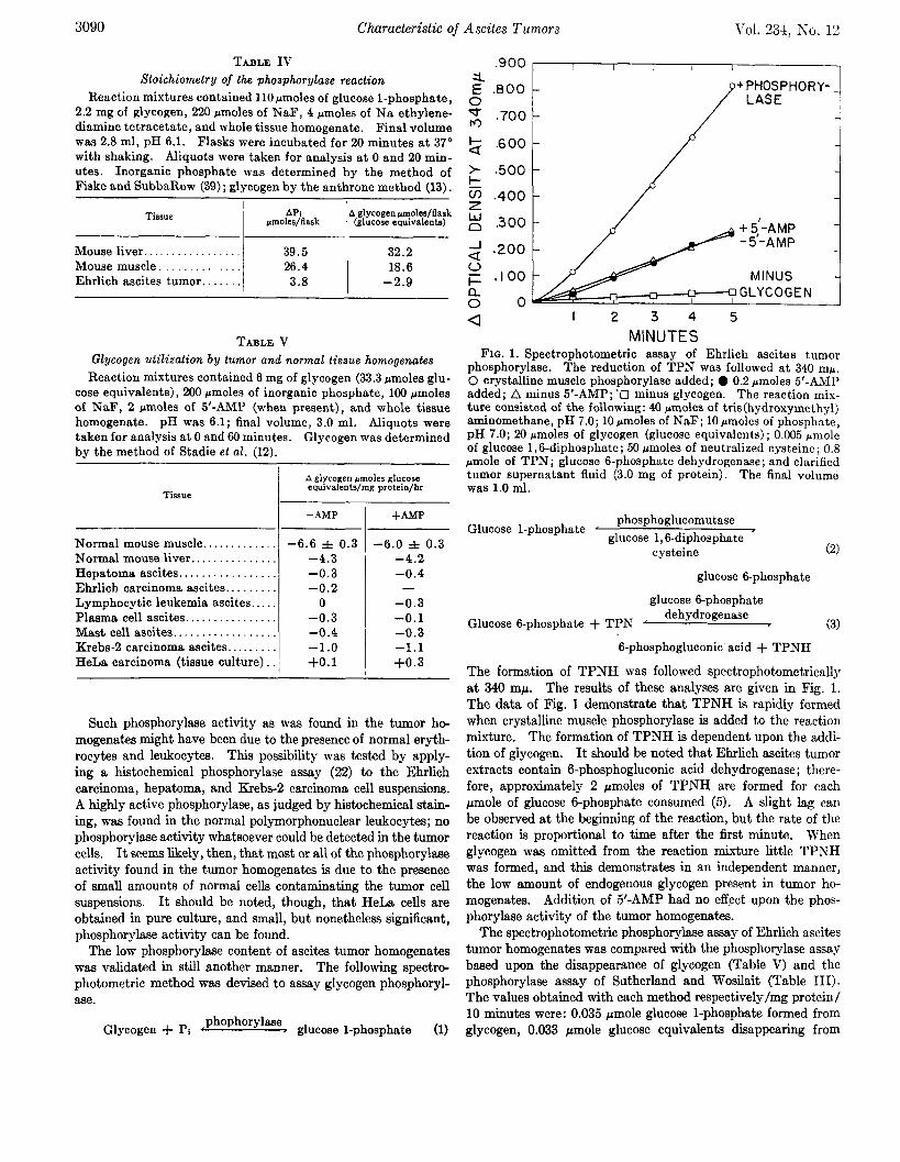
M INUTES FIG. 1. Spectrophotometric assay of Ehrlich ascites tumor
phosphorylase. The reduction of TPN was followed at 340 mp. 0 crystalline muscle phosphorylase added; 0 0.2 pmoles 5’-AMP added; A minus 5’-AMP; ‘13 minus glycogen. The reaction mix- ture consisted of the following: 40 pmoles of tris(hydroxymethy1) aminomethane, pH 7.0; 10 pmoles of NaF; 10 rmoles of phosphate, pH 7.0; 20 rmoles of glycogen (glucose equivalents) ; 0.005 pmole of glucose 1,6-diphosphate; 50 crmoles of neutralized cysteine ; 0.8 pmole of TPN; glucose 6-phosphate dehydrogenase; and clarified tumor supernatant fluid (3.0 mg of protein). The final volume was 1.0 ml.
Glucose l-phosphate ’ phosphoglucomutase
glucose 1,6diphosphate cysteine (2)
glucose 6-phosphate
glucose 6phosphate
Glucose g-phosphate + TPN ’ dehydrogenase
6-phosphogluconic acid + TPNH
The formation of TPNH was followed spectrophotometricallJ at 340 rnp. The results of these analyses are given in Fig. 1. The data of Fig. 1 demonstrate that, TPNH is rapidly formed when crystalline muscle phosphorylase is added to the reaction mixture. The formation of TPNH is dependent upon the addi- tion of glycogen. It should be noted that Ehrlich ascites tumor extracts contain 6-phosphogluconic acid dehydrogenase; there- fore, approximately 2 pmoles of TPNH are formed for each pmole of glucose 6-phosphate consumed (5). A slight lag can be observed at the beginning of the reaction, but the rate of the reaction is proportional to time after the first minute. When glycogen was omitted from the reaction mixture little TPNH was formed, and this demonstrates in an independent manner, the low amount of endogenous glycogen present in tumor ho- mogenates. Addition of 5’-AMP had no effect upon the phos- phorylase activity of the tumor homogenatei.
The spectrophotometric phosphorylase assay of Ehrlich ascites tumor homogenates was compared with the phosphorylase assay based upon the disappearance of glycogen (Table V) and the phosphorylase assay of Sutherland and Wosilait (Table III). The values obtained with each method respectively/mg protein/ 10 minutes were: 0.035 pmole glucose l-phosphate formed from glycogen, 0.033 rmole glucose equivalents diappearing from

December 1959 M. W. Nirenberg
glycogen, and 0.081 Lcmole Pi released from glucose l-phosphate. Separate Ehrlich ascites tumor homogenates were prepared for each analysis, yet the results of the three diierent types of assays essentially agree with each other. It seems reasonable to con- clude that Ehrlich ascites tumo; homogenates have 3 to 10% of the phosphorylase activity of normal liver or muscle and that most or all of the observed activity is derived from normal leukocytes present in the ascites suspensions.
Ehrlich ascites tumor homogenates can utilize glucose l-phos- phate. Supplementation with glucose 1,6diphosphate and cysteine was necessary for optimal phosphoglucomutase activity; both were routinely added for phosphorylase assays. The results of Fig. 1 demonstrate that phosphoglucomutase is present and is not rate limiting in glycogen degradation by Ehrlich ascites tumor homogenates. Phosphoglucomutase has also been dem- onstrated in the Novikoff hepatoma (23).
Phosphorylase Activity of Tumor of Viral Origin
The phosphorylase content of a virus-induced tumor, the Rous sarcoma, and normal chicken muscle are presented in Table III. Both samples of tissue were removed from the same animal. The Rous sarcoma was a solid tumor and was not studied as com- pletely as the other tumors, but nonetheless low phosphorylase levels, comparable to the ascites tumors and the HeLa carcinoma, were found.
Activation of Phosphorylase by Epinephrine and Glucagon
Since epinephrine and glucagon arc involved in the activation of phosphorylase, it seemed logical to determine whether either hormone could facilitate the activation of phosphorylase in hepatoma and Ehrlich ascites tumor cells. The addition of 50 pg/ml of epinephrine and 25 pgg/ml of glucagon to mouse liver slices resulted in a marked and rapid reactivation of phosphoryl- ase. Additions of epinephrine and glucagon to hepatoma and epinephrine to Ehrlich ascites tumor cells had no effect upon phosphorylase activation. These data demonstrate the absence of this hormonally controlled enzymatic response in these tumors.
Studies with Phosphorylase dctivating System
Since the tumor cells had negligible phosphorylase activities and were not responsive to epinephrine and glucagon, it was of interest to determine whether the phosphorylase activating en- zymes were present. The data of Table V.1 demonstrate that HeLa carcinoma, hepatoma, and Ehrlich carcinoma homogenates can convert dephosphophosphorylase to phosphorylase. Addi- tion of dephosphophosphorylase kinase had no effect upon phos- phorylase activation in HeLa and Ehrlich carcinoma homog- enates, but increased the phosphorylasc activity of the hepatoma homogenate. The conversion of dcphosphophosphorylase to phosphorylase was not proportional to the amount of homog- enate added, possibly due to the involvement of adenosine-3’-5’- cyclic phosphate in the over-all reaction. The activation of phosphorylase was dependent upon the prcscnce of ATP. The results of Table VI demonstrate that the tumors possess a vigor- ous dcphosphophosphorylase-activating system and a relative absence of dephosphophosphorylase.
Phosphorylase Inactivating Enzyme
The question may be asked, “Does the Ehrlich ascites tumor have a high phosphorylase inactivating enzyme activity?” The
TABLET VI Activation of Dephosphophosphorylase by HeLa, Hepatoma
and Ehrlich Homogenates Reaction mixtures contained 4 pmoles of tris(hydroxymethy1)
aminomethane, pH 7.4,0.34 pmole of ATP, 0.5 pmole of MgSO,, 0.039 mole of epinephrine Cl., whole homogenate, and where indicated, dog liver dephosphophosphorylase and dog liver de- phosphophosphorylase kinase. 0.05 ml of the HeLa and hepatoma homogenates contained 2.37 and 1.90 mg of protein respectively. 0.15 ml of Ehrlich homogenate contained 9.04 mg of protein. Total volume was 0.2 ml. Reaction mixtures were incubated at 30” for 5 minutes. 1 ml of the phosphorylase reagent (14) contain- ing 2.0 pmoles 5’-AMP was then added, and the tubes were incu- bated at, 37” for 20 minutes. Samples were deproteinized by tri- chloroacetic acid precipitation at 0 and 20 minutes.
NO.
8
9
10
Homo- genate
ml 0.05 0.05 0.10 0.10 0.15 0.15 0.15
0.15
0.15
0.15
.4ddition
None + Dephosphophosphorylase None + Dephosphophosphorylase None + Dephosphophosphorylase + Dephosphophosphorylase + Dephosphophosphorylase
kinase + Dephosphophosphorylase - ATP - Dephosphophosphorylase + Dephosphophosphorylase
kinase - Homogenate + dephos-
phophosphorylase + de- phosphophosphorylase ki- nase
&La carci- noma
Ehrlich carci- noma
A ,molcs Pi/20 min. 0.19( 6.48 0.34f
15.7 0.765
19.7
0.033 0.85C 0.54s
11.7 1.19
15.8
20.9 28.0
3.96 2.01
1
1.68 13.2
12.3
conditions for assaying liver phosphorylase inactivating enzyme have been described (17). Purified preparations of dog liver phosphorylase were added to homogenates of normal mouse liver and Ehrlich tumor cells and the disappearance of phospho- rylase activity was measured at 10 and 20 minutes. The specific activity of the phosphorylase inactivating enzyme in homog- enates of dog liver is reported to be 1.2 to 1.6 (17). Under identical conditions the specific activity of this enzyme in mouse liver was 0.04 and in Ehrlich ascites tumor, 0.02. The low value obtained in mouse liver, as compared to dog liver possibly may be a species difference. These experiments demonstrate that mouse liver can inactivate phosphorylase at approsimately twice the rate of Ehrlich ascites tumor. The possibility that the tumor extract contains a powerful inhibitor of phosphorylase can there- fore be excluded.
DISCUSSION
Tumors exhibit such diversity in form and type that it would seem highly unlikely to expect a relative absence of phosphorylase in all tumors. The results obtained with ascites tumors should not be extrapolated to other types of tumors. Phosphorylase has been demonstrated in two types of solid tumors (24) ; however, considerably decreased phosphorylase levels have also been at- tributed to a solid hepatoma (25). Since solid tumors contain
3092 Characteristic of Ascites Tumors Vol. 234, So. 12
variable numbers of normal cells such as connective tissue, as- cites tumors were used primarily in this study. One advantage of ascites tumors is the relative purity of cell type which can be obtained.
Glycogen synthesis has been shown to proceed in a variety of tissues by Lcloir and others (26-29) by an irreversible UDP glucose transferase reaction. This, rather than phosphorylase, may be the main route of glycogen synthesis. Dr. R. Wu2 has found UDP-glucose transferase in HeLa cells. Under certain growth conditions the cells can accumulate glycogen; then low levels of phosphorylase, about 1 y0 that of an equivalent amount of muscle, can be demonstrated. Since HeLa carcinoma homog- enates can rapidly reactivate added dephosphophosphorylase (Table VI), the rate-limiting factor appears to be the availability of dephosphophosphorylase. UDP-glucose transferase has not been looked for in ascites tumors. Although no stored glycogen can be found in these tumor cells, the absence of glycogen need not always go hand in hand with the absence of phosphorylase. The possibility exists that some cells contain UDP-glucose trans- ferase but lack phosphorylase. This situation might result in a marked accumulation of glycogen.
Rat muscle and brain have little phosphorylase at birth, and after approximately 10 days the phosphorylase activities rise to adult levels (30). The phosphorylase content of rat liver 1 day after term also is greatly reduced.3 Fetal guinea pig liver, how- ever, contains adult quantities of phosphorylase (31). Some, but not all embryonic tissues, therefore, have greatly reduced phosphorylase levels when compared to the corresponding adult tissues.
Although both glycogen and phosphorylase are present in al- most all adult mammalian tissues, every cell type need not con- tain these substances. Hi&chemical studies have demon- strated, for example, the uneven distribution of phosphorylase activity among different cell types of a given tissue (22). Pre- liminary work with a virus-induced tumor, the Rous sarcoma, has revealed remarkably low phosphorylase levels when com- pared with normal chicken muscle taken from the same animal. Although this tumor is a sarcoma, chicken muscle may not be an adequate control, for the Rous sarcoma can arise from in- fected avian fibroblasts (32). Although no answer is available, the question should be raised, “DO certain types of normal cells such as fibroblasts also have low phosphorylase levels, and, if so, are ascites tumors derived primarily from these cell types?”
The breakdown of glycogen in both liver and muscle is clearly regulated by a complex hormonal mechanism. The extremely rapid interconversion of dephosphophosphorylase and phospho- rylase in resting versus contracting muscle has been emphasized (33), and it is possible that the activation and deactivation of phosphorylase controls the release of distinct waves of glucose- l-phosphate which can be converted quickly to lactate either with the concomitant production of pulses of ATP, if it is metab- olied via the Embden-Meyerhof pathway, or with the produc- tion of waves of TPNH if it is metabolized via the hexose mono- phosphate shunt. Although ascites tumor cells have exceedingly high rates of carbohydrate metabolism, they lack to a large extent this hormonal regulatory mechanism. The suggestion has been made (34) that some tumors become insensitive to certain controlling forces, such as hormonal regulation, through loss or inhibition of particular enzyme pathways. Transhydro-
1 Personal communication. *M. W. Niremberg, unpublished results.
genase is present in normal liver but has not been found in a number of ascites tumors (35), including a hepatoma. Addition of epinephrine and glucagon to hepatoma cells in this study did not result in an increased level of phosphorylase, possibly because of the low amount of dcphosphophosphorylase available.
Since it is always difficult to validate a negative finding, such as the absence of an enzyme, an attempt has been made to in- vestigate thoroughly the parameters of the phosphorylase assny. It seems reasonable to conclude that the phosphorylase contents of the ascites tumors studied, such as the hcpatoma, are very low when compared with normal liver or muscle. It is not pos- sible with the methods available to ascribe a total absence of phosphorylase to ascites tumors. It should be noted that the phosphorylase levels herein ascribed to sscites tumors undoubt- edly represent maximal values, for histological esamination re- vealed high phosphorylase activity in normal leukocytes also present in ascites suspensions. No phosphorylase whatsoever could be detected histologically in the tumor cells.
It seems striking that these tumors, utilizing monosaccharides at such rapid rates; should be relatively unable to degrade gly- cogen. In normal cells glycogcn appears to serve the cell as a hormonally controlled reservoir, or buffer, for “energy” and sub- strates. Clearly, the tumor cells studied neither possess a car- bohydrate reserve nor have, to any appreciable cstcnt, the ron- trol mechanism which may release pulses of intracellular glucose l-phosphate. It is not known what effect this may have upon the metabolism and economy of these neoplastic cells.
SUMMARY
The glycogen phosphorylase activities of seven types of ascites tumors, a tumor grown in tissue culture, and a solid virus-induced tumor were determined by chemical and histochemical tech- niques. Negligible phosphorylase activities were found com- pared to normal control tissues. Although glycogen is present in some neoplastic cells, little or no glycogen could be found in the ascites tumors. The phosphorylase activating enzymes of three tumors were studied. All contained ATP-dependent phosphorylase activating enzymes but had negligible amounts of dephosphophosphorylase. No epinephrine or glucagon-induced activation of phosphorylase was observed.
It is not known whether this enzymatic defect in ascites tumors can be extrapolated to other types of tumors. The relative absence of a hormonal mechanism regulating stored carbohydrate utilization in ascites tumors was discussed and was held in con- trast to the rapid degradation of monosaccharides by these tu- mors.
AckwwEedgmcnt.s-The help of Dr. Samuel Spicer in perform- ing the histochemical assays, Dr. Earl Sutherland for generously supplying purified preparations of dog liver enzyme, and Dr. Harry Eagle for supplying HeLa cell cultures is gratefully ac- knowledged. The author wishes to express his appreciation to Dr. Dewitt Stetten, Jr., and the members of the Section on Intermediary Metabolism for their continued helpful advice and encouragement.
REFERENCES 1. RALL, T. W., AND SUTHEBLAND, E. W., J. Biol. Chm., XX%
1065 (1958). 2. SUTHERLAND, E. W., AND RALL, T. W., J. Biol. Chem. 232,
1077 (1958). 3. WAEBIJRQ,~., Science, 123, 309 (1956).

December 1959 M. W. Nirenberg 3003
4. NIRENBEM, M. W., AND HOGG, J. F., J. Am. Chem. Sot., 78, 6210 (1956).
5. NIRENBERO, M. W., AND HOGG, J. F., J. Am. Chem. Sot. 80, 4407 (1958).
6. NIRENBERG, M. W., AND HOGG, J. F., Cancer Research, 18,518 (1958).
7. NIRENBERG, M. W., Federation Proc., i7.283 (1958). 8. NIRENBERG, M. W., Biochim. el Biophya. Acta, 30. 203 (1958). 9. FRENCH, C. S., AND MILNER, H., in S. P. COLOWICK AND N. 0.
KAPLAN (Editors), Methods in enzymology, Vol. I, Academic Press, Inc., New York, 1955, p. 64.
16. NOSSAL, P. M., Australian J. Exptl. Biol. &fed. Sci., 31, 583 (1953).
11. HORECKER, B. L., AND SMYRNIOTIS, P. Z., in S. P. COLOWICK AND N. 0. KAPLAN (Editors), Methods in enzymology, Vol. I, Academic PresB, Inc., New York, 1955, p. 323.
12. STADIE, W. C., HAUOAARD, N., AND MARSH, J. B., J. Biol. Chem., 183, 167 (1951).
13. CARROLL, N. V., LONGLEY, R. W., AND ROE, J. H., J. Biol. Chem., 220, 583 (1956).
14 SUTHERL.~ND, E. W., AND WOYILAIT, W. D., J. Biol. Chem., 218, 459 (1956).
15. CORI, G. T., ILLINGWORTH, B., AND KELLER, P. J., in S. P. COLOWICK AND N. 0. KAPL.~N (Editors), Methods in enzy- mology, Vol. I, Academic Press, Inc., New York, 1955, p. 200.
16. ROLL, T. W., SUTHERLAND, E. W., AND BERTHET, J., J: Biol. Chem., 224, 463 (1957).
17. WOSILAIT, W. D., AND SUTHERLAND, E. W., J. Biol. Chem., 218, 469 (1956).
18. BUCHER, T., Biochim. et Biophys. Acta, 1, 292 (1947). 19. LILLIE, R. D., Histopathologic technic and practical histochem-
istry, 2nd edition, Blakiston Company, New York, 1954, p. 120.
20. KORN, E. D., J. Am. Chem. Sot., 30, 1520 (1958).
21.
22.
23. 24.
25.
26.
27.
28.
29.
30. 31.
32. 33.
34. 35.
36.
37.
38.
39.
CORI, C. F., CORK, G. T., AND GREEN, A. A., J. Biol. Chrtn. 161, 39 (1943).
TAKEUCHI, T., AND KURIAKI, H., J. Histochem. Cytochcm., 3, 153 (1955).
WEB&, G.; AND CANTERO, A., Cancer Research, 17,995 (1957). GORANSON, E. S., MCBRIDE, J., AND WEBER, G., Cancer I?e-
search, 14, 227 (1954). HADJIOLOV, A. A., AND DANCHEVA, K. I., Nature, 181, 547
(1958). LELOIR, L. F., AND CARDINI, C. E., J. Am. Chem. Sot., 79,
6340 (1957). LELOIR, L. F., OLAVARRIA, J. M., GOLDEMBERO, S. H. AND
CARMINATTI, H., Arch. B&hem. Biophys., 81, 508 (1959‘). ROBBINS, P. W., TROUT, R. R., AND LIPMANN,%‘., Proc. Natl.
Acad. Sci. U. S., 46. 6 (1959). VILLAR-PALASI, C., AND LARNER, J., Biochim. et Biophys.
Acta, 30, 449 (1958). SHAPIRO, B., AND WERTHEIMER, E., Biochem. J., 37,397 (19.13). NEMETH, A. M., INSULL, W. JR., AND FLEXNER, L. B., J. Iliol.
Chem., 208, 765 (1954). RUBIN, H., Virology, 1, 445 (1955). CORI, C. F., in 0. H. GAEBLER (Editor), Enzymes: units of
biological structure and junction, Academic Press, Inc., New York, 1956, p. 573.
SWA~VN, M. M., Cancer Research, 18, 1118 (1958). REYNAFARJE, B., AND POTTER, V. R., Cancer Research, 17,
1112 (1957). SATO, fi., B~;LKIN, M., AND ESSNER, E., J. Natl. Cancer ZfL,Tt,,
17, 1 (1956). POTTER, M.,. AND Law, L. W., J. Natl. Cancer. Inst., 18. ,413
(1957). DUNN, T. B., AND POTTER, M., J. hrutl. Cancer Inst., 18, 587
(1957). FISKE, C. H., AND SUBBARO~, Y., J. Biol. Chem., 66, 375
(1925).