99m -DTPA Study to Validate an Experimental Model of ...
6
Hindawi Publishing Corporation Advances in Urology Volume 2013, Article ID 929620, 5 pages http://dx.doi.org/10.1155/2013/929620 Research Article 99m Tc-DTPA Study to Validate an Experimental Model of Ureteral Obstruction in Rabbits: Preliminary Results Marcelo Lopes de Lima, 1 Rodolfo Bertti, 1 Juliano César Moro, 1 Fábio Coltro Neto, 1 Ricardo Miyaoka, 1 Adriano Fregonesi, 1 Mariana da Cunha Lopes de Lima, 2 and Celso Darío Ramos 2 1 Division of Urology, Department of Surgery, Campinas State University, Avenida Vital Brasil 251, Caixa Postal 6142, Cidade Universit´ aria Zeferino Vaz, 13083-888 Campinas, SP, Brazil 2 Nuclear Medicine Division, Department of Radiology, Campinas State University, Avenida Vital Brasil 251, Caixa Postal 6142, Cidade Universit´ aria Zeferino Vaz, 13083-888 Campinas, SP, Brazil Correspondence should be addressed to Marcelo Lopes de Lima; [email protected] Received 25 June 2013; Revised 11 October 2013; Accepted 15 October 2013 Academic Editor: Maxwell V. Meng Copyright © 2013 Marcelo Lopes de Lima et al. is is an open access article distributed under the Creative Commons Attribution License, which permits unrestricted use, distribution, and reproduction in any medium, provided the original work is properly cited. Objective. To create a ureteral obstruction experimental model that can be proved through 99m Tc-DTPA renal scintigraphy and histopathological studies, without causing total renal function loss. Materials and Methods. Ten New Zealand white rabbits were submitted to a surgical experiment to create a model of unilateral obstruction to urinary flow. Surgery procedure provided unilateral ureteral obstruction (leſt kidney) to urinary flow and posteriorly was evaluated by 99m Tc-DTPA renal scintigraphy and histopathological study. 99m Tc-DTPA renal study was performed to detect and quantify signs of obstruction and to evaluate renal function. Statistical analysis was performed through the Student t-test with a significance level of < 0.05. Results. Nine of the ten rabbits presented leſt renal unit obstruction and one nonobstructive on the 99m Tc-DTPA and histopathological studies. All the right renal units, which were not submitted to surgical procedure, were nonobstructed by the studies. ere was a general agreement between scintigraphy and histopathological results in both groups. Conclusion. e experimental model promoted the creation of ureteral obstruction in rabbits, confirmed by nuclear medicine scintigraphy and histopathology, and could be used in further studies to better understand urinary obstruction. 1. Introduction Pelvi-ureteric junction (PUJ) obstruction is one of the most frequent congenital anomalies of the urinary tract system. It is associated with pain, hydronephrosis, urinary tract infections, and eventually loss of renal function [1, 2]. It affects around 40% to 60% of all newborns with hydronephrosis [3], two times more common in males, and may be bilateral in 5% to 15% of cases [1, 4]. PUJ obstruction may be caused by intrinsic factors, like aperistaltic ureteral segment, obstructive fold mucosa, ureteral polyp, or ureteral stenosis [5]. Among extrinsic factors stands inferior renal polar vessel crossing anteriorly the PUJ [6]. Treatment varies from clinical observation to surgery. ere are some surgical modalities available to correct the PUJ obstruction, differing from the open pyeloplasty to the latest in technology such as robotic assisted surgeries and endourological procedures [7–9]. Currently scientific literature is short in ureteral obstruc- tion models that accurately reproduce the clinical and micro- scopic features of this infirmity [10–12]. e creation of a standardized experimental model that would be able to cause obstructive disturbance without leading to renal function loss would provide information capable of enhancing not only the diagnosis but also the treatment choices.
Transcript of 99m -DTPA Study to Validate an Experimental Model of ...
Hindawi Publishing CorporationAdvances in UrologyVolume 2013, Article ID 929620, 5 pageshttp://dx.doi.org/10.1155/2013/929620
Research Article99mTc-DTPA Study to Validate an Experimental Model ofUreteral Obstruction in Rabbits: Preliminary Results
Marcelo Lopes de Lima,1 Rodolfo Bertti,1 Juliano César Moro,1
Fábio Coltro Neto,1 Ricardo Miyaoka,1 Adriano Fregonesi,1
Mariana da Cunha Lopes de Lima,2 and Celso Darío Ramos2
1 Division of Urology, Department of Surgery, Campinas State University, Avenida Vital Brasil 251, Caixa Postal 6142,Cidade Universitaria Zeferino Vaz, 13083-888 Campinas, SP, Brazil
2 Nuclear Medicine Division, Department of Radiology, Campinas State University, Avenida Vital Brasil 251, Caixa Postal 6142,Cidade Universitaria Zeferino Vaz, 13083-888 Campinas, SP, Brazil
Correspondence should be addressed to Marcelo Lopes de Lima; [email protected]
Received 25 June 2013; Revised 11 October 2013; Accepted 15 October 2013
Academic Editor: Maxwell V. Meng
Copyright © 2013 Marcelo Lopes de Lima et al. This is an open access article distributed under the Creative Commons AttributionLicense, which permits unrestricted use, distribution, and reproduction in any medium, provided the original work is properlycited.
Objective. To create a ureteral obstruction experimental model that can be proved through 99mTc-DTPA renal scintigraphy andhistopathological studies, without causing total renal function loss. Materials and Methods. Ten New Zealand white rabbits weresubmitted to a surgical experiment to create a model of unilateral obstruction to urinary flow. Surgery procedure providedunilateral ureteral obstruction (left kidney) to urinary flow and posteriorly was evaluated by 99mTc-DTPA renal scintigraphy andhistopathological study. 99mTc-DTPA renal study was performed to detect and quantify signs of obstruction and to evaluate renalfunction. Statistical analysis was performed through the Student t-test with a significance level of 𝑃 < 0.05. Results. Nine of the tenrabbits presented left renal unit obstruction and one nonobstructive on the 99mTc-DTPA and histopathological studies. All the rightrenal units, which were not submitted to surgical procedure, were nonobstructed by the studies. There was a general agreementbetween scintigraphy and histopathological results in both groups. Conclusion. The experimental model promoted the creationof ureteral obstruction in rabbits, confirmed by nuclear medicine scintigraphy and histopathology, and could be used in furtherstudies to better understand urinary obstruction.
1. Introduction
Pelvi-ureteric junction (PUJ) obstruction is one of the mostfrequent congenital anomalies of the urinary tract system.It is associated with pain, hydronephrosis, urinary tractinfections, and eventually loss of renal function [1, 2]. It affectsaround 40% to 60% of all newborns with hydronephrosis [3],two timesmore common inmales, andmay be bilateral in 5%to 15% of cases [1, 4].
PUJ obstruction may be caused by intrinsic factors,like aperistaltic ureteral segment, obstructive fold mucosa,ureteral polyp, or ureteral stenosis [5]. Among extrinsicfactors stands inferior renal polar vessel crossing anteriorlythe PUJ [6].
Treatment varies from clinical observation to surgery.There are some surgical modalities available to correct thePUJ obstruction, differing from the open pyeloplasty to thelatest in technology such as robotic assisted surgeries andendourological procedures [7–9].
Currently scientific literature is short in ureteral obstruc-tionmodels that accurately reproduce the clinical andmicro-scopic features of this infirmity [10–12].
The creation of a standardized experimental model thatwould be able to cause obstructive disturbance withoutleading to renal function loss would provide informationcapable of enhancing not only the diagnosis but also thetreatment choices.
2 Advances in Urology
Through this model, one would be able to test drugs, seekbiochemical markers, assess radiopharmaceutical produces,research pathological features related to ureteral obstruction,and evaluate therapeutic methods.
The objective of the present study is to create a ureteralobstruction experimental model that can be proved throughrenogram with technetium-99m diethylenetriaminepentaa-cetic acid ( 99mTc-DTPA) and histopathological studies, with-out causing total renal function loss.
2. Materials and Methods
The protocol for the research project was approved by theinstitution research and ethical commission, within whichthe work was undertaken.
After approval by the research and ethical commission,ten New Zealand white rabbits were submitted to the presentsurgical experiment. The animals were all 3 months old andfemale and weighted 3.5 Kg on average.
After general anesthesia acquired with intramuscularinjection of ketamine (30mg/kg) and xylazine (5mg/kg), therabbits’ abdominal cavities were opened through a midlineabdominal incision of 15 cm in length and their left ureter wasidentified and dissected out, preserving its vascularization.Subsequently, a 1.5 cm long incision was made into the leftpsoas muscle starting 1.5 cm below the inferior pole of leftkidney, creating a longitudinal groove.
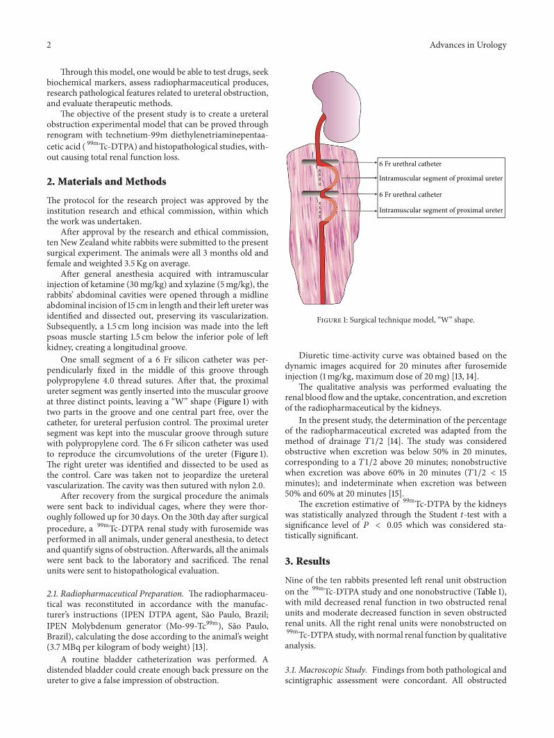
One small segment of a 6 Fr silicon catheter was per-pendicularly fixed in the middle of this groove throughpolypropylene 4.0 thread sutures. After that, the proximalureter segment was gently inserted into the muscular grooveat three distinct points, leaving a “W” shape (Figure 1) withtwo parts in the groove and one central part free, over thecatheter, for ureteral perfusion control. The proximal uretersegment was kept into the muscular groove through suturewith polypropylene cord. The 6 Fr silicon catheter was usedto reproduce the circumvolutions of the ureter (Figure 1).The right ureter was identified and dissected to be used asthe control. Care was taken not to jeopardize the ureteralvascularization. The cavity was then sutured with nylon 2.0.
After recovery from the surgical procedure the animalswere sent back to individual cages, where they were thor-oughly followed up for 30 days. On the 30th day after surgicalprocedure, a 99mTc-DTPA renal study with furosemide wasperformed in all animals, under general anesthesia, to detectand quantify signs of obstruction. Afterwards, all the animalswere sent back to the laboratory and sacrificed. The renalunits were sent to histopathological evaluation.
2.1. Radiopharmaceutical Preparation. The radiopharmaceu-tical was reconstituted in accordance with the manufac-turer’s instructions (IPEN DTPA agent, Sao Paulo, Brazil;IPEN Molybdenum generator (Mo-99-Tc99m), Sao Paulo,Brazil), calculating the dose according to the animal’s weight(3.7MBq per kilogram of body weight) [13].
A routine bladder catheterization was performed. Adistended bladder could create enough back pressure on theureter to give a false impression of obstruction.
6 Fr urethral catheter
Intramuscular segment of proximal ureter
6 Fr urethral catheter
Intramuscular segment of proximal ureter
Figure 1: Surgical technique model, “W” shape.
Diuretic time-activity curve was obtained based on thedynamic images acquired for 20 minutes after furosemideinjection (1mg/kg, maximum dose of 20mg) [13, 14].
The qualitative analysis was performed evaluating therenal blood flow and the uptake, concentration, and excretionof the radiopharmaceutical by the kidneys.
In the present study, the determination of the percentageof the radiopharmaceutical excreted was adapted from themethod of drainage 𝑇1/2 [14]. The study was consideredobstructive when excretion was below 50% in 20 minutes,corresponding to a 𝑇1/2 above 20 minutes; nonobstructivewhen excretion was above 60% in 20 minutes (𝑇1/2 < 15minutes); and indeterminate when excretion was between50% and 60% at 20 minutes [15].
The excretion estimative of 99mTc-DTPA by the kidneyswas statistically analyzed through the Student 𝑡-test with asignificance level of 𝑃 < 0.05 which was considered sta-tistically significant.
3. Results
Nine of the ten rabbits presented left renal unit obstructionon the 99mTc-DTPA study and one nonobstructive (Table 1),with mild decreased renal function in two obstructed renalunits and moderate decreased function in seven obstructedrenal units. All the right renal units were nonobstructed on99mTc-DTPA study, with normal renal function by qualitativeanalysis.
3.1. Macroscopic Study. Findings from both pathological andscintigraphic assessment were concordant. All obstructed

Advances in Urology 3
Table 1: Results of the scintigraphic study with 99mTc-DTPA.
Rabbits 99mTc-DTPA excretion (%)1 24.0% (O)2 74.0% (N)3 40.0% (O)4 24.0% (O)5 38.0% (O)6 30.0% (O)7 21.0% (O)8 30.0% (O)9 35.0% (O)10 25.2% (O)†N: nonobstructed; O: obstructed.
kidneys according to scintigraphy presented hydronephrosis.Of these, two units were classified as mild hydronephrosiswhilst the remaining seven units were classified as mod-erate. No specimen presented severe hydronephrosis. Mildhydronephrosis was considered when proximal ureteral seg-ments showed a dilation of 2 : 1 ratio when compared withproximal ureteral diameter of control kidneys. Similarly,moderate hydronephrosis was determined as a 3 : 1 to 4 : 1ratio dilation. Control kidneys weighed 10 g in average andcortical width varied from 0.5 cm to 0.6 cm. All left sidekidneys classified as hydronephrotic (𝑛 = 9) weighed 13.5 gin average and cortex varied from 0.3 cm to 0.4 cm with anaverage weight gain of 34.7% (10–55%)when compared to theright side (𝑃 < 0.05).
3.2. Microscopic Study. Parenchymal renal alterations inhydronephrotic specimens were mostly noted in upper andlower poles where composed papillae are found. Medialportion of the kidney where simple papillae predominatewas less affected. Renal cortex is minimally changed undermicroscopic view in mild hydronephrosis: ectasia, tubularatrophy, and mild nephritis. On the other hand, in mod-erate hydronephrosis, dilation alterations were more obvi-ous: atrophy and ectasia seen in tubules, collecting ducts,and contorted tubules; Tamm-Horsfall protein accumulationin peritubular, tubular, and Bowman’s spaces; and chronicnephritis, as previously described in renal obstruction byHeptinstall [16].
Besides the above mentioned alterations, moderatehydronephrotic cases also presented a “thyroidization” pro-cess, which is a tubular atrophy development with accumula-tion of eosinophilic amorphous substance in its lumen, givingit a thyroid gland-like aspect. Within a 30-day period, nosigns of severe hydronephrosis could be seen.
Scar tissue (fibroblasts) and inflammatory cells were notobserved in ureteral segments specimens that were wrappedby psoas muscle plicature.
4. Discussion
PUJ obstruction represents approximately 40% of the uro-logic diseases diagnosed in the prenatal period. Frequentlyit does not require invasive treatment, but only clinical and
laboratorial followup. In about 20% of the cases, a postnatalsurgical correction of the PUJ obstruction is necessary inorder to preserve renal function. If left untreated, it maylead to a syndrome with blockade and reduction of theurine flow, dilatation of the urinary tract, hydronephro-sis, and other symptoms [1–3]. Several aspects of urinaryobstruction have been studied. Urinary tract obstructionresults in renal compensatory mechanisms and may causeirrecoverable functional loss and histological alterations. Thepathophysiology of this progression is poorly understood[17].
Several factorsmay interfere in the process of obstruction.Wang et al. [18] have demonstrated that neonatally inducedpartial unilateral ureteral obstruction in rats is associatedwith changes in the abundance of renal acid-base transportersthat are paralleled by reduction in renal function, dependingon the severity and duration of obstruction. MacLellanet al. [17] have demonstrated that urinary tract obstruc-tion is associated with elevated urinary levels of alanine,succinate, dimethylglycine, creatinine, taurine, choline-likecompounds, hippurate, and lactate and decreased urinarylevels of 2-oxoglutarate and citrate. Despite these reports onbiochemical alterations, histological changes during chronicpartial ureteral obstruction were not well studied [19]. Long-term results confirmed that partial ureteral obstructionin newborn mice produces fibrotic lesions of the renalparenchyma, however with no correlation with dilatationof the upper tract [20] and substances like atorvastatinameliorated fibrosis, and helped preserve kidney filtrationfunction [19]. Experimental models in animals would permittesting of new drugs and observation of histological changesin the urinary obstruction.
There are some experimental models of ureteral obstruc-tion [10–12, 21, 22]. However, none have confirmed partialobstruction and the small size of these animals may hindernew surgical techniques evaluations. The objective of thepresent study was to develop a new experimental model ofureter stenosis, expanding the existing settings, although abigger animal model was used, as rabbits were included inthe research.
The rabbit is an attractive model for kidney studies,including renal obstruction and transplantation, in view ofits docility, convenient size, easy maintenance, low cost, andrenal anatomical and functional similarity to human kidneys[23–26]. Although the rabbit kidney is unipapillary and thehuman kidney is multipapillary [27], there have been severalstudies in the literature that have used rabbits as animalmodel, evaluating different renal diseases, stating that therabbit is suitable biological model for lesions of the kidney[26, 27].
It is estimated that rabbit life expectancy is around 8 years.If we were to make a linear interpolation from that figureto human life expectancy, we might say that one rabbit yearis roughly equivalent to one human decade [28, 29]. Then,we considered that 30 days were sufficient time to observechanges of obstruction.
The renal scintigraphy was performed to confirm theurinary obstruction and evaluate the renal function mainte-nance, allowing the validation of the method and permitting

4 Advances in Urology
the safe use of the model in the future. Considering thissimilarity and previous studies in the literature [23, 26], wehave considered themethod of𝑇1/2 adequate for rabbits, cal-culating the adequate doses of DTPA-99mTc and furosemideaccording to the weight of the animals.
The renal scintigraphy performed in the sample clearlyshowed that nine renal units presented obstruction. Noneof the left units showed total loss of function. The only unitwithout obstruction could be a result of a fail in the creationof circumvolutions. The diagnosis of ureteral obstructionis commonly made by 99mTc-DTPA diuretic renogram [14,15]. 99mTc-DTPA is one of the most widely used radio-pharmaceuticals to assess renal function of patients withsuspected urinary obstruction, with excretion predominantlyby glomerular filtration. Renal scintigraphywas preferentiallyused because it is a noninvasive and quick exam, providingrapid dynamic imaging of the kidneys, including renal flow,function, and urinary outflow evaluation [14, 15].
The utilized surgical technique in the present study isa modification of Ulm and Miller’s [10] procedure, authorswho first described the fixation of the upper ureter to thepsoas muscle. Our PUJ model uses circumvolutions of theureter to promote a local alteration of the peristaltic wave, notan extrinsic constriction, and through these ups and downs,a functional alteration may have occurred in the ureteralemptying. As Hammad et al. [30] have demonstrated, aftercomplete or partial ureteral obstruction, there are immediate,significant changes in the propagation of electrical impulsesin the proximal and distal ureter, which are generally lessmarked after partial obstruction than after complete obstruc-tion.
The present model has the ability to control the degreeof obstruction by the tension force applied in the ureter. Onecan anticipate that when the obstruction is undone, simply byliberating the ureter from the muscle without resections andanastomosis, it will make it easier to analyze what happens tothis renal unit while recovering. This finding is of relevanceto improve data and knowledge on the behavior of thePUJ obstruction and also on the sensitivity of diagnostictests with regard to different degrees of obstruction that aredifficult to counsel and to treat the patients efficiently. Basedon the need for a trusting model of PUJ obstruction thepresent study aimed to create an ease-to-do upper ureteralobstruction model. To date there have been some studies onPUJ obstruction regarding the creation of an animal modeleither for better understanding of the disease biology or forsurgical training purposes. Studies varied vastly from thetechnique used to generate the obstruction to the size ofanimals submitted to the experiments [31]. Moreover, thehistological datawas not provided by all of those experiments,and the ones that did mention them lacked sustainablefindings. This proposed model of partial ureteral obstructionenables chronic evaluation studies of morphological, func-tional, and histological changes of the obstructed kidney [32].We believe that further studies could be developed based onthemodel described and could reference the findings, thoughpreliminary, of the model described here.
5. Conclusions
The experimental model developed promoted the creationof ureteral obstruction in rabbits, confirmed by nuclearmedicine and histopathological exams, and could be used infurther studies to better understand urinary obstruction.
Abbreviations
PUJ: Pelvi-ureteric junction99mTc-DTPA: Technetium-99m
diethylenetriaminepentaacetic acid.
Conflict of Interests
The authors declare no conflict of interests.
Acknowledgments
The authors acknowledge Ms. Mercedes Santos from the“Apoio Didatico, Cientıfico e Computacional,” CampinasState University, who assisted in preparing the figure of thispaper, and Ana Cristina de Morais and Willian AdalbertoSilva, from the “Experimental Surgery Laboratory, Depart-ment of Surgery, Campinas State University,” who assisted inthe surgical experiment.
References
[1] S. Halachmi and G. Pillar, “Congenital urological anomaliesdiagnosed in adulthood—management considerations,” Journalof Pediatric Urology, vol. 4, no. 1, pp. 2–7, 2008.
[2] K. R. Anderson and R. M. Weiss, “Physiology and evaluationof ureteropelvic junction obstruction,” Journal of Endourology,vol. 10, no. 2, pp. 87–91, 1996.
[3] C.-C. Liang, P.-J. Cheng, C.-J. Lin, H.-W. Chen, A.-S. Chao,and S.-D. Chang, “Outcome of prenatally diagnosed fetalhydronephrosis,” The Journal of Reproductive Medicine for theObstetrician and Gynecologist, vol. 47, no. 1, pp. 27–32, 2002.
[4] D. M. Feldman, M. Decambre, E. Kong et al., “Evaluation andfollow-up of fetal hydronephrosis,” Journal of Ultrasound inMedicine, vol. 20, no. 10, pp. 1065–1069, 2001.
[5] C.Mendelsohn, “Functional obstruction: the renal pelvis rules,”The Journal of Clinical Investigation, vol. 113, no. 7, pp. 957–959,2004.
[6] J. H. Yiee, S. Johnson-Welch, L. A. Baker, andD. T.Wilcox, “His-tologic differences between extrinsic and intrinsic ureteropelvicjunction obstruction,” Urology, vol. 76, no. 1, pp. 181–184, 2010.
[7] M. C. Ost, J. D. Kaye, M. J. Guttman, B. R. Lee, and A. D. Smith,“Laparoscopic pyeloplasty versus antegrade endopyelotomy:comparison in 100 patients and a new algorithm for the mini-mally invasive treatment of ureteropelvic junction obstruction,”Urology, vol. 66, no. 5, pp. 47–51, 2005.
[8] J. P. Yurkanin and G. J. Fuchs, “Laparoscopic dismemberedpyeloureteroplasty: a single institution’s 3-year experience,”Journal of Endourology, vol. 18, no. 8, pp. 765–769, 2004.
[9] S. Chuanyu, X. Guowei, X. Ke, D. Qiang, and Z. Yuanfang,“Retroperitoneal laparoscopic dismembered Anderson-Hynespyeloplasty in treatment of ureteropelvic junction obstruction

Advances in Urology 5
(report of 150 cases),” Urology, vol. 74, no. 5, pp. 1036–1040,2009.
[10] A. H. Ulm and F.Miller, “An operation to produce experimentalreversible hydronephrosis in dog,” The Journal of Urology, vol.88, pp. 337–341, 1962.
[11] P. C. Ryan and J. M. Fitzpatrick, “Partial ureteric obstruction:a new variable canine experimental model,” The Journal ofUrology, vol. 137, no. 5, pp. 1034–1038, 1987.
[12] K. M. Kim, D. K. Kim, and H. Choi, “Partial ureteral obstruc-tion: a new experimental model in rats,” Seoul Journal ofMedicine, vol. 34, no. 1, pp. 55–61, 1993.
[13] B. L. Shulkin, G. A. Mandell, J. A. Cooper et al., “Procedureguideline for diuretic renography in children 3.0,” Journal ofNuclear Medicine Technology, vol. 36, no. 3, pp. 162–168, 2008.
[14] E. J. Fine, “Interventions in renal scintirenography,” Seminars inNuclear Medicine, vol. 29, no. 2, pp. 128–145, 1999.
[15] M. C. L. Lima, M. L. De Lima, C. F. V. Pepe et al., “Technetium-99m-L,L-ethylenedicysteine is more effective than technetium-99m diethylenetriamine penta-acetic acid for excludingobstruction in patients with pyelocalicinal dilation,” Urology,vol. 76, no. 2, pp. 283–288, 2010.
[16] G. S. Hill, “Calcium and the Kidney, Hydronephrosis,” inHeptinstall’s Pathology of the Kidney, J. C. Jennette, J. L. Olson,M. M. Schwartz, and F. G. Silva, Eds., pp. 891–936, Lippincott-Raven Publishers, Philadelphia, Pa, USA, 1998.
[17] D. L. MacLellan, D. Mataija, A. Doucette et al., “Alterations inurinary metabolites due to unilateral ureteral obstruction in arodent model,”Molecular BioSystems, vol. 7, no. 7, pp. 2181–2188,2011.
[18] G. Wang, W. Yuan, T.-H. Kwon et al., “Age-related changes inexpression in renal AQPs in response to congenital, partial,unilateral ureteral obstruction in rats,”PediatricNephrology, vol.27, no. 1, pp. 83–94, 2012.
[19] J. P. Fitzgerald, S.-Y. Chou, I. Franco et al., “Atorvastatin ame-liorates tubulointerstitial fibrosis and protects renal functionin chronic partial ureteral obstruction cases,” The Journal ofUrology, vol. 182, no. 4, pp. 1860–1868, 2009.
[20] N. Botto, R. Azoulay, M. Peuchmaur, and A. El Ghoneimi,“Renal parenchymal fibrosis and atrophy are not correlatedwith upper tract dilatation: long-term study of partial unilateralureteral obstruction in neonatal mice,” Journal of PediatricUrology, vol. 7, no. 3, pp. 310–316, 2011.
[21] S. Josephson, B. Robertson, G. Claesson, and I. Wikstad,“Experimental obstructive hydronephrosis in newborn rats. I.Surgical technique and long-term morphologic effects,” Inves-tigative Urology, vol. 17, no. 6, pp. 478–483, 1980.
[22] H. Huland, D. Gonnermann, B. Werner, and U. Possin, “Anew test to predict reversibility of hydronephrotic atrophy afterstable partial unilateral ureteral obstruction,” The Journal ofUrology, vol. 140, no. 6, pp. 1591–1594, 1988.
[23] M. Kekomaki, H. Rikalainen, P. Ruotsalainen, and C.Bertenyi, “Correlates of diuretic renography in experimentalhydronephrosis,” The Journal of Urology, vol. 141, no. 2, pp.391–394, 1989.
[24] P.M. Andrews, B. S. Khirabadi, and B. C. Bengs, “Using tandemscanning confocal microscopy to predict the status of donorkidneys,” Nephron, vol. 91, no. 1, pp. 148–155, 2002.
[25] I. A. Sammut, K. Burton, E. Balogun et al., “Time-dependentimpairment of mitochondrial function after storage and trans-plantation of rabbit kidneys,” Transplantation, vol. 69, no. 7, pp.1265–1275, 2000.
[26] M. J. Hanley, “Studies on acute disease models,”Kidney Interna-tional, vol. 22, no. 5, pp. 536–545, 1982.
[27] R. Dimitrov, D. Kostov, K. Stamatova, and V. Yordanova,“Anatomotopographical and morphological analysis of normalkidneys of rabbits (Oryctolagus cuniculus),” Trakia Journal ofSciences, vol. 10, no. 2, pp. 79–84, 2012.
[28] D. S. Woodruff-Pak, “Aging and classical conditioning: parallelstudies in rabbits and humans,”Neurobiology of Aging, vol. 9, no.5-6, pp. 511–522, 1988.
[29] R. R. Fox, “The rabbit (Oryctolagus cuniculus) and research onaging,” Experimental Aging Research, vol. 6, no. 3, pp. 235–248,1980.
[30] F. T. Hammad, W. J. Lammers, B. Stephen, and L. Lubbad,“Propagation of the electrical impulse in reversible unilateralureteral obstruction as determined at high electrophysiologicalresolution,”The Journal of Urology, vol. 185, no. 2, pp. 744–750,2011.
[31] J. Fichtner, F. G. Boineau, J. E. Lewy, R. K. Sibley, R. C. Vari, andL. M. D. Shortliffe, “Congenital unilateral hydronephrosis in arat model: continuous renal pelvic and bladder pressures,” TheJournal of Urology, vol. 152, no. 2, pp. 652–657, 1994.
[32] M. Lima, R. Miyaoka, J. Moro, and C. D’Ancona, “Laparo-scopic nephrectomy for xanthogranulomatous pyelonephritis-are there predictive factors for success?” Clinics, vol. 67, no. 8,pp. 907–909, 2012.

Submit your manuscripts athttp://www.hindawi.com
Stem CellsInternational
Hindawi Publishing Corporationhttp://www.hindawi.com Volume 2014
Hindawi Publishing Corporationhttp://www.hindawi.com Volume 2014
MEDIATORSINFLAMMATION
of
Hindawi Publishing Corporationhttp://www.hindawi.com Volume 2014
Behavioural Neurology
EndocrinologyInternational Journal of
Hindawi Publishing Corporationhttp://www.hindawi.com Volume 2014
Hindawi Publishing Corporationhttp://www.hindawi.com Volume 2014
Disease Markers
Hindawi Publishing Corporationhttp://www.hindawi.com Volume 2014
BioMed Research International
OncologyJournal of
Hindawi Publishing Corporationhttp://www.hindawi.com Volume 2014
Hindawi Publishing Corporationhttp://www.hindawi.com Volume 2014
Oxidative Medicine and Cellular Longevity
Hindawi Publishing Corporationhttp://www.hindawi.com Volume 2014
PPAR Research
The Scientific World JournalHindawi Publishing Corporation http://www.hindawi.com Volume 2014
Immunology ResearchHindawi Publishing Corporationhttp://www.hindawi.com Volume 2014
Journal of
ObesityJournal of
Hindawi Publishing Corporationhttp://www.hindawi.com Volume 2014
Hindawi Publishing Corporationhttp://www.hindawi.com Volume 2014
Computational and Mathematical Methods in Medicine
OphthalmologyJournal of
Hindawi Publishing Corporationhttp://www.hindawi.com Volume 2014
Diabetes ResearchJournal of
Hindawi Publishing Corporationhttp://www.hindawi.com Volume 2014
Hindawi Publishing Corporationhttp://www.hindawi.com Volume 2014
Research and TreatmentAIDS
Hindawi Publishing Corporationhttp://www.hindawi.com Volume 2014
Gastroenterology Research and Practice
Hindawi Publishing Corporationhttp://www.hindawi.com Volume 2014
Parkinson’s Disease
Evidence-Based Complementary and Alternative Medicine
Volume 2014Hindawi Publishing Corporationhttp://www.hindawi.com