9) Senescence and programmed cell death (PCD)aix-slx.upol.cz/~fellner/doc/PMP_9a-e.pdf · PMP 9)...
55
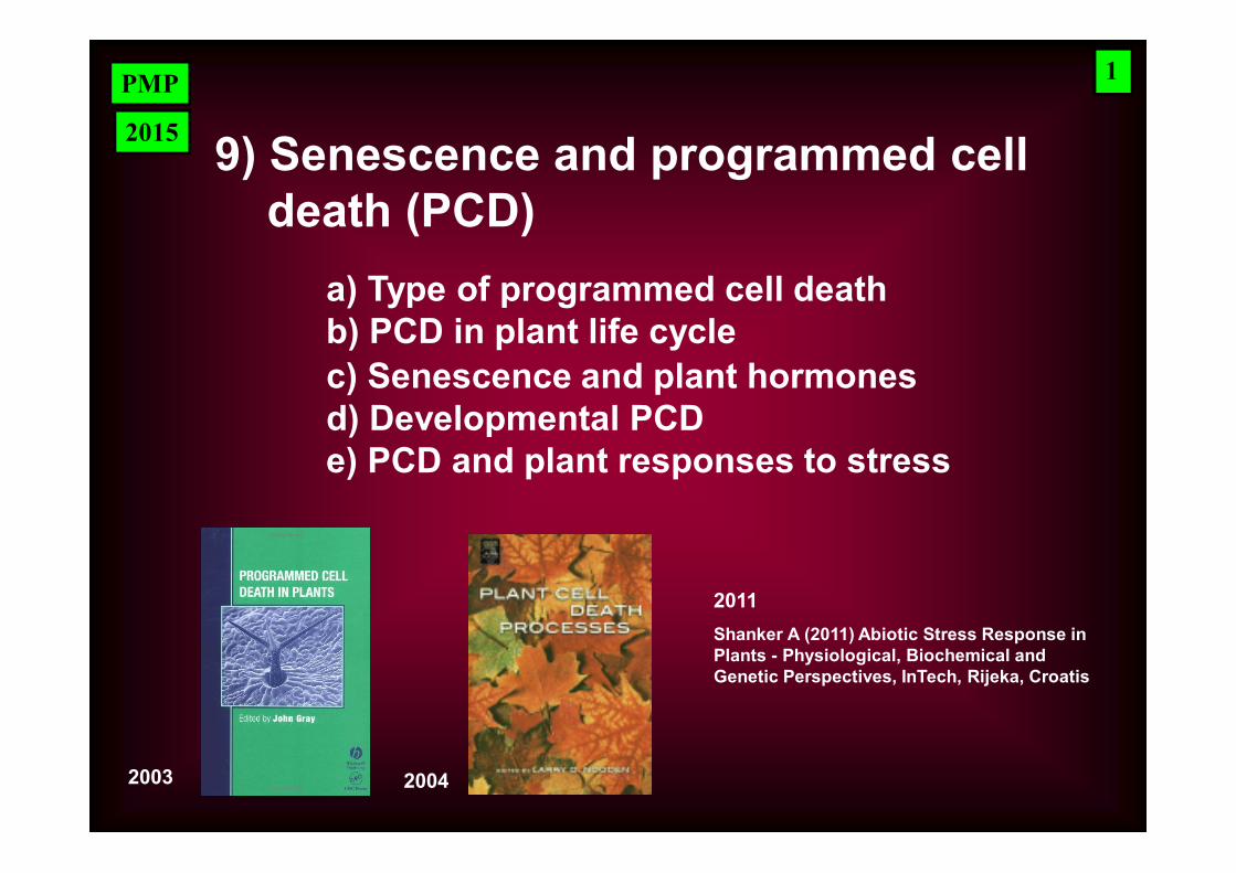
PMP 9) Senescence and programmed cell death (PCD) a) Type of programmed cell death b) PCD in plant life cycle 1 2015 c) Senescence and plant hormones d) Developmental PCD e) PCD and plant responses to stress 2003 2004 Shanker A (2011) Abiotic Stress Response in Plants - Physiological, Biochemical and Genetic Perspectives, InTech, Rijeka, Croatis 2011
Transcript of 9) Senescence and programmed cell death (PCD)aix-slx.upol.cz/~fellner/doc/PMP_9a-e.pdf · PMP 9)...
PMP
9) Senescence and programmed cell death (PCD)
a) Type of programmed cell deathb) PCD in plant life cycle
1
2015
c) Senescence and plant hormonesd) Developmental PCDe) PCD and plant responses to stress
2003 2004
Shanker A (2011) Abiotic Stress Response in Plants - Physiological, Biochemical and Genetic Perspectives, InTech, Rijeka, Croatis
2011
PMP
Programmed death - Programmed Cell Death (PCD) – is essential part of the growth and development of eukaryotic organisms and Their responses to stress
Organism itself controls initiation and process of death => „programmed death“
Examples of PCD in plants:
- senescence- Cell death associated with hypersensitive response
2
PMP
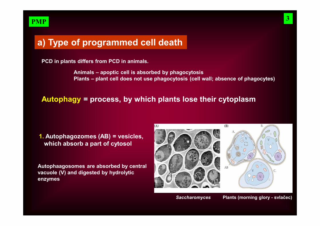
a) Type of programmed cell death
PCD in plants differs from PCD in animals.
Animals – apoptic cell is absorbed by phagocytosisPlants – plant cell does not use phagocytosis (cell wall; absence of phagocytes)
Autophagy = process, by which plants lose their cytoplasm
1. Autophagozomes (AB) = vesicles, which absorb a part of cytosol
Saccharomyces Plants (morning glory - svlačec)
3
Autophaagosomes are absorbed by central vacuole (V) and digested by hydrolytic enzymes
PMP
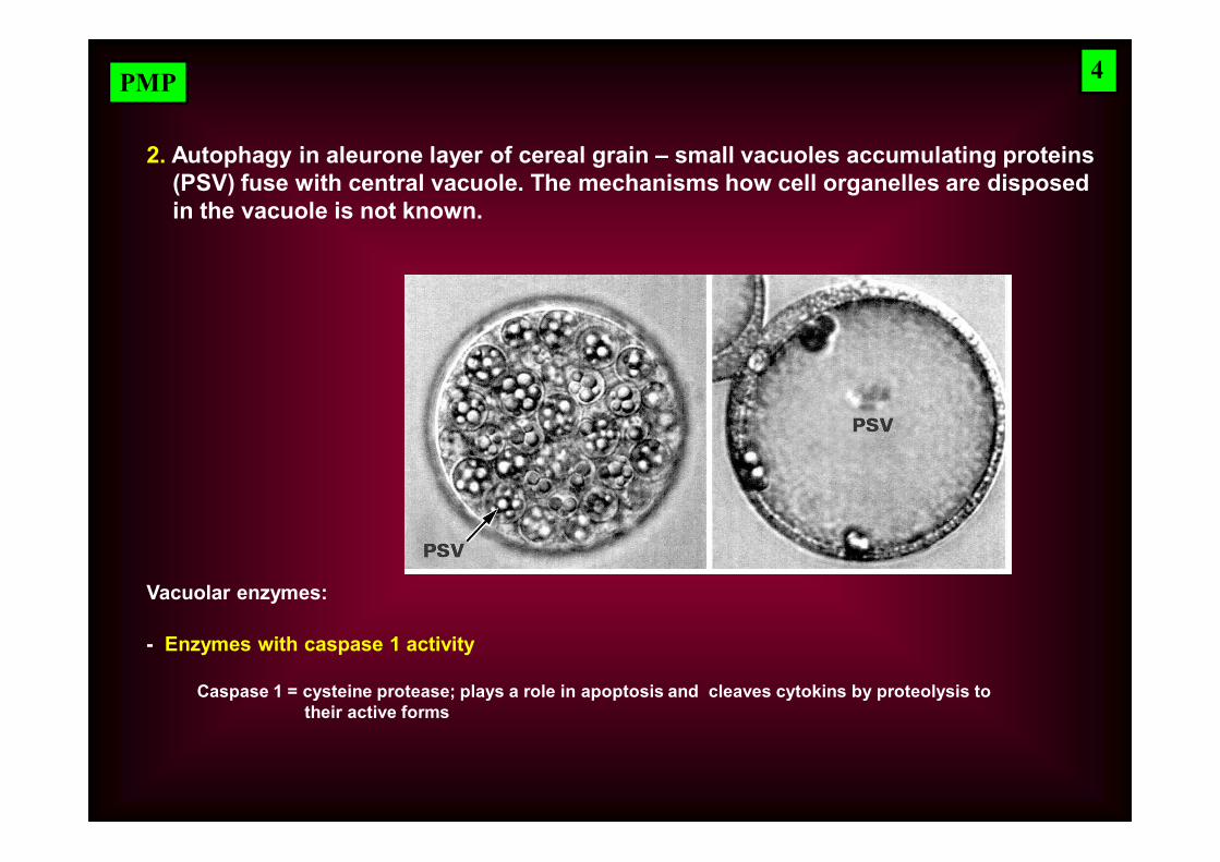
2. Autophagy in aleurone layer of cereal grain – small vacuoles accumulating proteins (PSV) fuse with central vacuole. The mechanisms how cell organelles are disposedin the vacuole is not known.
Vacuolar enzymes:
- Enzymes with caspase 1 activity
Caspase 1 = cysteine protease; plays a role in apoptosis and cleaves cytokins by proteolysis to their active forms
4
PMP
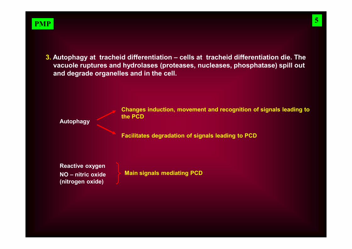
3. Autophagy at tracheid differentiation – cells at tracheid differentiation die. The vacuole ruptures and hydrolases (proteases, nucleases, phosphatase) spill out and degrade organelles and in the cell.
5
Autophagy
Changes induction, movement and recognition of signals leading to the PCD
Facilitates degradation of signals leading to PCD
Reactive oxygenNO – nitric oxide(nitrogen oxide)
Main signals mediating PCD
PMP
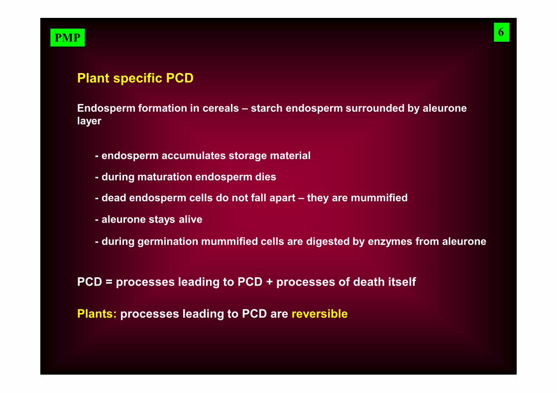
Plant specific PCD
Endosperm formation in cereals – starch endosperm surrounded by aleurone layer
PCD = processes leading to PCD + processes of death itself
Plants: processes leading to PCD are reversible
6
- endosperm accumulates storage material
- during maturation endosperm dies
- dead endosperm cells do not fall apart – they are mummified
- aleurone stays alive
- during germination mummified cells are digested by enzymes from aleurone
PMP
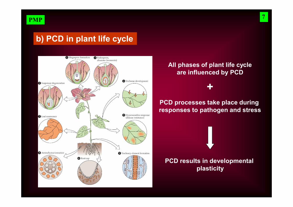
b) PCD in plant life cycle
All phases of plant life cycleare influenced by PCD
PCD processes take place during responses to pathogen and stress
+
PCD results in developmental plasticity
7
PMP
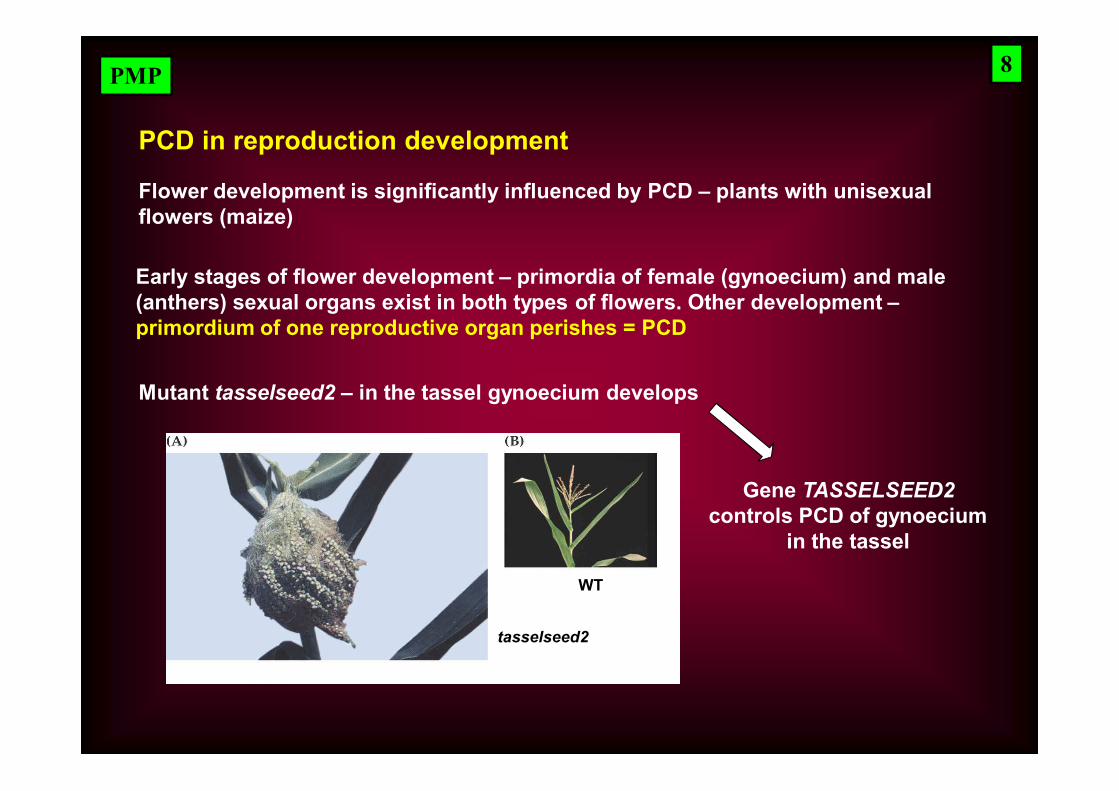
PCD in reproduction development
Flower development is significantly influenced by PCD – plants with unisexual flowers (maize)
Early stages of flower development – primordia of female (gynoecium) and male (anthers) sexual organs exist in both types of flowers. Other development –primordium of one reproductive organ perishes = PCD
Mutant tasselseed2 – in the tassel gynoecium develops
Gene TASSELSEED2 controls PCD of gynoecium
in the tassel
WT
tasselseed2
8
PMP
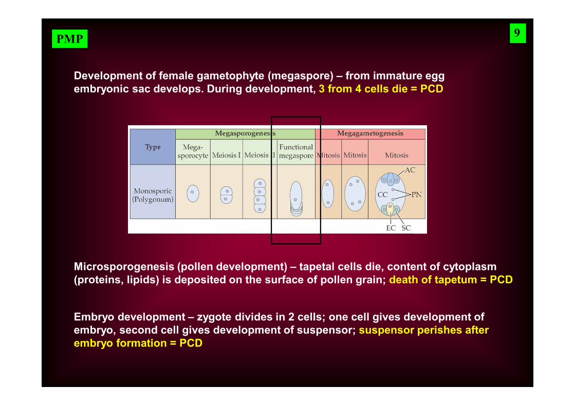
Development of female gametophyte (megaspore) – from immature egg embryonic sac develops. During development, 3 from 4 cells die = PCD
Microsporogenesis (pollen development) – tapetal cells die, content of cytoplasm (proteins, lipids) is deposited on the surface of pollen grain; death of tapetum = PCD
Embryo development – zygote divides in 2 cells; one cell gives development of embryo, second cell gives development of suspensor; suspensor perishes after embryo formation = PCD
9
PMP
PCD in vegetative development
Growth of embryo – before germination, growth of embryo is mechanically limited by endosperm cells; once endosperm cells die, embryo can grow; death of endosperm cells = PCD
Differentiation of xylem tracheids – live tracheal cells have no conductive function; cytoplasm of tracheid elements dies and is removed; dead cells with only secondary cell wall function as tracheids; death of treacheid elements = PCD
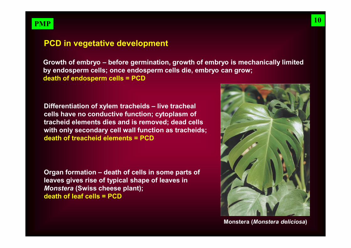
Organ formation – death of cells in some parts of leaves gives rise of typical shape of leaves in Monstera (Swiss cheese plant); death of leaf cells = PCD
10
Monstera (Monstera deliciosa)
PMP
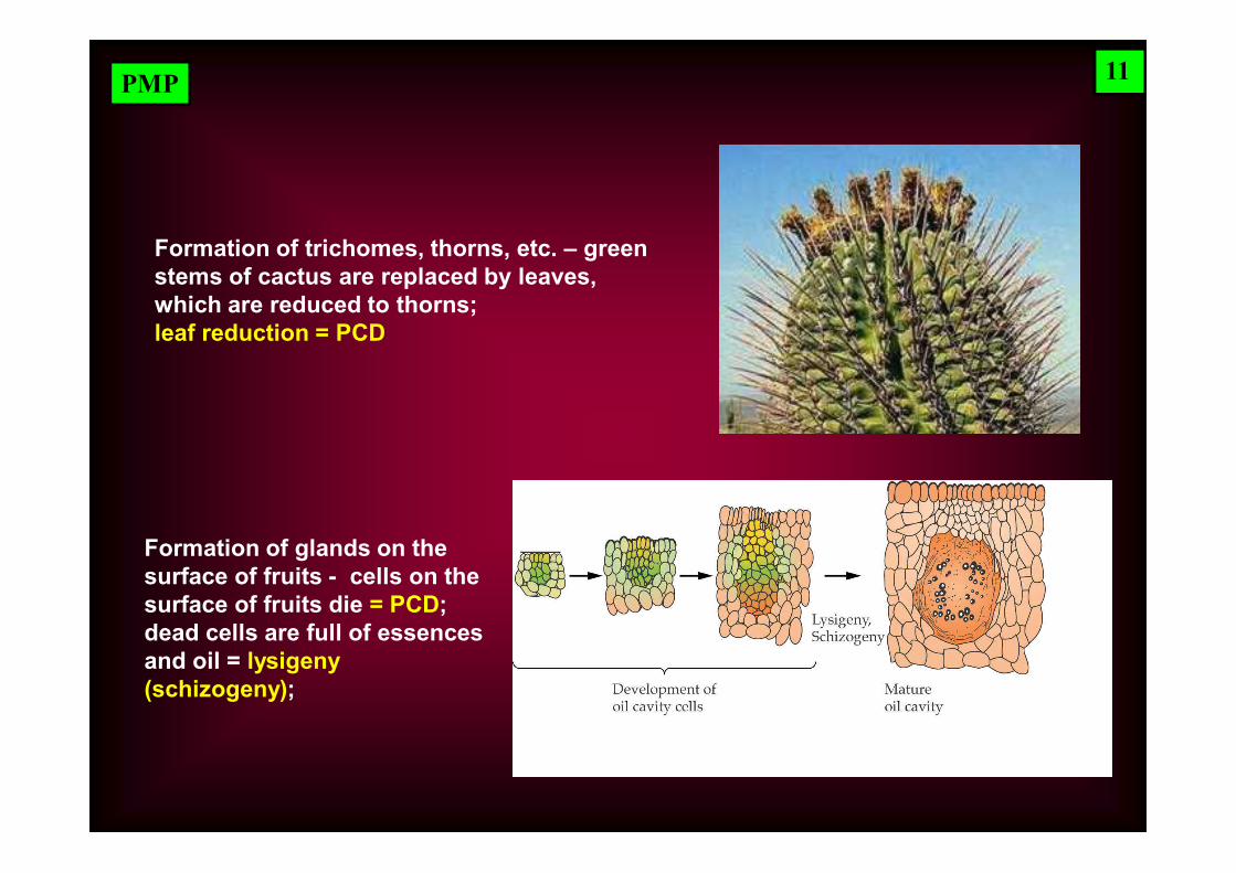
Formation of trichomes, thorns, etc. – green stems of cactus are replaced by leaves, which are reduced to thorns; leaf reduction = PCD
Formation of glands on the surface of fruits - cells on the surface of fruits die = PCD; dead cells are full of essences and oil = lysigeny (schizogeny);
11
PMP
PCD as a part of plant responses to stress
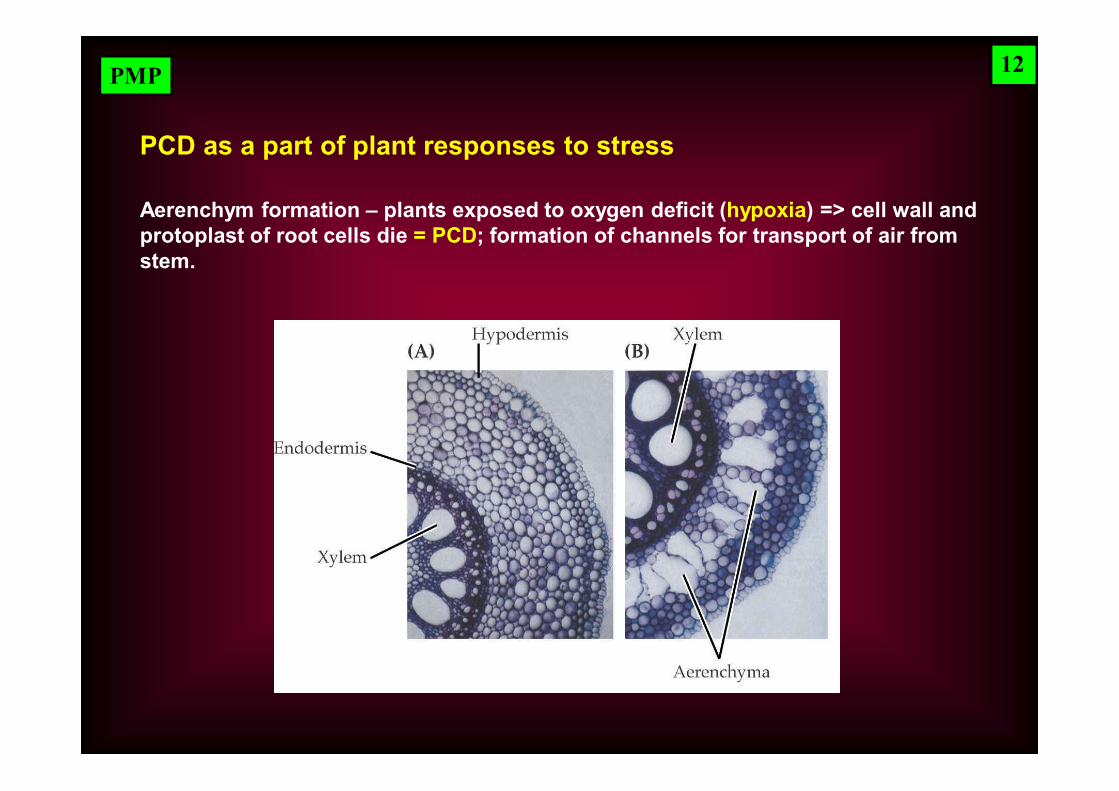
Aerenchym formation – plants exposed to oxygen deficit (hypoxia) => cell wall and protoplast of root cells die = PCD; formation of channels for transport of air from stem.
12
PMP
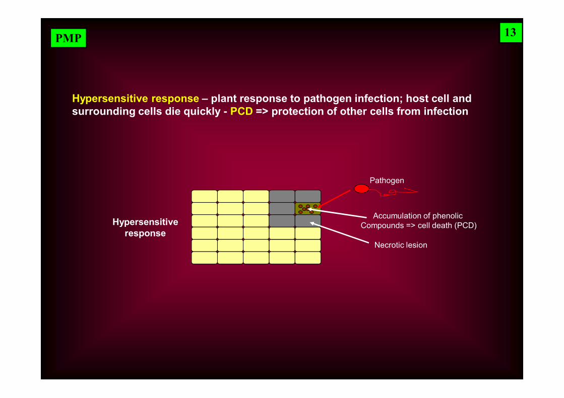
Hypersensitive response – plant response to pathogen infection; host cell and surrounding cells die quickly - PCD => protection of other cells from infection
Pathogen
Accumulation of phenolic Compounds => cell death (PCD)
Necrotic lesion
Hypersensitiveresponse
13
PMP
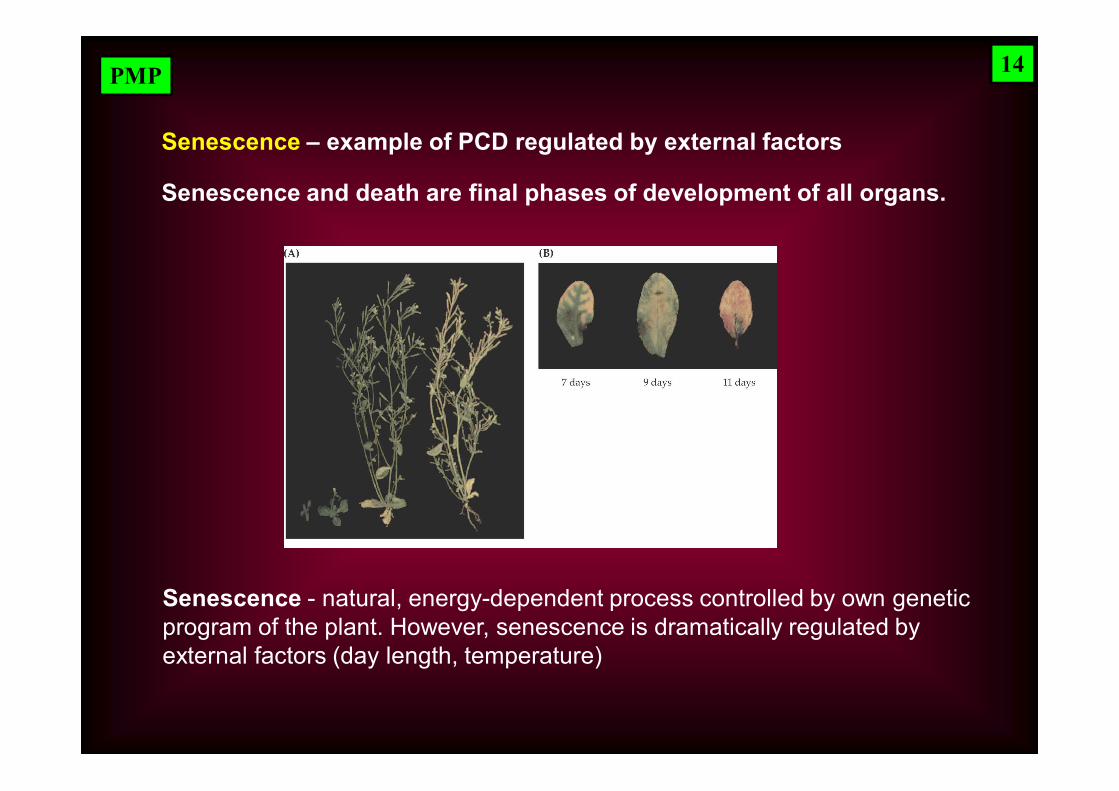
Senescence - natural, energy-dependent process controlled by own genetic program of the plant. However, senescence is dramatically regulated by external factors (day length, temperature)
Senescence and death are final phases of development of all organs.
14
Senescence – example of PCD regulated by external factors
PMP
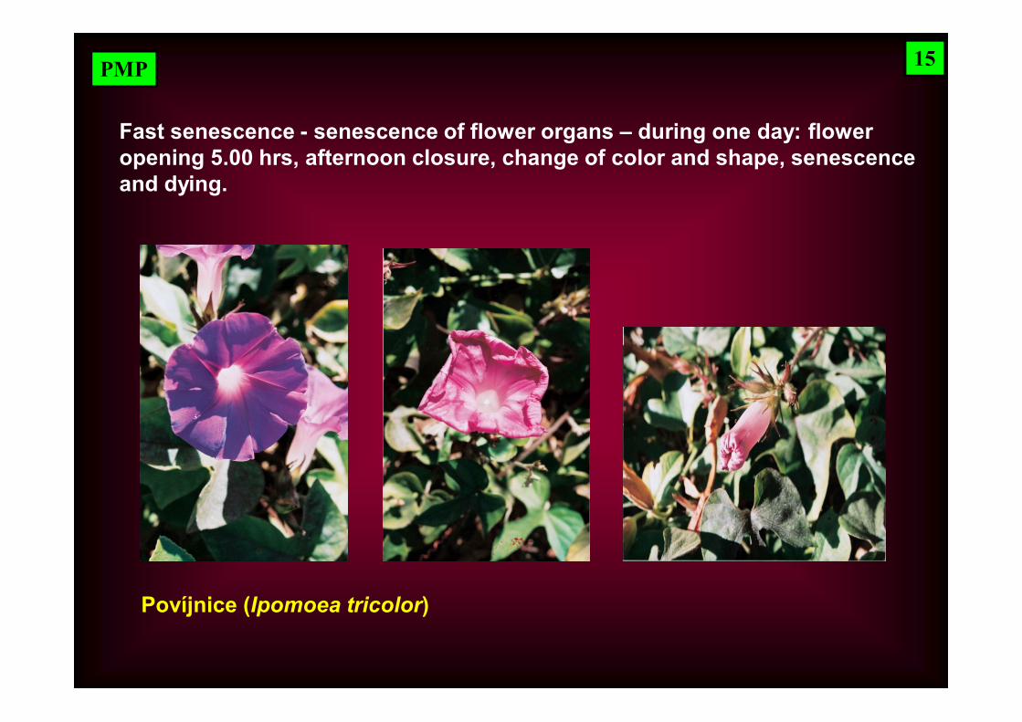
Fast senescence - senescence of flower organs – during one day: floweropening 5.00 hrs, afternoon closure, change of color and shape, senescenceand dying.
Povíjnice (Ipomoea tricolor)
15
PMP
Slow senescence - leaves (needle) of pine Pinus longaeva are replaced after 45 years
Mechanism of integration of senescence programs in development and life of organs or whole plants is not known.
Hypothesis „die now“ – signal „die now“ is continuously present – cells, tissues, organs respond to it in the moment when their individual program gives the command.
Signal „die now“ of particular cells can induce senescence in other cells.
16
PMP
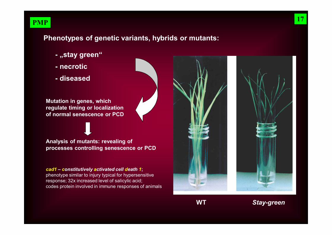
Phenotypes of genetic variants, hybrids or mutants:
- „stay green“ - necrotic - diseased
Mutation in genes, which regulate timing or localization of normal senescence or PCD
Analysis of mutants: revealing of processes controlling senescence or PCD
WT Stay-green
cad1 – constitutively activated cell death 1; phenotype similar to injury typical for hypersensitive response; 32x increased level of salicylic acid; codes protein involved in immune responses of animals
17
PMP
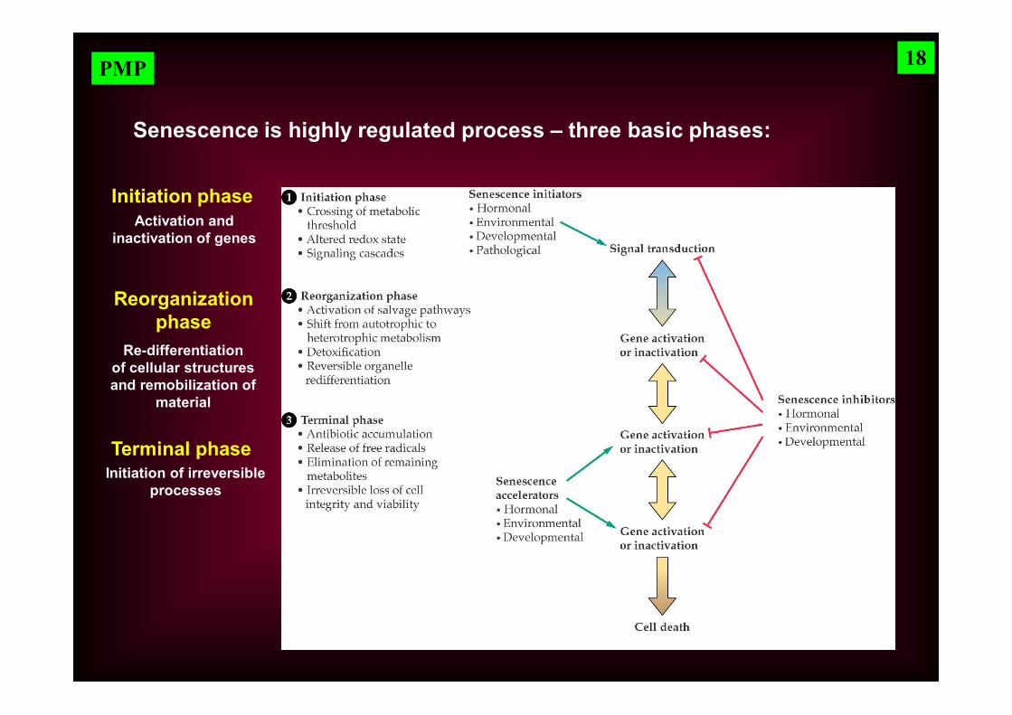
Initiation phase
Reorganizationphase
Terminal phase
Senescence is highly regulated process – three basic phases:
Re-differentiationof cellular structures and remobilization of
material
Initiation of irreversible processes
Activation and inactivation of genes
18
PMP
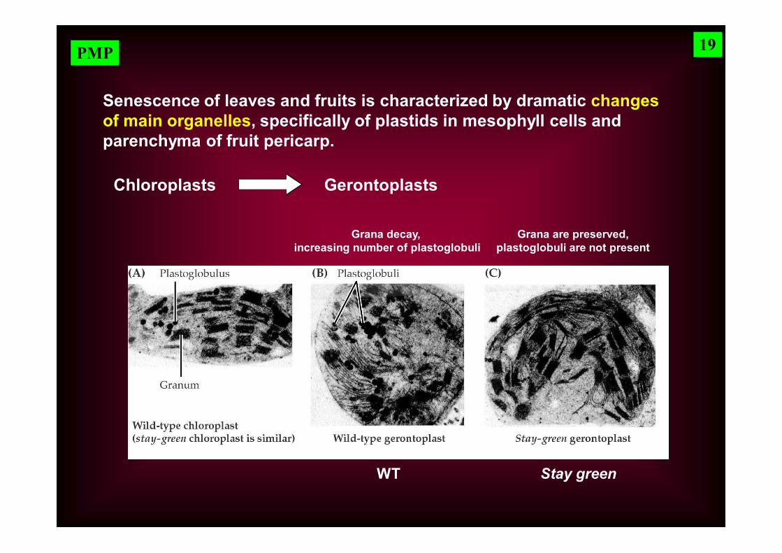
Senescence of leaves and fruits is characterized by dramatic changes of main organelles, specifically of plastids in mesophyll cells and parenchyma of fruit pericarp.
Chloroplasts Gerontoplasts
Grana decay, increasing number of plastoglobuli
Grana are preserved,plastoglobuli are not present
WT Stay green
19
PMP
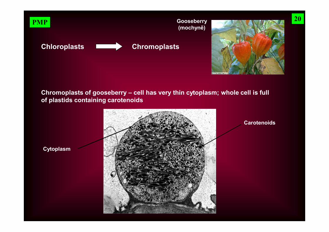
Chloroplasts Chromoplasts
Carotenoids
Chromoplasts of gooseberry – cell has very thin cytoplasm; whole cell is full of plastids containing carotenoids
Cytoplasm
20Gooseberry(mochyně)
PMP
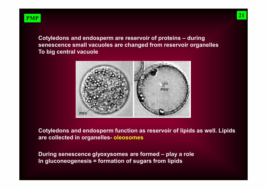
Cotyledons and endosperm function as reservoir of lipids as well. Lipids are collected in organelles- oleosomes
During senescence glyoxysomes are formed – play a role In gluconeogenesis = formation of sugars from lipids
Cotyledons and endosperm are reservoir of proteins – during senescence small vacuoles are changed from reservoir organelles To big central vacuole
21
PMP
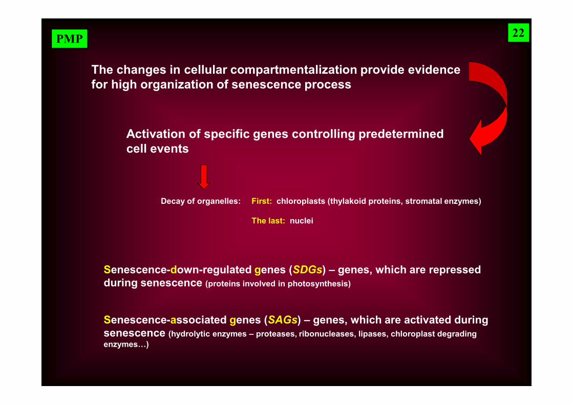
Activation of specific genes controlling predetermined cell events
Senescence-associated genes (SAGs) – genes, which are activated during senescence (hydrolytic enzymes – proteases, ribonucleases, lipases, chloroplast degradingenzymes…)
The changes in cellular compartmentalization provide evidence for high organization of senescence process
Senescence-down-regulated genes (SDGs) – genes, which are repressed during senescence (proteins involved in photosynthesis)
22
Decay of organelles: First: chloroplasts (thylakoid proteins, stromatal enzymes)
The last: nuclei
PMP
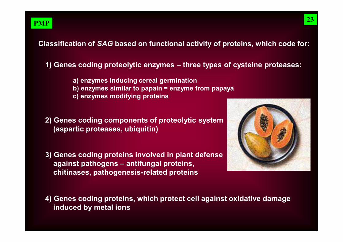
Classification of SAG based on functional activity of proteins, which code for:
1) Genes coding proteolytic enzymes – three types of cysteine proteases:
a) enzymes inducing cereal germinationb) enzymes similar to papain = enzyme from papayac) enzymes modifying proteins
2) Genes coding components of proteolytic system (aspartic proteases, ubiquitin)
3) Genes coding proteins involved in plant defenseagainst pathogens – antifungal proteins,chitinases, pathogenesis-related proteins
4) Genes coding proteins, which protect cell against oxidative damageinduced by metal ions
23
PMP
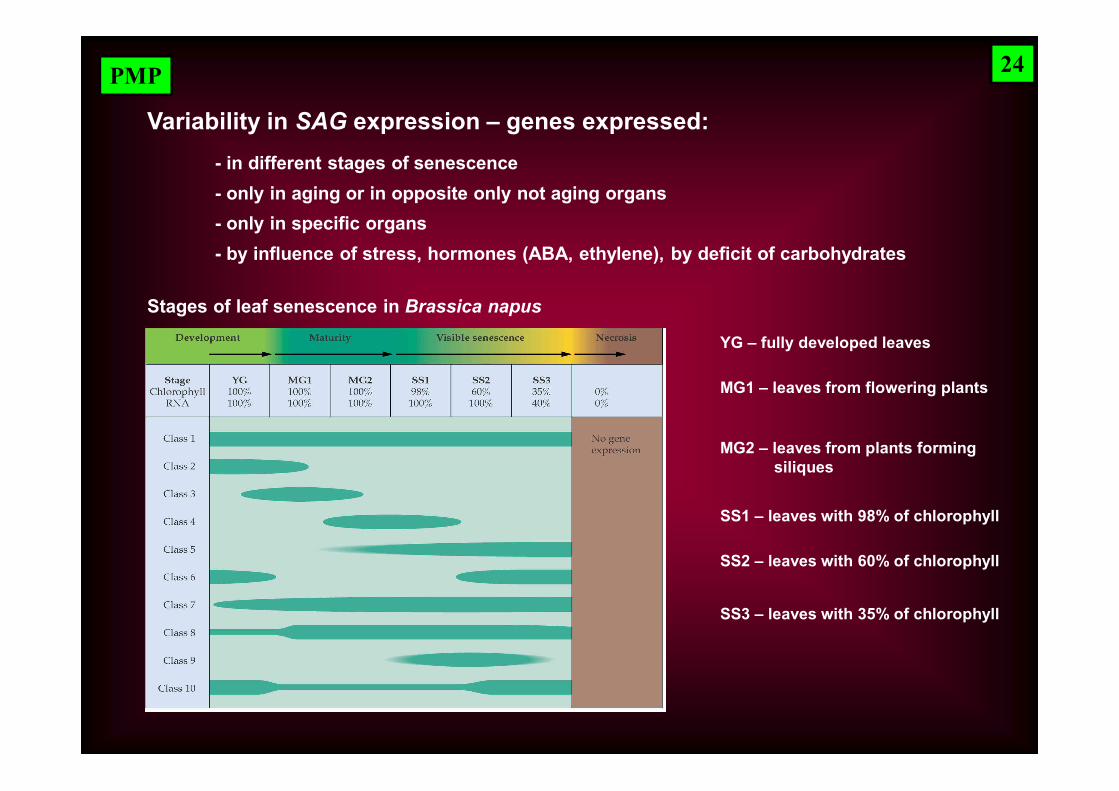
Variability in SAG expression – genes expressed:
Stages of leaf senescence in Brassica napus
YG – fully developed leaves
MG1 – leaves from flowering plants
MG2 – leaves from plants forming siliques
SS1 – leaves with 98% of chlorophyll
SS2 – leaves with 60% of chlorophyll
SS3 – leaves with 35% of chlorophyll
- in different stages of senescence- only in aging or in opposite only not aging organs - only in specific organs- by influence of stress, hormones (ABA, ethylene), by deficit of carbohydrates
24
PMP
Mutants in genes involved in senescence
- genes coding individual enzymes of metabolic pathways = genes functioning later in signaling pathway
- genes regulating initiation of whole senescence program = genes functioning at the beginning of senescence signaling pathways
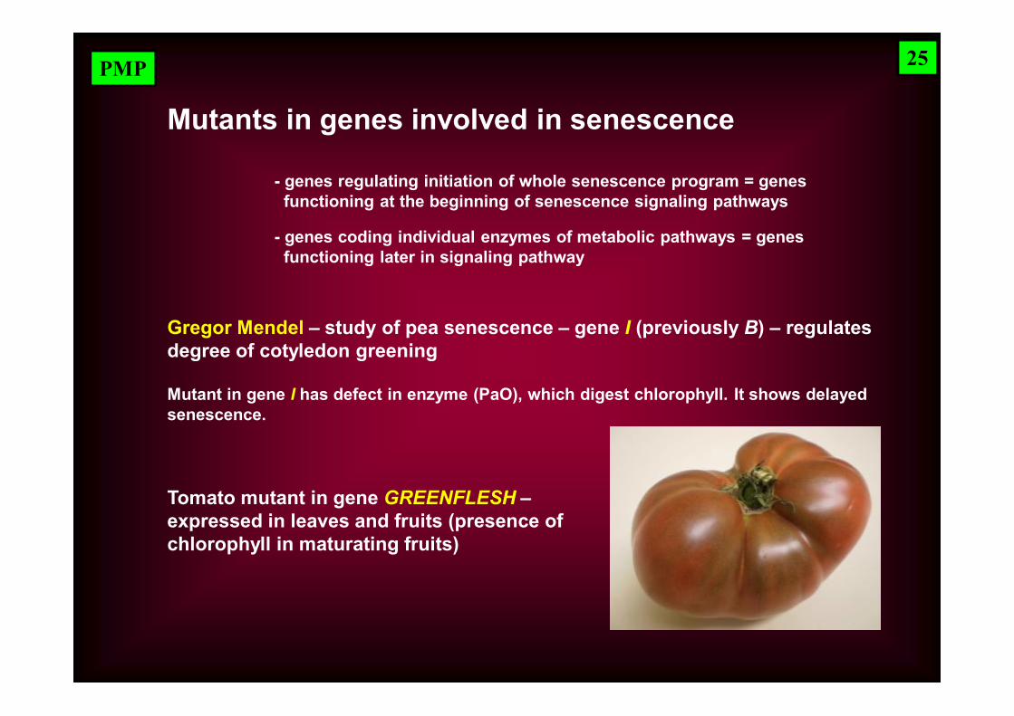
Gregor Mendel – study of pea senescence – gene I (previously B) – regulates degree of cotyledon greening
Mutant in gene I has defect in enzyme (PaO), which digest chlorophyll. It shows delayed senescence.
Tomato mutant in gene GREENFLESH –expressed in leaves and fruits (presence of chlorophyll in maturating fruits)
25
PMP
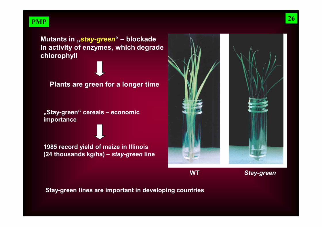
Mutants in „stay-green“ – blockadeIn activity of enzymes, which degradechlorophyll
WT Stay-green
„Stay-green“ cereals – economic importance
1985 record yield of maize in Illinois(24 thousands kg/ha) – stay-green line
Plants are green for a longer time
Stay-green lines are important in developing countries
26
PMP
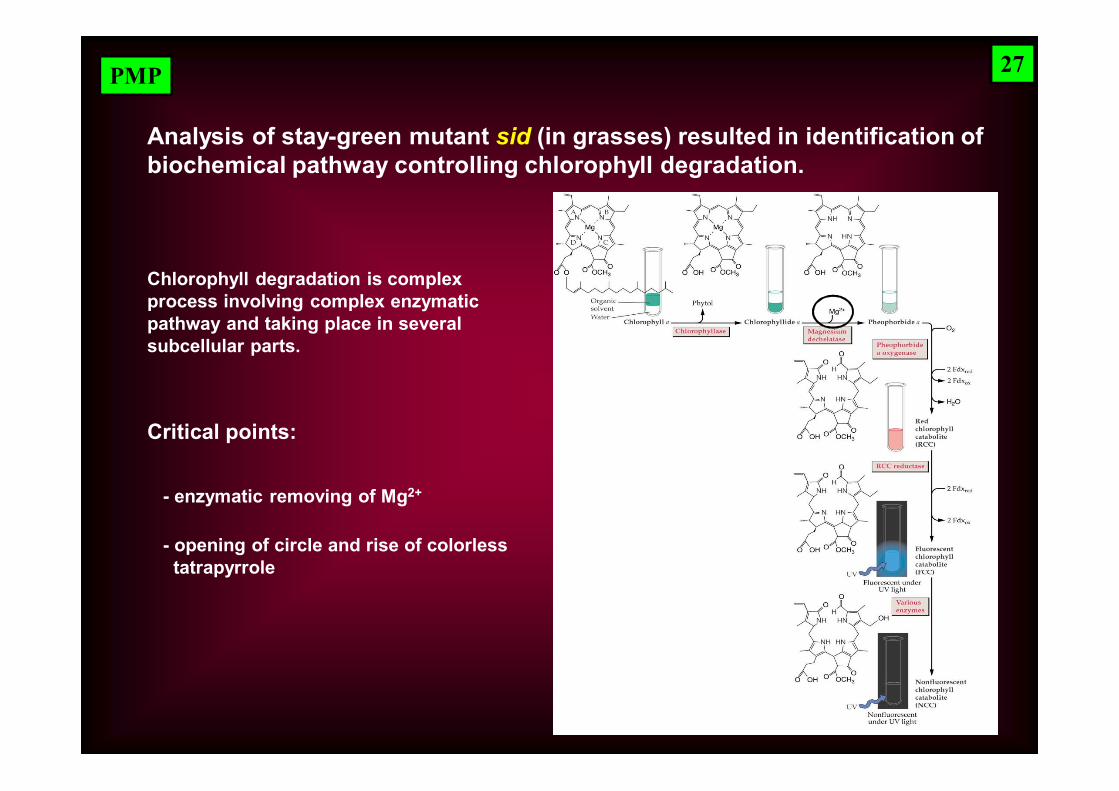
Analysis of stay-green mutant sid (in grasses) resulted in identification of biochemical pathway controlling chlorophyll degradation.
Chlorophyll degradation is complex process involving complex enzymatic pathway and taking place in several subcellular parts.
Critical points:
- enzymatic removing of Mg2+
- opening of circle and rise of colorlesstatrapyrrole
27
PMP
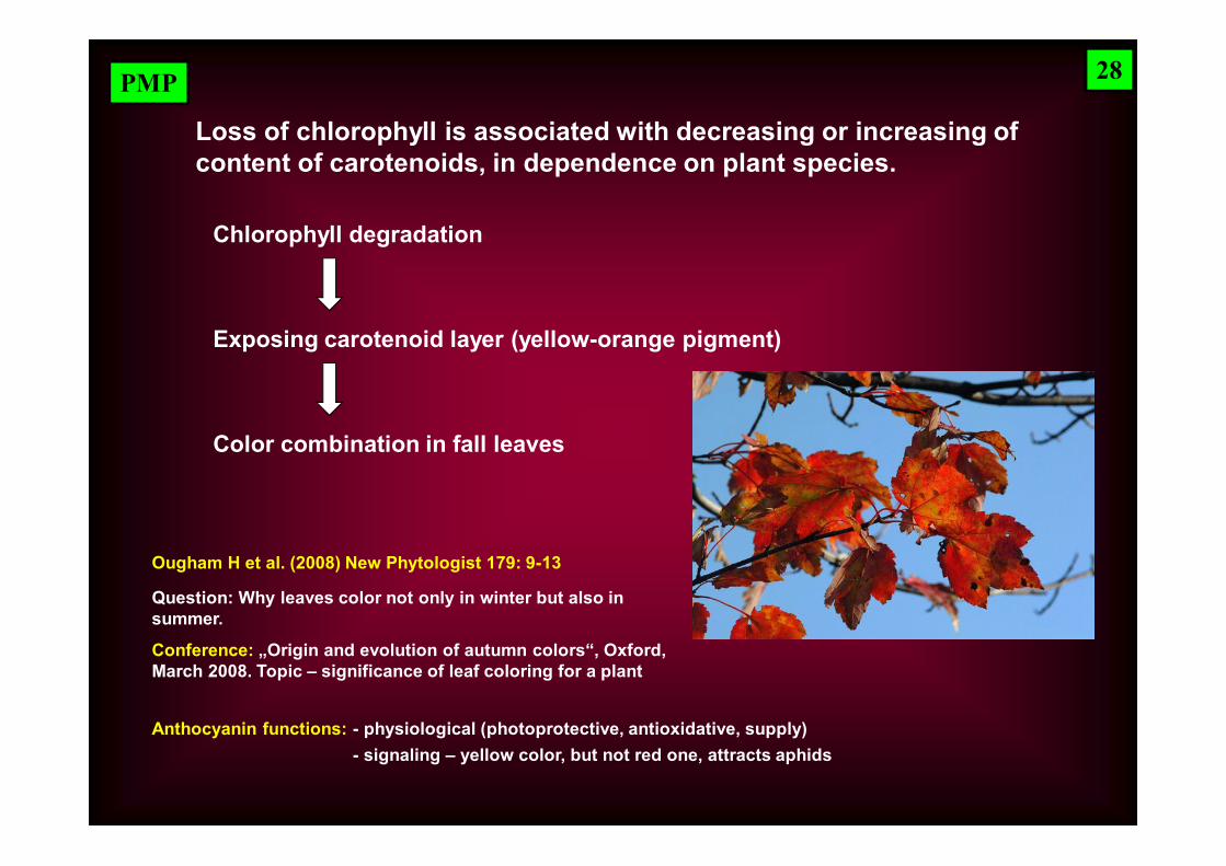
Loss of chlorophyll is associated with decreasing or increasing of content of carotenoids, in dependence on plant species.
Chlorophyll degradation
Exposing carotenoid layer (yellow-orange pigment)
Color combination in fall leaves
28
Ougham H et al. (2008) New Phytologist 179: 9-13
Question: Why leaves color not only in winter but also in summer.
Conference: „Origin and evolution of autumn colors“, Oxford,March 2008. Topic – significance of leaf coloring for a plant
Anthocyanin functions: - physiological (photoprotective, antioxidative, supply) - signaling – yellow color, but not red one, attracts aphids
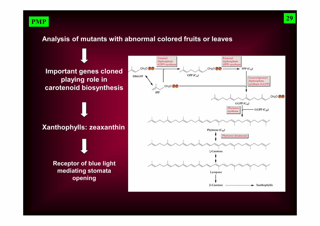
PMP
Important genes cloned playing role in
carotenoid biosynthesis
Analysis of mutants with abnormal colored fruits or leaves
Xanthophylls: zeaxanthin
Receptor of blue light mediating stomata
opening
29
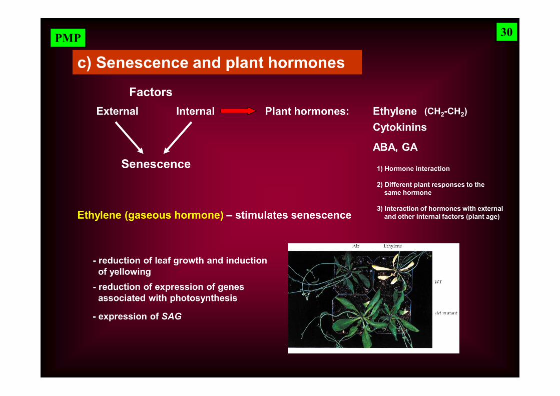
PMP
Senescence
c) Senescence and plant hormones
FactorsExternal Internal Plant hormones: Ethylene
Cytokinins
ABA, GA
1) Hormone interaction
2) Different plant responses to the same hormone
Ethylene (gaseous hormone) – stimulates senescence
- reduction of leaf growth and inductionof yellowing
- reduction of expression of genes associated with photosynthesis
- expression of SAG
(CH2-CH2)
3) Interaction of hormones with external and other internal factors (plant age)
30
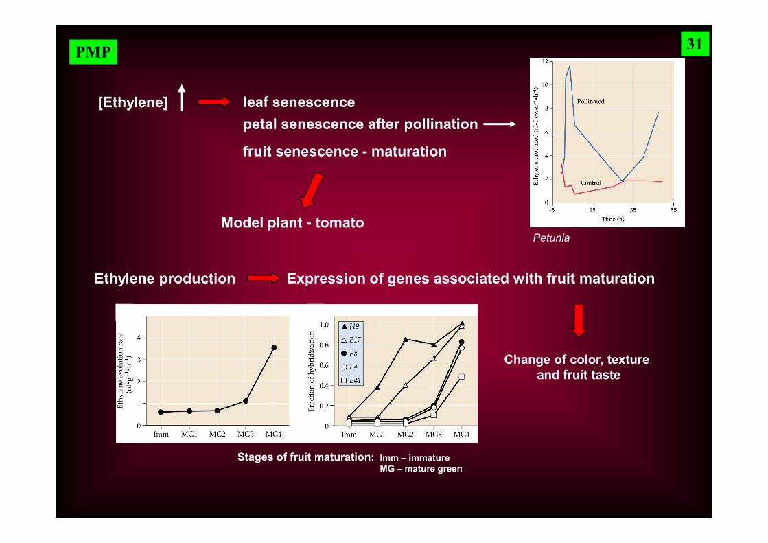
PMP
leaf senescence[Ethylene]petal senescence after pollination
PetuniaModel plant - tomato
fruit senescence - maturation
Ethylene production Expression of genes associated with fruit maturation
Change of color, texture and fruit taste
Stages of fruit maturation: Imm – immature MG – mature green
31
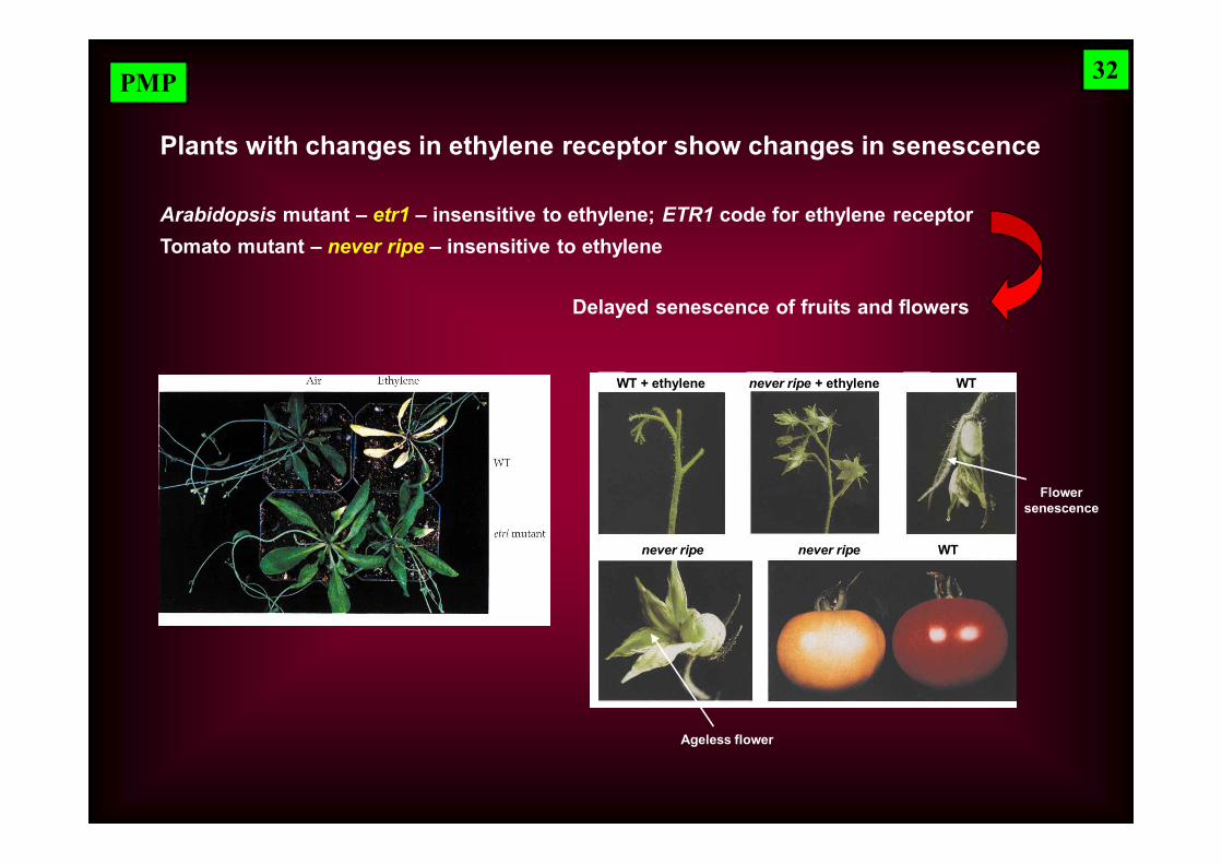
PMP
Plants with changes in ethylene receptor show changes in senescence
Arabidopsis mutant – etr1 – insensitive to ethylene; ETR1 code for ethylene receptorTomato mutant – never ripe – insensitive to ethylene
Delayed senescence of fruits and flowers
WT + ethylene WT
Flower senescence
never ripe
never ripe + ethylene
Ageless flower
never ripe WT
32
PMP
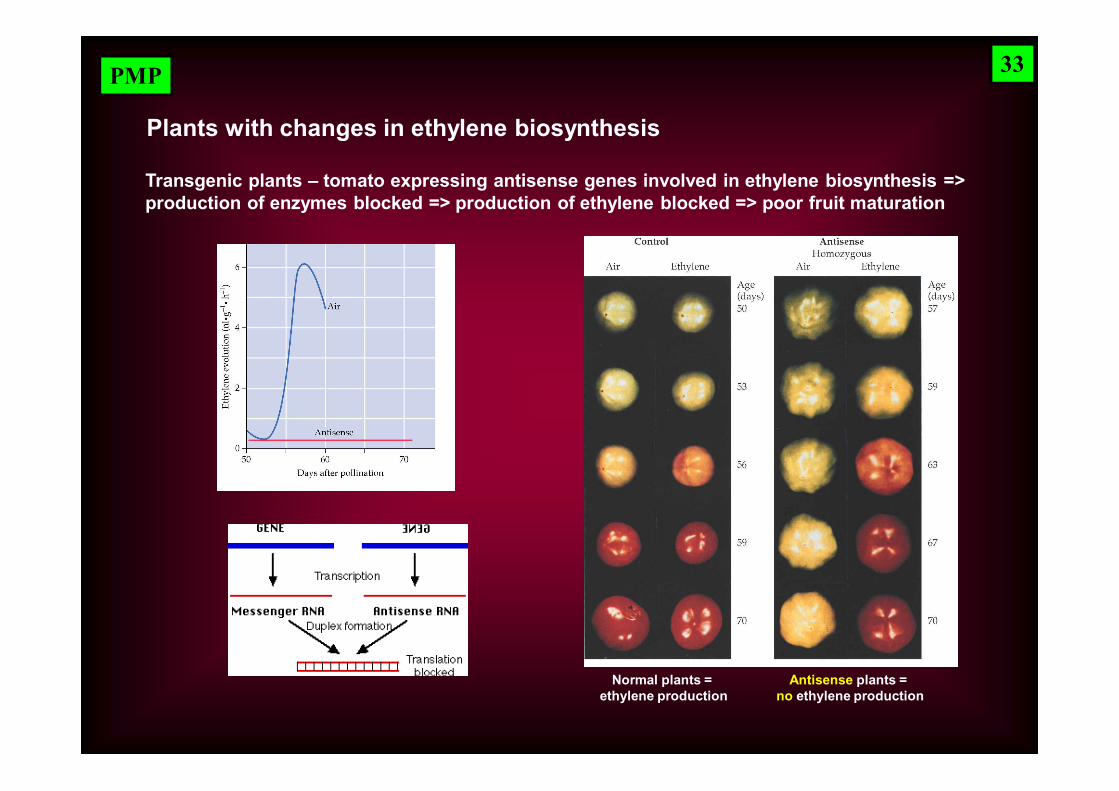
Plants with changes in ethylene biosynthesis
Transgenic plants – tomato expressing antisense genes involved in ethylene biosynthesis => production of enzymes blocked => production of ethylene blocked => poor fruit maturation
Normal plants = ethylene production
Antisense plants = no ethylene production
33
PMP
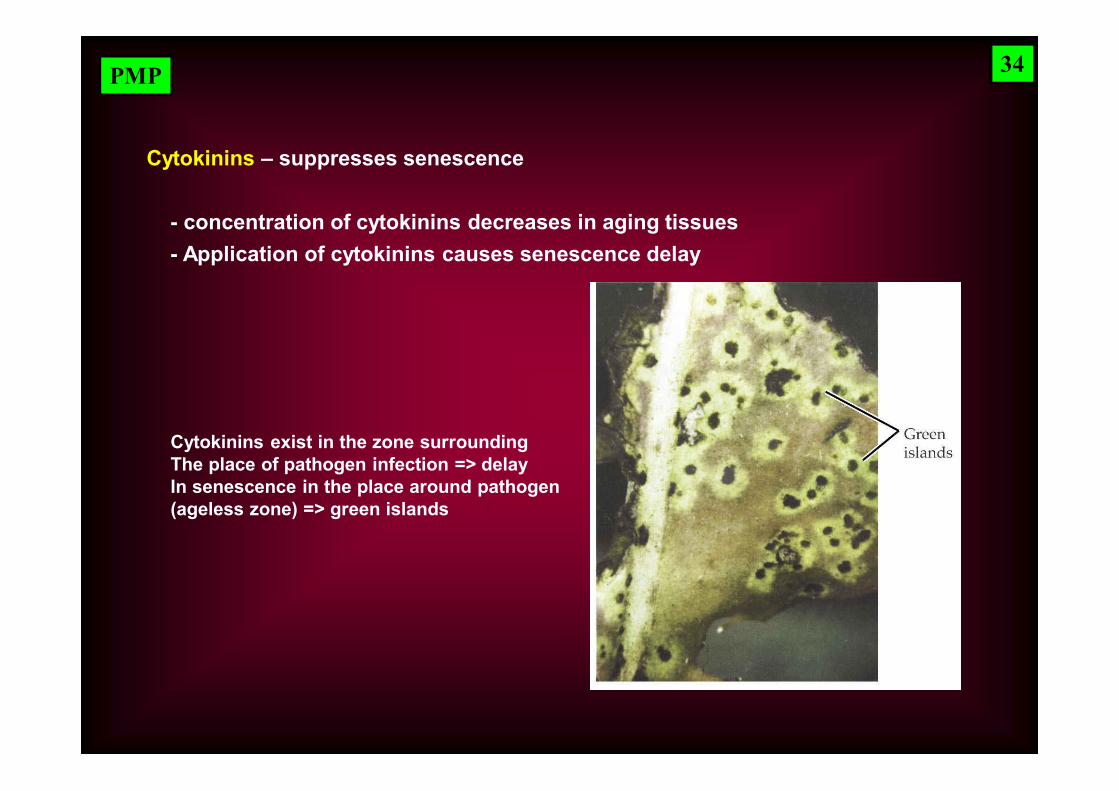
Cytokinins – suppresses senescence
- concentration of cytokinins decreases in aging tissues- Application of cytokinins causes senescence delay
Cytokinins exist in the zone surrounding The place of pathogen infection => delay In senescence in the place around pathogen (ageless zone) => green islands
34
PMP
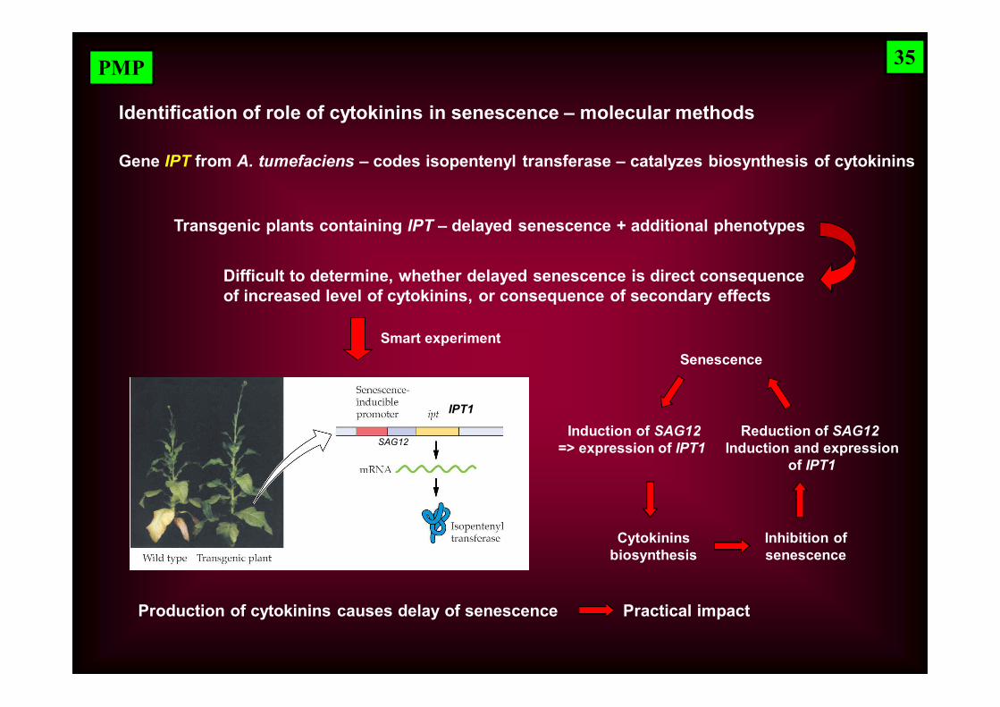
Identification of role of cytokinins in senescence – molecular methods
Gene IPT from A. tumefaciens – codes isopentenyl transferase – catalyzes biosynthesis of cytokinins
Transgenic plants containing IPT – delayed senescence + additional phenotypes
Difficult to determine, whether delayed senescence is direct consequence of increased level of cytokinins, or consequence of secondary effects
Senescence
Induction of SAG12=> expression of IPT1
Cytokininsbiosynthesis
Inhibition ofsenescence
Reduction of SAG12Induction and expression
of IPT1
Smart experiment
Production of cytokinins causes delay of senescence Practical impact
35
SAG12
IPT1
PMP
Possible explanation of antisenescence activity of cytokinins
1) Tissue with higher level of cytokinins play a role in metabolite economy of plants => accumulation of nutrition => senescence does not occur
2) Cytokinins can suppress expression of key genes: SAG
Duál aktivity of cytokinins:
- action on long distances (stimulation of differentiation and metabolite economy)
- local effect (in aging cells cytokinins suppress senescence)
36
3) Cytokinins activate transcription of chloroplast genes. Activity of cytokinins depend on light and age of cells and leaves
PMP
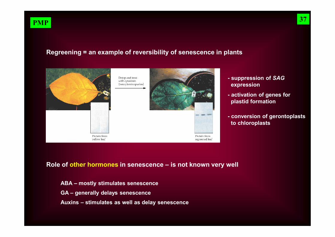
Regreening = an example of reversibility of senescence in plants
- suppression of SAG expression
- activation of genes for plastid formation
- conversion of gerontoplasts to chloroplasts
Role of other hormones in senescence – is not known very well
ABA – mostly stimulates senescenceGA – generally delays senescenceAuxins – stimulates as well as delay senescence
37
PMP
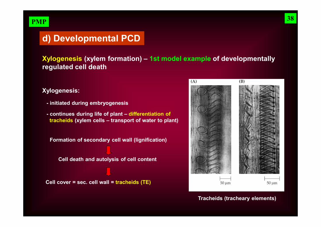
d) Developmental PCD
Xylogenesis (xylem formation) – 1st model example of developmentallyregulated cell death
Xylogenesis:
Tracheids (tracheary elements)
Formation of secondary cell wall (lignification)
Cell death and autolysis of cell content
- initiated during embryogenesis
- continues during life of plant – differentiation of tracheids (xylem cells – transport of water to plant)
Cell cover = sec. cell wall = tracheids (TE)
38
PMP
Development of TE from mesophyll cells of Zinnia
Culture in vitro
+ auxin
+ cytokinins
Induction by hormones
Dedifferentiation PrecursorTE cell
Immature TEwith sec. cell wall
(lignification)
Tonoplastrupture
TE = celltracheids
Degradation
39
Zinnia (Ostálka)

PMP
3 phases of tracheid differentiation tracheid ~ 4 days
Expression of genes involved in responses to damage(PI - Protease inhibitors)
(RP – Ribosomal proteins)(EF – Elongation factors)
Expression of TE differentiation-related genes
(TED, unknown function)
Expression of genes participating in synthesis of cytoskeleton components
(TUB - tubulin).Activation of genes coding
proteins of cell wall(arabinogalactan-like, extensin-like)
In vitro process of differentiation of TE in cell population happens synchronously
Possibility of biochem. and mol. analyses
40
PMP
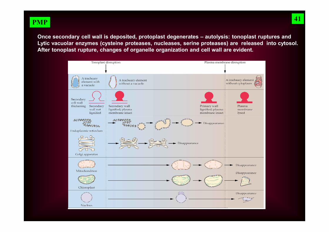
Once secondary cell wall is deposited, protoplast degenerates – autolysis: tonoplast ruptures andLytic vacuolar enzymes (cysteine proteases, nucleases, serine proteases) are released into cytosol.After tonoplast rupture, changes of organelle organization and cell wall are evident.
41
PMP
Regulation of PCD during tracheid formation
Auxins
Recent results – involvement of NO (nitric oxide)
NO – reactive water and lipid soluble gas; involved in many biological processes:– stomata closure
2002 – cytokinins induce formation of NO in Arabidopsis, tobacco, parsley, etc.
Cytokinins – role in induction of PCD in plants and animals – 2002; elements of signaling pathway are known very poorly
– seed germination
– root development
– expression of defense genes
Cytokinins induce synthesis of NO in xylem cells => inhibition of respiration =>=> PCD (tracheid formation)
42
PMP
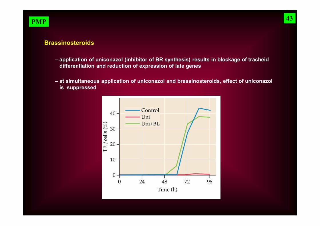
Brassinosteroids
– at simultaneous application of uniconazol and brassinosteroids, effect of uniconazol is suppressed
– application of uniconazol (inhibitor of BR synthesis) results in blockage of tracheid differentiation and reduction of expression of late genes
43

PMP
PCD of endosperm and aleurone cells -- 2nd model example of developmentally
regulated cell death
2 types of cells – 2 different ways of PCD
– aleurone cells– starch endosperm
Starch endosperm – dead cells, but their content is not degraded – cell is mummified; at germination, endosperm is degraded by enzymes released from aleurone
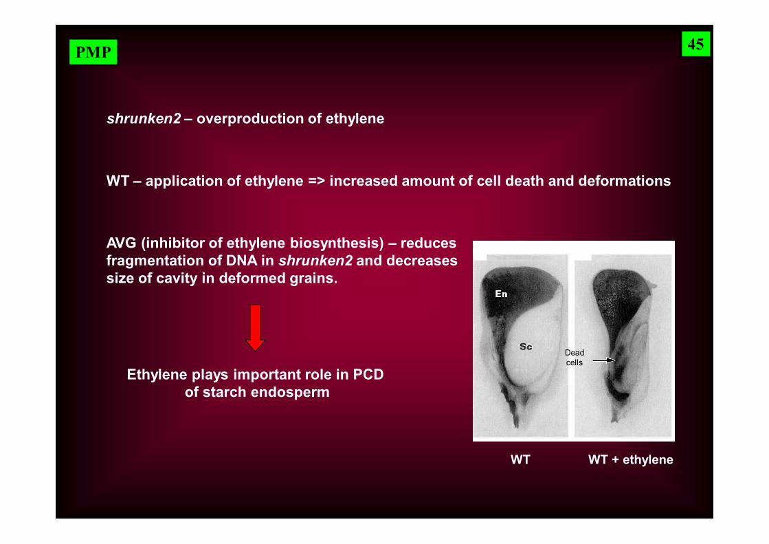
shrunken2 – maize mutant, precocious death of starch endospermand cell degradation; during PCD nuclear DNA is cut to big fragments; differentially from WT, shrunken2 cells autolyseand endosperm shrunkens – rise of cavities (*)
WT shrunken2
28
32
40
Days after pollination
44
PMP
shrunken2 – overproduction of ethylene
WT – application of ethylene => increased amount of cell death and deformations
AVG (inhibitor of ethylene biosynthesis) – reducesfragmentation of DNA in shrunken2 and decreases size of cavity in deformed grains.
Ethylene plays important role in PCD of starch endosperm
WT WT + ethylene
Deadcells
45

PMP
Aleurone cells – stay alive until germination and till all reserves of endosperm are mobilized
End of germination changes in aleurone: - vacuolization- death- protoplast disintegration
Plant hormones ABA and GA regulate PCD of aleurone:
GA – stimulates beginning of PCD – causescell death 8 days after application
ABA – delays PCD – causes delay ofcell death about 6 months
46

PMP
e) PCD and plant responses to stress
Hypoxia – starts at soil submersion
- aerenchym formation – fast process, consisting in removing of cortical cellsincluding cell wall and formation of spaces (channels) for oxygen transport
HypoxiaActivity of ACC synthase
High level of cellulases
Aerenchymy
47

PMP
Cells undergoing hypoxia show higher level of Ca2+ in cytosol.
Changes in Ca2+ concentration is fast
Role of cytosolic Ca2+ in hypoxia
[Ca] i – cytosolic Ca2+
On – oxidization of medium switched on
48
Off – oxidization of medium switched off –hypoxia starts
Ca1 – 1 mM external Ca2+
Ca10 – 10 mM external Ca2+ Changes in levels of cytosolic Ca2+ in cultured cells of maize
PMP
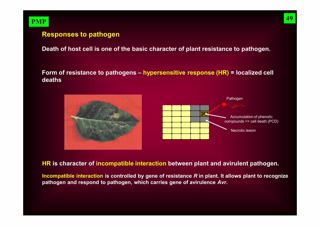
Responses to pathogen
Death of host cell is one of the basic character of plant resistance to pathogen.
Form of resistance to pathogens – hypersensitive response (HR) = localized cell deaths
HR is character of incompatible interaction between plant and avirulent pathogen.
Incompatible interaction is controlled by gene of resistance R in plant. It allows plant to recognizepathogen and respond to pathogen, which carries gene of avirulence Avr.
Pathogen
Accumulation of phenoliccompounds => cell death (PCD)
Necrotic lesion
49
PMP
In the absence of one of gene R or Avr compatible interaction occurs – plant is notable to recognize pathogen and disease break out.
Cell death may be symptoms of disease during compatible interaction. This form of cell death is not programmed and it is a consequence of killing the hostby pathogen (toxins secreted by pathogen).
Basic question:
Is the cell death during HR suicide (geneticall programmed death) or murder (death as a consequence of toxicity of products produced by pathogen)?
Recent research leans to hypothesis of suicide.
50
PMP
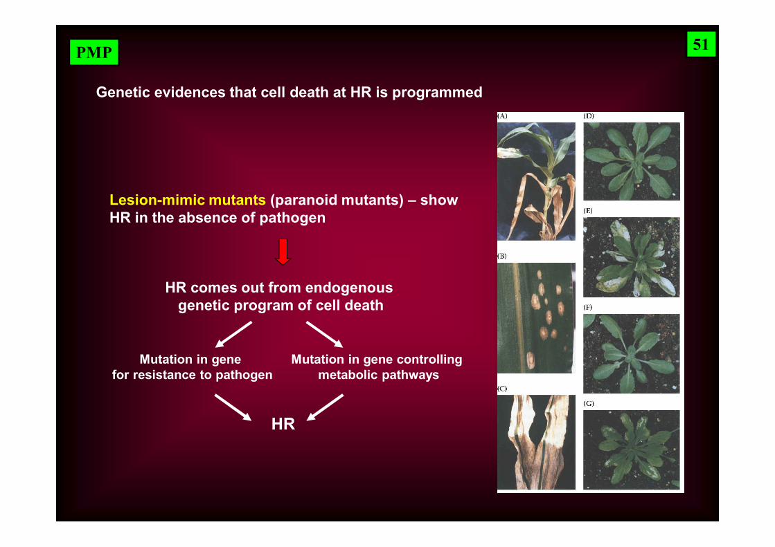
Genetic evidences that cell death at HR is programmed
Lesion-mimic mutants (paranoid mutants) – showHR in the absence of pathogen
HR comes out from endogenous genetic program of cell death
Mutation in gene for resistance to pathogen
Mutation in gene controlling metabolic pathways
HR
51
PMP
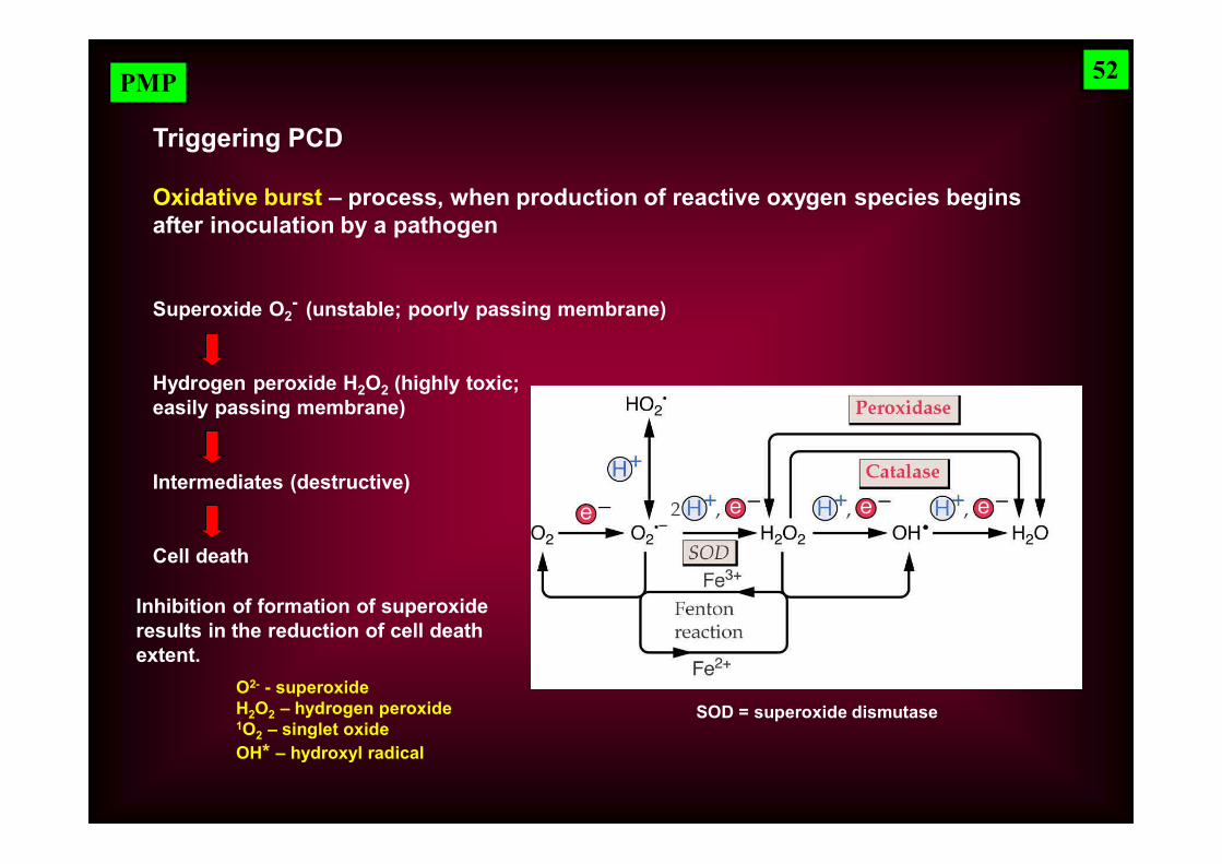
Triggering PCD
Oxidative burst – process, when production of reactive oxygen species begins after inoculation by a pathogen
Superoxide O2- (unstable; poorly passing membrane)
Hydrogen peroxide H2O2 (highly toxic; easily passing membrane)
Intermediates (destructive)
Cell death
Inhibition of formation of superoxide results in the reduction of cell death extent.
52
SOD = superoxide dismutaseO2- - superoxideH2O2 – hydrogen peroxide1O2 – singlet oxideOH* – hydroxyl radical
PMP
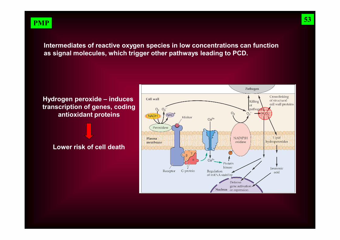
Intermediates of reactive oxygen species in low concentrations can function as signal molecules, which trigger other pathways leading to PCD.
Hydrogen peroxide – induces transcription of genes, coding
antioxidant proteins
Lower risk of cell death
53
PMP
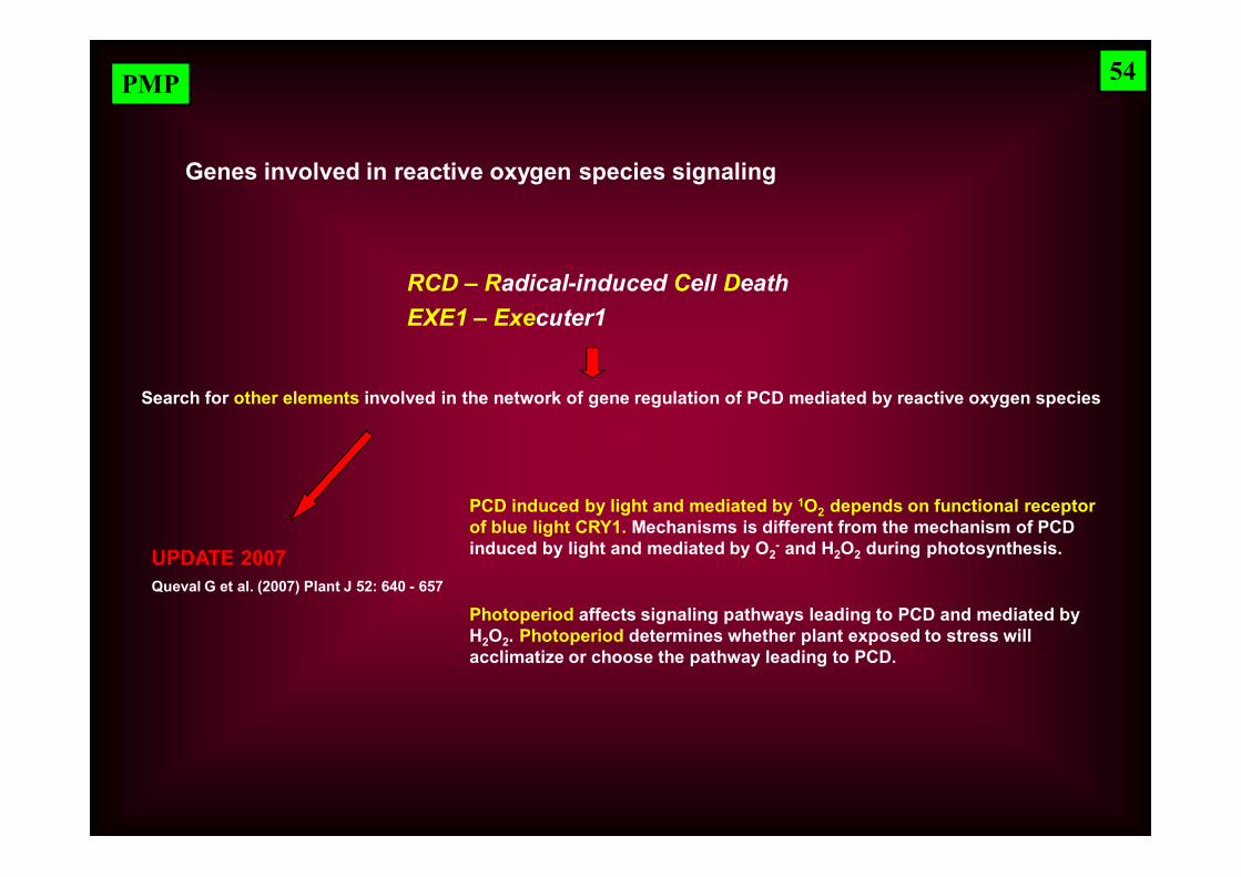
Genes involved in reactive oxygen species signaling
RCD – Radical-induced Cell DeathEXE1 – Executer1
Search for other elements involved in the network of gene regulation of PCD mediated by reactive oxygen species
54
PCD induced by light and mediated by 1O2 depends on functional receptor of blue light CRY1. Mechanisms is different from the mechanism of PCD induced by light and mediated by O2
- and H2O2 during photosynthesis.UPDATE 2007Queval G et al. (2007) Plant J 52: 640 - 657
Photoperiod affects signaling pathways leading to PCD and mediated byH2O2. Photoperiod determines whether plant exposed to stress will acclimatize or choose the pathway leading to PCD.

PMP 55NecrosisPCD
Chloroplasts
Mitochondria
Peroxisomes
Oxidases-peroxidases
O2-
H2O2
1O2
OH-Post
-tran
slat
ion
chan
ges
NO
Ethylen,JA, SA
EXE1
RCD1
Antioxidants
Genes of PCD
Proteases
Nucleases
MAPK
Gene expression Sensors - targets Production
Pathogen
Abiotic stress
Developmentalstimuli
Stimuli
MAPK – mitogen-activated protein kinase