Unit V: Movement Muscle Contraction - Part II Chapter 9 – pg 293-307.
Upload
mariama-malangCategory
view
116download
0
Skeletal Muscle II
SR2002
Dr. Arimantas Lionikas
October 24, 2013

Muscle IIPlan
• Muscle Innervation• Neuromuscular transmission• Contractile properties• Electrical stimulation• Muscle fibre types
• Reading list: 1. Enoka R. Neuromechanics of human movement. 2002. Publishers: Human Kinetics, p. 257-278 2. MacIntosh, B.R., Gardiner, P.F. McComas, A.J. Skeletal muscle, 2nd edition. 2006. Publishers: Human Kinetics, p. 32-39, 126-150.3. McArdle W.D. et al. Exercise Physiology: energy, nutrition, human performance. 2001. Publisher: Lippincott Williams & Wilkins, p. p. 358-382.
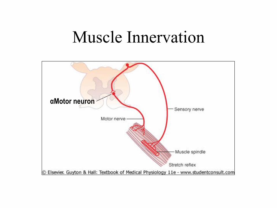
Muscle Innervation
αMotor neuron

• One motor nerve innervates a group of muscle fibres
• Motor nerve divides into terminals that form neuromuscular junctions on muscle fibres
• Neuromuscular junctions are located in the middle of the fibres
Muscle Innervation (cont)
• Microscopic analysis: Three muscle fibres innervated by branches of a motor nerve
• Diphtheria toxin damages myelin sheaths of nerve fibres (shown by arrows)

Neuromuscular transmission
• Nerve fibres (axons) conduct action potentials (APs) at a fast rate (~40-80 m/s)
• Terminals of motor nerves form neuromuscular junctions (also called motor end plates)
• The key function of the neuromuscular junctions is transmission of activation from nerve to muscle fibres

Neuromuscular transmission• Arrival of the AP at the nerve
terminal triggers release of acetylcholine (Ach) into the synaptic cleft (space between nerve and muscle fibre)
• Ach is made from Ac-CoA from mitochondria and choline in nerve terminals
• Released Ach binds to receptors on the muscle fibre and triggers influx of Na+ followed by generation of action potential (AP) in muscle fibres
• Ach acts for a short time since it is degraded by Ach esterase enzyme
• Choline is taken up by nerve terminals for re-synthesis of Ach

Szule JA, Harlow ML, Jung JH, De-Miguel FF, et al. (2012)
Synaptic vesicles of axon terminals
Synaptic vesicles
The active zone
The process leading to the release of Ach involves docking of the synaptic vesicles to the active zone , fusion with the membrane and release of the mediator.

Neuromuscular transmissionClinical relevance
• Myasthenia gravis due to low levels of acetylcholine (Ach):
• Eyelids are dropped due to muscle weakness
• Injection of a drug which blocks Ach esterase helps to regain muscle strength: eyelids open
Before treatment After treatment • Myasthenia gravis
(autoimmune disease) can be due to low levels of:
• (1) acetylcholine (Ach) • (2) Ach receptor
• Drugs blocking neuromuscular transmission are muscle relaxants used during operations (ex.: suxamethonium)
• Tubocurorine is found in some plants. Indians used it in hunting (poisoned arrows)

• After depolarization of postsynaptic membranes, action potential (AP) is generated
• AP spreads to both ends of muscle fibresAction potential (AP)
Action potential (AP)

Contractile propertiesHuman muscles
• Contractile properties can be investigated in humans muscles
• Electrical stimulation is applied over surface electrodes
• Torque in isometric contraction is usually recorded
• The set up is for studies of the quadriceps muscle
Electrodes
Dynamometerstrap
Force
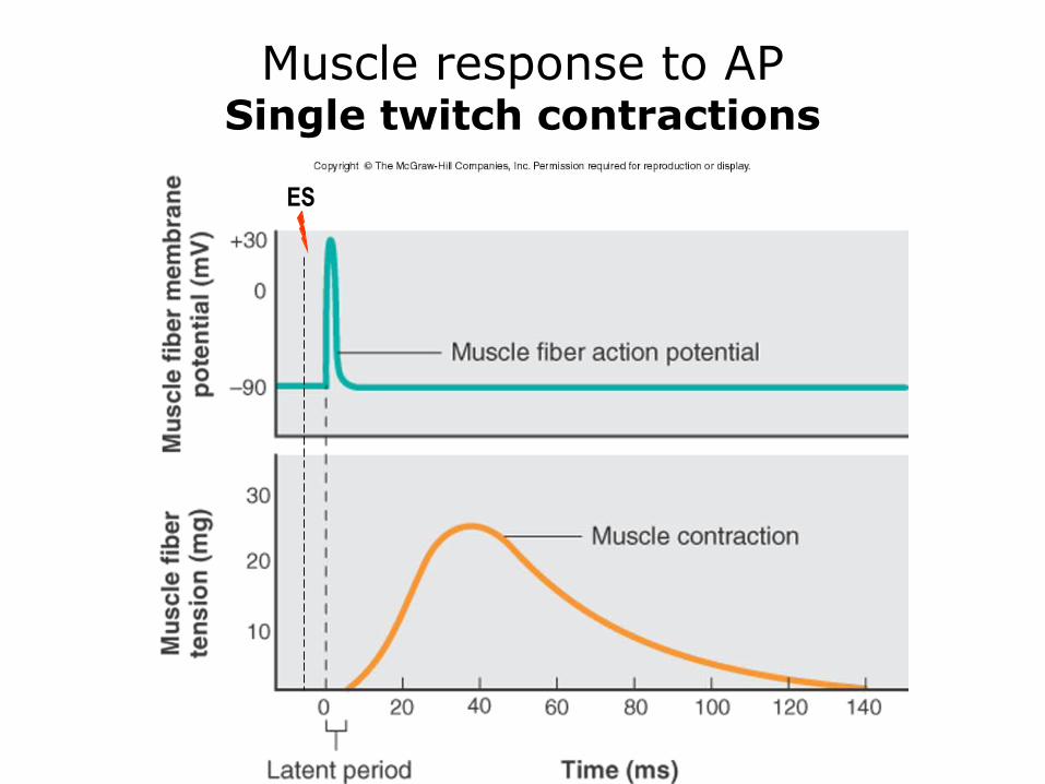
Muscle response to APSingle twitch contractions
ES
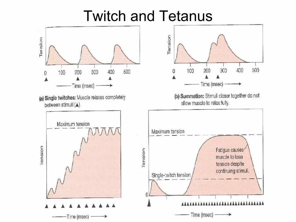
Twitch and Tetanus

Contractile propertiesHuman muscles
• Muscle force depends on the frequency of stimulation
• Unfused contractions are associated with fluctuation in force during a contraction
• Muscle fibres are activated at 5-100 Hz frequencies in voluntary contractions
• Electrical stimulation can be used for training of muscles
Westerblad et al. 2002
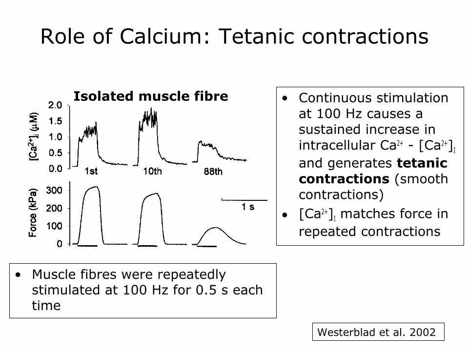
Role of Calcium: Tetanic contractions
• Continuous stimulation at 100 Hz causes a sustained increase in intracellular Ca2+ - [Ca2+]I and generates tetanic contractions (smooth contractions)
• [Ca2+]I matches force in repeated contractions
Isolated muscle fibre
• Muscle fibres were repeatedly stimulated at 100 Hz for 0.5 s each time
Westerblad et al. 2002

Applications of Electrical Stimulation• Neuromuscular electrical stimulation (NMES) is used for
functional and therapeutic applications in subjects with spinal cord injury or stroke.
A transcutaneous multichannel neuroprosthesis system allows persons with paraplegia unbraced ambulation for home andshort community distances. (Parastep I System User, courtesy of Sigmedic Inc., Fairborn, OH.)SHEFFLER & CHAE 2007

Muscle fibre types
• Skeletal muscle is not a homogeneous tissue.
• Muscle fibres that muscle consists of can be subdivided in two main types: type I and type II (the latter is further divided into IIA & IIX).
• Type of myosin heavy chains expressed in the fibres is the primary determinant of the fibre type and their contractile properties.
Mouse soleus. Acid preincubation pH 4.47
I IIA
Eriksson et al 2005
50 µm

Properties of fibre types
Characteristic Type I Type IIA Type IIX(B)
Morphology Fibre ∅ Small* Intermediate Large
Capillaries/mm2 High Intermediate Low
Mitochondrial vol High Intermediate Low
Histo- & Bio-chemistry
Myosin ATPase Low High High
Glycolytic capacity Low High High
Oxidative capacity High High/Medium Low
Contractility Contraction Slow Fast Fast
Relaxation Slow Fast Fast
Fatigue resistance High Moderate/High Low
Adaptation# Increase in fibre area Low Moderate Moderate
* Not always; depends on the muscle. Size between type I and II is about the same in soleus# Adaptation to several weeks of strength training.

Fibre type distribution in various muscles• Proportion of fibre types differ among muscles.• Type I fibres dominate in postural muscles (e.g.
soleus) and muscles involved in fine movements (e.g. adductor pollicis).
• Type II fibres dominate muscles involved in movements requiring quick and / or powerful contractions (e.g. orbicularis oculi, biceps brachii).
Harridge, Bottinelli et al. 1996
120 ms
68 ms
81 ms
Muscle % type I
Orbicularis oculi 15
Tricep brachii 37
Quadriceps femoris 52
Adductor pollicis 80
Soleus 80

Variation in fibre types among individuals
• Even though on average vastus lateralis muscle consists of ~50 of type I fibres, there is a broad range of variation between individuals (from ~20% to >80% of type I fibres).
• Important consequences of variation of fibre type composition:– Prevalence of certain fibre type is favourable for some athletic
activities (e.g. type I – endurance events, type II – speed and power events).
– Apparently increasing proportion of type II fibres is associated with prevalence to weight gain and cardiovascular risk.

Factors affecting proportion of fibre types• Genetic factors play important role in proportion of fibre types.• Moderate changes may occur as adaptation to physical activity or
lack of it.• Drastic change in degree of activity leads to marked shift in
proportion of fibre types.
Type IIType I
Endurance TrainingImmobilization
Spinal Cord Injury
Proportion of Fibre Types

Muscle IISummary
• One motor nerve innervates many muscle fibres
• Acetylcholine plays a key role in transmission of excitation from nerve to muscle
• For muscle contraction to occur release of calcium from sarcoplasmic reticulum is required
• Proportion of type I and type II fibres are important determinants of contractile properties.