9 Mehra and Jain JDT
14
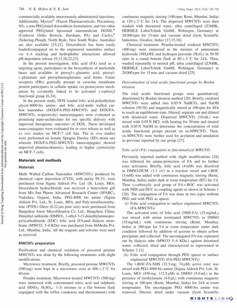
2013 http://informahealthcare.com/drt ISSN: 1061-186X (print), 1029-2330 (electronic) J Drug Target, 2013; 21(8): 745–758 ! 2013 Informa UK Ltd. DOI: 10.3109/1061186X.2013.813028 ORIGINAL ARTICLE Development, characterization and cancer targeting potential of surface engineered carbon nanotubes Neelesh Kumar Mehra and N. K. Jain Pharmaceutics Research Laboratory, Department of Pharmaceutical Sciences, Dr. H. S. Gour Central University, Sagar (M.P.), India Abstract The aim of the present study was to assess the in vitro and in vivo potential of doxorubicin- loaded, folic acid appended engineered multi-walled carbon nanotubes (DOX/FA-PEG- MWCNTs) for efficient tumor targeting. The loading efficiency was determined to be 92.0 0.92 (DOX/FA-PEG-MWCNTs) in phosphate buffer solution (pH 7.4) ascribed to p–p stacking interaction. The developed nanoconjugates were evaluated for in vitro DOX release, erythrocytes toxicity, ex vivo cytotoxicity and cell uptake studies on MCF-7 (breast cancer cell line). The DOX/FA-PEG-MWCNTs nanoconjugate affords higher efficacy in tumor growth suppression due to its stealth nature and most preferentially taken up by the cultured MCF-7 through caveolae-mediated endocytosis as compared to free DOX. The in vivo studies were performed to determine the pharmacokinetics, biodistribution and antitumor efficacy on tumor bearing female Sprague Dawley rats and improved pharmacokinetics confirm the function of FA-PEG conjugated CNTs. The median survival time for tumor bearing rats treated with DOX/FA-PEG-MWCNTs (30 d) was extended very significantly as compared to free DOX (p50.001). The results concluded that developed water-soluble nano-conjugates might emerge as ‘‘safe and effective’’ nano-medicine in cancer treatment by minimizing the side effects with and Generally Regarded as Safe prominence. Keywords Anti-tumor activity, carbon nanotubes, doxorubicin, drug targeting, folic acid, MCF-7 cells, pharmacokinetic History Received 12 March 2013 Revised 5 June 2013 Accepted 5 June 2013 Published online 3 July 2013 Introduction Cancer is amongst the top three killers in modern society, next to heart and cerebrovascular diseases, tuberculosis and acquired immune deficiency syndrome (AIDS). Breast cancer is the most common and second leading cause of cancer deaths today in women worldwide, both in the developed and developing countries. Despite the high incidence rates, in Western countries, 89% of women diagnosed with breast cancer are still alive 5 year after diagnosis, which is due to detection and treatment. In 2010, nearly 1.5 million people were told ‘‘you have breast cancer’’. It has been continuously rising due to the increase in life expectancy, urbanization and adoption of western life styles. The Canadian Cancer Society in 2011 reported that an estimated 23 400 women were diagnosed and 5100 died with breast cancer, moreover approximately 190 men were also diagnosed and 55 died with breast cancer [1] (World Health Organization fact sheet. Available from the URL http://www.who.int/cancer/en/; http://www.worldwidebreast cancer.com/learn/breast-cancer-statistics-worldwide/ Accessed date 28th April 2013). Although the significant progress has been made in the development of new, safe nanomedicines for cancer therapy, yet we still strongly need a complete and reliable cure of cancer. In the current scenario, carbon nanotubes (CNTs) have attracted escalating attention and are under investigation with surface modification with targeting ligand to offer a sustained/controlled level of drug and to accomplish cellular target with enhanced specificity. CNTs are unique, three dimensional sp 2 hybridized carbon nanomaterial have attracted tremendous attention as valuable, promising, alternative ‘‘safe and effective’’ nano-architecture to biomed- ical applications due to their unique physicochemical proper- ties such as biocompatibility, non-immunogenicity, high loading efficiency, high aspect ratio, structural flexibility, non-cytotoxic and non-biodegradable nature [2–10]. The pristine CNTs (first generation) are not suitable for drug delivery due to their hydrophobic nature, impurities and toxic in nature. These major hurdles have been easily sorted out by surface engineering with either covalent or non-covalent modification strategies, thus surface engineered CNTs have been designed and tested for targeted delivery by conjugating targeting moieties and have proven non-cytotoxic to human cells [2,5,11–18]. Doxorubicin (DOX) is a potent anthracycline cytostatic antibiotic used in the treatment of several mali- gnancies by intercalating with the DNA or DNA topoisom- erase II [15,19,20]. Adriamycin Õ and Rubex Õ are two Address for correspondence: Professor N. K. Jain, Pharmaceutics Research Laboratory, Department of Pharmaceutical Sciences, Dr. H.S. Gour University, Sagar (M.P.) 470 003, India. Tel/Fax: +91-7582- 265055. E-mail: [email protected], [email protected] Journal of Drug Targeting Downloaded from informahealthcare.com by Copyright Clearance Center on 09/11/13 For personal use only.
-
Upload
dr-neelesh-mehra -
Category
Documents
-
view
281 -
download
0
Transcript of 9 Mehra and Jain JDT
2013
http://informahealthcare.com/drtISSN: 1061-186X (print), 1029-2330 (electronic)
J Drug Target, 2013; 21(8): 745–758! 2013 Informa UK Ltd. DOI: 10.3109/1061186X.2013.813028
ORIGINAL ARTICLE
Development, characterization and cancer targeting potential of surfaceengineered carbon nanotubes
Neelesh Kumar Mehra and N. K. Jain
Pharmaceutics Research Laboratory, Department of Pharmaceutical Sciences, Dr. H. S. Gour Central University, Sagar (M.P.), India
Abstract
The aim of the present study was to assess the in vitro and in vivo potential of doxorubicin-loaded, folic acid appended engineered multi-walled carbon nanotubes (DOX/FA-PEG-MWCNTs) for efficient tumor targeting. The loading efficiency was determined to be92.0� 0.92 (DOX/FA-PEG-MWCNTs) in phosphate buffer solution (pH 7.4) ascribed to p–pstacking interaction. The developed nanoconjugates were evaluated for in vitro DOX release,erythrocytes toxicity, ex vivo cytotoxicity and cell uptake studies on MCF-7 (breast cancer cellline). The DOX/FA-PEG-MWCNTs nanoconjugate affords higher efficacy in tumor growthsuppression due to its stealth nature and most preferentially taken up by the cultured MCF-7through caveolae-mediated endocytosis as compared to free DOX. The in vivo studies wereperformed to determine the pharmacokinetics, biodistribution and antitumor efficacy on tumorbearing female Sprague Dawley rats and improved pharmacokinetics confirm the functionof FA-PEG conjugated CNTs. The median survival time for tumor bearing rats treated withDOX/FA-PEG-MWCNTs (30 d) was extended very significantly as compared to free DOX(p50.001). The results concluded that developed water-soluble nano-conjugates mightemerge as ‘‘safe and effective’’ nano-medicine in cancer treatment by minimizing the sideeffects with and Generally Regarded as Safe prominence.
Keywords
Anti-tumor activity, carbon nanotubes,doxorubicin, drug targeting, folic acid,MCF-7 cells, pharmacokinetic
History
Received 12 March 2013Revised 5 June 2013Accepted 5 June 2013Published online 3 July 2013
Introduction
Cancer is amongst the top three killers in modern society, next
to heart and cerebrovascular diseases, tuberculosis and
acquired immune deficiency syndrome (AIDS). Breast
cancer is the most common and second leading cause of
cancer deaths today in women worldwide, both in the
developed and developing countries. Despite the high
incidence rates, in Western countries, 89% of women
diagnosed with breast cancer are still alive 5 year after
diagnosis, which is due to detection and treatment. In 2010,
nearly 1.5 million people were told ‘‘you have breast cancer’’.
It has been continuously rising due to the increase in life
expectancy, urbanization and adoption of western life styles.
The Canadian Cancer Society in 2011 reported that an
estimated 23 400 women were diagnosed and 5100 died with
breast cancer, moreover approximately 190 men were
also diagnosed and 55 died with breast cancer [1] (World
Health Organization fact sheet. Available from the URL
http://www.who.int/cancer/en/; http://www.worldwidebreast
cancer.com/learn/breast-cancer-statistics-worldwide/ Accessed
date 28th April 2013). Although the significant progress has
been made in the development of new, safe nanomedicines for
cancer therapy, yet we still strongly need a complete and
reliable cure of cancer. In the current scenario, carbon
nanotubes (CNTs) have attracted escalating attention and are
under investigation with surface modification with targeting
ligand to offer a sustained/controlled level of drug and to
accomplish cellular target with enhanced specificity. CNTs are
unique, three dimensional sp2 hybridized carbon nanomaterial
have attracted tremendous attention as valuable, promising,
alternative ‘‘safe and effective’’ nano-architecture to biomed-
ical applications due to their unique physicochemical proper-
ties such as biocompatibility, non-immunogenicity, high
loading efficiency, high aspect ratio, structural flexibility,
non-cytotoxic and non-biodegradable nature [2–10]. The
pristine CNTs (first generation) are not suitable for drug
delivery due to their hydrophobic nature, impurities and toxic
in nature. These major hurdles have been easily sorted out by
surface engineering with either covalent or non-covalent
modification strategies, thus surface engineered CNTs have
been designed and tested for targeted delivery by conjugating
targeting moieties and have proven non-cytotoxic to human
cells [2,5,11–18].
Doxorubicin (DOX) is a potent anthracycline
cytostatic antibiotic used in the treatment of several mali-
gnancies by intercalating with the DNA or DNA topoisom-
erase II [15,19,20]. Adriamycin� and Rubex� are two
Address for correspondence: Professor N. K. Jain, PharmaceuticsResearch Laboratory, Department of Pharmaceutical Sciences, Dr. H.S.Gour University, Sagar (M.P.) 470 003, India. Tel/Fax: +91-7582-265055. E-mail: [email protected], [email protected]
Jour
nal o
f D
rug
Tar
getin
g D
ownl
oade
d fr
om in
form
ahea
lthca
re.c
om b
y C
opyr
ight
Cle
aran
ce C
ente
r on
09/
11/1
3Fo
r pe
rson
al u
se o
nly.
commercially available intravenously administered injections.
Additionally, Myocet� (Enzon Pharmaceuticals, Piscataway,
NJ), a non-PEGylated doxorubicin formulation; and two other
approved PEGylated liposomal nanomedicine DOXIL�
(Centocor Ortho Biotech, Horsham, PA) and Caelyx�
(Schering-Plough, North Ryde, New South Wales, Australia)
are also available [15,21]. Doxorubicin has been easily
loaded/conjugated on to the engineered nanotubes surface
via p–p stacking and hydrophobic interactions allowed
pH-dependent release [9,15,20,22,23].
In the present investigation, folic acid (FA) used as a
targeting agent, participates in the biosynthesis of nucleotide
bases and available in pteroyl-L-glutamic acid, pteroyl-
L-glutamate and pteroylmonoglutamic acid forms. Folate
receptors (FRs) generally present in caveolae membrane
protein participates in cellular uptake via protocytosis mech-
anism by covalently linked to its activated g-carboxyl
functional group [6,24].
In the present study, DOX loaded folic acid-polyethylene
glycol-4000-bis amine- and folic acid-multi walled car-
bon nanotubes (DOX/FA-PEG-MWCNTs and DOX/FA-
MWCNTs, respectively) nanoconjugates were evaluated as
promising nano-architecture for site specific delivery with
improved therapeutic outcomes of DOX. These developed
nano-conjugates were evaluated for in vitro release as well as
ex vivo studies on MCF-7 cell line. The in vivo studies
were performed on female Sprague Dawley (SD) strain rats
wherein DOX/FA-PEG-MWCNTs nanoconjugates showed
improved pharmacokinetics, leading to higher cytotoxicity
on MCF-7 cells.
Materials and methods
Materials
Multi Walled Carbon Nanotubes (MWCNTs) produced by
chemical vapor deposition (CVD), with purity 99.3%, were
purchased from Sigma Aldrich Pvt. Ltd. (St. Louis, MO).
Doxorubicin hydrochloride was received a benevolent gift
from M/s Sun Pharm Advanced Research Centre (SPARC),
Vadodara, Gujarat, India. PEG-4000 bis amine (Sigma
Aldrich Pvt. Ltd., St. Louis, MO), and Poly-tetrafluoroethy-
lene (PTFE) filters (0.22 mm pore size) were purchased from
Hangzhou Anow Microfiltration Co. Ltd., Hangzhou, China.
Dimethyl sulfoxide (DMSO), 1-ethyl-3-(3-dimethylaminopro-
pyl)carbodiimide (EDC) folic acid (FA)and dialysis mem-
brane (MWCO, 5–6 KDa) was purchased from HiMedia Pvt.
Ltd., Mumbai, India. All the reagents and solvents were used
as received.
MWCNTs preparation
Purification and chemical oxidation of procured pristine
MWCNTs was done by the following treatments with slight
modifications.
Microwave treatment. Briefly, procured pristine MWCNTs
(500 mg) were kept in a microwave oven at 400� 2 �C for
60 min.
Piranha treatment. Microwave-treated MWCNTs (500 mg)
were immersed with concentrated nitric acid and sulphuric
acid (HNO3: H2SO4:: 1:3) mixture in a flat bottom flask
(equipped with the reflux condenser and thermometer) with
continuous magnetic stirring (100 rpm; Remi, Mumbai, India)
at 120� 2 �C for 24 h. The dispersed MWCNTs were then
washed with deionized water, ultra centrifuged (Z36HK,
HERMLE LaborTchnik GmbH, Wehingen, Germany) at
20 000 rpm for 15 min and vacuum dried (Jyoti Scientific
Industries, Gwalior, India) [13,15,16].
Chemical treatment. Piranha-treated oxidized MWCNTs
(400 mg) were immersed in the mixture of ammonium
hydroxide (NH4OH) and hydrogen peroxide (H2O2) in 50:50
ratio in a round bottom flask at 80� 5 �C for 24 h. Then,
washed repeatedly to neutral pH, ultra centrifuged (Z36HK,
HERMLE LaborTchnik GmbH, Wehingen, Germany) at
20 000 rpm for 15 min and vacuum dried [25].
Determination of total acidic functional groups by Boehm
titration
The total acidic functional groups were quantitatively
determined by Boehm titration method [26]. Briefly, oxidized
MWCNTs were added into 0.05 N NaHCO3 and NaOH
solution (50:50) and magnetically stirred at 100 rpm for 48 h
to reach an equilibrium state, filtered, separate out and diluted
with deionized water. Dispersed MWCNTs (10 mL) was
mixed with 0.05 N HCL with heating for 30 min and titrated
with 0.05 N NaOH to determine the concentration of total
acidic functional groups present on ox-MWCNTs. Then,
ox-MWCNTs were further used for acylation and amidation
as previous reported by our group [27].
Folic acid (FA) conjugation to functionalized MWCNTs
Previously reported method with slight modifications [28]
was followed for amine-protection of FA and its further
ester activation. Briefly, folic acid (4 mM) was dissolved
in DMSO:DCM: (1:1 v/v) in a reaction vessel and t-BOC
(5 mM) was added with continuous magnetic stirring (Remi,
Mumbai, India) under dark at room temperature (RT) for 3 d.
Then g-carboxylic acid group of FA-t-BOC was activated
with NHS and DCC as coupling agents as shown in Scheme 1
[28]. The conjugation of FA was done in two types without
PEG and with PEG as spacer.
(i) Folic acid conjugation to surface engineered MWCNTs
(FA-MWCNTs)
The activated ester of folic acid (NHS-FA) (25 mg/mL)
was mixed with amine terminated MWCNTs in DMSO
(10 mg/mL) with continuous stirring (Remi, Mumbai,
India) at 200 rpm for 5 d at room temperature under dark
condition followed by addition of acetone to obtain yellow
precipitate and collected. The unconjugated FA was separated
out by dialysis tube (MWCO 5–6 KDa,) against deionized
water, collected, dried and characterized as represented in
Scheme 2 [1].
(ii) Folic acid conjugation through PEG spacer to surface
engineered MWCNTs (FA-PEG-MWCNTs)
The t-BOC-FA-NHS (32.5 mg; 74 mM) active ester was
mixed with PEG-4000 bis amine (Sigma Aldrich Pvt. Ltd., St.
Louis, MO) (450 mg; 112.5mM) in DMSO (5.0 mL) in the
presence of triethylamine (4.0 mL) with continuous magnetic
stirring at 100 rpm (Remi, Mumbai, India) for 24 h at room
temperature. The unconjugate PEG 4000-bis amine was
removed, filtered, dried under vacuum (Jyoti Scientific
746 N. K. Mehra & N. K. Jain J Drug Target, 2013; 21(8): 745–758
Jour
nal o
f D
rug
Tar
getin
g D
ownl
oade
d fr
om in
form
ahea
lthca
re.c
om b
y C
opyr
ight
Cle
aran
ce C
ente
r on
09/
11/1
3Fo
r pe
rson
al u
se o
nly.

Scheme 1. Synthesis and activation of folicacid [24].
Scheme 2. Synthesis of folic acid-MWCNTs nano-conjugate from NHS-folic acid conjugate.
DOI: 10.3109/1061186X.2013.813028 Development, characterization and cancer targeting potential 747
Jour
nal o
f D
rug
Tar
getin
g D
ownl
oade
d fr
om in
form
ahea
lthca
re.c
om b
y C
opyr
ight
Cle
aran
ce C
ente
r on
09/
11/1
3Fo
r pe
rson
al u
se o
nly.

Industries, Gwalior, India) to yield folate-conjugate (t-BOC-
FA-PEG-NH2) as pale yellow solid and detected using UV/
Vis spectrophotometer at lmax 363 nm (Shimadzu, 1601,
Kyoto, Japan) [24].
Carboxylated MWCNTs (33.36 mg) were dispersed
in DMSO and EDC dissolved in DMSO (6.41 mg/mL) was
added to it with continuous magnetic stirring (100 rpm; Remi,
Mumbai, India) for 6 h, followed by addition of t-BOC-FA-
PEG-NH2 (4.60 mg/mL). The reaction was continued under
vigorous stirring upto 5 d and remaining un-conjugated
FA-PEG-NH2 was removed by dialysis (MWCO, 5–6 KDa,
HiMedia, Mumbai, India); the product was collected, dried
and characterized by FTIR spectroscopy (Scheme 3)
[1,24,29]. The FTIR spectroscopy was performed by KBr
pellet method after absorption of small amount of FA-
MWCNTs and FA-PEG-MWCNTs (Perkin Elmer 783,
Pyrogen 1000 Spectrophotometer, Shelton, CT) and scanned
in the range from 4000 to 500 cm�1 [15].
Physicochemical characterization
Briefly, DOX (30 mg) in triethylamine (TEA) solution was
mixed with FA-MWCNTs and FA-PEG-MWCNTs (10 mg)
dispersions in phosphate buffer solution (PBS; pH 7.4) with
continuous magnetic stirring (Remi, Mumbai, India) up to 48 h
at room temperature in dark condition. Addition of TEA is a
very crucial step which converts salt form of DOX into its free
base to facilitate loading of the drug. Then free, unbound DOX
was removed through dialysis membrane (MWCO 5–6 KDa,
HiMedia, Mumbai, India) against deionized water, and prod-
ucts denoted (DOX/FA-MWCNTS and DOX/FA-PEG-
MWCNTS) were lyophilized (Heto dry winner, Denamrk,
Germany) and the amount of DOX was determined by UV/
visible spectrophotometrically at wavelength lmax 480.0 nm
(UV/Vis, 1601, Shimadzu, Kyoto, Japan). Where, standard
DOX solution (100mg/mL) was prepared for quantitative
analysis and loading efficiency was calculated as follows: [22].
% Loading efficiency
¼Weight of loaded DOX�Weight of free DOX
Weight of loaded DOX� 100
Characterizations of engineered MWCNTs
The size and surface morphology were characterized by
Transmission Electron Microscopy (TEM; Morgani 268-D,
Fei, Holland) after drying on carbon-coated copper grid and
staining negatively by 1% PTA by metal shadowing tech-
nique. Similarly, the surface fracture was performed using
SEM (Philips XL 30 FEG FE SEM, New Jersey) of all
samples. The surface potential of functionalized MWCNTs
was determined by zeta potential (z) using Malvern Zetasizer
4, 10 (Malvern Instrument, Worcestershire, UK) [15,16].
pH-responsive in vitro release studies
The in vitro release of DOX from DOX/FA-MWCNTs and
DOX/FA-PEG-MWCNTs nanoconjugates was determined in
PBS (pH 7.4 and 5.3) as recipient media while maintaining
the physiological temperature 37� 0.5 �C throughout the
study using dialysis tube diffusion technique. The definite
amount (10 mL) of developed nanoconjugates (DOX/FA-
MWCNTs and DOX/FA-PEG-MWCNTs) free from any DOX
molecule was placed in the dialysis tube (MWCO 5–6 KDa,
HiMedia, Mumbai, India), hermetically tied at both ends and
immediately suspended in the receptor medium maintaining
Scheme 3. Synthesis of folic acid-PEG-MWCNTs nano-conjugate.
748 N. K. Mehra & N. K. Jain J Drug Target, 2013; 21(8): 745–758
Jour
nal o
f D
rug
Tar
getin
g D
ownl
oade
d fr
om in
form
ahea
lthca
re.c
om b
y C
opyr
ight
Cle
aran
ce C
ente
r on
09/
11/1
3Fo
r pe
rson
al u
se o
nly.
strict sink conditions with constant stirring (100 RPM; Remi,
Mumbai, India). Samples were withdrawn at different time
points and determined by UV/Visible spectrophotometer at
lmax 480.0 nm (UV/Vis, Shimadzu 1601, Kyoto, Japan)
[1,15,19,24].
Hemolytic toxicity
Hemolytic toxicity was performed according to a previously
reported method with slight modifications [6,15,16,30].
Briefly, whole human blood was collected in Hi-clot vial
(HiMedia, Mumbai, India), centrifuged (3000 rpm; Remi,
Mumbai, India) for 15 min and red blood corpuscles (RBCs)
were separated out, washed, and resuspended in normal saline
solution (0.9% w/v) to obtain a suspension. The RBCs
suspension (1 mL) was mixed with the 0.9% w/v normal saline
(4.5 mL), free DOX, DOX/FA-MWCNTs and DOX/FA-PEG-
MWCNTs dispersions (0.5 mL) incubated to 60 min, and
allowed to interact. Then, appropriate dilutions have been
made and absorbance was taken at 480.0 nm (Shimadzu 1601,
Kyoto, Japan) considering 0.9% NaCl solution (normal saline)
and deionized water as nil and 100% hemolysis, respectively.
The percent hemolysis was calculated using the formula.
Hemolysis % ¼ ðAbs� Abs0ÞðAbs100 � Abs0Þ
� 100
where, Abs, Abs0 and Abs100 represent the absorbance of
samples, a solution of 0% hemolysis and a solution of 100%
hemolysis, respectively.
Cell culture studies
The MCF-7 (human breast cancer cell lines) cell line was
cultured in Dulbecco’s Modified Eagle Medium (DMEM;
HiMedia, Mumbai, India) supplemented with 10% heat-
inactivated fetal calf serum (FCS; HiMedia, Mumbai, India),
2 mM l-glutamine, 1% penicillin-streptomycin mixture
(Sigma, St Louis, MO) to discourage the growth of micro-
organism and maintained in a humidified atmosphere at 5%
CO2 at 37� 0.5 �C grown to 80% confluence in tissue culture
grade flasks and subcultured after discarding the used medium,
leaving the cells adhered to the bottom of the flask. These
adherent cells were further used for the determination of
cytotoxicity and induction of tumor in animals [1,31].
Cell viability assay
The cytotoxicity study was performed by the cleavage of
tetrazolium salt [{3-(4,5 dimethyl thiazole-2 yl)-2,5-diphenyl
tetrazolium bromide} (MTT)] to a blue formazan derivative
by living cells [6,9,15,23]. Exponentially grown cells were
seeded at 2� 105 cell/mL in different 96 well flat-bottomed
tissue culture plates (IIwaki, Glass, Tokyo, Japan). The cells
were separately treated with increasing concentration (1–
100mM) of DOX (Free DOX, DOX/FA-MWCNTs and DOX/
FA-PEG-MWCNTs) simultaneously under controlled envir-
onment for 24 h at 37� 0.5 �C in humidified atmosphere with
5% CO2. Subsequently, MTT solution (5 mg/mL) in PBS (pH
7.4) was added to each well and incubated for 4 h at
37� 0.5 �C, facilitating MTT to be reduced by viable cells
with the formation of purple formazan crystals. The formazan
crystals were dissolved in DMSO (100 mL) and the absorb-
ance was noted at 570 nm with the help of an ELISA plate
reader (Medispec Ins. Ltd, Mumbai, India) and the relative
(%) cell viability was calculated with the following formula:
Cell viability ð%Þ ¼ ½A�test
½A�control
� 100
where, [A]test is the absorbance of the test sample and
[A]control is the absorbance of control samples.
Hematological study
Hematological parameters such as erythrocytes (RBCs and
WBCs) and differential counts (monocytes, lymphocytes
and neutrophiles) were analyzed in female Sprague Dawley
strain rats having uniform weight and size. The four groups
comprised three rats in each group (n¼ 3) were divided
and free DOX, DOX/FA-MWCNTs, and DOX/FA-PEG-
MWCNTs dispersion containing 250 mg/mL equivalent to
DOX were administered intravenously into first, second and
third groups, respectively; and fourth group served as control;
all animals maintained on same regular diets upto 7 d. After
7 d blood samples were collected through retro-orbital plexus
from the animal eye and RBCs, WBCs and differential count
were determined [1,19].
Accelerated stability study
The DOX/FA-MWCNTs and DOX/FA-PEG-MWCNTs dis-
persions were stored in amber color and colorless vials
at 4� 0.5 �C, room temperature (25� 0.5 �C) and 35� 0.5 �Cup to 7 weeks in dark condition. The formulations were
analyzed initially and periodically every week upto 7 weeks
for any changes in color, precipitation, turbidity, crystalliza-
tion and consistency [6,15].
In vivo studies
In vivo experimental studies were carried out on Albino rats
(female Sprague Dawley strain rats, 8–9 weeks old, weighing
120� 10 g) in accordance with standard institutional guiding
principles duly approved by the Committee for the Purpose of
Control and Supervision of Experiments on Animals
(CPCSEA) of Dr. Hari Singh Gour University, Sagar (M.P.),
India. Animals were housed in plastic cages and access to
water ad libitum by maintaining hygienic and ventilated cage
and fed a special low-folate diet (casein 100 g/kg, soya protein
100 g/kg, soyabean 70 g/kg, cellulose 47.5 g/kg. cornstarch
170 g/kg, sucrose 450 g/kg, mineral mix 45, folate-free
vitamin mix 12.68 g/kg, choline 1.5 g/kg, BHT 0.014 g/kg,
L-cystine 3.3) and acclimatized at temperature 25� 2 �C and
50–60% relative humidity under natural light/dark condition
prior to in vivo study [24].
Anti-tumor activity
The in vivo antitumor activity of the developed nanoconju-
gates was evaluated in Female Sprague Dawley (SD) rats. The
tumor model was generated by cultured, serum-free MCF-7
cells (2� 106 cells in 50 mL) using hypodermic needle into the
subcutaneous portion in the right shoulder of animals and
routinely monitored for tumor development by palpating the
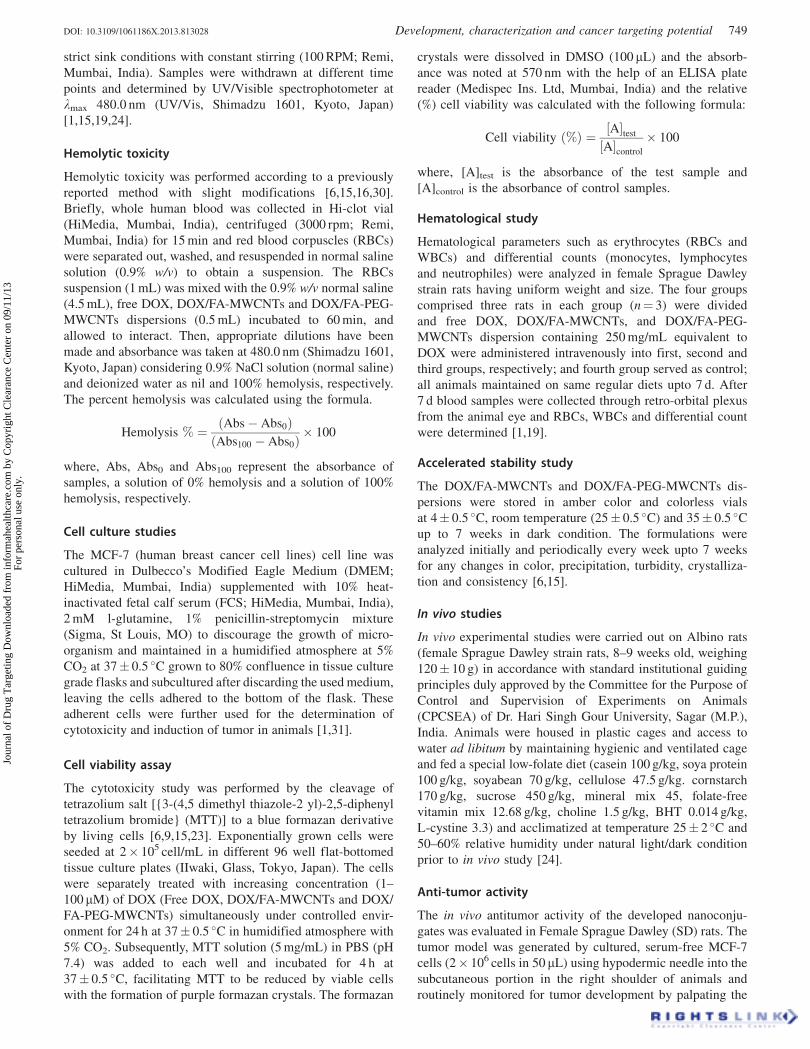
DOI: 10.3109/1061186X.2013.813028 Development, characterization and cancer targeting potential 749
Jour
nal o
f D
rug
Tar
getin
g D
ownl
oade
d fr
om in
form
ahea
lthca
re.c
om b
y C
opyr
ight
Cle
aran
ce C
ente
r on
09/
11/1
3Fo
r pe
rson
al u
se o
nly.

injected area with index finger and thumb. The tumor bearing
animals were randomly divided into four treatment groups
(Control, free DOX, DOX/FA-MWCNTs and DOX/FA-PEG-
MWCNTs) for treatment with 5 mg/kg body weight dose
equivalent to DOX. At predetermined time intervals tumor
volume was measured by measuring its dimension (major and
minor axis) using electronic digital caliper and computed
according to the formula: Tumor volume (mm3)¼Width�(length/2)2 up to 30 d. All animals were accommodated in
a pathogen-free laboratory environment during the studies.
Survival of the tumor bearing animals was also monitored
in the separate group up to 30 d [32–34].
Biodistribution study
Animals were divided into three groups and sterilized free
DOX, DOX/FA-MWCNTs and DOX/FA-PEG-MWCNTs
dispersion in normal saline (0.9% w/v) were administered
intravenously through caudal tail vein route (equivalent dose
of DOX¼ 5.0 mg/kg body weight) into animals.
Group I: DOX HCL served as control (free DOX).
Group II: DOX loaded FA-MWCNTs dispersion (DOX/
FA-MWCNTs).
Group III: DOX loaded FA-MWCNTs dispersion (DOX/
FA-PEG-MWCNTs).
Each group was administered the same i. v. dose of free
DOX, DOX/FA-MWCNTs and DOX/FA-PEG-MWCNTs
dispersions, animals carefully sacrificed by decapitation
method at time intervals of 1, 6, 12 and 24 h. Subsequently,
the different organs such as liver, spleen, kidney, heart and
tumor were carefully separated out, washed, weighed and
stored under freezed condition till used. Then contents treated
with 100mL of 10% TCA solution, vortexed (Superfit
vortexer, India) for 2 min, methanol was added and cen-
trifuged (3000 rpm, 10 min; Remi, Mumbai, India) and
supernatant was decanted into another vial and evaporated
to dryness at 60� 2 �C [19,33,35]. The dried residue was
collected in vials and analyzed for DOX content by HPLC
(Shimadzu, C18, Kyoto, Japan) methods reported by Agrawal
et al. [19] and Reddy and Murthy [35]. In which a mixture of
buffer pH 4.0/acetonitrile/methanol (60:24:16; v/v/v) used as
mobile phase with flow rate 1.2 mL/min at pressure of 102/
101 bars with adjusting 20 min runtime and peak at 480.2 nm
was consider with its retention time (RT) and area.
In vivo pharmacokinetics after intravenousadministration
The pharmacokinetics of DOX in plasma was measured
from plasma-concentration curve in healthy female Sprague
Dawley rats after intravenous injections (5.0 mg/kg body
weight dose) of developed formulations (free DOX, DOX/FA-
MWCNTs and DOX/FA-PEG-MWCNTs). The animals were
acclimatized at room temperature by maintaining the relative
humidity (RH) 55–60% under natural light/dark condition
prior to studies. The blood samples were collected from the
retro-orbital plexus of rat eyes under the mild anesthesia
into the Hi-Anticlot blood collecting vials (HiMedia,
Mumbai, India) at predetermined data points
(0.25, 0.5, 1, 2, 3, 6, 12, 18, 24 and 48 h) and centrifuged to
separate the RBCs, and serum, and supernatant (serum) was
collected. Then 100 mL of 10% w/v trichloro acetic acid in
methanol was added and vortexed (Superfit, Mumbai, India)
and ultracentrifuged (Z36HK, HERMLE LaborTchnik
GmbH, Wehingen, Germany) to obtained the clear super-
natant. The clear supernatant was collected in HPLC vials
(HiMedia, Mumbai, India) and analyzed by HPLC method.
The pharmacokinetic parameters such as peak plasma
concentration (Cmax) were calculated from the plasma con-
centration curve. The area under the curve (AUC0�t), area
under the first moment curve (AUMC), mean residence time
(MRT), plasma half-life (t1/2), apparent volume of distribution
at steady state (Vss) and at terminal phase (Vz) and half value
duration (HVD) were also calculated [19,33].
Pharmacokinetic data analysis
The pharmacokinetic data analysis of plasma concentration
time profile was conducted using the Kinetica 5.0 PK/PD
analysis software (Thermo Fischer Scientific, West Palm
Beach, FL) followed by non-compartment analysis.
Statistical analysis
The results are expressed as mean� standard deviation (�SD)
(n¼ 3) and statistical analysis was performed with Graph Pad
Instat Software (Version 3.00, Graph Pad Software, San Diego,
CA) by one-way ANOVA followed by the Tukey–Kramer
test for multiple comparisons. A probability p� 0.05 was
considered while significant and p� 0.001 was considered as
extremely significant.
Results and discussion
In the context of targeted drug delivery, first generation
(pristine) CNTs are not suitable due to inherent aqueous
insolubility and presence of impurities. The procured pristine
MWCNTs from Sigma Aldrich Pvt. Ltd. (St. Louis, MO) were
purified in a microwave oven and subsequently strong acid
treatment (H2SO4:HNO3) followed by NH4OH and H2O2 to
remove any metallic or amorphous impurities and also to
generate the carboxylic acid (–COOH) groups on to the
surfaces of MWCNTs.
The direct acid–base titration analysis was performed to
determine the total acidic functional groups present on
oxidized MWCNTS by Boehm titration method using
Table 1. Quantitative analysis of total functional group by Boehm titration on oxidized MWCNTs.
Samples TreatmentTotal functional
group (mmol–1/g)Carboxylic group
(mmol–1/g) Lengths (nm)
Pristine MWCNTs Microwave treated 10.2� 0.82 4.22� 0.55 960� 0.57Microwave-treated MWCNTs HNO3:H2SO4 treated 18.5� 0.20 10� 0.14 400� 0.85Carboxylated-MWCNTs H2O2/NaOH treated 24� 0.72 16� 0.33 80� 0.08
Values represent mean� SD (n¼ 3).
750 N. K. Mehra & N. K. Jain J Drug Target, 2013; 21(8): 745–758
Jour
nal o
f D
rug
Tar
getin
g D
ownl
oade
d fr
om in
form
ahea
lthca
re.c
om b
y C
opyr
ight
Cle
aran
ce C
ente
r on
09/
11/1
3Fo
r pe
rson
al u
se o
nly.

0.05 N NaOH and observation are presented in Table 1 [26].
Microwave-treated MWCNTs gave 10.2� 0.82 mmol–1/g and
4.22� 0.55 mmol–1/g total functional groups and carboxylic
groups, respectively, whereas HNO3:H2SO4 and H2O2:NH4OH
treated MWCNTs gave 18.5� 0.20, 10� 0.14 mmol–1/g and
24� 0.72 and 16� 0.33 mmol–1/g total functional groups
and carboxylic groups, respectively. The determination of
free acidic functional groups by Boehm titration is based on the
fact that the 0.05 N NaOH neutralizes carboxylic, phenolic-
and lactone groups present on the oxidized MWCNTS,
whereas 0.05 N NaHCO3 neutralizes only the carboxylic acid
functional group [25–27]. Our results are in line with the
previously published reports [25–27], but MWCNTs treatment
by ammonium hydroxide (NH4OH) and hydrogen peroxide
(H2O2) (50:50) followed by initial microwave oven treatment is
a debut study. This combined treatment approach may
drastically increase the total concentration of acidic functional
groups (phenolic, lactone and carboxylic functional groups) on
nanotubes. Further, these ox-MWCNTs were subjected to
acylation and amidation process as reported previously [13].
FA was conjugated either without spacer or with PEG-bis-
4000 amine as spacer to NH2 terminated and carboxylated
MWCNTs, respectively. The FTIR spectra of FA-MWCNTs
and FA-PEG-MWCNTs are shown in Figure 1(A) and (B).
Figure 1(A) shows the peak of aromatic C–H bending
at 832 cm�1, esters unconjugated C¼O at 1243.2 cm�1,
aromatic C¼C bending and stretching at 1637.2 cm�1
suggesting the attachment of folic acid to the MWCNTs,
which contained aromatic rings. However, Figure 1(B) shows
the prominent peaks at 3436.7 cm�1, 2916.0 cm�1,
1652.0 cm�1, 1437.2 cm�1, 1315.1 cm�1, a strong and sharp
peak at 1025.0 cm�1 of C-O stretch ether linkage wherein
peak of C–O stretch of ether linkage was found to be strong
and sharp at 1025.0 cm�1 due to the polyether backbone of
PEG and remaining peaks of aromatic compounds indicated
the presence of folic acid (Supporting information Table S1).
The morphology and size of carboxylated and DOX/FA-
PEG-MWCNTs were characterized by Transmission Electron
Microscope (TEM) and are shown in Figure 2(A) and (B).
The TEM observations clearly depict that the CNTs are
tubular in shape with open ends and in nanometric size range.
Moreover the images suggest that there was no change in their
tubular structure even after conjugation of FA and PEG.
The surface charge of the pristine, ox�, DOX/FA-
MWCNTs and DOX/FA-PEG-MWCNTs nanoconjugates
was determined from their electrophoresis mobility at
acidic, neutral and alkaline pH by zeta potential (z) according
to the Helmholtz-Smoluchowski equation. The ox-MWCNTs
depicted the slightly negative zeta potential (�10 mV), which
could be due to the generation of acidic functional groups
during the oxidation. The free COOH– was ionized at alkaline
pH and thus negative zeta potential was observed. The
DOX/FA-PEG-MWCNTs nanoconjugate showed positive
zeta potential of þ5.0, þ3.8 and þ4.8 at acidic, neutral and
alkaline medium, respectively [13]. PEG being non-ionic
could decrease the zeta potential of the formulations due to its
presence on the surface of MWCNTs.
The anthracycline antibiotic DOX was physically loaded
by simple mixing in DOX/FA-PEG-MWCNTs and DOX/FA-
MWCNTs nanoconjugates as evidenced by reddish color.
The % loading efficiency (% LE) was calculated in PBS
(pH 7.4) at 480.0 nm using UV/Vis spectrophotometer
(Shimadzu, 1601, Kyoto, Japan) and found to be
90.2� 0.22 and 92� 0.92 (n¼ 3) for DOX/FA-MWCNTs
and DOX/FA-PEG-MWCNTs, respectively. UV/Vis spectro-
photometry data of MWCNTs formulations suggest that
DOX can easily adsorb on to the surface of MWCNTs
probably through strong p–p stacking interactions of quinine
part of DOX and CNTs and accordingly greatest loading was
found to be 92� 0.92 in DOX/FA-PEG-MWCNTs nano-
conjugte. Further, endohedral entrapment into the interior
cavity of nanotubes structure leading to higher entrapment is
also expected, however measurement technique is not
investigated yet. The observed data could possibly be
ascribed to loading of cationic DOX in and around PEG
based micro domains also via p–p stacking at pH 7.4
Figure 1. Fourier transform infra-red (FTIR)spectra of (A) FA-MWCNTs and(B) FA-PEG-MWCNTs nano-conjugates.
DOI: 10.3109/1061186X.2013.813028 Development, characterization and cancer targeting potential 751
Jour
nal o
f D
rug
Tar
getin
g D
ownl
oade
d fr
om in
form
ahea
lthca
re.c
om b
y C
opyr
ight
Cle
aran
ce C
ente
r on
09/
11/1
3Fo
r pe
rson
al u
se o
nly.

(Scheme 4). The general loading efficiency (LE) of DOX in
different dispersion followed the order:
DOX=FA�MWCNTs! DOX=FA� PEG�MWCNTs
ðLeast crowding ends! greatest crowding endsÞ
The high loading efficiency of engineered nanotubes
makes it a better carrier with better stability of DOX complex
at normal pH and sustained release in acidic microenviron-
ments (lower pH). The sustained release behaviour of the
drug from the nanotubes at acidic pH is an important factor
in tumor specific targeted drug delivery. Recently, Huang
et al. reported approximately 91% DOX loading efficiency in
functionalized CNTs [22].
The in vitro release of DOX from DOX/FA-MWCNTs and
DOX/FA-PEG-MWCNTs dispersions was performed at pH
(7.4 and 5.3) through a dialysis membrane (MWCO 5–6 KDa,
HiMedia, Mumbai, India) at 37� 0.5 �C. The in vitro release
suggests sustained release at lysosomal pH (5.3) ascribed to
the greater hydrophilicity, and by cleavage of the interactions
between the DOX molecules and engineered CNTs. On
comparing the release of DOX/FA-PEG-MWCNTs to other
formulations, effect of PEG modification could be appre-
ciated and the order of release was as follows:
DOX=FA� PEG�MWCNTs! DOX=FA�MWCNTs
ðSustained Release! Faster ReleaseÞ
The initial burst release achieved due to diffusion or the
adsorbed DOX followed by the sustained released may
possibly suggest lesser exposure of loaded drug to external
microenvironment that could be due to greater steric
hindrance on ends and side walls, resulting in sustained
release pattern of the loaded drug following initial faster
release as shown in Figure 3(A) and (B). The DOX release
data best fits into the Higuchian release kinetic. Our in vitro
release data of DOX are in line with the previous reports
[8,9,22]. The in vitro DOX release pattern depends on several
factors like pH, surface charge characteristics, degradation
rate, particle size, rate of hydration and dehydration and
interaction force of DOX binding to the surface of nanotubes.
The initial fast release was attributed to the rapid swelling
of DOX associated with diffusion, another reason being the
chemical interaction through hydrogen bonding between
DOX and nanotubes surface leading to sustain release pattern
[22]. Zhang et al. similarly reported pH-triggered drug release
response from the modified nanotubes under normal physio-
logical conditions and release at reduced pH typical of micro-
environments of intracellular lysosomes or endosome or
cancerous tissue [23]. It is clearly depicted that engineered
CNTs may show the pH-responsive DOX release.
The % hemolysis data of free DOX (15.7� 0.5), pristine
MWCNTs (18.0� 0.5), DOX/FA-MWCNTs (12.5� 0.5)
and DOX/FA-PEG-MWCNTs (9.0� 0.5) were compared.
Pristine MWCNTs shows highest (18.0� 0.5) while
Figure 2. Photomicrographs of (A and C)carboxylated MWCNTs and (B and D)FA-PEG-MWCNTs.
752 N. K. Mehra & N. K. Jain J Drug Target, 2013; 21(8): 745–758
Jour
nal o
f D
rug
Tar
getin
g D
ownl
oade
d fr
om in
form
ahea
lthca
re.c
om b
y C
opyr
ight
Cle
aran
ce C
ente
r on
09/
11/1
3Fo
r pe
rson
al u
se o
nly.
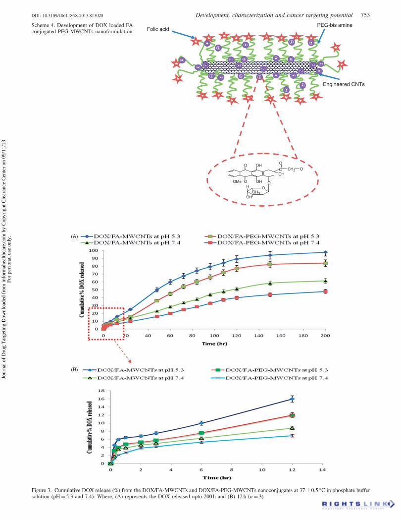
Scheme 4. Development of DOX loaded FAconjugated PEG-MWCNTs nanoformulation. Folic acid
PEG-bis amine
Engineered CNTs
OMe O
O
OOH
OH
OHC
O
O
OCH3
CH2
H
OH
Figure 3. Cumulative DOX release (%) from the DOX/FA-MWCNTs and DOX/FA-PEG-MWCNTs nanoconjugates at 37� 0.5 �C in phosphate buffersolution (pH¼ 5.3 and 7.4). Where, (A) represents the DOX released upto 200 h and (B) 12 h (n¼ 3).
DOI: 10.3109/1061186X.2013.813028 Development, characterization and cancer targeting potential 753
Jour
nal o
f D
rug
Tar
getin
g D
ownl
oade
d fr
om in
form
ahea
lthca
re.c
om b
y C
opyr
ight
Cle
aran
ce C
ente
r on
09/
11/1
3Fo
r pe
rson
al u
se o
nly.
DOX/FA-PEG-MWCNTs shows minimum (9.0� 0.5) hemo-
lytic toxicity. The hemolytic toxicity of pristine MWCNTs
was enough to limit its use as drug delivery system. Pristine
MWCNTs shows (18.0� 0.5) highest hemolytic toxicity due
to the presence of some metallic impurities; however on
functionalization it was reduced to 12.5� 0.5 in case of DOX/
FA-MWCNTs. Recently, Sachar and Saxena reported that
pristine and acid-treated CNTs were toxic to mouse blood-
derived erythrocytes in vitro as well as in vivo [36]. It is well
reported that the pristine MWCNTs (first generation CNTs)
are not suitable for drug delivery, but their compatibility may
be improved through functionalization. However, our hemo-
lytic toxicity results clearly suggest that functionalization
or PEGylation considerably reduced the hemolysis upto
9.0� 0.5 possibly due to non availability of any free
positively charged functional moieties. PEGylation make
nanotubes more biocompatible module in comparison to
pristine and acid-treated ones.
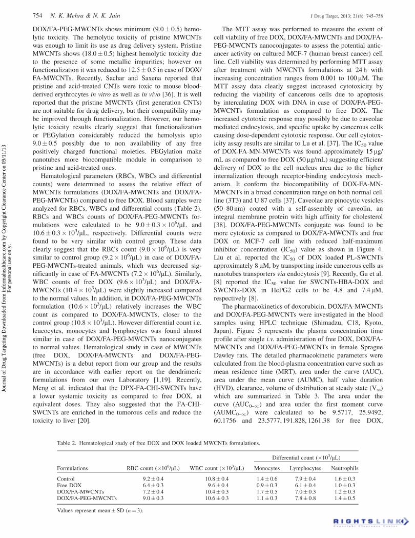
Hematological parameters (RBCs, WBCs and differential
counts) were determined to assess the relative effect of
MWCNTs formulations (DOX/FA-MWCNTs and DOX/FA-
PEG-MWCNTs) compared to free DOX. Blood samples were
analyzed for RBCs, WBCs and differential counts (Table 2).
RBCs and WBCs counts of DOX/FA-PEG-MWCNTs for-
mulations were calculated to be 9.0� 0.3� 106/mL and
10.6� 0.3� 103/mL, respectively. Differential counts were
found to be very similar with control group. These data
clearly suggest that the RBCs count (9.0� 106/mL) is very
similar to control group (9.2� 106/mL) in case of DOX/FA-
PEG-MWCNTs-treated animals, which was decreased sig-
nificantly in case of FA-MWCNTs (7.2� 106/mL). Similarly,
WBC counts of free DOX (9.6� 103/mL) and DOX/FA-
MWCNTs (10.4� 103/mL) were slightly increased compared
to the normal values. In addition, in DOX/FA-PEG-MWCNTs
formulation (10.6� 103/mL) relatively increases the WBC
count as compared to DOX/FA-MWCNTs, closer to the
control group (10.8� 103/mL). However differential count i.e.
leucocytes, monocytes and lymphocytes was found almost
similar in case of DOX/FA-PEG-MWCNTs nanoconjugates
to normal values. Hematological study in case of MWCNTs
(free DOX, DOX/FA-MWCNTs and DOX/FA-PEG-
MWCNTs) is a debut report from our group and the results
are in accordance with earlier report on the dendrimeric
formulations from our own Laboratory [1,19]. Recently,
Meng et al. indicated that the DPX-FA-CHI-SWCNTs have
a lower systemic toxicity as compared to free DOX, at
equivalent doses. They also suggested that the FA-CHI-
SWCNTs are enriched in the tumorous cells and reduce the
toxicity to liver [20].
The MTT assay was performed to measure the extent of
cell viability of free DOX, DOX/FA-MWCNTs and DOX/FA-
PEG-MWCNTs nanoconjugates to assess the potential antic-
ancer activity on cultured MCF-7 (human breast cancer) cell
line. Cell viability was determined by performing MTT assay
after treatment with MWCNTs formulations at 24 h with
increasing concentration ranges from 0.001 to 100 mM. The
MTT assay data clearly suggest increased cytotoxicity by
reducing the viability of cancerous cells due to apoptosis
by intercalating DOX with DNA in case of DOX/FA-PEG-
MWCNTs formulation as compared to free DOX. The
increased cytotoxic response may possibly be due to caveolae
mediated endocytosis, and specific uptake by cancerous cells
causing dose-dependent cytotoxic response. Our cell cytotox-
icity assay results are similar to Lu et al. [37]. The IC50 value
of DOX-FA-MN-MWCNTs was found approximately 15 mg/
mL as compared to free DOX (50mg/mL) suggesting efficient
delivery of DOX to the cell nucleus area due to the higher
internalization through receptor-binding endocytosis mech-
anism. It conform the biocompatibility of DOX-FA-MN-
MWCNTs in a broad concentration range on both normal cell
line (3T3) and U 87 cells [37]. Caveolae are pinocytic vesicles
(50–80 nm) coated with a self-assembly of caveolin, an
integral membrane protein with high affinity for cholesterol
[38]. DOX/FA-PEG-MWCNTs conjugate was found to be
more cytotoxic as compared to DOX/FA-MWCNTs and free
DOX on MCF-7 cell line with reduced half-maximum
inhibitor concentration (IC50) value as shown in Figure 4.
Liu et al. reported the IC50 of DOX loaded PL-SWCNTs
approximately 8 mM, by transporting inside cancerous cells as
nanotubes transporters via endocytosis [9]. Recently, Gu et al.
[8] reported the IC50 value for SWCNTs-HBA-DOX and
SWCNTs-DOX in HePG2 cells to be 4.8 and 7.4mM,
respectively [8].
The pharmacokinetics of doxorubicin, DOX/FA-MWCNTs
and DOX/FA-PEG-MWCNTs were investigated in the blood
samples using HPLC technique (Shimadzu, C18, Kyoto,
Japan). Figure 5 represents the plasma concentration time
profile after single i.v. administration of free DOX, DOX/FA-
MWCNTs and DOX/FA-PEG-MWCNTs in female Sprague
Dawley rats. The detailed pharmacokinetic parameters were
calculated from the blood-plasma concentration curve such as
mean residence time (MRT), area under the curve (AUC),
area under the mean curve (AUMC), half value duration
(HVD), clearance, volume of distribution at steady state (Vss)
which are summarized in Table 3. The area under the
curve (AUC0–1) and area under the first moment curve
(AUMC0–1) were calculated to be 9.5717, 25.9492,
60.1756 and 23.5777, 191.828, 1261.38 for free DOX,
Table 2. Hematological study of free DOX and DOX loaded MWCNTs formulations.
Differential count (�103/mL)
Formulations RBC count (�106/mL) WBC count (�103/mL) Monocytes Lymphocytes Neutrophils
Control 9.2� 0.4 10.8� 0.4 1.4� 0.6 7.9� 0.4 1.6� 0.3Free DOX 6.4� 0.3 9.6� 0.4 0.9� 0.3 6.1� 0.4 1.0� 0.3DOX/FA-MWCNTs 7.2� 0.4 10.4� 0.3 1.7� 0.5 7.0� 0.3 1.2� 0.3DOX/FA-PEG-MWCNTs 9.0� 0.3 10.6� 0.3 1.1� 0.3 7.8� 0.8 1.4� 0.5
Values represent mean� SD (n¼ 3).
754 N. K. Mehra & N. K. Jain J Drug Target, 2013; 21(8): 745–758
Jour
nal o
f D
rug
Tar
getin
g D
ownl
oade
d fr
om in
form
ahea
lthca
re.c
om b
y C
opyr
ight
Cle
aran
ce C
ente
r on
09/
11/1
3Fo
r pe
rson
al u
se o
nly.

DOX/FA-MWCNTs and DOX/FA-PEG-MWCNTs, respect-
ively. The AUC0–1) and AUMC(0–1) of DOX/FA-PEG-
MWCNTs were approximately 6-fold and 53-fold higher as
compared to free DOX, respectively. The elimination half-life
(t1/2) of DOX/FA-PEG-MWCNTs, DOX/FA-MWCNTs and
free DOX was found to be 14.956, 4.8432 and 1.8846 while
MRT was found to be 20.9616, 7.3924 and 2.4632, respect-
ively. In contrast with t1/2 of DOX/FA-PEG-MWCNTs
(14.956) were 3 and 8 times (p50.005), while MRT was 3
and 8 times longer as compared to DOX/FA-MWCNTs and
free DOX, respectively. The prolonged t1/2 clearly depicted
the DOX/FA-PEG-MWCNTs in the systemic circulation.
The obtained results are ascribed to biocompatibility of
engineered nanotubes upon PEGylation to reside it for longer
time inside the body. Our pharmacokinetics data clearly
suggest the improved bioavailability of DOX as compared to
free DOX, which make nanotubes a most promising alterna-
tive, smart nanobiomedicine in targeted drug delivery. Liu
et al. reported long-term fate of PEG functionalized SWCNTs
by intravenous administration in animals and found longest
blood circulation upto 1 d and near-complete clearance of
SWCNTs from the main organs approximately in 2 months
[38]. Cherukari et al. reported the low acute toxicity and
long circulation of disaggregated SWCNTs by low dose of
nanotubes [39]. The intrinsic stability and structural flexibil-
ity of surface engineered CNTs may enhances the circulation
time as well as the bioavailability of drug molecules [40,41].
Huang et al. only described a new family of folate-decorated
and carbon nanotubes mediated delivery system encapsulating
doxorubicin for controlled release [22].
Recently, Jain and co-investigators reported Amphotericin
B loaded mannosylated MWCNTs (AmBitubes) was released
in a controlled manner at different pH environment with
increased cell uptake and higher disposition in macrophages
rich organs using J774 cell line indicating the site-specific
drug delivery. Authors suggested that the AmBitubes could
be employed as efficient nano-carrier for anti-leishmanial
therapy [16].
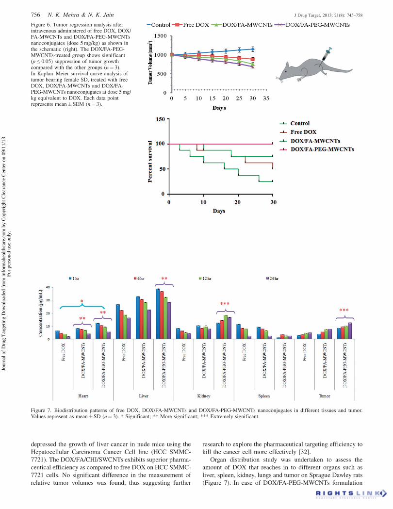
The anti-tumor activity of free DOX, DOX/FA-MWCNTs
and DOX/FA-PEG-MWCNTs nanoconjugates was studied in
tumor bearing female Sprague Dawley strain rats and tumor
growth inhibition rate in terms of mean tumor volume (mm3)
as represented in Figure 6. The results suggested that DOX/
FA-PEG-MWCNTs nanoconjugate reduced extremely signifi-
cant tumor volume compared to free DOX (p50.001) after
tumor implantation. Survival of tumor bearing SD rats after
treatments with the nanotubes conjugates are represented in
Kaplan Meier survival curves, which suggested that the
median survival time with DOX/FA-PEG-MWCNTs-treated
animals (30 d) significantly (p� 0.001) as compared to free
DOX and control group due to their biocompatible and long
circulatory nature. Liu et al., 2008, reported that no obvious
toxicity or negative health effects observed over 3 months by
injected i.v. PEGylated SWCNTs and no mortality or loss of
body weight were seen in any mice [33]. Recently, Ji et al.
developed a new type of targeted drug delivery system
(TDDS) using chitosan modified SWCNTs for controlled
release of DOX by constructing folic acid (FA) modified
chitosan encapsulating doxorubicin (DOX/FA/CHI/SWCNTs)
wherein FA was bound to the outer CHI layer and effectively
Table 3. Pharmacokinetic parameters of free DOX, DOX/FA- MWCNTs and DOX/FA-PEG-MWCNTs dispersion.
ParametersCmax
(mg/mL)HVD(h)
AUC(0–t)
(mg.h/mL)AUC(0–1)
(mg.h/mL)AUMC(0–t)
(mg. hr2/mL)AUMC(0–1)
(mg.h2/mL)t1/2
(h)MRT(h) Clz Vz Vss
Free DOX 6.11 0.3564 9.0445 9.5717 17.926 23.5777 1.8846 2.4632 10.4474 28.4065 25.7346DOX/FA-MWCNTs 6.06 0.8329 25.0625 25.9492 164.351 191.828 4.8432 7.3924 3.8536 26.9279 28.4881DOX/FA-PEG-MWCNTs 6.50 1.7482 53.6275 60.1756 805.781 1261.38 14.956 20.9616 1.6618 35.8567 34.8341
Probability p50.001; standard deviation55%.Cmax¼ peak plasma concentration; Tmax¼ time taken to reach Cmax; t1/2¼ elimination half life; MRT¼mean residence time; AUC(0–1)¼ area under
plasma drug concentration over time curve; HVD¼ half value duration; Clz¼ clearance; Vz¼Volume of distribution; Vss¼Volume of distributionat steady state.
Mean� SD (n¼ 3).
Figure 4. Percent cell viability of MCF-7 cell after treated with freeDOX, DOX/FA-MWCNTs and DOX/FA-PEG-MWCNTs at 24 h (n¼ 3).
Figure 5. Serum concentration of DOX obtained from free DOX, DOX/FA-MWCNTs and DOX/FA-PEG-MWCNTs at different data points.Mean� SD (n¼ 6; p� 0.001).
DOI: 10.3109/1061186X.2013.813028 Development, characterization and cancer targeting potential 755
Jour
nal o
f D
rug
Tar
getin
g D
ownl
oade
d fr
om in
form
ahea
lthca
re.c
om b
y C
opyr
ight
Cle
aran
ce C
ente
r on
09/
11/1
3Fo
r pe
rson
al u
se o
nly.
depressed the growth of liver cancer in nude mice using the
Hepatocellular Carcinoma Cancer Cell line (HCC SMMC-
7721). The DOX/FA/CHI/SWCNTs exhibits superior pharma-
ceutical efficiency as compared to free DOX on HCC SMMC-
7721 cells. No significant difference in the measurement of
relative tumor volumes was found, thus suggesting further
research to explore the pharmaceutical targeting efficiency to
kill the cancer cell more effectively [32].
Organ distribution study was undertaken to assess the
amount of DOX that reaches in to different organs such as
liver, spleen, kidney, lungs and tumor on Sprague Dawley rats
(Figure 7). In case of DOX/FA-PEG-MWCNTs formulation
Figure 6. Tumor regression analysis afterintravenous administered of free DOX, DOX/FA-MWCNTs and DOX/FA-PEG-MWCNTsnanoconjugates (dose 5 mg/kg) as shown inthe schematic (right). The DOX/FA-PEG-MWCNTs-treated group shows significant(p� 0.05) suppression of tumor growthcompared with the other groups (n¼ 3).In Kaplan–Meier survival curve analysis oftumor bearing female SD, treated with freeDOX, DOX/FA-MWCNTs and DOX/FA-PEG-MWCNTs nanoconjugates at dose 5 mg/kg equivalent to DOX. Each data pointrepresents mean� SEM (n¼ 3).
Figure 7. Biodistribution patterns of free DOX, DOX/FA-MWCNTs and DOX/FA-PEG-MWCNTs nanoconjugates in different tissues and tumor.Values represent as mean� SD (n¼ 3). * Significant; ** More significant; *** Extremely significant.
756 N. K. Mehra & N. K. Jain J Drug Target, 2013; 21(8): 745–758
Jour
nal o
f D
rug
Tar
getin
g D
ownl
oade
d fr
om in
form
ahea
lthca
re.c
om b
y C
opyr
ight
Cle
aran
ce C
ente
r on
09/
11/1
3Fo
r pe
rson
al u
se o
nly.

high uptake of DOX was observed in tumor, liver, kidney and
spleen at time intervals upto 24 h. The high levels of DOX
was found after 1 h of administered dose in liver, tumor and
kidney and rapid decline in the overall formulation, thereafter
indicating that most of MWCNTs were eliminated through the
renal excretion route. The amount of DOX was found to be
remarkably increased at tumor site with time in case of DOX/
FA-PEG-MWCNTs formulation due to receptor-mediated
endocytosis (RME) mechanism. Our results are in accordance
with previous Leading Opinion by Meng et al. [20] in
targeting doxorubicin to tumors using raw and treated carbon
nanotubes. In vitro drug release data suggested initial rapid
release followed by gradual slow release, similar pattern was
observed in in vivo study. The variation in quantity of drug
estimated in vivo is due to biological effects on the bioactive
that predominate its biodistribution pattern. Biodistribution
study data suggested that the DOX/FA-PEG-MWCNTs
nanoconjugate could deliver drug selectively at the tumor
cells.
Recently, our laboratory developed and characterized
the dexamethasone conjugated MWCNTs for controlled
DOX delivery with reduced toxicity using ‘‘A-549’’
lung epithelial cancer cell line where the DOX loaded
DEX-MWCNTs showed less hemolytic and more cytotoxic
as compared to free DOX [15]. Our ex vivo and in vivo
results are in accordance with the previous published
reports [42,43].
Stability of the formulations (DOX, DOX/FA-MWCNTs
and DOX/FA-PEG-MWCNTs) was studied at different con-
ditions of temperature (4� 0.5 �C, 25� 0.5� �C and 50 �C)
after keeping in dark (amber color bottle) and light (colorless
vials) up to 7 weeks [15]. The formulations were found to be
most stable in dark at 4� 0.5 �C (Table 4). Stability of the
DOX/FA-PEG-MWCNTs formulations was observed at dif-
ferent conditions of temperature (4� 0.5 �C, 25� 0.5� �Cand 50 �C) after keeping in dark (amber color bottle) and light
(colorless vials) and evaluated every week upto 7 weeks.
Among all formulations DOX/FA-PEG-MWCNTs was found
to be most stable in dark at RT. In terms of stability profile
f-MWCNTs could possibly present themselves as a most
stable system due to p–p stacking interaction in all tempera-
ture ranges and environment required for biological applica-
tions. Thus we conclude that the DOX/FA-PEG-MWCNTs
formulation is more stable than other MWCNTs formulation
at 4� 0.5 �C, in dark suggesting that the developed nanotubes
formulation may be suitably stored in amber color bottle or
vials at a cool place.
Conclusions
To best of our knowledge, this is the complete study
report with evidence of improved selective treatment of
cancer using DOX/FA-PEG-MWCNTs formulations most
suitable as controlled and targeted drug delivery. The results
suggested that the DOX/FA-PEG-MWCNTs formulation
showed the better targeting response using MCF-7 breast
cancer cell line through cavaeolin-mediated endocytosis
mechanism. From the outcomes of our present research
studies, it can be concluded that the DOX loaded surface
modified MWCNTs showed better in vitro, ex vivo and
biocompatibility profile as compared to other nano-carriers
depicting higher loading (92.0� 0.92) and sustained release
profile especially at acidic microenvironment corresponding
to conditions existing at cancerous tissues/sites. The improved
kinetics of nanotubes formulation upon PEGylation such
as MRT, t1/2, HVD and AUMC(0–1) 20.9616, 14.956, 1.7582
and 1261.38, respectively for DOX/FA-PEG-MWCNTs as
compared to free DOX may be considered significantly
effective for intravenous administration. However, folate
conjugation makes it more targetable approach precluding
the non-target sites such as existing nanoparticles, liposomes
and dendrimers [1,24,44–46]. Thus optimal therapeutic
response and improved bioavailability may be achieved with
minimized side effects associated with the carrier and
anticancer drug.
Acknowledgements
The authors gratefully acknowledge M/s Sun Pharmaceutical
Advanced Research Center (SPARC), Vadodara, India, for
a gift sample of Doxorubicin hydrochloride, All India
Institute of Medical Sciences (AIIMS), New Delhi, India,
for Transmission Electron Microscopy (TEM) and Scanning
Electron Microscopy (SEM). One of the authors (Neelesh
Kumar Mehra) is thankful to the University Grants
Commission (UGC), New Delhi, India for providing the
Junior Research Fellowship (JRF) during the tenure of these
studies.
Declaration of interest
The authors report no conflict of interest.
Table 4. Accelerated stability testing of nanotubes formulations.
Dark (�C) Light (�C)
Parameters Formulations 4� 0.5 25� 0.5 35� 0.5 4� 0.5 25� 0.5 35� 0.5
Turbidity DOX/FA-MWCNTs – – þþ þ þþ þþþDOX/FA-PEG-MWCNTs – – þþ þ þþ þþþ
Precipitation DOX/FA-MWCNTs – – þ – þ þþDOX/FA-PEG-MWCNTs – – þ – þ þþ
Change in colour DOX/FA-MWCNTs – þ þ – þ þþDOX/FA-PEG-MWCNTs – þ þ – þ þþ
Crystallization DOX/FA-MWCNTs – – þ – þ þDOX/FA-PEG-MWCNTs – – þ – þ þ
Change in consistency DOX/FA-MWCNTs – þ þþ – þ þþDOX/FA-PEG-MWCNTs – þ þþ – þ þþ
(–) no change; (þ) small change; (þþ) considerable change; (þþþ) enough change.
DOI: 10.3109/1061186X.2013.813028 Development, characterization and cancer targeting potential 757
Jour
nal o
f D
rug
Tar
getin
g D
ownl
oade
d fr
om in
form
ahea
lthca
re.c
om b
y C
opyr
ight
Cle
aran
ce C
ente
r on
09/
11/1
3Fo
r pe
rson
al u
se o
nly.
References
1. Gupta U, Dwivedi SKD, Bid HK, et al. Ligand anchoreddendrimers based nanoconstructs for effective targeting to cancercells. Int J Pharm 2010;393:185–96.
2. Jain NK, Mishra V, Mehra NK. Targeted drug delivery tomacrophages. Exp Opinion Drug Deliv 2013;10:353–67.
3. Bianco A, Kostarelos K, Prato M. Making carbon nanotubesbiocompatible and biodegradable. Chem Commun 2011;47:10182–8.
4. Mehra NK, Jain AK, Lodhi N, et al. Challenges in the use of carbonnanotubes in biomedical applications. Crit Rev Ther Drug Carr Syst2008;25:169–206.
5. Mehra NK, Mishra V, Jain NK. A review on receptor basedtherapeutic targeting. Ther Deliv 2013;4:369–94.
6. Singh R, Mehra NK, Jain V, Jain NK. Gemcitabine-loadedsmart carbon nanotubes for effective targeting to cancer cell.J Drug Target 2013. [Epub ahead of print]. doi: 10.3109/1061186X.2013.778264.
7. Mehra NK, Sharma S, Singhai AK, Kumar V. Carbon nanotubes:a new line to drug delivery. Pharm Rev 2011;9:75–80.
8. Gu YJ, Cheng J, Jin J, et al. Development and evaluation of pH-responsive single-walled carbon nanotube-doxorubicin complexesin cancer cells. Int J Nanomed 2011;6:2889–98.
9. Liu Z, Sun X, Nakayama-Ratchford N, Dai H. Supramolecularchemistry on water soluble carbon nanotubes for drug loading anddelivery. ACS Nano 2007;11:50–6.
10. Pantarotto D, Hoebeke J, Graff R. Synthesis, structural character-ization and immunological properties of carbon nanotubesfunctionalized with peptides. J Am Chem Soc 2003;125:6160–4.
11. Villa CH, Dao T, Ahearn I, et al. Single-walled carbon nanotubesdeliver peptide antigen into dendritic cells and enhance IgGresponses to tumor-associated antigens. ACS Nano 2011;5:5300–12.
12. Wang L, Zhang WL, Zhang M, et al. Synergistic enhancement ofcancer therapy using a combination of docetaxel and photothermalablation induced by singe-walled carbon nanotubes. Int J Nanomed2011;6:2641–52.
13. Jain AK, Dubey V, Mehra NK, et al. Carbohydrate conjugated multiwalled carbon nanotubes: Development and characterization.Nanomed: Nanotech Biol Med 2009;5:432–42.
14. Varkouhi K, Foillard S, Lammers T, et al. SiRNA delivery withfunctionalized carbon nanotubes. Int J Pharm 2011;416:419–25.
15. Lodhi N, Mehra NK, Jain NK. Development and characterizationof dexamethasone mesylate anchored on multi walled carbonnanotubes. J Drug Target 2013;21:67–76.
16. Pruthi J, Mehra NK, Jain NK. Macrophages targeting of ampho-tericin B through mannosylated multi walled carbon nanotubes.J Drug Target 2012;20:593–604.
17. Shvedova AA, Tkach AV, Kisin ER, et al. Carbon nanotubesenhance metastatic growth of lung carcinoma via up-regulation ofmyeloid-derived suppressor cells. Small 2013;9:1691–5.
18. Simenova PP. Update on carbon nanotubes toxicity. Fut Nanomed2009;4:373–75.
19. Agrawal A, Gupta U, Asthana A, Jain NK. Dextran conjugateddendrite nanoconstructs as potential vectors for anti-cancer agent.Biomaterials 2009;30:3588–96.
20. Meng L, Zhang X, Lu Q, et al. Single walled carbon nanotubes asdrug delivery vehicles: targeting doxorubicin to tumors.Biomaterials 2012;33:1689–98.
21. Kaminskas LM, McLeod VM, Kelly BD, et al. A comparisonof changes to doxorubicin pharmacokinetics, antitumor activityand toxicity mediated by PEGylated dendrimer and PEGylatedliposome drug delivery systems. Nanomed: Nanotech Biol Med2011;8:103–11.
22. Huang H, Yuan Q, Shah JS, Misra RDK. A new family of folate-decorated and carbon nanotube-mediated drug delivery system:synthesis and drug delivery response. Adv Drug Deliv Rev 2011;63:1332–9.
23. Zhang X, Meng L, Lu Q, et al. Targeted delivery and controlledrelease of doxorubicin to cancer cells using modified single wallcarbon nanotubes. Biomaterials 2009;30:6041–7.
24. Singh P, Gupta U, Asthana A, Jain NK. Folate and folate–PEG-PAMAM dendrimer: synthesis, characterization, and targeted
anticancer drug delivery potential in tumor bearing mice. BioconjChem 2008;19:2239–52.
25. Datsyuk V, Kalyva M, Papagelis K, et al. Chemical oxidation ofmulti walled carbon nanotubes. Carbon 2008;46:833–40.
26. Boehm HP. Surface oxides on carbon and their analysis: a criticalassessment. Carbon 2012;40:145–9.
27. Yudianti R, Onggo H, Sudiraman Y, et al. Analysis of functionalgroup sited on multi-wall carbon nanotubes. The Open Mat Sc2011;5:242–7.
28. Lee RJ, Low PS. Folate-mediated tumor cell targeting of liposome-entrapped doxorubicin in vitro. Biochim Biophys Acta 1995;1233:134–44.
29. Shi X, Wang SH, Shen M, et al. Multifunctional dendrimer-modified multiwalled carbon nanotubes: synthesis, characterizationand in vitro cancer cell targeting and imaging. Biomacromolecules2009;10:1744–50.
30. Mishra V, Gupta U, Jain NK. Influence of different generationsof poly (propylene imine) dendrimers on human erythrocytes.Pharmazie 2010;65:891–5.
31. Prickett WM, Rite BDV, Resasco DE, Harrison RG. Vasculartargeted single-walled carbon nanotubes for near-infrared lighttherapy of cancer. Nanotechnology 2011;22:455101:1–7.
32. Ji Z, Lin G, Lu Q, et al. Targeted therapy of SMMC-7721liver cancer in vitro and in vivo with carbon nanotubesbased drug delivery system. J Colloid Interface Sci 2012;365:143–9.
33. Liu Z, Chen K, Davis C, et al. Drug delivery with carbon nanotubesfor in vivo cancer treatment. Cancer Res 2008;68:6652–60.
34. Bhirde AA, Patel V, Gavard J, et al. Targeted killing of cancer cellsin vivo and in vitro with EGF-directed carbon nanotubes-baseddrug delivery. ACS Nano 2009;3:307–16.
35. Reddy LH, Murthy RSR. Pharmacokinetics and biodistributionstudies of doxorubicin loaded poly (butylcyanoacrylate) nanopar-ticles synthesized by two different techniques. Biomed Pap 2004;148:161–6.
36. Sachar S, Saxena RK. Cytotoxic effect of poly-dispersed singlewalled carbon nanotubes on erythrocytes in vitro and in vivo. PLoSONE 2011;6:e22032(1–8).
37. Lu YJ, Wei KC, Ma CCM, et al. Dual targeted deliveryof doxorubicin to cancer cells using folate-conjugated magneticmulti-walled carbon nanotubes. Colloids Surfaces B: Biointerfaces2012;89:1–9.
38. Liu Z, Davis C, Cai W, et al. Circulation and long-term fate offunctionalized biocompatible single-walled carbon nanotubesin mice probed by raman spectroscopy. Proc Nat Acad Sci USA2008;105:1410–15.
39. Cherukari P, Gannon CJ, Leeuw TK, et al. Mammalian pharma-cokinetics of carbon nanotubes using intrinsic near-infrared fluor-escence. Proc Nat Acad Sci USA 2006;103:18882–6.
40. Chen J, Chen S, Zhao X, et al. Functionalized single-walled carbonnanotubes as rationally designed vehicle for tumor-targeted drugdelivery. J Am Chem Soc 2008;130:16778–85.
41. Liu Z, Cai W, He L, et al. In-vivo biodistribution and highlyefficient tumor targeting of carbon nanotubes in mice. NatNanotech 2007;2:47–52.
42. Ren J, Shen S, Wang D, et al. The targeted delivery of anticancerdrugs to brain glioma by PEGylated oxidised multi-walled carbonnanotubes modified with angiopep-2. Biomaterials 2012;33:3324–33.
43. Shen S, Ren J, Chen J, et al. Development of magnetic multi walledcarbon nanotubes combined with near-infrared radiation-assisteddesorption for the determination of tissue distribution of doxorubi-cin liposome injects in rats. J Chromatography A 2011;1218:4619–26.
44. Prabharan M, Grailer JJ, Pilla S, et al. Folate-conjugated amphi-philic hyperbranched block copolymers based on Boltorn� H40,poly (L-lactide) and poly (ethylene glycol) for tumor-targeted drugdelivery. Biomaterials 2009;30:3009–19.
45. Park J, Fong PM, Lu J, et al. PEGylated PLGA nanoparticles for theimproved delivery of doxorubicin. Nanomed: Nanotech Biol Med2009;5:410–18.
46. Rose PG. Pegylated liposomal doxorubicin: optimizing the dosingschedule in ovarian cancer. Oncologist 2005;10:205–14.
758 N. K. Mehra & N. K. Jain J Drug Target, 2013; 21(8): 745–758
Jour
nal o
f D
rug
Tar
getin
g D
ownl
oade
d fr
om in
form
ahea
lthca
re.c
om b
y C
opyr
ight
Cle
aran
ce C
ente
r on
09/
11/1
3Fo
r pe
rson
al u
se o
nly.