
8/25/20151 Floor Calls Bonnie K. Dwyer, MD Maternal Fetal Medicine Palo Alto Medical Foundation.
73
03/27/22 1 Floor Calls Bonnie K. Dwyer, MD Maternal Fetal Medicine Palo Alto Medical Foundation
-
Upload
lindsey-hill -
Category
Documents
-
view
212 -
download
0
Transcript of 8/25/20151 Floor Calls Bonnie K. Dwyer, MD Maternal Fetal Medicine Palo Alto Medical Foundation.

04/19/23 1
Floor CallsBonnie K. Dwyer, MD
Maternal Fetal Medicine
Palo Alto Medical Foundation

04/19/23 2
IntroductionWords of Wisdom
All of the answers lie in the
Differential Diagnosis

04/19/23 3
Topics
• General Principles• Fever- Intra Partum, Post Partum, General
• Low Urine Output
• Shortness of Breath
• Chest Pain

04/19/23 4
General Principles
• Does the patient need to be seen?– What are the patient’s vitals?– Is there an abnormal vital sign?– Is the patient symptomatic?
• Does the patient need to be seen NOW?
• Decide if you need help.

04/19/23 5
General Principles
• RUN vs. WALK– Run for any unstable vital sign– Go immediately for SOB /Chest Pain/Altered
Mental Status

04/19/23 6
General Principles
• While running or walking– Think about the differential diagnosis– Think about what more information you will
need to diagnose the problem– Decide on a plan of action

04/19/23 7
General Principles
• Be systematic in your thinking• Divide every problem into the following
categories:– Differential diagnosis– Diagnostic plan– Treatment plan
• Have a memorized or “Rote” diagnostic plan for each problem– you may later adjust it according to circumstance

04/19/23 8
Fever
The definition and management of fever is different depending on the setting
Intra-partumPost-Partum
General
04/19/23 9
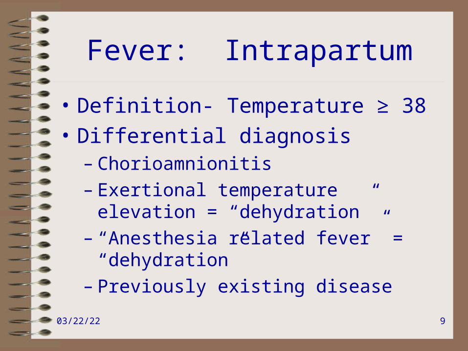
Fever: Intrapartum
• Definition- Temperature ≥ 38
• Differential diagnosis– Chorioamnionitis– Exertional temperature elevation =
“dehydration”– “Anesthesia related fever” = “dehydration”– Previously existing disease

04/19/23 10
Fever: IntrapartumDiagnostic Plan
• Physical exam– Exertional temperature elevation/ “anesthesia related
fever”- includes only low grade temperatures, ie T< 38.0 (F100.4)– Research definition of “chorio” includes maternal
fever and one more sign/symptom including maternal tachycardia (>100 bpm), fetal tachycardia, foul smelling lochia, or tender uterus
– Clinical definition, “chorio” is T ≥ 38.0 (F100.4)

04/19/23 11
Fever: IntrapartumTreatment
• Diagnosis determines treatment– Exertional temperature elevation“Bolus”– Chorioamnionitis Ampicillin/Gentamicin
during labor• PCN allergic-->Kefzol • If PCN anaphylaxis-->clinda/erythro if known GBS
sensitivities available. Vanco if unknown.• If C/S is performed, add anaerobic coverage. Generally
continued for 48 hours post-op.• Studies have shown that a single dose of antibiotic post
vaginal delivery is as good as 24 hour doses.

04/19/23 12
Fever: Post Partum
• Whole different world!
• Definition– Temperature greater than 38.5 X1, or– Temperature greater than 38.0 X2 after the
first 24 hours post partum

04/19/23 13
Fever: Post PartumDiagnosis
• Differential Diagnosis (head to toe)– Mastitis– Atelectasis/Pneumonia—aspiration or hospital acquired– Endometritis– Pyelonephritis– Cellulitis/Wound Abscess– Vaginal hematoma/abscess– DVT/other thrombosis (septic pelvic thrombophlebitis)– Drugs and other usual suspects

04/19/23 14
Fever: Post PartumDiagnosis
• Endometritis-– Uterine tenderness, foul smelling lochia– Absence of other obvious source– Know your bugs- On Creogs
• Polymicrobial• 80% involve anaerobic organisms—peptostreptococci,
bacteroides, etc.• Gram neg rods (E.coli), Gram pos cocci (GBS), etc.• Late endometritis—that is two weeks out may involve
chlamydia—so add doxy to this regimen

04/19/23 15
Fever: Post PartumDiagnostic Plan
• Physical Exam
• +/- U/A, Ucx
• +/- CBC
• +/- Blood cultures X2
• +/- CXR
• +/- stool culture
04/19/23 16
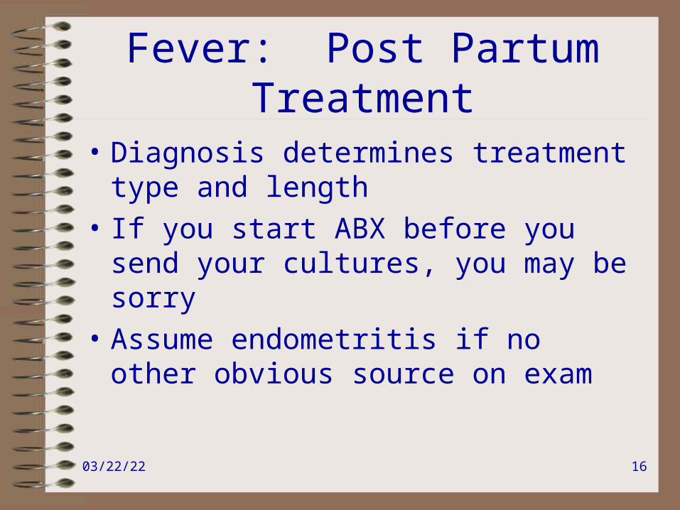
Fever: Post PartumTreatment
• Diagnosis determines treatment type and length
• If you start ABX before you send your cultures, you may be sorry
• Assume endometritis if no other obvious source on exam

04/19/23 17
Fever: Post PartumTreatment
• Endometritis– This is the only bacterial infection that I know of for
which you stop ABX when pt. is afebrile!!– Most will stop ABX when a pt. has been afebrile for
24-48 hours. If the pt. is s/p C/S—usually 48 hours.– Traditional antibiotics are “Triples,” but other broad
spectrum antibiotics have been shown to be just as efficacious-Amp/Gent/Clinda—daily or thrice daily dosing-Clinda/Gent alone – recommended by ACOG-Zosyn, Unasyn, Cefotetan, Augmetin (po!!)

04/19/23 18
Fever: Post PartumEndometritis
• Blood cultures are done in a patient with endometritis to direct care if the patient NOT responding.
• 10-20% of endometritis will have positive blood cultures.
• 10-20% of endometritis will be secondary to inadequately covered enterococcus.
• Although most cultures reveal a single organism, the infection is STILL polymicrobial!

04/19/23 19
Fever: Post PartumTreatment
• Pyelonephritis– Traditional treatment is Amp/gent, new studies show
Cephalosporins also OK—Kefzol and Ceftriaxone are fine.
– When afebrile X 24 hours, change to po’s, need 14 day course
(if pt. not breast feeding, fluroquinolones ok, then only need 7 days)(+ blood cultures help with diagnosis, but do not alter treatment)
NO MACRODANTIN for PYELO!!!!

04/19/23 20
Fever: Post PartumTreatment
• Mastitis- Typically T≥38.3 with systemic symptoms– Dicloxicillin or Keflex (traditional)—both OK for breast feeding
and cover staph and strep. (Nafcillin or Kefzol if IV ABX needed.)
– New emphasis to cover MRSA if recent hospitalization, consider clindamycin 300 mg qid
– 10-14 day course– Breast feeding or pumping hastens recovery.– NSAIDS– Abscesses must be drained and can be diagnosed by ultrasound

04/19/23 21
Fever: General
• Rote– Physical Exam– Blood culture X2– U/A, Ucx– +/- CXR– +/- stool cultures, ie C.diff

04/19/23 22
Fever: GeneralDifferent World!
• Definition- Temperature >38.5 (101.5)• Differential Diagnosis
– Infection– Drug– Thrombus- DVT-upper or lower extremity/PE– Atelectasis– Cancer– Inflammatory disease/Vasculitis/Other

04/19/23 23
Fever: GeneralDiagnostic Plan
Individualize according to the patient. Think through anatomically:
– Head: Sinusitis, Meningitis, otitis/pharyngitis
– Heart: Endocarditis
– Lungs: Pneumonia, pleural effusion
– Chest: Line infection
– Abdomen- abscess, pyelonephritis, biliary, infectious diarrhea,
spontaneous or secondary bacterial peritonitis
– Pelvis- PID/TOA, abscess
– Back- Decubitus ulcers, rectal abscess
– Extremities- cellulitis, septic thrombus, line infection, osteomyelitis

04/19/23 24
Fever: GeneralDiagnostic Plan
• If the patient is immunocompromised, expand your differential diagnosis
• If no obvious source of bacterial infection, think about viral causes of fever and the rest of the differential diagnosis

04/19/23 25
Fever: GeneralTreatment Plan
• Diagnosis determines treatment type, dose, and duration.
• Empiric treatment only if patient is septic or in danger of sepsis or life threatening complication.

04/19/23 26
Fever: GeneralTreatment Plan
• Broad spectrum antibiotics – Know what category of bug each antibiotic covers, ie gram
positive, negative, anaerobic, atypicals– Neutropenia: Each institution has its own hierarchy of Broad
spectrum coverage.– Chronic illness or hospitalization: Add coverage for resistant
gram positives with Vanco– If pt. in danger of dying or has a nosocomial infection, consider
“double coverage” of gram negatives, specifically pseudomonas– Traditional Pseudomonal ABXs include: Gent/Tobra, Ceftaz,
Cefepime, Zosyn/Timentin, Cipro, Imipenem/Meropenem, Aztreonam

04/19/23 27
Low Urine Output
Low urine output is not the problem, it signifies a problem
Your goal is not to make
the patient pee, but to figure out why she is not peeing

04/19/23 28
Low Urine OutputDefinition
• Low Urine Output-– Less than 0.5cc/kg/hr (30-40cc/hr in a typical
woman)
• Oliguria- 400-500 cc/day
• Anuria- Less than 50cc/day

04/19/23 29
Low Urine Output
• Differential Diagnosis– Intravascularly dry-
• True hypovolemia: intravascular depletion
• Hypervolemia with intravascular depletion: 3rd spacing or low albumin states
• “Intravascularly Dry”: low cardiac output, or low SVR (the kidney thinks the body is intravascularly dry)
– Acute kidney injury (Acute renal failure)
– Obstruction/Mechanical problem-outlet obstruction, ie FOLEY BLOCKADE, or hole in the bladder

04/19/23 30
Low Urine OutputDiagnostic Plan: Rote
• On the phone- rule out easy things first– Does the pt. have a foley
• If yes—flush foley
• If no- Place foley and call me with the output
• Determine volume status– Vital signs- HR, BP, O2 sat– Physicial exam- mucous membranes, neck
veins, lungs, extremities

04/19/23 31
Low Urine OutputDiagnostic Plan- Extras
Still can’t figure out volume status?
Here are some tools:– Blood- BUN/Cr, Na+, HCO3– Urine – sp. Gravitiy, urine Na+, urine
creatinine (calculate your FeNa!!!)– CVP if you have a central line in place

04/19/23 32
Low Urine OutputTreatment
• Intravasculary Dry: True Hypovolemia, including 3rd spacing and low albumin states– Give volume
• NS or LR
• Hesban or albumin
– Avoid nephrotoxins, specifically NSAIDS, ACEI’s, contrast dye
– Follow volume status on exam, O2 sat, I’s/O’s, daily wt.s very closely

04/19/23 33
Low Urine OutputTreatment
• “Intravascularly Dry”- CHF, Cirrhosis, sepsis– Treatment is illness and circumstance specific– You have to make the kidney see more
perfusion– ie increase cardiac output, increase SVR, and/or increase intravascular volume
– Avoid Nephrotoxins as above

04/19/23 34
Low Urine OutputTreatment
• Acute Kidney Injury (Acute renal failure)– Pre-renal azotemia- see Intravascularly dry above– Intra renal- in the hospital usually ATN
• ATN- – If secondary to pre-renal azotemia- fluid may help some,
but beware of fluid overload– Avoid nephrotoxins- NSAIDS, ACEI’s, contrast dye,
Aminoglycosides, Ampho B, Vanco• Interstitial Nephritis- avoid nephrotoxins- NSAIDS,
PCN/Cephalosporins• Glomerulonephritis/Vascular lesion—much less common
“hospital acquired problem”– Post-renal (ureteral/bladder/urethral obstruction)- see next

04/19/23 35
Low Urine OutputTreatment
• ATN can either be oliguric (no pee) or non-oliguric (yes pee)– Lasix can convert oliguric to non-oliguric but will not
change the renal prognosis
– Lasix will only help you control volume status/electrolytes, NOT IMPROVE RENAL FUNCTION
– ATN is managed supportively. Typical duration is 7-21 days, but may be months. A pt. may need dialysis for this time.

04/19/23 36
Low Urine OutputTreatment
Again !!!!• Lasix is used to treat symptoms of volume
overload– not low urine output
• Remember, low urine output is not your problem, it is what is causing the low urine output that is your problem

04/19/23 37
Low Urine OutputTreatment
• Obstruction/Mechanical
-You can treat this by removing or circumventing the obstruction
- After an obstruction is fixed, a pt. can develop “post-obstruction diuresis” which is an inappropriate diuresis– causing a pt. to become intravascularly dry if not monitored appropriately

04/19/23 38
Shortness of Breath
Differential Diagnosis:• LOW O2 SAT
– Hypoxemia
• Normal O2 SAT– Airway obstruction– Irritation of the pleura/lung parenchyma– Metabolic- Acidosis, Sepsis– Cardiac Ischemia equivalent– Anemia– Anxiety

04/19/23 39
Shortness of BreathDifferential Diagnosis
• Hypoxemia• Pulmonary edema- cardiogenic, non-cardiogenic
• Pneumonia
• Pulmonary embolism
• Atelectasis
• Pleural Effusion
• Pneumothorax
• Large Airway Obstruction
• Reactive Airway Disease/ COPD
• Restrictive Pulmonary Disease

04/19/23 40
SOB: Diagnostic PlanRote
• Current Vital signs, including a
ROOM AIR SAT
• Evaluate the patient immediately

04/19/23 41
Diagnostic Rote Plan
• Physical Exam- SICK vs. NOT SICK– Is the pt. in distress? – Diaphoretic? Tachypneic?– Altered Mental Status?– Cardiac exam- Tachycardic? Neck Veins?– Lung exam- Crackles? Wheeze?– Abdomen- Pain?– Extremities- Symmetric? DVT?

04/19/23 42
SOB: Diagnostic PlanRote
• If the pt. is sick- by virtue of vital signs or physical exam– CXR– EKG– Room Air ABG—if pt. too hypoxic to take off
oxygen, an ABG on O2 is still useful to evaluate ventilation

04/19/23 43
SOB: Diagnostic PlanRote
• CXR– Pulmonary infiltrates- Water, pus, or blood
(pulmonary edema, pneumonia, diffuse alveolar hemorrhage)
– Low lung volumes- poor breath, atelectasis, pleural effusion, pneumothorax
– Large lung volumes COPD– Normal lung fields think PE – Heart size

04/19/23 44
SOB: Diagnostic PlanRote
• EKG– Rate– Rhythm– Evidence of ischemia– Evidence of cardiac strain- via hypertrophy
and axis– Evidence of PE

04/19/23 45
SOB: Diagnostic PlanRoteABG
• Two components of respiratory distress– Oxygenation- Calculate the Aa gradient (on room air)– Ventilation- What is the pCO2?
• If the pCO2 is low (<40)– this is appropriate for someone who is hypoxic and trying to compensate with respiratory rate
• If the pCO2 is normal or high (near 40 or above)- – Is normal appropriate?—if the pt. appears to be working hard
to breathe, a nl or elevated pCO2 may represent resp. failure– This may be secondary to chronic pCO2 retention from COPD
You can check the HCO3-, if elevated you’re OK
04/19/23 46
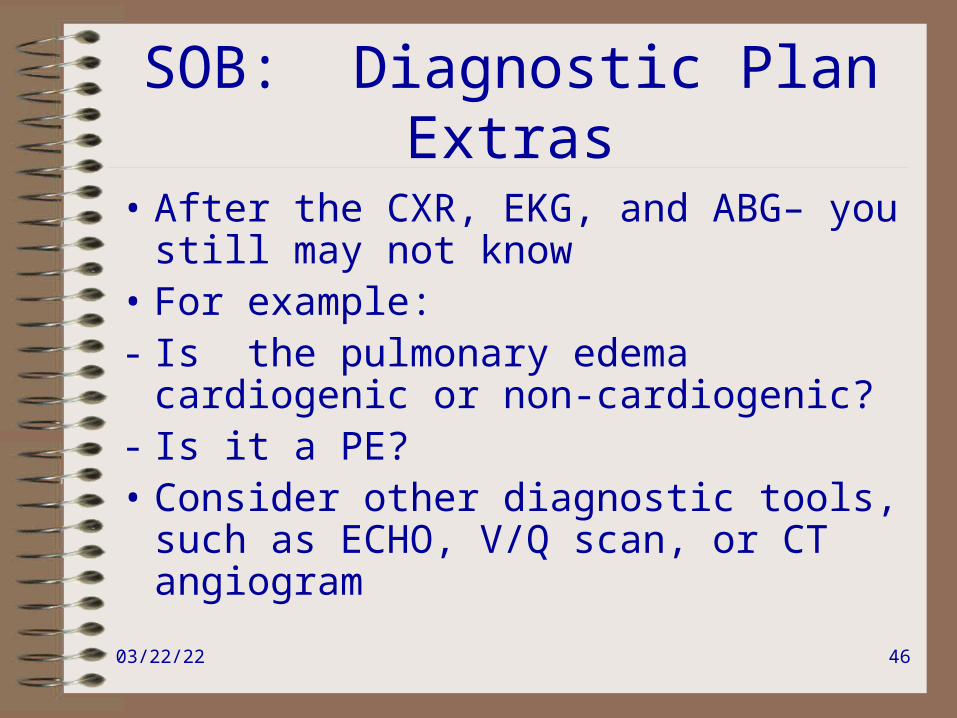
SOB: Diagnostic PlanExtras
• After the CXR, EKG, and ABG– you still may not know
• For example:- Is the pulmonary edema cardiogenic or
non-cardiogenic?- Is it a PE?• Consider other diagnostic tools, such as
ECHO, V/Q scan, or CT angiogram
04/19/23 47
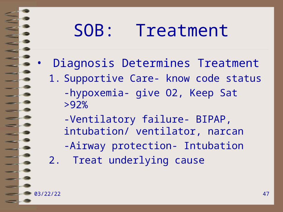
SOB: Treatment
• Diagnosis Determines Treatment1. Supportive Care- know code status
-hypoxemia- give O2, Keep Sat >92%
-Ventilatory failure- BIPAP, intubation/ ventilator, narcan
-Airway protection- Intubation
2. Treat underlying cause

04/19/23 48
SOB: Treat Underlying Cause
• Pulmonary edema- may need ECHO or SWAN to distinguish. These have different treatments and different prognoses.– Cardiogenic- Diurese, if pt. not in Sinus rhythm- convert
or slow to nl rate• Ask yourself, why she decompensated• If pt. on Mg++--Turn off the Mg++, give Ca gluconcate• ?MI, arrythmia, fluid overload, valvular lesion, peripartum
cardiomyopathy
– Non-Cardiogenic- Diuresis may help• Otherwise known as acute lung injury (ALI) or ARDS– depending
on extent• Treat underlying cause/Treatment primarily supportive
04/19/23 49
SOB: Treat Underlying Cause
• Pneumonia- Supportive care and ABX• Inpt.- 10- 14 day course of ABX, generally empiric
treatment. – Community Acquired-
1. cefotaxime/ cetriaxone/unasyn AND macrolide (azithro/clarithro/erythro) OR
2. Fluoroquinolones (moxi, gemi, levofloxicin)– ICU-
1. beta lactam AND azithro 2. Beta lactam AND fluoroquinolone3. Aztreonam AND fluoroquinolone
– Aspiration- Zosyn (Clinda OK for outpt. Aspiration)

04/19/23 50
SOB: Treat Underlying Cause
• Pneumonia– Outpt. Community Acquired PNA
– OK, if pt. <65, can take Po’s, has nl O2 sat, has capability of aquiring and taking ABX, has no comorbid illness, and is not pregnant
– May be bacterial or viral or mycobacterial!
– For bacterial: Azithro/doxy/fluoroquinolone OR Amoxicillin/Augmentin AND macrolide— 10-14 day course

04/19/23 51
SOB: Treat Underlying Cause
• Pulmonary Embolism- Think PE until proven otherwise– Risk Factors- ALL OF YOUR PTs.—any one who is
pregnant, post-op, or has cancer
– Work up may or may not show large Aa gradient, right axis / S1Q3T3 on EKG– pregnant women are especially tricky
– D-dimer ELISA is great for screening (great negative predictive value)—but will not work in pt.s who are pregnant, post-op, or who have cancer!!!

04/19/23 52
SOB: Treat Underlying Cause
• Think PE until proven otherwise– especially with a negative CXR– Anticoagulate immediately if suspicion is high enough
to get a definitive study (pretest probability>30%)– Lovenox 1mg/kg bid is treatment dose– Use unfractionated Heparin if worried about bleeding,
if pt. has renal disease, or if pt. very obese– Do not feel bad for anticoagulating or getting a
definitive study if the scan is negative—you still did the right thing

04/19/23 53
SOB: Treat Underlying Cause
• PE– the definitive study– CT angio vs. V/Q scan- The better test
depends on the radiologist and the institution– At Stanford they are equally good– If the pt. has a Cr>1.5, choose V/Q– If the pt. has underlying lung parenchymal
disease, choose CT angio

04/19/23 54
SOB: Treat Underlying Cause
• Asthma– Identify triggers and remove them– Albuterol immediately/add atrovent– if severe, may
need epi– Long acting β-agonist– Steroid inhaler– If severe, systemic steroids—Solumedrol or
Prednisone- most start with 30-60 mg qd and then do a rapid taper
****NOT all wheezes are “asthma”—wheeze can be heard with pulmonary edema

04/19/23 55
Chest PainDifferential Diagnosis
• Differential Diagnosis– Cardiac: Cardiac Ischemia/Pericarditis/Aortic
Dissection– Pulmonary: Pulmonary Embolism/ Pneumonia/
Pulmonary edema/Pleuritis/Pneumothorax– Musculoskeletal: Muscle spasm/Costochondritis/
Herpes Zoster– GI: GERD/gastritis/PUD/Esophageal
spasm/Pancreatitis/Biliary Disease– Pre-eclampsia– Anxiety—Diagnosis of exclusion

04/19/23 56
Chest Pain: Rote Diagnostic Plan
• Get vital signs from the nurse
• Order an EKG over the phone—STAT
• Think about a relevant DDx on your way!!

04/19/23 57
Chest Pain: RoteDiagnostic Plan
• Everybody gets an
EKG
And
Usually a CXR

04/19/23 58
Chest Pain: RoteDiagnostic Plan
• When you arrive at the scene
Rule out, Rule in:– You are basically taking a systematic approach
—• Is it Deadly?—Call for help.
• Is it Sick or Not Sick?
• Is it Cardiac/Pulmonary/GI/other?

04/19/23 59
Chest Pain: RoteDiagnostic Plan
• Get the EKG, ask for the nurse to obtain an old one
• Obtain vitals at bedside• Physical Exam
– Is the pt. in distress? Diaphoretic? Tachypneic?
– Is the pt.’s pain pleuritic? Reproducible with external pressure or limb movement?
– Heart exam- rate, rhythm, JVP
– Lungs- decreased breath sounds? Air movement?
– Abdomen- Acute abdomen?
– Extremities- Symmetric?

04/19/23 60
Chest Pain: RoteDiagnostic Plan
• History- As you are performing the exam, ask questions which relate to what you are examining.
• These questions are ROTE and memorized. They do not have long answers.
• Just interrupt the patient• If the pt. cannot answer– don’t waste time
here.

04/19/23 61
Chest Pain: RoteDiagnostic Plan
• Heart exam- Pain=Pressure=Discomfort– Have you ever had anything like this before?– CADRFs– What were you doing when it started? – Does it radiate to the arm, back, or neck? – Is it assoc. with Nausea/ Diaphoresis/ SOB/palpitations? – How long has it been present? – Does it come and go, or is it constant?– Out of 10, how bad is it? – Does it get worse with a deep breath?

04/19/23 62
Chest Pain: RoteDiagnostic Plan
• EKG- Obtain this as soon as possible, keep asking for it/help obtain it
• You need an old EKG. Just make a rule—ALL PATIENTS OVER 50 OR WITH HISTORY OF DIABETES OR CARDIAC DISEASE GET A BASELINE EKG ON ADMISSION– or you will be sorry when she develops Chest Pain.
• Strongly consider CXR

04/19/23 63
Chest Pain:Diagnostic Plan
• Use what you have learned in your evaluation- even if you are still waiting for studies.
• Identify the organ system involved- Cardiac/ Pulmonary/ GI/ Musculoskeletal/ other
• This will help determine treatment

04/19/23 64
Chest Pain:Treatment
• Deadly?—Call for help- more Nurses, Senior Resident, Medicine, Cardiology, or Code?
• Sick, Not Sick?-- Determine level of care.
• Organ system?— Hedge your bets, begin to treat while you are figuring it out.

04/19/23 65
Chest Pain:Treatment
• Diagnosis Determines Treatment
• Treatment Includes:– Treating Underlying Disease– Giving analgesics

04/19/23 66
Chest Pain: Treatment
• Think of BAD things first.• Consider treating these empirically, if they are
low risk interventions. – Give O2– Consider ASA– if no contraindications, will decrease
mortality by 23-50%, if unstable angina or true MI--pt.s may chew it.
– Consider Maalox/Nitroglycerin—for diagnosis/ treatment.
– Turn off the Mg++ if it is on.

04/19/23 67
Chest Pain:Cardiac Ischemia
• Treatment-– Oxygen– Morphine for pain– Nitroglycerin for pain- SL/paste/drip- typically 0.4 mg SL given
q 5 min. X3, then another route should be used—hold for SBP<100—obtain post pain EKG
– ASA to decrease risk of MI – Try to decrease myocardial work/increase O2 delivery– Consider beta blockade (with MI, decreases mortality by 15-
30%)– Consider transfusion if Hct<30– Bring HR and BP to normal range

04/19/23 68
Chest Pain:Cardiac Ischemia
• Call Cards to help decide- +/- Lovenox, IIb/IIIa inhibitor, Cath lab, or TPA/thrombolysis– these are EKG dependent
• “Time is Myocardium!”
• Aortic Dissection is a contraindication to heparinization, etc.

04/19/23 69
Chest Pain:Pleuritic
• Treat underlying cause- ie. ABX, lasix, chest tube etc.
• If you suspect PE >30% pre-test probability give lovenox—rule out aortic dissection first.
• Treat with analgesics– Narcotics are good for air hunger—but careful if
worried about drive to breathe– NSAIDs are good for pleurisy—careful if concerned
about bleeding

04/19/23 70
Chest Pain: GI
• GERD/PUD—Maaolx good for acute discomfort, consider Pepcid, PPI– May need additional outpatient diagnostic and
treatment follow up
• GI disaster- perforated viscous, ischemic bowel, pancreatitis—individualize treatment

04/19/23 71
Chest Pain:Other
• Musculoskeletal- NSAIDs• Pre-eclampsia- True Abd. Pain implies
severe disease and end organ complications
• Anxiety- Reassurance, consider Benzo.– This is a true diagnosis of exclusion– If panic attacks– pt. may need outpt. diagnosis
and treatment—SSRIs generally used

04/19/23 72
Floor Calls
THE END

04/19/23 73