7a.1 7a. Structure Elucidation: IR and C-NMR ... · PDF file7a.2 • Regardless of...
15
© 2009, Department of Chemistry, The University of Western Ontario 7a.1 7a. Structure Elucidation: IR and 13 C-NMR Spectroscopies (text 11.1 – 11.5, 12.1 – 12.5, 12.10) A. Electromagnetic Radiation • Energy is transmitted through space in the form of electromagnetic radiation, which are oscillating waves of electric and magnetic fields, and this is term used to describe all the component waves found in a continuous spectrum known as the electromagnetic spectrum. • The spectrum stretches from the very short wavelength (high energy) gamma rays to the very long wavelength (low energy) radio waves. • Humans can see the 400 – 700 nm visible region (colour).
Transcript of 7a.1 7a. Structure Elucidation: IR and C-NMR ... · PDF file7a.2 • Regardless of...

© 2009, Department of Chemistry, The University of Western Ontario 7a.1
7a. Structure Elucidation: IR and 13C-NMR Spectroscopies (text 11.1 – 11.5, 12.1 – 12.5, 12.10) A. Electromagnetic Radiation • Energy is transmitted through space in the form of
electromagnetic radiation, which are oscillating waves of electric and magnetic fields, and this is term used to describe all the component waves found in a continuous spectrum known as the electromagnetic spectrum.
• The spectrum stretches from the very short wavelength (high energy) gamma rays to the very long wavelength (low energy) radio waves.
• Humans can see the 400 – 700 nm visible region (colour).
7a.2
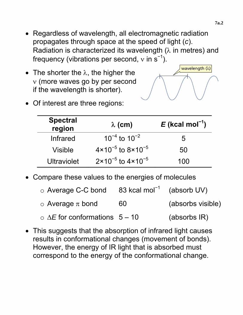
• Regardless of wavelength, all electromagnetic radiation propagates through space at the speed of light (c). Radiation is characterized its wavelength (λ in metres) and frequency (vibrations per second, ν in s−1).
• The shorter the λ, the higher the ν (more waves go by per second if the wavelength is shorter).
• Of interest are three regions:
Spectral region λ (cm) E (kcal mol−1)
Infrared 10−4 to 10−2 5 Visible 4×10⁻⁵ to 8×10⁻⁵ 50
Ultraviolet 2×10⁻⁵ to 4×10⁻⁵ 100
• Compare these values to the energies of molecules
o Average C-C bond 83 kcal mol−1 (absorb UV)
o Average π bond 60 (absorbs visible)
o ΔE for conformations 5 – 10 (absorbs IR)
• This suggests that the absorption of infrared light causes results in conformational changes (movement of bonds). However, the energy of IR light that is absorbed must correspond to the energy of the conformational change.

7a.3
A. IR Spectroscopy • As we have seen in first-year chem, the absorption of IR
light causes the vibrational excitation of bonds. The energy of the IR light absorbed depends on the functional group.
• Recall the functional group region and the fingerprint region of IR spectra, which are normally scanned from 4000 to 600 cm−1 (wavenumbers, reciprocal of λ when measured in cm). Absorption appears as peaks of lowered transmittance.

7a.4
1. IR spectra of alkanes, alkenes, and alkynes a. alkanes
• Saturated alkanes have relatively simple spectra, with peaks originating from C–H bonds
o Strong peak with multiple peaklets between 2850 – 3000 cm−1 (C–H stretch)
o Medium peak around 1450 cm−1 (CH2 bend)
o Weak peak around 1400 cm−1 (CH3 bend)
• These peaks are common to all organic compounds that contain saturated alkyl groups (i.e. not very useful!).
Aspirin

7a.5
b. alkenes
• Peaks due to C=C can be difficult to notice in IR. They are:
o A weak/medium peak around 1600 – 1680 cm−1 (C=C stretch)
o A weak/medium peak around 3000 – 3100 cm−1 (=C–H stretch); only present if there is an H on the C
c. alkynes
• Characteristic alkyne absorptions are:
o Weak peak between 2100 – 2160 cm−1 (C≡C stretch)
o A weak/medium peak around 3300 cm−1 for alkynes with an H on the C≡C atoms (≡C–H stretch).
7a.6
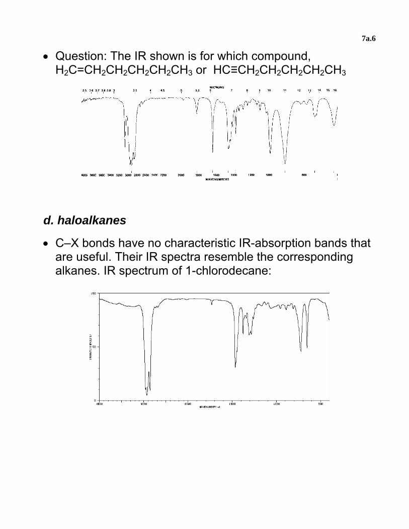
• Question: The IR shown is for which compound, H2C=CH2CH2CH2CH2CH3 or HC≡CH2CH2CH2CH2CH3
d. haloalkanes
• C–X bonds have no characteristic IR-absorption bands that are useful. Their IR spectra resemble the corresponding alkanes. IR spectrum of 1-chlorodecane:

7a.7
C. Nuclear Magnetic Resonance Spectroscopy 1. Energy absorption of nuclei
• Similarly to the spin of electrons, protons and neutrons can have spin quantum numbers of ± ½.
• This number depends on the total number of protons and neutrons in the nucleus. Atomic nuclei will have an overall nuclear spin and a spin number (I).
• All nuclei with an odd mass number (odd number of total neutrons and protons) can have two states, I = ± ½. That is, two energy spin states, ½ and −½ .
o Examples include 1H, 13C, 19F, 31P
• For nuclei with states I = ± ½, the spinning nucleus can be viewed as a spinning bar magnet. If such nuclei are placed in a magnetic field (B0), the two spin values assume two different energy states.
• The lower-energy spin state (+ ½) aligns its magnetic moment with the external field applied, and the higher-energy state (− ½) is aligned opposed to the applied field.

7a.8
• Therefore, in a strong magnetic field, nuclei with spin will absorb radiation of energy equal to the energy difference between the two states.
o Absorption of radiation by nuclei in a magnetic field is called nuclear magnetic resonance, NMR.
o For organic chemists, the two most useful types are
1H , termed proton NMR (1H NMR), which we will examine later on
13C, termed carbon NMR (13C NMR)
• For all types of NMR, the stronger the magnetic field (B0), the greater the energy separation between the two states.
• Very strong magnetic fields are required to make the separation large enough to be useful. For example, 13C
o At B0 = 7.5 T (Tesla), ΔE = 0.0072 cal/mol
o This energy separation corresponds to an electromagnetic frequency of 75 MHz and a wavelength of 1000 m (radiofrequency waves).
• Most commercial NMR instruments consist of a fixed magnetic field, a radiofrequency generator to “radiate” the nuclei, a receiver to detect the absorption, and a recorder.

7a.9
• The sample is dissolved in a solvent that does not interfere with the magnetic field. The sample is placed in a tube and spun rapidly in the machine’s field.
• The solvent chosen must be not contain nuclei with spin, so no hydrogens! Common solvents used are CCl4 and CDCl3.
2. 13C NMR
• The most abundant isotope of carbon is 12C (98.9%), which has no nuclear spin. But all substances containing carbon will contain the 13C isotope (1.1%), which does have spin and will give a signal in an NMR instrument.
• The local magnetic fields of carbon atoms in a molecule are affected by all neighbouring atoms. This makes the actual magnetic field around a specific atom slightly different than the applied field.

7a.10
• Therefore, every different carbon environment in a molecule leads to the absorption of a different radiofrequency
• We can determine the number of different C atoms (and thus 13C signals) by looking at molecular symmetry.
O
O
OHO
OOH
O OH
O
HO
1 signal
3 signals
citric acid
4 signals
• Instead of plotting signals on the x-axis of radiofrequency energy, absorption signals are plotted relative to a single 13C absorption signal of a reference compound, TMS.
Sitetramethylsilane (TMS)
• The signal of the TMS is arbitrarily set at zero, and all the signals of the molecule being analyzed absorb at different chemical shifts (units of delta δ).
o δ is measured in parts per million. Each ppm corresponds to one part per million in the frequency of radiation absorbed (e.g. 75 Hz in a 75 MHz machine).
• EWG’s, and greater s-character in hybrid C orbitals, shift 13C signals to the left of TMS. This is termed downfield.

7a.11
• Therefore, C with different magnetic environments will have a different chemical shift relative to TMS.
• In all 13C-NMR spectra, if a signal is visible at 0 ppm, it corresponds to the TMS signal.
• Thus, 13C-NMR spectra reveal the number of magnetically non-equivalent C atoms and their identity (chemical shifts).

7a.12
• Question: Predict the number of signals for the following compounds. Do the spectra agree with their structure?
OHCl
4-chlorophenol1-pentene
• Important note: signal heights on 13C-NMR spectra do not correspond with the number of C atoms they represent (see 1-pentene). However, higher signals usually result from greater numbers of equivalent nuclei (see 4-chlorophenol).
• The analysis of IR and 13C-NMR spectra, together with info about units of unsaturation, often gives enough information to elucidate molecular structure (see assigned problems).
• Summary: o C11: 304 – 312. C12: 329 – 334 and 342 – 346. o Problems: 11.9 – 11.12, 11.20, 12.9, 12.10, 12.14 o Dec 05: 16 – 18. Dec 06: 14, 15, 17.
Dec 07: 14 – 16. Dec 08: 14 – 16.
7a.13
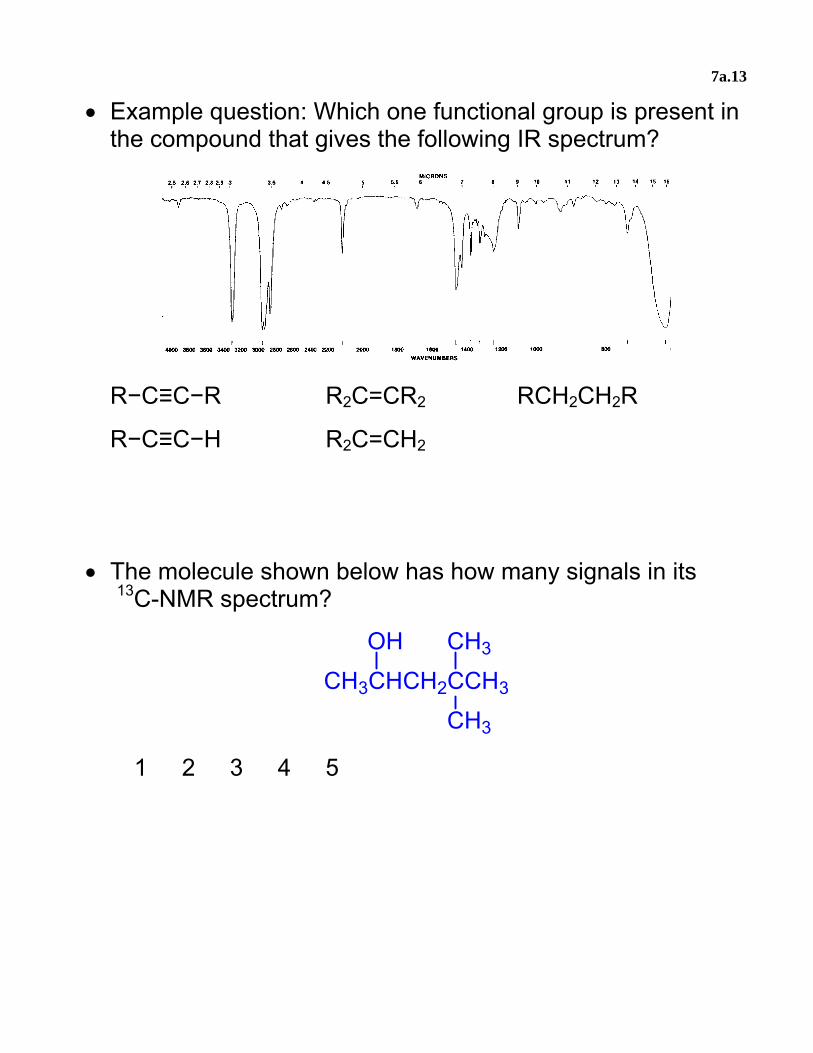
• Example question: Which one functional group is present in the compound that gives the following IR spectrum?
R−C≡C−R R2C=CR2 RCH2CH2R
R−C≡C−H R2C=CH2
• The molecule shown below has how many signals in its 13C-NMR spectrum?
CH3CHCH2CCH3
OH CH3
CH3
1 2 3 4 5
7a.14
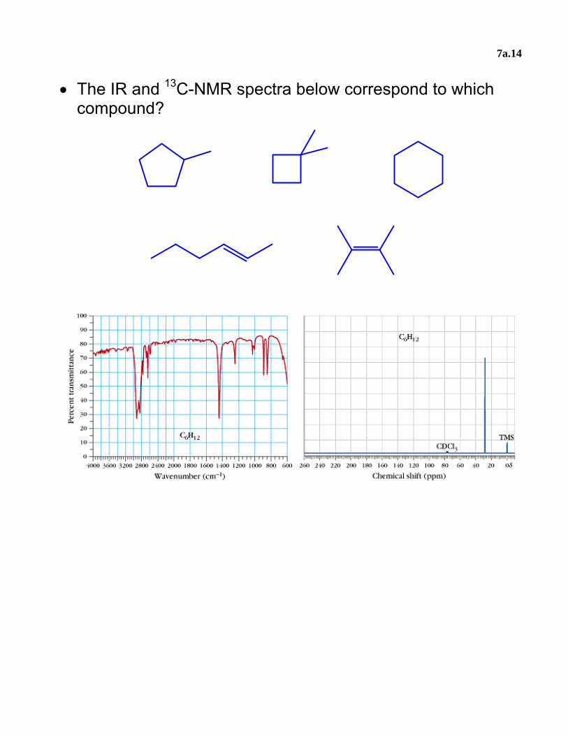
• The IR and 13C-NMR spectra below correspond to which compound?
7a.15
• MCAT questions
o What change occurs in a molecule when IR irradiation is absorbed?
• The spin of the electron flips • The vibrations of the bonds change • The rotation around the bond axes change • The translational motion changes
o What causes a peak in an NMR spectrum?
• A deflection of a molecule by a magnetic field • The absorption of electromagnetic radiation • The reorientation of a molecule in a magnetic
field • A reflection of a frequency of electromagnetic
radiation o A stretch at 1700 cm−1 in an IR spectrum indicates the
presence of which functional group? • ketone • alcohol • alkene • alkyne