
7 tract Infections of the respiratory - uib.cat · 7.1 Pathogenesis 71 7.2 Diagnosis 72 7.3...
15
7.1 Pathogenesis 71 7.2 Diagnosis 72 7.3 Management 72 7.4 Diseases and syndromes 73 7.5 Organisms 79 Self-assessment: questions 80 Self-assessment: answers 83 7.1 Pathogenesis The principal function of the respiratory tract is gas exchange. It is therefore constantly exposed to the gaseous environment, including particulate organic material, such as bacteria, viruses and spores (Ch. 3). Although the entire respiratory tract is constantly exposed to air, the majority of particles are filtered out in the nasal hairs and by inertial impaction with mucus- covered surfaces in the posterior nasopharynx (Fig. 11). The epiglottis, its closure reflex and the cough reflex all reduce the risk of microorganisms reaching the lower respiratory tract. Particles small enough to reach the tra- chea and bronchi stick to the respiratory mucus lining their walls and are propelled towards the oropharynx by the action of cilia (the ‘mucociliary escalator’). Antimicrobial factors present in respiratory secretions further disable inhaled microorganisms. They include lysozyme, lactoferrin and secretory IgA. Particles in the size range 5–10 µm may penetrate further into the lungs and even reach the alveolar air spaces. Here, alveolar macrophages are available to phagocytose potential pathogens, and if these are overwhelmed neutrophils can be recruited via the inflammatory response. The defences of the respira- tory tract are a reflection of its vulnerability to micro- bial attack. Acquisition of microbial pathogens is 71 Infections of the respiratory tract 7 Learning objectives You should: ● understand the mechanisms by which respiratory infections occur ● know how pathogens overcome host defences ● understand what factors increase vulnerability to respiratory infections. Overview This chapter deals with infections of structures that constitute the upper and lower respiratory tract. The general population commonly experiences upper respiratory tract infections, which are often seen in general practice. Lower respiratory tract infections are less common but are more likely to cause serious illness and death. Diagnosis and specific chemotherapy of respiratory tract infections present a particular challenge to both the clinician and the laboratory staff. Successful preventive strategies are available for several respiratory infections. Fig. 11 Defences of the respiratory tract.
-
Upload
phungnguyet -
Category
Documents
-
view
217 -
download
0
Transcript of 7 tract Infections of the respiratory - uib.cat · 7.1 Pathogenesis 71 7.2 Diagnosis 72 7.3...
7.1 Pathogenesis 71
7.2 Diagnosis 72
7.3 Management 72
7.4 Diseases and syndromes 73
7.5 Organisms 79
Self-assessment: questions 80
Self-assessment: answers 83
7.1 Pathogenesis
The principal function of the respiratory tract is gasexchange. It is therefore constantly exposed to thegaseous environment, including particulate organicmaterial, such as bacteria, viruses and spores (Ch. 3).Although the entire respiratory tract is constantlyexposed to air, the majority of particles are filtered out in
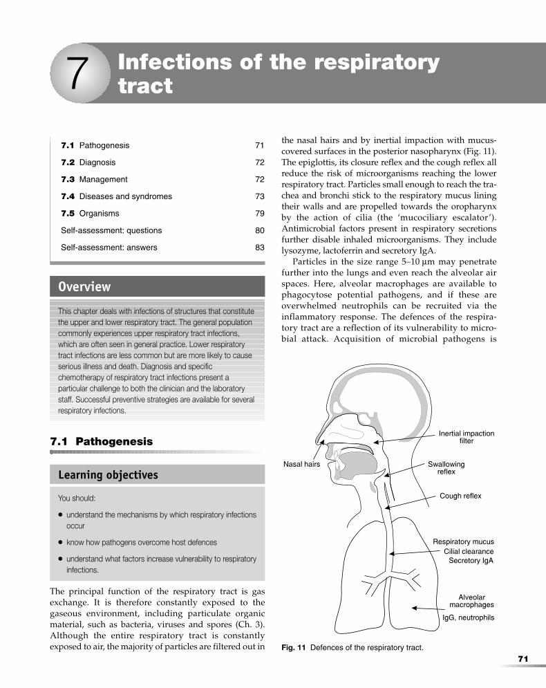
the nasal hairs and by inertial impaction with mucus-covered surfaces in the posterior nasopharynx (Fig. 11).The epiglottis, its closure reflex and the cough reflex allreduce the risk of microorganisms reaching the lowerrespiratory tract. Particles small enough to reach the tra-chea and bronchi stick to the respiratory mucus liningtheir walls and are propelled towards the oropharynxby the action of cilia (the ‘mucociliary escalator’).Antimicrobial factors present in respiratory secretionsfurther disable inhaled microorganisms. They includelysozyme, lactoferrin and secretory IgA.
Particles in the size range 5–10 µm may penetratefurther into the lungs and even reach the alveolar airspaces. Here, alveolar macrophages are available tophagocytose potential pathogens, and if these areoverwhelmed neutrophils can be recruited via theinflammatory response. The defences of the respira-tory tract are a reflection of its vulnerability to micro-bial attack. Acquisition of microbial pathogens is
71
Infections of the respiratorytract7
Learning objectives
You should:
● understand the mechanisms by which respiratory infectionsoccur
● know how pathogens overcome host defences
● understand what factors increase vulnerability to respiratoryinfections.
Overview
This chapter deals with infections of structures that constitutethe upper and lower respiratory tract. The general populationcommonly experiences upper respiratory tract infections,which are often seen in general practice. Lower respiratorytract infections are less common but are more likely to causeserious illness and death. Diagnosis and specificchemotherapy of respiratory tract infections present aparticular challenge to both the clinician and the laboratorystaff. Successful preventive strategies are available for severalrespiratory infections.
Fig. 11 Defences of the respiratory tract.
F70954-07.qxd 12/10/02 7:36 AM Page 71
primarily by inhalation, but aspiration and mucosaland haematogenous spread also occur. Individualswith healthy lungs rarely have any bacteria beyondthe carina.
Respiratory pathogens have developed a range ofstrategies to overcome host defences. Influenza virus,for example, has specific surface antigens that adhere tomucosal epithelial cells. The virus also undergoes peri-odic genetic reassortment resulting in expression ofnovel adhesins to which the general population has noeffective immunity. Streptococcus pneumoniae andHaemophilus influenzae both produce an enzyme (IgAprotease) capable of disabling mucosal IgA. Both thesespecies, other capsulated bacteria and mycobacteria areall resistant to phagocytosis. Penetration of local tissuesis usually required before damage occurs, althoughviruses causing the common cold appear to be an excep-tion. In some lower respiratory tract infections, the hostresponse is the principal cause of damage.
Human behaviour can also increase the risk of re-spiratory infection. Tobacco smoking has this effect byreducing the efficiency of cilial function and by causingthe production of more viscous respiratory secretions.Tracheal intubation for prolonged periods in the critical-ly ill bypasses the upper respiratory tract and provides aconduit for microbial access directly into the lungs.
7.2 Diagnosis
Clinical features
The features of different respiratory tract infectionslargely depend on the structures where inflammation islocalised and the extent to which function is altered. So,infection of the nasopharynx will result in a nasal dis-charge, bronchitis in cough and sputum production, andpneumonia in cough and sputum, but also in increasedrespiratory rate and chest radiograph changes.
Most upper respiratory tract infections are caused byviruses and are self-limiting. A specific aetiological diag-nosis would not alter treatment and would be costly.The role of the physician is limited to reassuring the
patient and recognising the more serious bacterial infec-tions that require specific antimicrobial chemotherapyor more extensive supportive treatment.
Lower respiratory tract infection should always betaken seriously since it is more likely to cause seriousmorbidity or even death.
Laboratory tests
History, physical examination, X-rays and laboratoryinvestigations focus on two issues: the degree of res-piratory compromise and the identity of the causalpathogen. Since a wide range of candidate pathogensmay have to be considered, the number of likely candi-dates should be reduced as far as possible by searchingfor clues in the history, examination and preliminaryresults. A history of tobacco consumption, recent travel,occupation, pets, and contacts with similar symptomsshould be sought.
Diagnostic specimens can be obtained from the res-piratory tract with deceptive ease, but their valueis often limited by contamination by the indigenousflora of the oral cavity. To prevent contaminationof lower respiratory tract specimens, the upper res-piratory tract must be bypassed. Chest X-rays are afundamental part of evaluation of lower respiratorytract infections and provide evidence of the distribu-tion and extent of disease more reliably than signselicited by auscultation. Postero-anterior views aremost commonly used, but a lateral view can providevaluable additional information.
Blood gas analysis should be performed if there isany suspicion of acute respiratory compromise. The keyindicators of disease severity in pneumonia are raisedrespiratory rate (> 30 beats/min), hypoxia, hypercapnia,bilateral or recently enlarging radiographic opacities,shock, renal failure and confusion.
7.3 Management
Chemotherapy
The antimicrobial therapy of respiratory tract infec-tion depends not only on the likely microbial cause of
72
Infections of the respiratory tract7
Learning objectives
You should:
● know which features indicate that a specific area of therespiratory tract is infected
● know how to assess respiratory compromise
● know how to identify the pathogen.
Learning objectives
You should:
● know when chemotherapy is indicated
● know how to choose the most suitable drug
● know how to prevent infection and the spread of infections.
F70954-07.qxd 12/10/02 7:36 AM Page 72

infection but also on the primary site involved and theseverity of disease. The commoner upper respiratorytract infections are rarely life threatening and in manycases are self-limiting. It is therefore possible to man-age many of these infections without specificchemotherapy, thereby avoiding all the possibleadverse effects. However, even apparently trivialinfections such as pharyngitis may require specificantibiotic treatment in some cases. The problem is inknowing who and when to treat with antimicrobialagents.
Lower respiratory infections are less of a problemin this respect, since infection is much more likely tocause significant morbidity and mortality. Antibioticsshould be used as early as possible in the course ofinfection. The problem here is in knowing which of awide range to choose. It is often necessary to make a‘best guess’ or presumptive choice in severely illpatients, based on the most likely microbial agent. Theinitial choice of chemotherapy may have to be sub-stantially modified in the light of laboratory results.Patients with pneumonia who are ill enough to requirehospitalisation usually require parenteral antibiotics.A syndrome-based choice of therapy has become thepreferred approach, since antibiotic choice and deci-sions on the need for hospital admission and activesupportive care do not have to wait for a laboratory-based aetiological diagnosis.
Prevention
The ease with which respiratory infections can bespread and their associated morbidity has led to thedevelopment of specific preventive approaches.Influenza can be prevented by immunisation with alive attenuated vaccine. The changes in epidemicstrains of influenza virus necessitate periodic changesin vaccine composition and revaccination of high-riskgroups such as the elderly and patients with cardiac orrenal failure. Pneumococcal infection can also be pre-vented by vaccination. Like influenza, changes in pre-vailing infective strains (capsular polysaccharidetypes) require alterations in the composition of thepolyvalent vaccine. Again, vaccination is restricted tohigh-risk groups. Infection with Mycobacterium tuber-culosis can be prevented by vaccination with a live-attenuated strain (BCG; bacillus Calmette–Guérin),although protection against pulmonary infection maybe only partial in some populations. In hospitals, thespread of respiratory infection from known cases ofinfluenza and pneumonia can be prevented by infec-tion control procedures. These are referred to as ‘addi-tional precautions’ and include nursing the patient in aseparate side ward, away from other patients and non-immune staff. Filter-type masks and aprons are also
worn by staff and other visitors. At a personal level,covering the mouth when coughing or sneezing is asimple but effective means of preventing the spread ofrespiratory pathogens.
7.4 Diseases and syndromes
The main infectious diseases of the respiratory tract arelisted in Table 9.
Pharyngitis
Pharyngitis is an inflammation of the throat, resulting inpain on swallowing and swollen, red pharyngealmucosa. It is most often caused by a respiratory virus(rhinovirus, coronavirus, adenovirus, influenza virus,parainfluenza viruses, respiratory syncytial virus),Epstein–Barr virus or coxsackievirus.
Aetiological clues include:
● conjunctivitis: adenovirus ● constitutional symptoms (lethargy and malaise) and
tonsillar exudate: Epstein–Barr virus ● posterior palatal ulcers: coxsackievirus ● abrupt onset, ‘doughnut’ pharyngeal lesions and
beefy uvula: Streptococcus pyogenes (group Astreptococcus)
● grey pharyngeal pseudomembrane in unvaccinatedsubject: Corynebacterium diphtheriae.
Bacterial pharyngitis Bacterial pharyngitis is less common and its single mostfrequent cause is S. pyogenes. Other rare bacterial causesinclude Neisseria gonorrhoeae, Mycoplasma pneumoniae, C.diphtheriae and Arcanobacterium haemolyticum. Peak inci-dence is between autumn and spring in temperate cli-mates, and during the rainy season in the tropics.Transmission is more rapid among groups sharingcrowded living quarters and is by droplet spread ordirect transmission.
Viral pharyngitis Viral pharyngitis is a self-limiting condition that doesnot usually require a specific aetiological diagnosis.
73
7Diseases and syndromes
Learning objectives
You should:
● know the major infections of the respiratory tract
● know the factors contributing to their occurrence
● understand the basis of their clinical management.
F70954-07.qxd 12/10/02 7:36 AM Page 73

Diagnosis When Epstein–Barr virus infection (infectious mononu-cleosis) is suspected, full blood count, blood film andPaul–Bunnell test for heterophile antibodies should berequested. This is not sensitive in Asians; in this groupIgM to viral capsid antigen should be sought. The inves-tigation most frequently requested for pharyngitis isdetection of S. pyogenes. This species is detected eitherby culture on blood agar and subsequent latex aggluti-nation reaction for group-specific polysaccharide, or bydirect antigen detection. Neither method can distin-guish oropharyngeal colonisation from true infection,but only culture allows antibiotic susceptibility testing.Suspicion of infection with N. gonorrhoea, Mycoplasmaspp., Arcanobacterium sp. or Corynebacterium spp. shouldbe communicated to the laboratory so that specialist,non-routine culture media can be used.
Treatment An oral penicillin or erythromycin is used to treat strep-tococcal pharyngitis. Treatment may not alter the courseof the primary pharyngeal infection, but it shouldreduce the risk of major non-infective sequelae suchas rheumatic heart disease, poststreptococcal glomeru-lonephritis and Sydenham’s chorea. The need forantibiotic treatment of streptococcal pharyngitis hasbeen questioned in developed countries, since the non-infective sequelae of streptococcal infection are all rare;but the recent increase in streptococcal infection inEurope and North America may change this view.
The other complications of streptococcal pharyngitisinclude scarlet fever (less common than in the past indeveloped countries), streptococcal toxic shock syn-
drome (both caused by toxin) and quinsy (paratonsillarabscess). In quinsy, there may be secondary infectionwith oral anaerobic bacteria, but these are often peni-cillin sensitive. Drainage of purulent foci is required.
Common cold
The common cold is a frequent occurrence, especially inyoung children and their parents during theautumn–spring period. The condition is caused mainlyby rhinoviruses. The size of the rhinoviral group, andthe causal role of other respiratory viruses in a minorityof common colds, has prevented the development of aneffective vaccine. There is a nasal discharge, nasalobstruction and sneezing. Pharyngitis and cough maybe present, but fever and myalgia are both rare features.There is no reason to use antimicrobial agents, and treat-ment should be restricted to alleviation of symptoms.
Influenza
Epidemic and endemic influenza occurs, caused byinfluenza virus groups A–C. Some of the features of acommon cold may be present, but systemic and res-piratory symptoms are more pronounced. Fever, lethargyand myalgia are all common. The influenza virus is anRNA virus with a segmented genome. Two major surfaceantigens are used in typing epidemic strains: haemagglu-tinin and neuraminidase. The different types of influenzavirus noted in successive epidemics are the result ofgenetic reassortment which causes an antigenic shift.Minor changes in antigenic makeup occur between epi-demics. These are referred to as antigenic drift. Antigenicshift results in influenza epidemics because it renders
74
Infections of the respiratory tract7Table 9 Infectious diseases of the respiratory tract
Infection Features
Pharyngitis Acute inflammation of the throat, resulting in pain on swallowing and swollen, red pharyngeal mucosa
Common cold Self-limiting rhinitis, causing nasal discharge, nasal obstruction, discomfort and sneezing Influenza Acute, usually self-limiting, viral infection with respiratory and systemic features Otitis media Acute inflammation of the middle ear Otitis externa Inflammation of the external auditory meatus Acute sinusitis Inflammation of the maxillary, frontal, ethmoid or sphenoidal sinuses Laryngitis Inflammation of the larynx, with hoarseness and loss of voice Bronchitis Cough and sputum production; can be acute or chronic Pneumonia Acute, community-acquired Occurs prior to or immediately after hospital admission; cough, chest signs and fever Acute, hospital-acquired Occurs in vulnerable patients in hospital; onset gradual and symptoms unreliable for
diagnosis Chronic Insiduous onset, prolonged course; usually diagnosed by radiological findings In AIDS See Ch. 18
Pulmonary tuberculosis Coughing and sneezing occur, fever, night sweats, weight loss and coughing blood; chest X-ray demonstrates lung changes
Empyema Accumulation of purulent fluid in the pleural space Croup See Ch. 16 Epiglottis See Ch. 16 Bronchiolitis See Ch. 16
F70954-07.qxd 12/10/02 7:36 AM Page 74
pre-existing specific immunity to influenza virus anti-gens obsolete. High mortality rates have been recordedduring influenza epidemics as a result of cardio-respiratory failure or secondary bacterial pneumonia(caused by Staphylococcus aureus or S. pneumoniae).
Diagnosis Diagnosis is usually clinical, with serology reserved forepidemiological studies and pandemic surveillance.
Treatment Treatment is aimed at symptomatic relief and at compli-cations if they occur. However, amantidine treatmentmay be of benefit if commenced early during infectionwith epidemic type A strains.
New treatments for influenza infection, such as theneuraminidase inhibitor oseltamivir, may reduce theduration of symptoms in a proportion of patients.
A vaccine is available, but it is only effective againstpreviously isolated strains. The vaccine is thereforeoffered to those at high risk of complications, i.e. theelderly, those with cardiac or respiratory disease, thosewith renal failure, the inhabitants of residential institu-tions and those in high-risk occupations (e.g. health care).
Otitis media
Otitis media is an acute inflammation of the middle ear.It is most frequent in the younger child, whose eusta-chian tube is shorter and more horizontal. It is also moreprone to blockage by hypertrophic lymphoid tissue atthe proximal end, as a result of prior respiratory tractinfection. Purulent fluid accumulates behind a tense, redtympanic membrane and may discharge externally afterrupture of the membrane. Infection is most often causedby S. pneumoniae or H. influenzae. Fever and local painare common features. Common complications includesecretory otitis media and impaired hearing. Much rarercomplications are meningitis and mastoiditis.
Diagnosis Diagnosis is mainly clinical. Auroscopic examinationof both tympanic membranes should be performed.Aetiological diagnosis is possible only if purulent exu-date from the middle ear is cultured, either following dis-charge via the eardrum or following tympanocentesis.
Treatment Antimicrobial treatment is with an antibacterial agent(e.g. oral ampicillin or erythromycin for 7–10 days).Some authorities recommend decongestant therapy asan alternative in uncomplicated acute otitis media.
Otitis externa
Inflammation of the external auditory meatus is most oftencaused by the hyphae-forming fungus Aspergillus niger.
Diagnosis is by culture of fungus from exudate. Aural toilet and treatment with a topical agent such
as aluminium acetate may be sufficient. Topical antibiot-ic preparations should be avoided. A rare, sometimeslife-threatening variant, called malignant otitis externa,occurs in diabetics and is caused by Pseudomonas aerugi-nosa. Therapy with agents effective against Pseudomonasspp. should be used.
Acute sinusitis
Infection of the axillary, frontal, ethmoid or sphenoidalsinuses with bacteria from the nasopharynx followsimpaired drainage of sinus secretions as a result of aprior upper respiratory tract infection or similar cause.The bacteria most commonly implicated are S. pneumo-niae and H. influenzae. Infection causes the sinus to fill upwith mucopus, which alters the resonance of the voiceand causes a feeling of local discomfort.
Diagnosis Diagnosis is mainly from the symptoms, but specialradiographic views may show filling of a maxillarysinus. Representative bacteriological specimens are dif-ficult to obtain.
Treatment Treatment is with decongestants to improve drainage.Surgical procedures may be required in more severe orpersistent cases. Some authorities argue that oral antibi-otics (e.g. ampicillin or erythromycin) should be used inaddition.
Laryngitis
Laryngitis is caused by one of the ‘respiratory’ virusesand is a self-limiting condition of hoarseness and loss ofvoice. It may also be a feature of a common cold orinfluenza. No specific therapy is required.
Bronchitis
There are three related conditions: acute bronchitis (inthe strict sense), tracheobronchitis and acute exacerba-tion of chronic bronchitis.
Acute bronchitis. This condition involves a cough,sputum production (which is usually white to cream incolour) but no radiographic changes on chest X-ray.Infection is with M. pneumoniae.
Tracheobronchitis. Here, acute bouts of coughing arenot accompanied by significant sputum production.Infection is caused by influenza virus, and features of sys-temic infection such as fever and myalgia may be present.
Acute exacerbation of chronic bronchitis. A chronic pro-ductive cough changes to become productive of larger
75
7Diseases and syndromes
F70954-07.qxd 12/10/02 7:36 AM Page 75
quantities of newly purulent sputum. This may be theresult of infection with one of the respiratory viruses, S.pneumoniae or H. influenzae.
Diagnosis In practice, there is considerable overlap between thesethree conditions. Sputum culture is of limited diagnosticvalue. Some authorities recommend culture only whenthere is no response to treatment after 48 hours.
Treatment Some patients will benefit from a few days’ treatmentwith an antibacterial agent (e.g. oral ampicillin or erythro-mycin), but many patients will not experience any bene-fit from therapy.
Patients at risk of cardiac or respiratory failureshould be vaccinated against pneumococcal infectionand influenza.
Pneumonia: acute, community-acquired
Acute pneumonia has its onset either prior to or imme-diately after admission to hospital. It is one of the mostcommon infectious causes of death worldwide. Patientswith acute pneumonia usually have a cough, chest signsand fever. The cough may or may not be productiveof purulent sputum. Chest signs are variable and proneto subjective interpretation. They may indicate areasof consolidation, fluid in the air spaces or even thepresence of an effusion or cavity. The most importantconsequence of acute pneumonia is impairment ofrespiratory function, which should be assessed as a firstpriority. The identity of the likely infective agent willdetermine choice of antimicrobial therapy. A careful his-tory, thorough examination and appropriate chest X-raysshould provide some clues to the likely causative agent.
Four main clinico-pathological patterns of acutepneumonia are recognised:
● lobar pneumonia —pulmonary consolidation demarcated by border of
segment or lobe
—most often caused by S. pneumoniae—also caused by S. aureus, S. pyogenes (group A
streptococcus) and Legionella pneumophila● bronchopneumonia
—patchy consolidation around the larger airways —caused by S. pneumoniae, H. influenzae, S. aureus
and L. pneumophila● interstitial pneumonia
—fine areas of interstitial infiltration in lung fields —usually no sputum production at presentation —caused by Legionella sp., Mycoplasma spp. or virus —initial treatment is with erythromycin
● aspiration pneumonia —follows aspiration of oral or gastric contents —damage usually caused by chemical or mechanical
insult —chest X-ray changes either in lower right lobe or, if
supine, apex of right lower lobe —bacterial damage caused by oral streptococci or
anaerobes.
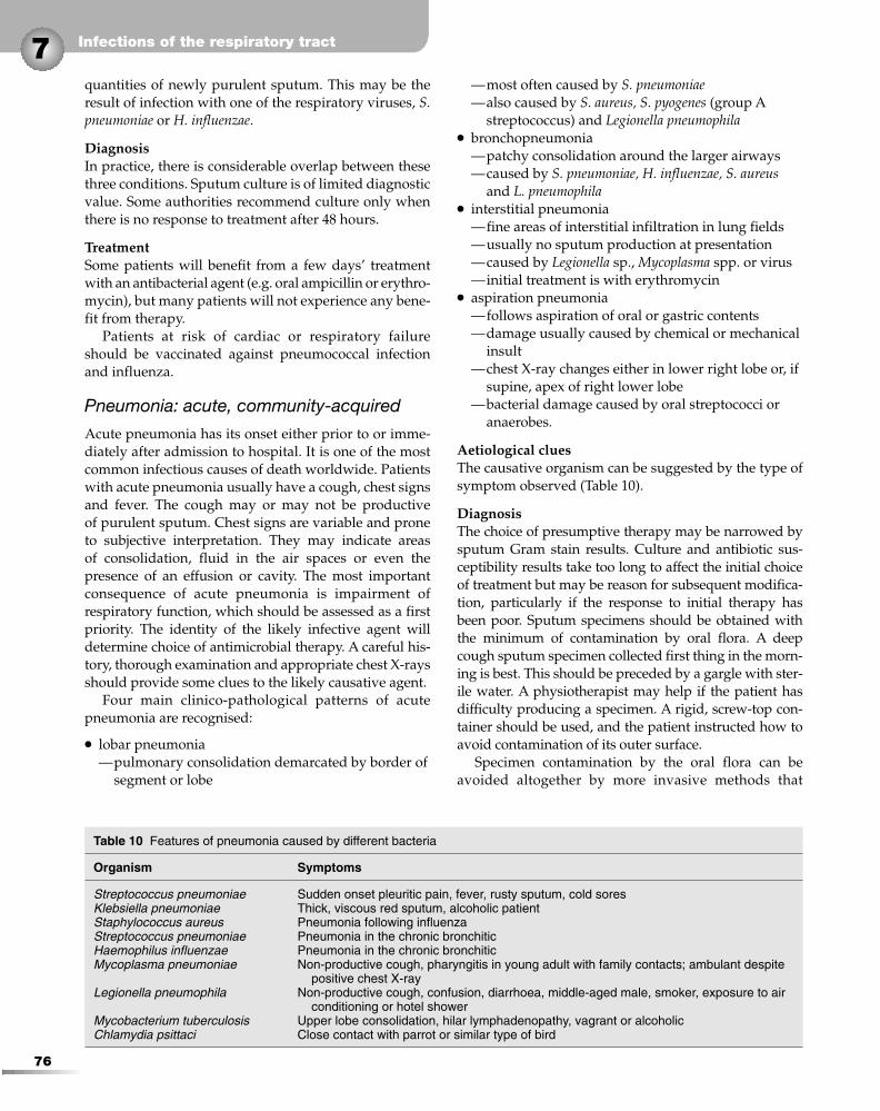
Aetiological clues The causative organism can be suggested by the type ofsymptom observed (Table 10).
Diagnosis The choice of presumptive therapy may be narrowed bysputum Gram stain results. Culture and antibiotic sus-ceptibility results take too long to affect the initial choiceof treatment but may be reason for subsequent modifica-tion, particularly if the response to initial therapy hasbeen poor. Sputum specimens should be obtained withthe minimum of contamination by oral flora. A deepcough sputum specimen collected first thing in the morn-ing is best. This should be preceded by a gargle with ster-ile water. A physiotherapist may help if the patient hasdifficulty producing a specimen. A rigid, screw-top con-tainer should be used, and the patient instructed how toavoid contamination of its outer surface.
Specimen contamination by the oral flora can beavoided altogether by more invasive methods that
76
Infections of the respiratory tract7
Table 10 Features of pneumonia caused by different bacteria
Organism Symptoms
Streptococcus pneumoniae Sudden onset pleuritic pain, fever, rusty sputum, cold sores Klebsiella pneumoniae Thick, viscous red sputum, alcoholic patient Staphylococcus aureus Pneumonia following influenza Streptococcus pneumoniae Pneumonia in the chronic bronchitic Haemophilus influenzae Pneumonia in the chronic bronchitic Mycoplasma pneumoniae Non-productive cough, pharyngitis in young adult with family contacts; ambulant despite
positive chest X-ray Legionella pneumophila Non-productive cough, confusion, diarrhoea, middle-aged male, smoker, exposure to air
conditioning or hotel shower Mycobacterium tuberculosis Upper lobe consolidation, hilar lymphadenopathy, vagrant or alcoholic Chlamydia psittaci Close contact with parrot or similar type of bird
F70954-07.qxd 12/10/02 7:36 AM Page 76
bypass the mouth. These include transtracheal aspir-ation, bronchoscopy with protected specimen collectiondevice and transbronchial or transthoracic biopsy.All these techniques require time, skill and specialequipment and may cause unwanted side effects.Blood culture should be performed if the patient has afever.
Preliminary result based on Gram stain can be pro-vided in minutes after the laboratory receives the speci-men. If the smear is full of neutrophil polymorphs and asingle type of organism (e.g. Gram-positive diplococ-cus), the result may make a timely contribution to clini-cal decision-making. Large quantities of saliva or thepresence of buccal epithelial cells in the smearsuggest that it is unsuitable for further bacteriologicalevaluation. It is important to alert the diagnostic labo-ratory to the possibility of Mycoplasma, Legionella orMycobacterium spp. because these organisms all requirenon-routine procedures for detection. Some laboratoriesoffer direct or indirect immunofluorescent detection ofLegionella and Chlamydia spp. Legionella and mycoplas-mas can be cultured, but there is a low rate of detectioncompared with serological methods. However, thedelay necessary for a second serum titre makes theinformation obtained of less use in patient management.
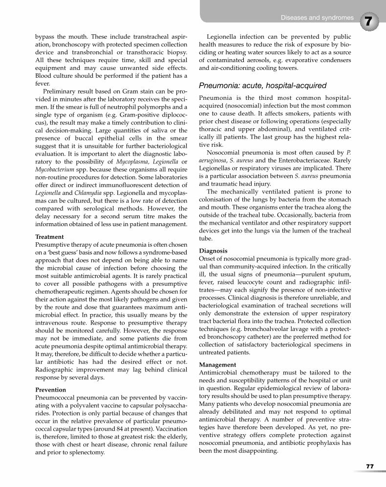
Treatment Presumptive therapy of acute pneumonia is often chosenon a ‘best guess’ basis and now follows a syndrome-basedapproach that does not depend on being able to namethe microbial cause of infection before choosing themost suitable antimicrobial agents. It is rarely practicalto cover all possible pathogens with a presumptivechemotherapeutic regimen. Agents should be chosen fortheir action against the most likely pathogens and givenby the route and dose that guarantees maximum anti-microbial effect. In practice, this usually means by theintravenous route. Response to presumptive therapyshould be monitored carefully. However, the responsemay not be immediate, and some patients die fromacute pneumonia despite optimal antimicrobial therapy.It may, therefore, be difficult to decide whether a particu-lar antibiotic has had the desired effect or not.Radiographic improvement may lag behind clinicalresponse by several days.
Prevention Pneumococcal pneumonia can be prevented by vaccin-ating with a polyvalent vaccine to capsular polysaccha-rides. Protection is only partial because of changes thatoccur in the relative prevalence of particular pneumo-coccal capsular types (around 84 at present). Vaccinationis, therefore, limited to those at greatest risk: the elderly,those with chest or heart disease, chronic renal failureand prior to splenectomy.
Legionella infection can be prevented by publichealth measures to reduce the risk of exposure by bio-ciding or heating water sources likely to act as a sourceof contaminated aerosols, e.g. evaporative condensersand air-conditioning cooling towers.
Pneumonia: acute, hospital-acquired
Pneumonia is the third most common hospital-acquired (nosocomial) infection but the most commonone to cause death. It affects smokers, patients withprior chest disease or following operations (especiallythoracic and upper abdominal), and ventilated crit-ically ill patients. The last group has the highest rela-tive risk.
Nosocomial pneumonia is most often caused by P.aeruginosa, S. aureus and the Enterobacteriaceae. RarelyLegionellas or respiratory viruses are implicated. Thereis a particular association between S. aureus pneumoniaand traumatic head injury.
The mechanically ventilated patient is prone tocolonisation of the lungs by bacteria from the stomachand mouth. These organisms enter the trachea along theoutside of the tracheal tube. Occasionally, bacteria fromthe mechanical ventilator and other respiratory supportdevices get into the lungs via the lumen of the trachealtube.
Diagnosis Onset of nosocomial pneumonia is typically more grad-ual than community-acquired infection. In the criticallyill, the usual signs of pneumonia—purulent sputum,fever, raised leucocyte count and radiographic infil-trates—may each signify the presence of non-infectiveprocesses. Clinical diagnosis is therefore unreliable, andbacteriological examination of tracheal secretions willonly demonstrate the extension of upper respiratorytract bacterial flora into the trachea. Protected collectiontechniques (e.g. bronchoalveolar lavage with a protect-ed bronchoscopy catheter) are the preferred method forcollection of satisfactory bacteriological specimens inuntreated patients.
Management Antimicrobial chemotherapy must be tailored to theneeds and susceptibility patterns of the hospital or unitin question. Regular epidemiological review of labora-tory results should be used to plan presumptive therapy.Many patients who develop nosocomial pneumonia arealready debilitated and may not respond to optimalantimicrobial therapy. A number of preventive stra-tegies have therefore been developed. As yet, no pre-ventive strategy offers complete protection againstnosocomial pneumonia, and antibiotic prophylaxis hasbeen the most disappointing.
77
7Diseases and syndromes
F70954-07.qxd 12/10/02 7:36 AM Page 77
Pneumonia: chronic
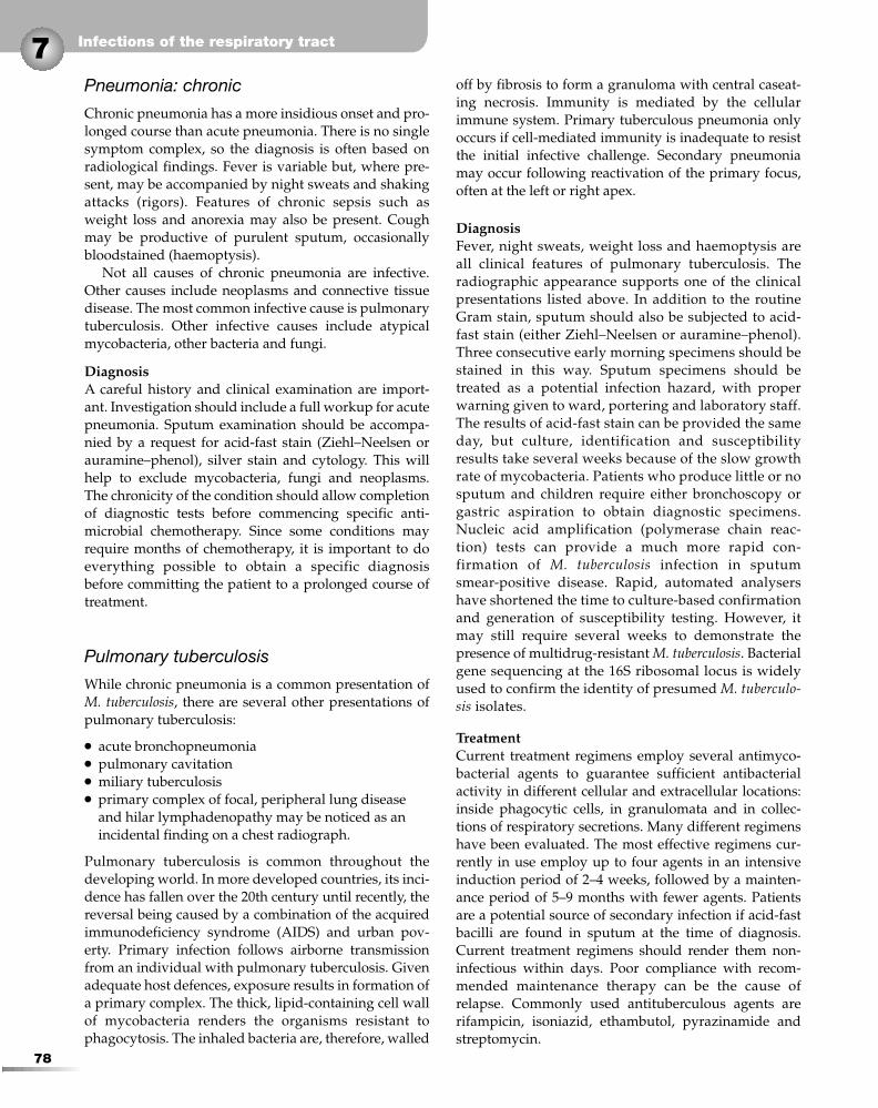
Chronic pneumonia has a more insidious onset and pro-longed course than acute pneumonia. There is no singlesymptom complex, so the diagnosis is often based onradiological findings. Fever is variable but, where pre-sent, may be accompanied by night sweats and shakingattacks (rigors). Features of chronic sepsis such asweight loss and anorexia may also be present. Coughmay be productive of purulent sputum, occasionallybloodstained (haemoptysis).
Not all causes of chronic pneumonia are infective.Other causes include neoplasms and connective tissuedisease. The most common infective cause is pulmonarytuberculosis. Other infective causes include atypicalmycobacteria, other bacteria and fungi.
Diagnosis A careful history and clinical examination are import-ant. Investigation should include a full workup for acutepneumonia. Sputum examination should be accompa-nied by a request for acid-fast stain (Ziehl–Neelsen orauramine–phenol), silver stain and cytology. This willhelp to exclude mycobacteria, fungi and neoplasms.The chronicity of the condition should allow completionof diagnostic tests before commencing specific anti-microbial chemotherapy. Since some conditions mayrequire months of chemotherapy, it is important to doeverything possible to obtain a specific diagnosisbefore committing the patient to a prolonged course oftreatment.
Pulmonary tuberculosis
While chronic pneumonia is a common presentation ofM. tuberculosis, there are several other presentations ofpulmonary tuberculosis:
● acute bronchopneumonia ● pulmonary cavitation ● miliary tuberculosis ● primary complex of focal, peripheral lung disease
and hilar lymphadenopathy may be noticed as anincidental finding on a chest radiograph.
Pulmonary tuberculosis is common throughout thedeveloping world. In more developed countries, its inci-dence has fallen over the 20th century until recently, thereversal being caused by a combination of the acquiredimmunodeficiency syndrome (AIDS) and urban pov-erty. Primary infection follows airborne transmissionfrom an individual with pulmonary tuberculosis. Givenadequate host defences, exposure results in formation ofa primary complex. The thick, lipid-containing cell wallof mycobacteria renders the organisms resistant tophagocytosis. The inhaled bacteria are, therefore, walled
off by fibrosis to form a granuloma with central caseat-ing necrosis. Immunity is mediated by the cellularimmune system. Primary tuberculous pneumonia onlyoccurs if cell-mediated immunity is inadequate to resistthe initial infective challenge. Secondary pneumoniamay occur following reactivation of the primary focus,often at the left or right apex.
Diagnosis Fever, night sweats, weight loss and haemoptysis areall clinical features of pulmonary tuberculosis. Theradiographic appearance supports one of the clinicalpresentations listed above. In addition to the routineGram stain, sputum should also be subjected to acid-fast stain (either Ziehl–Neelsen or auramine–phenol).Three consecutive early morning specimens should bestained in this way. Sputum specimens should betreated as a potential infection hazard, with properwarning given to ward, portering and laboratory staff.The results of acid-fast stain can be provided the sameday, but culture, identification and susceptibilityresults take several weeks because of the slow growthrate of mycobacteria. Patients who produce little or nosputum and children require either bronchoscopy orgastric aspiration to obtain diagnostic specimens.Nucleic acid amplification (polymerase chain reac-tion) tests can provide a much more rapid con-firmation of M. tuberculosis infection in sputumsmear-positive disease. Rapid, automated analysershave shortened the time to culture-based confirmationand generation of susceptibility testing. However, itmay still require several weeks to demonstrate thepresence of multidrug-resistant M. tuberculosis. Bacterialgene sequencing at the 16S ribosomal locus is widelyused to confirm the identity of presumed M. tuberculo-sis isolates.
Treatment Current treatment regimens employ several antimyco-bacterial agents to guarantee sufficient antibacterialactivity in different cellular and extracellular locations:inside phagocytic cells, in granulomata and in collec-tions of respiratory secretions. Many different regimenshave been evaluated. The most effective regimens cur-rently in use employ up to four agents in an intensiveinduction period of 2–4 weeks, followed by a mainten-ance period of 5–9 months with fewer agents. Patientsare a potential source of secondary infection if acid-fastbacilli are found in sputum at the time of diagnosis.Current treatment regimens should render them non-infectious within days. Poor compliance with recom-mended maintenance therapy can be the cause ofrelapse. Commonly used antituberculous agents arerifampicin, isoniazid, ethambutol, pyrazinamide andstreptomycin.
78
Infections of the respiratory tract7
F70954-07.qxd 12/10/02 7:36 AM Page 78

Prevention Prevention is by intradermal inoculation of a live attenu-ated strain of mycobacterium (BCG) after non-reactivityhas been demonstrated by tuberculin skin test. Sincethe main reservoir of disease in developed countriesis adults with untreated pulmonary tuberculosis, sec-ondary spread can be prevented by contact tracingand treatment. In some countries, cattle are a significantadditional reservoir. Pasteurisation of milk, meatinspection and establishment of a national tuberculosis-free cattle stock are all important approaches to preven-tion of zoonotic tuberculosis.
Empyema
Empyema is the accumulation of purulent fluid inthe pleural space. It is caused by direct extensionfrom underlying pneumonia, infection resulting frompenetrating thoracic trauma or haematogenous spreadfrom a distant focus. Infection may be caused by a vari-ety of bacteria including S. aureus, the Enterobacteriaceae,streptococci and obligate anaerobes.
Diagnosis Pleural effusion and a gas–fluid interface may be evi-dent on chest radiograph (a lateral view is a more sensi-tive means of detection), and there will also be dullnessto percussion over the affected area.
The collection of purulent fluid requires drainage fordiagnostic and therapeutic purposes. Anaerobic cultureshould be requested, preferably by communication withthe laboratory prior to undertaking the drainage pro-cedure. Athoracic surgical opinion should be sought early.
Treatment Presumptive antibiotic therapy depends on the resultsof Gram stain but should include an agent active againstobligate anaerobes, e.g. metronidazole.
7.5 Organisms
A checklist of the organisms discussed in this chapter isgiven in Box 2. Further information is given on thepages indicated.
79
7Organisms
Bacteria see page Fungi see pageStreptococcus pneumoniae 245 Aspergillus niger 269 Staphylococcus aureus 243 Corynebacterium diphtheriae 246–7 VirusesKlebsiella pneumoniae 248 Rhinoviruses 260 Pseudomonas aeruginosa 249–50 Coronaviruses 258Haemophilus influenzae 251 Coxsackieviruses 260 Legionella pneumophila 252 Adenoviruses 257 Mycoplasma pneumoniae 254 Influenza virus 259Chlamydia spp. 255 Parainfluenza viruses 259Streptococcus pyogenes 244 Respiratory syncytial virus 259 Mycobacterium tuberculosis 253–4 Epstein–Barr virus 257 Mycobacterium spp. 253–4 Arcanobacterium haemolyticum 73
Box 2 Organisms that infect the respiratory tract
F70954-07.qxd 12/10/02 7:36 AM Page 79
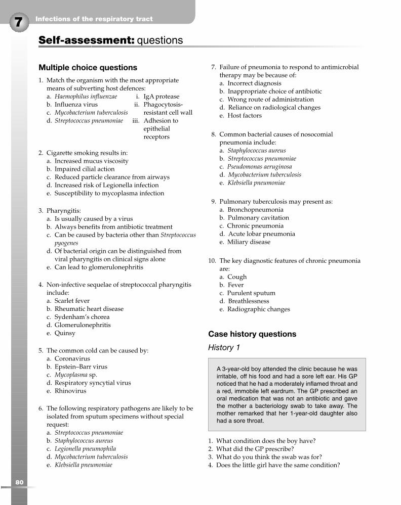
Multiple choice questions
1. Match the organism with the most appropriatemeans of subverting host defences: a. Haemophilus influenzae i. IgA protease b. Influenza virus ii. Phagocytosis-c. Mycobacterium tuberculosis resistant cell wall d. Streptococcus pneumoniae iii. Adhesion to
epithelialreceptors
2. Cigarette smoking results in: a. Increased mucus viscosity b. Impaired cilial action c. Reduced particle clearance from airways d. Increased risk of Legionella infection e. Susceptibility to mycoplasma infection
3. Pharyngitis: a. Is usually caused by a virus b. Always benefits from antibiotic treatment c. Can be caused by bacteria other than Streptococcus
pyogenesd. Of bacterial origin can be distinguished from
viral pharyngitis on clinical signs alone e. Can lead to glomerulonephritis
4. Non-infective sequelae of streptococcal pharyngitisinclude: a. Scarlet fever b. Rheumatic heart disease c. Sydenham’s chorea d. Glomerulonephritis e. Quinsy
5. The common cold can be caused by: a. Coronavirus b. Epstein–Barr virus c. Mycoplasma sp. d. Respiratory syncytial virus e. Rhinovirus
6. The following respiratory pathogens are likely to beisolated from sputum specimens without specialrequest: a. Streptococcus pneumoniaeb. Staphylococcus aureusc. Legionella pneumophilad. Mycobacterium tuberculosise. Klebsiella pneumoniae
7. Failure of pneumonia to respond to antimicrobialtherapy may be because of: a. Incorrect diagnosis b. Inappropriate choice of antibiotic c. Wrong route of administration d. Reliance on radiological changes e. Host factors
8. Common bacterial causes of nosocomialpneumonia include: a. Staphylococcus aureusb. Streptococcus pneumoniaec. Pseudomonas aeruginosad. Mycobacterium tuberculosise. Klebsiella pneumoniae
9. Pulmonary tuberculosis may present as: a. Bronchopneumonia b. Pulmonary cavitation c. Chronic pneumonia d. Acute lobar pneumonia e. Miliary disease
10. The key diagnostic features of chronic pneumoniaare: a. Cough b. Fever c. Purulent sputum d. Breathlessness e. Radiographic changes
Case history questions
History 1
1. What condition does the boy have? 2. What did the GP prescribe? 3. What do you think the swab was for? 4. Does the little girl have the same condition?
Infections of the respiratory tract7
80
Self-assessment: questions
A 3-year-old boy attended the clinic because he wasirritable, off his food and had a sore left ear. His GPnoticed that he had a moderately inflamed throat anda red, immobile left eardrum. The GP prescribed anoral medication that was not an antibiotic and gavethe mother a bacteriology swab to take away. Themother remarked that her 1-year-old daughter alsohad a sore throat.
F70954-07.qxd 12/10/02 7:36 AM Page 80

History 2
1. What was the first infection? 2. Why was flucloxacillin chosen? 3. How reliable is tracheal suction as a specimen
collection technique?
Data interpretation
Table 11 is the report relating to a 57-year-old malesmoker with fever, confusion, diarrhoea and non-pro-ductive cough.
1. Given the clinical features in this case, what possiblebacterial cause of this infection has not beenmentioned on this report?
2. Does a negative report rule out the possibility of thespecies mentioned in your answer to 1?
3. What is the explanation for the presence of each ofthe bacteria mentioned in this report?
4. What other microbiological investigations mighthelp you to establish an aetiological diagnosis in thiscase?
Objective structured clinical examination(OSCE)
A 48-year-old man with fever and a productive cough wasadmitted after he became increasingly short of breath. Hehad a temperature of 38.5˚C, a pulse of 120 beats/min anda respiratory rate of 22 breaths/min. Chest examinationrevealed reduced expansion on the right, dullness to per-cussion, quiet breath sounds and dullness to percussion inthe right midzone and green-coloured sputum. Chest X-ray showed a clearly demarcated opacity occupying theright middle lobe. Blood gases on arterial blood collectedwhile the patient was breathing room air confirmed ahypoxia and respiratory acidosis.
You are asked the following:
1. Does this patient have a lobar pneumonia? 2. Is his pneumonia most likely to be caused by
Streptococcus pneumoniae infection? 3. Do other bacteria such as Legionella pneumophila
cause lobar pneumonia? 4. Will bacteriological investigations assist the
immediate management of this infection? 5. Should ceftriaxone be used as a first choice of
antibiotic in resistant Streptococcus pneumoniaeinfection?
Short notes questions
Write short notes on the following:
1. Why the lungs are usually free from bacterialcontamination in healthy individuals
2. Methods you know for obtaining diagnosticmicrobiology specimens from the lower respiratorytract; describe how to prevent contamination withthe oral flora
3. Influenza and its complications 4. The clinical presentation of acute bronchitis and its
treatment
Viva questions
1. Are viral upper respiratory tract infectionsimportant?
2. What microbiological tests would you use todiagnose an acute, community-acquired
7Self-assessment: questions
81
A 16-year-old student was admitted to an intensivecare unit following a severe head injury in a road traf-fic accident. Four days after admission, he was still inneed of mechanical ventilation and had developed afever and raised leucocyte count. One of the nurseshad noticed that the patient had purulent and slightlybloodstained tracheal secretions and had sent themto the diagnostic laboratory. The Gram stain reportsaid: ‘Gram-positive cocci: further identification andsensitivities to follow’. Intravenous flucloxacillin wascommenced, and fucidic acid added 2 days laterwhen further results reached the intensive care unit.The patient had a further serious infection withPseudomonas aeruginosa 2 weeks later but survivedand eventually left hospital after almost a year.
Table 11 Report for data interpretation
Test Results
Bronchoalveolar lavage fluid Microscopy Leucocytes +++
Epithelial cells + Monocytes + Gram stain mixed bacteria Acid-fast stain: no acid-fast bacilli seen Mycobacteria: culture results will be
issued on a separate report Culture Mixed bacteria including Moraxella
catarrhalis, Pseudomonas aeruginosaand viridans group streptococci
Antibiotic susceptibilities of M. catarrhalis andP. aeruginosa
Amoxicillin R Augmentin S Doxycycline S Co-trimoxazole R SGentamicin S SCiprofloxacin S STimentin S S
R, resistant; S, sensitive.
F70954-07.qxd 12/10/02 7:36 AM Page 81

Infections of the respiratory tract7
82
pneumonia? How would the results influence yourchoice of antibiotic treatment?
3. In acute, community-acquired pneumonia, a specificaetiological diagnosis is now thought to be less
important as a guide to immediate clinicalmanagement. What key features will determine yourimmediate course of action?
F70954-07.qxd 12/10/02 7:36 AM Page 82
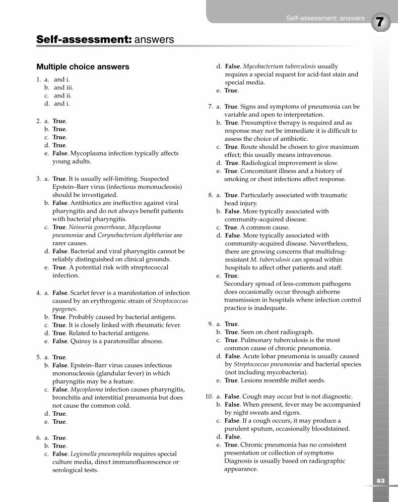
Multiple choice answers
1. a. and i. b. and iii. c. and ii. d. and i.
2. a. True. b. True. c. True. d. True. e. False. Mycoplasma infection typically affects
young adults.
3. a. True. It is usually self-limiting. SuspectedEpstein–Barr virus (infectious mononucleosis)should be investigated.
b. False. Antibiotics are ineffective against viralpharyngitis and do not always benefit patientswith bacterial pharyngitis.
c. True. Neisseria gonorrhoeae, Mycoplasmapneumoniae and Corynebacterium diphtheriae arerarer causes.
d. False. Bacterial and viral pharyngitis cannot bereliably distinguished on clinical grounds.
e. True. A potential risk with streptococcalinfection.
4. a. False. Scarlet fever is a manifestation of infectioncaused by an erythrogenic strain of Streptococcuspyogenes.
b. True. Probably caused by bacterial antigens. c. True. It is closely linked with rheumatic fever. d. True. Related to bacterial antigens. e. False. Quinsy is a paratonsillar abscess.
5. a. True. b. False. Epstein–Barr virus causes infectious
mononucleosis (glandular fever) in whichpharyngitis may be a feature.
c. False. Mycoplasma infection causes pharyngitis,bronchitis and interstitial pneumonia but doesnot cause the common cold.
d. True. e. True.
6. a. True. b. True. c. False. Legionella pneumophila requires special
culture media, direct immunofluorescence orserological tests.
d. False. Mycobacterium tuberculosis usuallyrequires a special request for acid-fast stain andspecial media.
e. True.
7. a. True. Signs and symptoms of pneumonia can bevariable and open to interpretation.
b. True. Presumptive therapy is required and asresponse may not be immediate it is difficult toassess the choice of antibiotic.
c. True. Route should be chosen to give maximumeffect; this usually means intravenous.
d. True. Radiological improvement is slow. e. True. Concomitant illness and a history of
smoking or chest infections affect response.
8. a. True. Particularly associated with traumatichead injury.
b. False. More typically associated withcommunity-acquired disease.
c. True. A common cause. d. False. More typically associated with
community-acquired disease. Nevertheless,there are growing concerns that multidrug-resistant M. tuberculosis can spread withinhospitals to affect other patients and staff.
e. True. Secondary spread of less-common pathogensdoes occasionally occur through airbornetransmission in hospitals where infection controlpractice is inadequate.
9. a. True. b. True. Seen on chest radiograph. c. True. Pulmonary tuberculosis is the most
common cause of chronic pneumonia. d. False. Acute lobar pneumonia is usually caused
by Streptococcus pneumoniae and bacterial species(not including mycobacteria).
e. True. Lesions resemble millet seeds.
10. a. False. Cough may occur but is not diagnostic. b. False. When present, fever may be accompanied
by night sweats and rigors. c. False. If a cough occurs, it may produce a
purulent sputum, occasionally bloodstained. d. False. e. True. Chronic pneumonia has no consistent
presentation or collection of symptomsDiagnosis is usually based on radiographicappearance.
7Self-assessment: answers
83
Self-assessment: answers
F70954-07.qxd 12/10/02 7:36 AM Page 83

Case history answers
History 1
1. This patient has acute otitis media. 2. It was initially treated with an oral decongestant. 3. The swab was provided so that the mother could
send in a specimen of pus from the affected ear ifrupture of the tympanic membrane occurred.
4. The sister probably had the same upper respiratorytract infection that predisposed the boy to secondaryotitis media. The mother was advised that herdaughter did not require ‘prophylactic’ antibiotics.
History 2
1. The first infection was a hospital-acquired(nosocomial) pneumonia, and since he wasmechanically ventilated, it could also be referred toas a ventilator-associated pneumonia.
2. Flucloxacillin was given because Staphylococcusaureus infection was suspected; an organism morecommon in patients with head injury. (The fusidicacid was added 2 days later when the presence of S.aureus was confirmed.)
3. Tracheal aspirates from mechanically ventilatedpatients are prone to contamination with bacteriafrom the upper trachea and are, therefore, notrepresentative of the smaller airways. Specialisedbronchoscopic techniques are preferred as a meansof specimen collection in ventilated patients inintensive care, but these techniques are onlyavailable in some centres.
Data interpretation answer
1. Legionella spp. 2. No. Neither culture-based methods nor nucleic acid
amplification is the most sensitive means ofdiagnosing Legionnaires’ disease. The urinaryantigen test is currently the most sensitive means ofconfirming L. pneumophila infection.
3. The bacteria reported here could have been carriedon the tip of the bronchoscope after contaminationduring passage through the oropharynx. M.catarrhalis and viridans group streptococci areoropharyngeal commensals. P. aeruginosa is acoloniser of the oropharynx in a proportion ofhospital patients, the percentage increasing withlength of hospital stay, severity of underlyingdisease and exposure to broad-spectrum antibiotics.
4. Legionnaires’ disease can be diagnosed using acombination of serology, culture-based methods,nucleic acid amplification by the polymerase chainreaction and urinary antigen test. Serological tests forother respiratory pathogens should also be performed.
OSCE answer
1. Yes. He has a right middle lobe pneumonia. 2. Yes. This is the most common cause of community-
acquired lobar pneumonia. 3. Yes. Other bacterial species including L. pneumophila
can cause lobar pneumonia. 4. Yes. A sputum Gram stain showing neutrophils and
many Gram-positive diplococci will increase thesuspicion that this is a S. pneumoniae infection. Theresult should be available within minutes ofreceiving the sample in the laboratory.
5. No. Moderate penicillin resistance does not result ina significant increase in risk of penicillin treatmentfailure for S. pneumoniae infection unless the patienthas meningitis. In this case, ceftriaxone would be asatisfactory choice of agent. But for lobarpneumonia, high-dose intravenous benzylpenicillinremains the treatment of choice.
Short notes answers
1. Review the anatomical, physiological and otherdefences of the respiratory tract.
2. Start with a list. A tabular answer would beacceptable.
3. Remember to mention pathogenesis, surface antigenvariation, epithelial damage and subsequentstaphylococcal pneumonia.
4. Three brief paragraphs on acute bronchitis (strictsense), tracheobronchitis and acute exacerbation ofchronic bronchitis. If recommending antimicrobialtherapy, justify in terms of pathogens and likelyoutcome.
Viva answers
1. Yes. They are the commonest infective reason formedical consultation and antibiotic prescription. Youshould mention the common cold and pharyngitis asa minimum. Mention local data on specific viralpathogens, epidemiology, public health issues andcomplications, if available.
2. Microscopy and culture of respiratory secretions,nucleic acid amplification tests (polymerase chainreaction (PCR)), serology, urinary antigen test forLegionella pneumophila. Only a clear-cut Gram oracid-fast stain result and a urinary antigen test canhave immediate impact on antibiotic choice. PCRtakes longer but will produce a specific result.Culture is even slower and often producesinconclusive results. Serology is rarely helpful inacute management as a rise in antibody titre maynot occur until the patient has begun to recover.
Infections of the respiratory tract7
84
F70954-07.qxd 12/10/02 7:36 AM Page 84

3. The severity of respiratory infection is now taken asthe main guide to whether the patient (i) needshospital admission, and (ii) requires intensiverespiratory care. Key features used to make these
decisions are respiratory rate, blood urea, fallingPaO2 (arterial partial pressure of oxygen), fallingblood pressure and involvement of both lungs ormultiple lobes on chest radiograph.
7Self-assessment: answers
85
F70954-07.qxd 12/10/02 7:36 AM Page 85